Dedication
Professor(Dr.)SrinivasanChandrasekaran, IndianInstituteofScience(IISc),Bangalore
1.Greensyntheticapproachesforbiologicallyrelevant heterocycles:advancedsynthetictechniques—an overview
GoutamBrahmachari
2.2.4HeteroDiels-Aldercycloadditions30
2.2.5Diels-Aldercycloadditionsofheterocyclesas dienophilecomponents33
2.3Dipolarcycloadditions 37
2.4[2 1 2]-Cycloadditions
3.Microwave-assistedsynthesisofmedicinally privilegedheterocycles
DebasishBandyopadhyayandBimalKrishnaBanik
3.1Introduction
3.2Microwaveirradiation:mechanism
3.3Microwave-inducedsynthesisofheterocyclesof medicinalinterests
3.3.1Three-memberedheterocycleswithoneheteroatom57
3.3.2Four-memberedheterocycleswithoneheteroatom58
3.3.3Five-memberedheterocycles61
3.3.4Six-memberedheterocycles87
3.3.5Seven-memberedheterocycles95
3.4Concludingremarks
4.Applicationofmicrowaveirradiationinthe synthesisofP-heterocycles
GyorgyKeglevich
4.1Introduction 111
4.2Functionalizationofcyclicphosphinicacids 112
4.2.1Directesterificationandamidationofcyclic phosphinicacids112
4.2.2Interpretationandmodelingtherateenhancing effectofmicrowaves118
4.2.3Alkylatingesterificationofcyclicphosphinicacids121
4.2.4Thepropylphosphonicanhydride-promoted esterificationofcyclicphosphinicacids122
4.2.5Hydrolysisofcyclicphosphinates124
4.3Diels Aldercycloadditions,fragmentation-related phosphorylations,andinverseWittig-typereactions 126
4.4Phospha-Michaelreactions 127
4.5Kabachnik Fieldsreactions
4.6Thedeoxygenationof2,5-dihydro-1H-phosphole1-oxides
4.7Synthesisandreactionsofnew7-phosphanorbornene derivatives
4.8Concludingremarks
5.Microwave-assistedmulticomponentreactions asagreensyntheticapproachtoheterocycles: specialreferencetoHantzsch,Biginelli,and Groebke Blackburn Bienaymereactions
FeliciaPheiLinLimandAntonV.Dolzhenko
5.1Introduction
5.2Hantzschreaction
5.3Biginellireaction
5.4Groebke
5.5Concludingremarks
6.Useofballmillingforthesynthesisofbiologically activeheterocycles
NirmalyaMukherjee,PintuMaityandBrindabanC.Ranu
6.1Introduction 167
6.2Synthesisofheterocycles 169
6.2.1Nitrogen-containingheterocycles169
6.2.2Oxygen-containingheterocycles176
6.2.3Nitrogen oxygen/sulfur-containingheterocycles179
6.2.4Boron-containingheterocycles181
6.3Generalgreenaspectsofmechanochemicalreaction 182
6.4Concludingremarks 182 Acknowledgments 183 References 183
7.RecentadvancesinphotocatalyticMiniscireaction: aneco-friendlyfunctionalizationofbiologically relevantheteroarenes
AndreaFiorati,CristianGambarotti,LucioMelone,NadiaPastori, CarloPunta,GiuseppinaRaffainiandAdaTruscello
7.1Introduction 189
7.2Miniscireaction:asynthetictoolforbioactivemolecules 190
7.2.1Mechanisticinsight190
7.2.2Applicationsinmedicinalchemistry191
7.3Miniscireaction:theTiO2-mediatedphotocatalytic approach 193
Contents
7.3.1TiO2-mediatedphotocatalyticMiniscireaction: thesyntheticimplications194
7.3.2AdsorptiononthemoststableTiO2 anatasesurface: moleculardynamicsstudy196
7.4Miniscireaction:recentadvancesinthephotocatalytic approach 199
7.4.1 N-Hydroxyphthalimideesters:versatile radicalprecursors199
7.4.2Visiblelight mediatedMiniscireactioncatalyzed byIr-photoredoxcatalyst201
7.5Concludingremarks 203
8.Recentadvancesforconstructionandlate-stage diversificationofindolecoreviaC Hbond activation/functionalization
ArunKumarSinhaandRakeshKumar
8.1Introduction 207
8.2Methodsforthesynthesisofindoles 208
8.3C Hbondactivation 209
8.4C Hbondactivationapproachesforsynthesisof functionalizedindoleframework 210
8.4.1Transition-metalmediatedconstructionofindole coreviaC Hbondactivation211
8.5Late-stagediversificationofindolesystem (C Hactivationofpreformedindoleframework) 223
8.5.1C HbondfunctionalizationofindoleatC4position224
8.5.2C HbondfunctionalizationofindoleatC5position230
8.6C HbondfunctionalizationofindoleatC6position 233
8.6.1C HbondfunctionalizationofindoleatC7position237
8.7Concludingremarks 242 References 243
9.Cycloadditionreactionsinionicliquidsforthe synthesisofbiologicallyrelevantheterocycles
BeeraiahBaire,RameshGardasandSoniyaGandhi
9.1Introduction 249
9.1.1Cycloadditionreactionsandtheir heteroatomicversions250
9.1.2Ionicliquids251
9.2Thehetero-cycloadditionreactionsinionicliquids 252
9.2.1[4 1 2]-Cycloadditionreactions252
9.2.21,3-Dipolar[3 1 2]-cycloadditionreactions260
9.2.3Thehetero-[2 1 1]-cycloadditionreactions inionicliquids275
9.2.4[2 1 2]-Cycloadditionreactions277
9.2.5Ionicliquidascatalystsinhetero-cycloaddition reactions279
9.3Concludingremarks
10.Advancesingreenerprocessesfortriazolesynthesis viaazide-alkynecycloadditionreactions
PrasunChoudhuryandBasudebBasu
10.1Introduction
10.2Methodsofsynthesisoftriazolesusinggreenerprocesses 299
10.2.1Reactionspromotedbysolid surface/heterogeneouscatalysts300
10.2.2Reactionspromotedbycarbonaceousnanomaterials304
10.2.3Polymersupportedreactions308
10.2.4“On-water”synthesis310
10.2.5Solvent-freesynthesis312
10.2.6Ionicliquidmediatedreactions313
10.2.7Microwave-assistedreactions315
10.2.8Photocatalyticreactions321
10.2.9Combinatorialapproaches322
10.2.10Copper-freecatalyticprocesses327
10.2.11Othercatalyticprocesses333
10.3Concludingremarks
11.Employingarynesintransition-metal-free synthesisofbenzo-fusedfiveandsix-membered heterocycles:anupdate
TonyRoyandAkkattuT.Biju
11.1Introduction 355
11.2Synthesisoffive-memberedheterocycles 356
11.2.1Cycloadditionreactions356
11.2.2Heterocyclicconstructioninitiatedbyinsertion reactions363
11.2.3Dominoaryneroutesforheterocyclessynthesis365
11.2.4Molecularrearrangements367
11.2.5Multicomponentreactions369
11.3Synthesisofsix-memberedheterocycles 370
11.3.1Cycloadditionreactions370
11.3.2Transitionmetal-catalyzedreactions372
11.3.3Insertionreactions375
11.3.4Multicomponentreactions375 11.3.5Molecularrearrangements375
11.4Selectedexamplesforthesynthesisofmedium-sized heterocycles
12.Multicomponentapproachforthesustainable synthesisoflawsone-basedheterocycles
AnkitaChaudhary,GarimaKhannaandJ.M.Khurana
12.1Introduction
12.2Synthesisoflawsone-basedheterocyclesvia multicomponentapproach
12.3Concludingremarks
13.Synthesisofbiologicallyrelevantheterocyclic skeletonsundersolvent-freecondition
GarimaTripathi,AbhijeetKumar,SanchayitaRajkhowa andVinodK.Tiwari
13.1Introduction
13.2Synthesisofnitrogen-containingheterocycles
13.2.1Solvent-freesynthesisof five-memberedheterocycles424
13.2.2Synthesisofnitrogencontainingsix-membered heterocycles437
13.3Synthesisofbiologicallyrelevantoxygenheterocycles 443 13.3.1Synthesisoffunctionalizedfurans443
13.3.2Synthesisofbenzopyransandtheirderivatives445 13.3.3Synthesisofsubstitutedchromonesunder solvent-freecondition447
13.4Synthesesofheterocycleswithtwodifferentheteroatoms 449 13.4.1Synthesisofbenzoxazolesandbenzothiazoles449 13.4.2Synthesisofbenzothiazole449
13.5Synthesisoflargerringsizeheterocycles 450
13.6Concludingremarks
14.Ultrasound-promotedmetal-catalyzedsynthesisof heterocycliccompoundsofmedicinalinterest
Mar´ıaA.Schiel,GustavoF.Silbestri,Mo ´ nicaB.Alvarez andClaudiaE.Domini
14.1Introduction
14.1.1Ultrasound462
14.1.2Cavitation462
14.1.3Instrumentation466
14.1.4Ultrasoundinorganicchemistry466
14.2Ultrasound-promotedmetal-catalyzedsynthesisof heterocyclesofmedicinalinterest 467
14.2.1Pyrroleandfuranderivatives467
14.2.2Pyrazoleandimidazolederivatives469
14.2.3Pirazolonederivatives471
14.2.4Pyridineandpyrimidinederivatives472
14.2.5Benzodiazepines474
14.2.6Triazolederivatives476
14.3Concludingremarks
References
15.Ultrasonicationundercatalyst-freecondition: anadvancedsynthetictechniquetowardthegreen synthesisofbioactiveheterocycles
RajivKarmakarandChhandaMukhopadhyay
15.1Introduction
15.2Ultrasoundwave-assistedcatalyst-freemulticomponent synthesisof N-/O-heterocycles 498
15.2.1Crisscrosscycloaddition498
15.2.2Condensationreactions500
15.2.3Spirocompoundsbasedonisatinmoieties510
15.2.4Synthesisof1,2,4-oxadiazoles514
15.2.5Synthesisofbiscoumarins516
15.2.6Synthesisofpyridazines516
15.2.7Synthesisofdithiocarbamates517
15.2.8Synthesisofhighlysubstitutedpyrazoles518
15.2.9Synthesisoftetrahydropyrazolopyridines519
15.2.10Synthesisofsubstitutedthiourea522
15.2.11Synthesisof6H-1-benzopyrano[4,3-b] quinolin-6-ones522
15.2.12Synthesisofketeneimines523
15.2.13Synthesisofrhodanines524
15.2.14Synthesisof2,20 -(1,4-phenylene)bis[1-acetyl-1, 2-dihydro-4H-3,1-benzoxazin-4-one]derivatives525
15.2.15Synthesisof N-(4-arylthiazol-2-yl)hydrazones526
15.2.16Synthesisofpyrazolo[3,4-b]pyridines527
15.2.17Synthesisofbenzo[c]acridin-9H-one528
15.2.18Synthesisofbenzofurans528
15.2.19Synthesisofdihydropyrano[2,3-c]pyrazoles529
15.2.20Synthesisofpyrimidopyrans531
15.2.21Synthesisof2-aminopyrano[3,2-b]pyrans531
15.2.22Synthesisof2-amino-4,6-diphenylnicotinonitriles531
15.2.23Synthesisofpyrazoloquinazolinones532
15.2.24Synthesisofspiro[acenaphthylene-1, 30 -pyrrolizine/pyrrolo-thiazoline]-2-onederivatives533
15.2.25Synthesisofpseudopeptidecontainingrhodanine534
15.2.26Synthesisoffunctionalized2-aminoselenopyridines536
15.2.27Synthesisof3,4-dimethyl-2,4-dihydropyrazolo [4,3-c][1,2]benzothiazine-5,5-dioxide537
15.2.28Synthesisofpyrrolidine Lobelia alkaloidsanalogs537
15.2.29Synthesisof N-acetyl-2-aryl-1,2-dihydro(4H)-3,1-benzoxazin-4-ones538
15.2.30Synthesisofchiral2-iminoselenazolines538
15.2.31Synthesisof1-substituted1H-1,2,3,4-tetrazoles539
15.2.32Synthesisofpyrazoleandpyrimidinederivatives541
15.2.33Synthesisofsubstituted1,3,4-oxadiazolederivatives542
15.2.34Improvedsynthesisofpyrazolederivatives542
15.2.35Synthesisofdihydrothiophenesderivatives543
15.2.36Synthesisofbenzothiazolederivatives544
15.3Concludingremarks 545 References 546
16.Self-catalytictechniquesforthesynthesisof biologicallyrelevantheterocyclicscaffoldsat roomtemperature:arecentupdate
GoutamBrahmachari
16.1Introduction 563
16.2Self-catalyticsyntheticendeavorsforbiologicallyrelevant heterocyclicscaffoldswithouttheaidofanycatalysts 565
16.2.1Synthesisoffunctionalizedpyrido[2,3-d:6,5-d0 ] dipyrimidine-4,6-diones565
16.2.2Synthesisoffunctionalized1H-benzo[6,7]chromeno [2,3-d]pyrimidines565
16.2.3Synthesisoffunctionalizedpyrano[3,2-c] chromen-5(4H)-ones568
16.2.4Synthesisofstructurallydiverse4H-benzo[d][1,3] thiazinesand3,4-dihydroquinazoline-2(1H)-thiones569
16.2.5Synthesisofsubstituted2-iminothiazolidines569
16.2.6Synthesisofsubstitutedtetrahydroquinolines571
16.2.7Synthesisofsubstituted1,2,3-triazole-fusedindoles andpyrroles572
16.2.8Synthesisofsubstitutedisoxazolidine-cis-fused phosphadihydrocoumarins573
16.2.9Synthesisof5-substitutedindolechromeno[2,3-b] pyridines573
16.2.10Synthesisofsubstituted2-amino-4H-pyran-3, 5-dicarbonitrilederivatives574
16.2.11Synthesisofsubstituted5,6,11,11b-tetrahydro-1 H-indolizino[8,7-b]indol-1-onederivatives574
16.2.12Synthesisofdiverselyfunctionalized4-hydroxy3-pyrazolylcoumarins575
16.3Concludingremarks
Listofcontributors
Mo ´ nicaB.Alvarez DepartamentodeQu´ımica,InstitutodeQu´ımicadelSur (INQUISUR),Seccio ´ nQu´ımicaAnal´ıtica,UniversidadNacionaldelSur,Bah´ıa Blanca,Argentina
BeeraiahBaire DepartmentofChemistry,IndianInstituteofTechnologyMadras, Chennai,India
DebasishBandyopadhyay DepartmentofChemistry,TheUniversityofTexasRio GrandeValley,Edinburg,TX,UnitedStates;SchoolofEarth,Environmental,and MarineSciences(SEEMS),TheUniversityofTexasRioGrandeValley, Edinburg,TX,UnitedStates
BimalKrishnaBanik CommunityHealthSystemsofSouthTexas,Edinburg,TX, UnitedStates;DepartmentofMathematicsandNaturalSciences,Collegeof SciencesandHumanStudies,DeanshipofResearch,PrinceMohammadBinFahd University,AlKhobar,KingdomofSaudiArabia
BasudebBasu DepartmentofChemistry,NorthBengalUniversity,Darjeeling, India;DepartmentofChemistry,RaiganjUniversity,Raiganj,India
AkkattuT.Biju DepartmentofOrganicChemistry,IndianInstituteofScience, Bangalore,India
GoutamBrahmachari LaboratoryofNaturalProducts&OrganicSynthesis, DepartmentofChemistry,Visva-Bharati(ACentralUniversity),Santiniketan, India
AnkitaChaudhary DepartmentofChemistry,MaitreyiCollege,BapuDham Complex,Chanakyapuri,NewDelhi,India
PrasunChoudhury DepartmentofChemistry,NorthBengalUniversity,Darjeeling, India
AntonV.Dolzhenko SchoolofPharmacy,MonashUniversityMalaysia,Selangor, Malaysia;SchoolofPharmacyandBiomedicalSciences,CurtinHealth InnovationResearchInstitute,CurtinUniversity,Perth,WA,Australia
ClaudiaE.Domini DepartamentodeQu´ımica,InstitutodeQu´ımicadelSur (INQUISUR),Seccio ´ nQu´ımicaAnal´ıtica,UniversidadNacionaldelSur,Bah´ıa Blanca,Argentina
AndreaFiorati DepartmentofChemistry,Materials,andChemicalEngineering “G.Natta,”PolitecnicodiMilano,Milan,Italy
CristianGambarotti DepartmentofChemistry,Materials,andChemical Engineering“G.Natta,”PolitecnicodiMilano,Milan,Italy
Another random document with no related content on Scribd:
treated by swabbing the throat with solution of sodii hyposulphis or weak caustics and antiseptics.
In young and in adult pigs pseudo-membranous pharyngitis is often only a manifestation of pneumo-enteritis. It therefore calls for no special description at this point. No exact investigation of the organisms which produce these forms of pharyngitis with false membrane formation has been made in veterinary surgery. We only know that these diseases are not true diphtheria due to “Klebs’ bacillus.” Treatment should be very energetic from the commencement, but otherwise it differs in no respect from that ordinarily adopted.
Tonics and stimulants, like alcohol, wine, coffee, etc., are indicated.
[The following account of the disease is summarised from Law’s “Veterinary Medicine,” Vol. II.]
“Pseudo-membranous pharyngitis has long been recognised as a contagious disease of swine, attacking especially swine kept in herds or in close, insanitary pens. Young pigs are more liable to attack than older animals, perhaps, owing to the older animals having suffered the disease in early life.
Modern observation shows that pharyngitis with false membranes is common in swine plague, and the present tendency is to refer all such cases to that category. It is, however, altogether probable that the occurrence of local irritation, with the addition of an irritant or septic microbe altogether distinct from that of swine plague or hog cholera, gives rise at times to this exudative angina. Certain it is that septic poisoning with the food is not at all uncommon in the hog, in the absence of these infectious diseases.
Symptoms are those of sore throat, with much prostration, a croaking cough, yellow discharge from nose and mouth, and marked muscular weakness. The tongue, tonsils and soft palate are red, swollen, and studded with patches of false membrane. The identification of swine plague may be made by the history of the outbreak, the number of animals affected, the tendency to pulmonary inflammation, the enlarged lymph glands, the presence of the non-motile bacillus, which does not generate gas in saccharine
media, and which readily kills rabbits and pigs with pure cultures of the germ.
Treatment. Isolation, cleansing and disinfection. Locally antiseptics and generally a febrifuge regimen will be advisable.”
PHARYNGEAL POLYPI.
The term “pharyngeal polypi” includes tumours of varying character, which affect the polypus form, and occur with considerable frequency in the bovine species. Many of these polypi are simply actinomycotic growths springing from the pillars of the fauces, from the upper parts of the palate or from its posterior surface. Less frequently they arise from the lateral walls or the free surface of the hard palate.
Symptoms. The symptoms are so characteristic that the diagnosis rarely presents much difficulty. They may shortly be described as indicative of repeated obstruction in the pharyngeal, œsophageal or laryngeal region. At the moment of deglutition, the polypus is thrust towards and obstructs the œsophageal orifice.
Reflex stimuli are thus excited, which prevent deglutition; an attack of coughing occurs, and food mixed with saliva is ejected from the mouth and nostrils. The attack of coughing displaces the polypus either in a forward or lateral direction, and swallowing then again becomes possible, until by changing its position the growth produces fresh signs of obstruction.
In other cases the polypus may only be of such small size as to impede the food passing through the pharynx on its way into the œsophagus or to cause difficulty in respiration by partially blocking the pharyngeal portion of the nasal cavities. In such cases deglutition is only checked and rendered slower.
Or again, the pedicle of the polypus may be sufficiently long to allow the growth at certain moments to fall in front of the laryngeal opening. Respiration is then painful, difficult and noisy. Unless the growth is displaced during the subsequent attack of coughing, asphyxia may appear imminent, or may even occur unless assistance is afforded.
Guided by these symptoms, the operator will explore the pharynx manually, and thus discover the position and size of the tumour. Tumours of the naso-pharynx produce very similar symptoms.
The prognosis is based on the information obtained by manually exploring the pharynx. It is relatively favourable if the polypus has a well-marked neck, but is very grave if the tumour is largely sessile and cannot be removed.
Treatment. Medical treatment appears useless except in cases of polypi due to the presence of actinomyces. The administration of iodine and iodide of potassium, in large doses, may then lead to resorption; but extirpation is often preferable.
In other cases extirpation is the only rational treatment. The operation necessitates the performance of provisional tracheotomy in order to avoid risk of asphyxia. The growth may be directly removed through the buccal cavity without incision, provided that it prove possible to pass the chain of an écraseur around the pedicle; or through the buccal cavity, with incision, after vertically or obliquely dividing the soft palate; or, lastly, through the larynx, after performing median laryngotomy, thus obtaining access to the pharynx.
Only the first method of intervention is to be recommended; the last two are more delicate. They necessitate after-treatment, and when the patients are in a condition for slaughter it is frequently preferable to send them to the butcher. The essential point is not to act without a full knowledge of the causes.
CHAPTER III. DISEASES OF THE ŒSOPHAGUS.
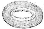
F��. 64.—Schema illustrating anatomy of the œsophagus: a strong external layer of muscle, intermediate cellulo-elastic layer, inner layer of mucous membrane lying in folds and capable of great dilatation.
The œsophageal tube is of very simple anatomical construction, and performs an equally simple physiological function; nevertheless, in the ox it is liable to a large number of diseased conditions. These conditions may affect only a circumscribed area of the mucous membrane or the entire extent of the tube. Again, both the muscular and mucous tissues may be affected, as in inflammation of the
œsophagus accompanied or followed by contraction, and in the formation of œsophageal abscesses and tumours; or the muscular tissue alone may be affected, as in cases of dilatation. Even where no lesion is apparent the normal rhythm of deglutition may be interfered with, either by the presence of a foreign body (obstruction) or by spasm of the muscular layers (œsophagismus) or by compression due to tissues surrounding the œsophagus (false contractions).
We shall successively study the different forms of œsophagitis, contraction, and dilatation, together with their complications; then obstructions, ruptures of the œsophagus, œsophagismus, and false contractions.
ŒSOPHAGITIS.
Inflammation of the œsophagus may be due to many different causes, and may occur in one of three different degrees of severity. It may be either superficial, i.e., limited to the epithelial layer of the mucous membrane; or deep, affecting the entire thickness of the mucous membrane (epithelium, corium, and œsophageal glands); or, finally, it may attack both the mucous and muscular layers. German authors recognise various divisions, such as erythematous, catarrhal, follicular, and phlegmonous œsophagitis. In reality these are not always different forms, but simply successive stages in the evolution of a single morbid condition. Here we shall only study the ordinary forms of œsophagitis, leaving on one side those which occur symptomatically during foot-and-mouth disease, cattle plague, gangrenous coryza, actinomycosis, etc.
Causation. The causes of œsophagitis may be divided into three groups of different character:—(a) Rough fodder (clover containing wrestharrow, thistles, thorns, furze, or splinters of wood, etc.) must be placed in the first rank, for its repeated action abrades and irritates the mucous membrane to such an extent as finally to produce inflammation. This inflammation usually remains superficial and of moderate intensity; its occurrence can be anticipated during years of scarcity, when the animals feed on rough and irritating material like fern, broom, heather, furze, etc.
(b) Hot drinks, whether in the nature of beverages or medicinal draughts, are a frequent cause of œsophagitis if administered by careless or inexperienced persons. The mucous membrane is scalded over a varying area and with different degrees of severity, or is destroyed by the chemical action of such drugs as ammonia, dilute acids, iodine solution, etc.
(c) Rough or clumsy manipulation in withdrawing or displacing foreign bodies, or merely passing the probang, produces that variety of œsophagitis termed traumatic. In clumsy hands œsophageal sounds or catheters may abrade or even tear the mucous membrane and subjacent tissues.
Symptoms. These vary to some extent, according to the intensity of the inflammatory phenomena. If the lesions are superficial and only implicate the epithelium, as in catarrhal œsophagitis, the symptoms often pass unnoticed, and only produce difficulty in swallowing. When inflammation has involved the entire thickness of the mucous membrane the immediate consequence is loss of appetite due to pain during swallowing. After the bolus of food has been masticated, and has passed into the pharynx, the animal stretches out its head and neck and seems to be making efforts to force it down the œsophageal canal. The progress of the bolus is slow and clearly difficult.
In œsophagitis due to scalding the blisters are soon broken by the passage of food, the corium is exposed, and the animal has equal difficulty in swallowing either solids or liquids. The reflex action provoked by the passage of the food over these lesions may be so violent that the ingesta never arrive at the stomach, but are violently rejected by a sudden and unexpected antiperistaltic contraction. Even saliva is returned. Moreover, in these cases the history is generally clear, and the animal is feverish or greatly depressed. These objective symptoms are very significant, and when, in addition, an abnormal and exceptional degree of sensibility is detected at some point by palpation, they unmistakably indicate the existence of œsophagitis.
The irregularity in deglutition, and therefore also in rumination, sometimes excites moderate tympanites without any very apparent cause. Should the condition still appear doubtful the œsophageal sound may be passed, but with great care. It generally aggravates the
pain and produces intense antiperistaltic movements, which the practitioner should not attempt to overcome.
Complications. If œsophagitis is moderate, recovery is the rule. The symptoms of pain gradually diminish.
When, on the contrary, inflammation is very intense, as in certain cases of traumatic œsophagitis, the injured spot may become infected and suppuration follow. The existing fever then persists or becomes more marked; the animal is extremely depressed; respiration may be difficult and accelerated, and appetite is entirely lost.
If the œsophageal abscess remains submucous the diagnosis is difficult, but it is often problematical, even when the abscess develops in the cervical region. The jugular furrow (usually on the left side) becomes the seat of a severe diffuse inflammatory swelling, the course of which clearly indicates the development of the symptoms. In exceptional cases fluctuation may be detected.
If from the first the abscess develops around the œsophagus or in the course of suppuration comes to occupy this position, swelling in the jugular furrows is more apparent and easier to detect, and in this case fluctuation may be localised. When the lesions are within the thorax no tangible symptoms can be detected. Death may occur in a few days, when an abscess in the lower cervical region breaks into the anterior mediastinum, or when an abscess in the thoracic region opens into the pleural cavity. In œsophagitis produced by scalding and from swallowing hot or caustic liquids the mucous membrane, and sometimes the muscular tissue, is destroyed, and ulcerations and cicatrices result, or the œsophagus may even be perforated, with rapidly fatal results; even when recovery occurs, cicatrices form and cause very grave contraction.
Diagnosis. The diagnosis is generally easy, provided that the symptoms noted are methodically analysed and the history of the case is taken into consideration.
Prognosis is favourable in ordinary cases. On the other hand, it may be very grave when general symptoms become marked, when the vital functions are disturbed and a deep-seated abscess appears to be forming.
Lesions. In the first degree the lesions are confined to inflammation and desquamation of the epithelium; in the second, to inflammation of the corium and of the mucous membrane; in the third, to infiltration of the submucous layers and of the muscular and periœsophageal tissues. Sloughing and perforation follow the administration of caustic liquids.
Treatment. As the direct application of medicines to the inflamed mucous membrane can only be of a momentary character, treatment is confined to administering emollient, anodyne, and slightly astringent drinks, the action of which is assisted by feeding with milk, farinaceous or mucilaginous foods. Under these circumstances recovery occurs in ten to fifteen days. The application of stimulant or blistering ointments along the jugular furrow may have a good effect.
When the general condition of the patient becomes aggravated, and the formation of an abscess appears certain, it is best to recommend slaughter. In the case of a submucous abscess the passage of the probang may, however, predispose or cause the abscess to open into the œsophagus, and thus lead rapidly to recovery, but this is exceptional. The “pointing” of the abscess and its opening towards the jugular furrow may be followed by temporary improvement, but at a later stage is followed by fistula formation, or by contraction of the œsophagus itself. From an economic standpoint it is better to slaughter.
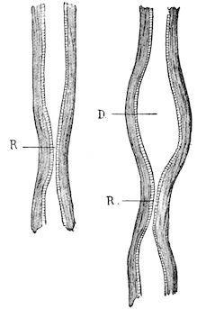
STRICTURE OF THE ŒSOPHAGUS.
Under normal conditions the cavity or lumen of the œsophageal tube is, so to speak, imaginary: the walls of the tube lie flatly together, and the mucous membrane is in folds. During the act of swallowing the tube becomes dilated to a degree varying with the size of the bolus of food, and again retracts as soon as deglutition is effected. Whenever the dilatability of the tube is markedly diminished by changes in its walls, and, in a much higher degree, when this dilatability has disappeared, true stricture exists. In the former case small boluses of food and liquids alone succeed in passing the stricture; in the latter, liquids alone can pass.
Causation. Strictures are never primary. They result from intense attacks of œsophagitis, ending in sclerosis of the mucous coat, extensive ulceration consequent on scalding, or interstitial inflammation affecting the muscular coats, which then become thickened or sclerosed.
Internal injuries due to attempts to withdraw or propel foreign bodies along the œsophagus may also cause strictures.
Lesions. In simple strictures the lesions are confined to the development in the depths of the mucous membrane and in the muscular layers of inflammatory tissue, which becomes denser with lapse of time. This alters the character of the walls and the structure of the tissues, and causes them to lose their elasticity. After extensive ulceration the tissue of the cicatrix contracts and hardens to a very varying degree.
Symptoms. The apparent symptoms are very clearly marked; the appetite is good, and the animal masticates as usual, but in the act of deglutition is seen to extend the head on the neck, and to make efforts to swallow, which prove unavailing when the contraction is too marked. A reflex antiperistaltic movement often causes the substances ingested to be at once rejected. These violent efforts, however, in time provoke dilatation above the stricture. A quantity of food accumulates in this dilatation, and the symptom so characteristic of œsophageal stricture then appears—viz., regular regurgitation. The second constant symptom associated with compression or obstruction of the œsophagus is tympanites after feeding, however trifling may be the amount swallowed. Rumination is suspended, and even eructation of gas is difficult. Finally, the characteristic sign of stricture is noted on passing the probang, which reveals the existence of the condition, indicates its position, and suggests its degree of development.
Diagnosis. Strictures only develop progressively and slowly, a fact which enables them to be differentiated from œsophagitis. It is more difficult to differentiate them from dilatations, because the stricture always ends by becoming complicated with dilatation; but this distinction is of little practical importance, the consequences being identical.
Prognosis. The prognosis is very grave, and there is no economic reason for attempting treatment except in special cases; the
F��. 65.—Schema of recent and old-standing contraction of the œsophagus. R, simple contraction; D, secondary dilatation.
indications are in the direction of slaughter.
From the economic standpoint there is no treatment. Basing their actions on human practice, the Germans have recommended progressive dilatation of the lumen of the œsophagus by passing a series of catheters of gradually increasing size. What, however, is justifiable in human medicine, where the only object is to keep the patient alive at any cost, may be highly objectionable in veterinary practice; and in the present instance this is the case. Except in very rare instances, which the practitioner alone can appreciate, dilatation is contraindicated, and the owner’s interest lies in slaughtering the animal before it has lost much condition.
DILATATION OF THE ŒSOPHAGUS.
Dilatations are more frequent than strictures. Their mode of origin is easily understood. When the muscular tissue has lost its tonicity and contractile power at a given point, or when, as a consequence of any form of inflammation, it has begun to undergo atrophy, the mucous membrane becomes herniated, because its circumference is not supported regularly during deglutition. The ectasia, which at first is of small size, becomes more marked in consequence of the tendency that exists for the food to accumulate in the dilated region. Dilatation is thus set up.
Localised attacks of œsophagitis, accidental injuries and fissuring of the œsophageal muscular tissue, produced by clumsy efforts to displace foreign bodies with the probang, are the principal causes of dilatation. When the probang is imprudently or clumsily manipulated, it may press excessively at any point where the œsophagus makes a slight bend, and thus split the contracted muscular coat without injuring the lax mucous membrane.
Œsophageal contractions, as we have seen, may form the point of origin of dilatations, but in this case the dilatations are more regular in form, and affect the entire circumference of the tube. The muscular tissue is still everywhere normal, and becomes dilated in consequence of equally-applied eccentric pressure.
Symptoms. When the dilatation develops slowly and progressively, as a consequence of muscular atrophy, the symptoms remain unnoticed for a long time, and the owner only begins to be anxious when the animal loses condition, or when the driver or cowman detects masses of half-chewed food mixed with the saliva in the manger.
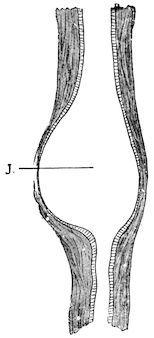
F��. 66.—Schema of œsophageal stricture (the muscular layer above the stricture has undergone atrophy; the mucous membrane is dilated).
Certain signs are pathognomonic; others may be regarded as of secondary importance. By carefully watching an animal which is feeding the following symptoms may be noted: As a general rule hunger is very marked, and the animal chews its food and swallows the first few mouthfuls in a perfectly normal way. Three, five, eight, or even ten mouthfuls may be swallowed; then the animal suddenly stops, appears a little anxious, extends its head and neck, an antiperistaltic contraction occurs, and one or two masses of food are rejected and fall into the manger. The discomfort being thus
momentarily relieved, the animal, which is dying of hunger, although faced with food which it is unable to swallow, returns to its meal, swallows one, two, or three boluses of food, regurgitation again occurs, and the whole process is repeated.
What is going on under these circumstances is easy to explain.
At the commencement of the meal the dilatation is usually empty, or nearly empty. A mouthful of food is swallowed. It descends the œsophagus until it arrives at the diverticulum, into which it partially or wholly passes, the peristaltic wave of contraction ceasing at this point. The second mouthful follows with the same result, then a third, a fourth, etc. The diverticulum soon becomes filled to repletion, and no more food can enter it. The food therefore accumulates in the upper portion of the œsophageal tube until the latter becomes nearly filled; but as this tube, provided its innervation is intact, is intolerant of the presence of any foreign body, and as efforts to swallow prove fruitless, a sudden antiperistaltic wave of contraction occurs, with the result that all the material contained in the tube above the dilatation is ejected into the mouth, whence it falls into the manger. The same result follows any further attempts to swallow during a particular feeding time. From this it will be seen that the animal can ingest at a given time only as much as the dilatation will contain.
In the intervals between meal times and under the action of the saliva and warmth, the food collected in the dilatation becomes softened, breaks down, and slowly moves onward towards the rumen. When the next feeding time arrives the dilatation is almost empty, and the same set of symptoms recurs.
If, instead of forage, the animal begins by taking gruel or very fluid material, deglutition appears normal, or at least fairly easy; but if drinking is deferred until after taking hard food, it becomes almost impossible, because the passage is obstructed. These symptoms are, so to speak, pathognomonic. Under any circumstances they are so significant that error in diagnosis is unlikely.
By careful examination œsophageal regurgitation can very easily be distinguished from true vomiting; the character of the rejected material shows that it has not come from the stomach, while the boluses of food preserve their cylindrical form, and are still saturated with saliva.
Some secondary signs also deserve to be mentioned, such as the animal’s anxiety and restlessness whilst its neighbours are feeding, the existence of trifling and intermittent tympanites due to suppressed eructation, suppression or irregularity of rumination, constipation, etc. At a later stage there is rapid wasting and disordered appetite, and finally the patients die slowly of hunger, whatever efforts are made to feed them.
When the seat of dilatation is in the cervical portion of the œsophagus, there are other symptoms which leave no doubt as to the condition. When empty the pouch cannot be detected; but during a meal the accumulation of food causes it to assume the appearance of a doughy, diffuse, indolent swelling, which alters the outline of the jugular furrow, yields to pressure, and sometimes produces respiratory disturbance by pressing on the trachea, the pneumogastric and inferior laryngeal nerves, etc.
When the dilatation is intra-thoracic and the above-described symptoms have been observed, the dilated spot can only be detected and localised by using the probang. The greatest possible prudence, however, is required in manipulating the instrument, in order to avoid rupturing the thin walls of the dilated portion.
The diagnosis is not always easy; when food is regurgitated, and one finds by auscultation that the sound usually produced by the passage of solids or liquids into the rumen is absent, there need be no hesitation in diagnosing either a dilatation or a stricture. The clinical consequences being the same, the possible error would be of little importance.
Prognosis. The animal’s life is rarely in immediate danger, but from the economic point of view the prognosis is extremely grave, and it is in the owner’s interest to slaughter the animal as soon as possible in order to avoid loss. Even in cases of dilatation in the cervical region, surgical interference is not advisable.
Treatment. As foreshadowed by what has been said, there is no rational economic treatment. When the dilatation is in the cervical region, one might in exceptional cases attempt to restore the regular calibre of the œsophagus by removing an elliptical portion of mucous membrane, and bringing the muscular tissue together with sutures; that is to say, when the rupture or fissuring of the muscular coat has been accidental. But as one is usually unable to remove the primary
cause, to which the change in the muscular tissue is essentially due, the dilatation would recur without the operation having conferred any benefit.
When an exact diagnosis has been made, the only useful indication is to confine the animal to very fluid food, which will not obstruct the œsophagus. Ordinary forage should be withheld.
ŒSOPHAGEAL OBSTRUCTIONS.
In this chapter we shall only consider such obstructions as occur in consequence of the animal having attempted to swallow without sufficiently chewing objects which become arrested in the œsophagus.
Obstruction is termed “total” or “partial,” according as the obstructing body fills the entire calibre of the œsophagus at the point of obstruction, or only occupies a part of the space. Partial obstructions produced by beet and turnip tops, etc., are usually but momentary; liquids and saliva are still able to pass between the obstruction and the walls of the tube, and as soon as the arrested food becomes a little softened it is displaced and the œsophagus again becomes patent.
Causation. The circumstances under which this accident occurs are extremely easy to understand. Obstructions are produced by apples, potatoes, turnips, carrots, cabbage-stalks, beetroots, etc., which, whether sliced or not, are swallowed gluttonously. Not having been sufficiently comminuted, and being of larger size than the œsophagus can readily accommodate, they become arrested at some point between the commencement of the œsophagus or a few inches behind the pharynx, or just in front of the point of entry of the gullet into the stomach. The latter is the commonest position, though not infrequently the obstruction occurs in the intra-thoracic portion.
It may occur in the stable, but is commoner in animals which, having broken loose, have entered orchards, gardens or potato or turnip fields and attempted to swallow apples, cabbages, potatoes, etc., found there.
In sheep, obstruction of the œsophagus is due to similar causes, but in their case the above-mentioned objects are replaced by small
wild apples, turnip shells, Jerusalem artichokes, horse-chestnuts, carrots, etc.
The symptoms may be divided into general and local.
General symptoms. As soon as the foreign body becomes fixed in position, the animal begins to make exceptional efforts to swallow. The head is extended on the neck, and the œsophagus and the muscles surrounding the trachea are violently contracted. These efforts proving fruitless, feeding is necessarily stopped, and the animal at once appears slightly anxious.
Very soon afterwards salivation sets in, saliva being continuously secreted. If the obstruction is total, the saliva cannot be swallowed, and is either returned in quantities by antiperistaltic movements or escapes in frothy filaments from the mouth.
Tympanites is not long in appearing. It is progressive, and results both from arrest of eructation and from continued fermentation in the rumen. It may eventually come to a standstill, or may continue and threaten to produce asphyxia.
Local symptoms. The local symptoms are difficult to appreciate, except in cases of cervical obstruction. Sometimes the foreign body produces a local swelling, which changes the outline of the jugular furrow, most frequently on the left side. In many cases it can only be detected by manipulating the parts between the trachea and the lower surface of the cervical vertebræ. When the obstruction is within the thorax, the probang alone can detect its position.
Diagnosis. The diagnosis is usually easy. The history and the observed symptoms are often very clear, and the suddenness with which the obstruction has made its appearance prevent the condition from being confused with dilatation or stricture.
The prognosis is very variable. It is often easy to remove the obstacle; in other cases intervention is difficult, and death may occur rapidly.
Treatment is confined to one essential point—removal of the obstruction. The chief difficulty lies in choosing the mode of intervention. Moreover, success depends on several factors, which, in the order of their importance, are as follows: the size of the obstructing body; the time which has elapsed since the accident
occurred; the bodily condition of the animal—i.e., whether it be fat or thin—and the extent to which tympanites has developed.
The first thing to do (and in favourable cases all that is required) is to puncture the rumen and leave the canula for some time in position. The onward progress of the foreign body, especially when the obstruction is in the intra-thoracic portion of the tube, is often impeded by the tympanites, which tends to thrust the object towards the pharynx, or at least to fix it in position. In consequence of a sudden change in the conditions of pressure the foreign body may move and pass into the rumen; all danger is then at an end.
Even though the obstruction does not immediately cease, puncture of the rumen, by removing the danger of asphyxia, allows one at least to wait for several hours, sometimes until next day, during which time the object may pass into the rumen without further extraneous assistance. The other methods may be grouped into four series:
I. External taxis. This is directed towards loosening the foreign body and thrusting it towards the pharynx and buccal cavity. It can only be used against obstructions in the cervical region. Two methods, although very ancient, are still practised.
(a) The first is carried out in the following way: the animal is fixed to a post or tree so that it cannot struggle, its head being drawn up as high as possible. The operator stands on the left side, with his back turned towards the patient’s head, his left hand is pressed into the right jugular furrow, his right hand is placed on the left jugular furrow immediately below the foreign body. By using the fingers the foreign body is moved, and is progressively thrust towards the pharynx, in spite of the animal’s efforts to swallow. In carrying out this manipulation it is absolutely indispensable not to let slip the obstructing body for a single instant, otherwise the peristaltic action will immediately return it to its former place. When it has been raised as far as the pharynx, an assistant passes his hand into the back of the mouth, as indicated in a former chapter, seizes the object and withdraws it; or, instead, the assistant takes over the operator’s duties, while the latter himself extracts the foreign body.
(b) In the second method the animal is fixed in a different position, the head being held about 10 to 12 inches from the ground, with the neck lowered and inclined towards the earth. As in this position the œsophagus is longitudinally relaxed, and can be dilated to its fullest
extent transversely, the difficulty of displacing the obstacle should be very much less. In this case the operator always stands on the left side of the neck, but with his back towards the animal’s body. The right arm is passed around the neck and the right hand pressed into the right jugular furrow, the left hand being similarly engaged in the left jugular furrow. The method of employing the fingers is identical, or instead of the fingers the thumbs may be used.
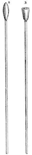
F��. 67. Œsophag eal sounds. Probangs.
When the obstructing object has been lifted as far as the pharynx it has a tendency to fall out of the mouth, and if it fail to do so it can be fixed in position and removed as in the preceding case.
II. Extraction. These methods are applicable to cases where the foreign body has become fixed in the cervical region, but more especially to obstructions in the intra-thoracic part of the œsophagus. In the majority of cases they are dangerous, and may lead to pinching, rupture, or perforation of the œsophageal mucous membrane. They should therefore be regarded as exceptional measures. Theoretically, the instruments described are perfect, but practically they do not secure the results anticipated, because one can never prevent displacement, wrinkling, and involution of the œsophageal mucous membrane.
The forceps probang has the drawback of seldom grasping smooth foreign bodies with sufficient firmness to permit of their extraction.
The corkscrew sound exposes one to the great danger of completely piercing the œsophagus, because it has to be managed blindly, and because one never knows at what depth the corkscrew portion should be protruded in order to obtain a proper hold of a foreign body.
III. Passage of the probang. When taxis fails or is inapplicable, we are forced to attempt thrusting the foreign body onwards. The method is much safer than the preceding, but, nevertheless, demands great tact, prudence, and gentleness. Suitable œsophageal sounds are made with cupped extremities, though in cases of emergency an instrument can often be successfully improvised from a cane, whip handle, or flexible stick,
about 4½ to 5 feet in length, securely wrapped at one end with cloth or tow and freely coated with some greasy material such as lard, vaseline, or oil.
The end of the sound having arrived in contact with the obstacle, the operator exercises moderate but permanent pressure. The obstacle may not move immediately, because of spasm of the œsophagus, which grasps it. It is therefore necessary to wait and to take advantage of a moment when the resistance is less, and even then the obstacle may not move.
Rough manipulation with improvised sounds may tear, fissure, or perforate the muscular and mucous coats, producing the gravest consequences.
IV. Crushing. The crushing of an obstruction in the cervical region was long ago suggested, and is still greatly commended by empirics and farriers. It is performed by means of a little mallet and a piece of board. The method is barbarous, and exposes the animal to such grave complications as crushing of the œsophageal walls, followed by necrosis, laceration of the connective tissue, and interstitial hæmorrhage, injuries of the superficial or deep-seated jugulars, of the carotid artery, pneumo-gastric nerve, etc. It should never be practised, even although attempts have been made to improve it by replacing the mallet and board by specially formed forceps intended for crushing potatoes or roots. Only in the rare cases where one is certain that the foreign body consists of a very ripe fruit could crushing be justified, and in this case there is no need to have recourse to special instruments, for the hands alone suffice.
Injection of alkaloids. The practitioner occasionally finds himself in the embarrassing position of having vainly tried all the above methods. Before adopting the last resource, viz., œsophagotomy, it is then worth while to test the action of certain alkaloids, injected subcutaneously, after having punctured the rumen.
We know that pilocarpine and eserine stimulate secretion and the action of the bowels. Injected under the skin they cause frequent swallowing efforts, and intense peristalsis extending throughout the length of the digestive tract. Doses of 1½ to 2 grains of pilocarpine and 1 to 1½ grains of eserine, according to the size of the animal,
sometimes produce excellent results, and rapidly remove obstructions.
Apomorphine, the effects of which are, so to speak, inverse, because they tend to produce anti-peristalsis and vomiting, may be tried in doses of 2 or 3 grains.
Œsophagotomy. The last resource is œsophagotomy, which, however, is only applicable in cases of obstruction of the cervical portion of the œsophagus. It should be performed as described in the section hereafter on operative manipulation. (See also Möller and Dollar’s “Regional Surgery,” p. 166.)
The point selected is necessarily governed by the position of the obstacle. There is no need to enter into full details. We may remark that it is not always necessary to perform the complete operation, and the third and fourth stages can sometimes be avoided by substituting for them attempts to break down the foreign body by submucous manipulation. The œsophagus, having been exposed and isolated, is punctured with a straight tenotome immediately below the obstacle. A curved tenotome is then introduced, and the root, potato, or fruit divided. As a rule, a little pressure from the outside then causes one or other of the fragments to move onwards and deglutition becomes normal.
Attempts have also been made to divide the obstructing body directly without previous incision and without isolating the œsophagus. It is much more difficult, for the least movement of the patient changes the relationships of the superposed layers and introduces obstacles to the manipulation of the blunt-pointed tenotome which is employed. More success often attends attempts to puncture the object with a fine trocar.
RUPTURES AND PERFORATIONS OF THE ŒSOPHAGUS.
Causation. Wounds of the œsophagus caused by external violence are rare, or at least secondary; lacerations produced from within, on the contrary, as a result of clumsy manipulation are relatively frequent. They may extend throughout the length of the tube, but in a far greater number of cases are found near the
entrance to the stomach at the point where the œsophagus turns towards the left.
The passage of the œsophageal sound or probang is apt to exaggerate this curvature, and if pushed violently the instrument may produce first a flexure, then a partial rupture or even a perforation of the tube.
In other cases a rough, irregular, infected foreign body may when swallowed penetrate the wall and cause inflammation, necrosis and perforation of the œsophagus.
The symptoms are always very grave, and of rapid development. They consist in local œdematous swelling, sero-sanguineous infiltration at the entrance to the chest, in the pretracheal region and along the jugular furrows.
The pneumo-gastric and inferior laryngeal nerves being compressed, dyspnœa results. If the œsophagus is perforated in the thoracic cavity septic pleurisy at once sets in.
Diagnosis. The diagnosis is easy, provided the history point to perforation of the œsophagus.
The prognosis is fatal whenever the perforation is within the thorax. It is sometimes possible to intervene in cases of perforation in the cervical region, but from the economic standpoint such intervention is of little value.
CHAPTER IV.
DEPRAVED APPETITE PICA THE LICKING HABIT.
Depraved appetite, causing animals to swallow bodies which cannot properly be described as food, is frequent. The condition is commonest in adult animals of the bovine species, in calves and in lambs. The consequences are sometimes very serious, so that although depraved appetite does not represent a well-defined morbid entity, it is important to be in a position to remedy it.
Depraved appetite does not appear under the same conditions in young and in old animals. In adults it often results from faulty feeding, or from some wasting disease which develops insidiously, or remains unrecognised; in young animals it is the result of insufficient nourishment.
Roloff & Röll hold that pica is the first symptom of osteomalacia (which see).
DEPRAVED APPETITE IN THE OX.
Causation. In the bovine species depraved appetite occurs in adult, debilitated animals, which are often, though not always, suffering from some well-marked digestive disturbance.
The frequency of this symptom, and the peculiarities in its occurrence, have caused it to be referred to a large number of different causes, among which may be mentioned bad hygiene, chronic gastro-enteritis, tuberculosis, osseous cachexia, pasteurellosis, gestation, etc.
It is very certain that the peculiarity in the appetite is, above all, the result of incomplete and irrational alimentation. The animal has certain special requirements, to meet which the food must be of suitable composition. If these alimentary and digestive conditions
are not fulfilled, depraved appetite may occur, even in animals which appear well nourished. Certain authors refer the appearance of this condition to want of certain soda salts in the daily ration, and, in support of this opinion, they point to the frequency of the disease in mountainous regions where the geological formation is chiefly granite, as in the Black Forest. Alluvial soils are supposed not to produce it. It certainly seems more common on soils lacking in certain constituents or exhausted by repeatedly growing certain crops. Nevertheless, in France it might be urged that pica occurs equally on all kinds of soil, and a German author, Lemke, ascribes this perversion of nutrition to the want of phosphorus. Haubner and Siedamgrotsky attribute it to a nervous disorder. All causes which exhaust the organism, especially all chronic diseases of digestive origin, may induce aberration of appetite.
Permanent stabling, confinement, absence of sunlight, want of exercise and pure air contribute to the general debility which predisposes to attack. Dry seasons, by reducing the supply of food, have a similar effect.
In tuberculosis and in pasteurellosis, it is the general organic decline which produces these puzzling changes in appetite. Similarly the influence of gestation depends on the superadded demands on the organism caused by the development of the fœtus.
Symptoms. The symptoms may be divided into two phases.
In the first phase, the animals still preserve their appetite, but whenever they have an opportunity they eat earth, sand, manure, litter saturated with urine, plaster, etc. They lick the walls, the boarding, the mangers and the trees, and they chew and swallow linen spread out to dry.
This phase may continue for a very long time, three to four months or more, provided no acute complication results from the eating of such foreign material. There is no fever, but the appetite, although well preserved, is often capricious, and the ordinary food is eaten slowly.
In the second phase, which frequently marks the development of complications produced by the passage, contact, or prolonged sojourn of various materials in the digestive tract, fever appears, little marked as a rule, but continuous in character.
The appetite is diminished. The animal wastes; the secretion of milk diminishes, and signs of chronic gastro-enteritis may be noted. The perversion of appetite still continues; rags, decomposing or filthy materials, pieces of old shoes, etc., are eaten, and it is not surprising that such substances should have an unfavourable effect on the mucous membrane of the digestive tract.
The wasting process slowly leads to marked emaciation, and after an interval of from six months to a year, or even two years, the patients die in a state of complete exhaustion. The lesions found on post-mortem examination are those of various diseases capable of producing depraved appetite or simply lesions of chronic gastroenteritis.
Diagnosis. The diagnosis presents no difficulty. The important point is to discover whether or not there exists some previously unrecognised primary disease.
Prognosis. The prognosis of this condition is grave, because depraved appetite is frequently only a symptom of some incurable disorder, or because the changes in the digestive mucous membrane are already too far advanced to permit of much improvement.
The lesions comprise: general emaciation, presence of a yellow serum in the fatty tissue, muscles pale and flabby, catarrh of the mucous membrane of the stomach and bowel. The blood seems less in quantity and coagulates feebly or not at all.
Treatment. The treatment should be directed against the primary cause, if such exists (osseous cachexia, pasteurellosis, gestation, etc.).
In other cases a change in management and in feeding, and the administration of food rich in mineral salts like chlorides, carbonates, and phosphates of lime, soda or potash, produces the best possible results. The leguminous foods, sainfoin, clover and lucern, are to be recommended. The animal, if formerly stabled, should be turned out and its living conditions entirely altered.
It is often useful to place a block of rock-salt in the manger; when hyperacidity of the stomach is suspected lime water, chalk, or magnesia should be given. Where digestion is weak or slow HCl, pepsin and vegetable bitters are indicated. Nevertheless, one sees cases which refuse to yield to any of the ordinary methods. In
treating these, Lemke has recommended the subcutaneous injection of chloride of apomorphine, a drug which may be regarded as a true specific. The doses vary between 1½ and 3 grains, and an injection is given once a week for three weeks in succession. After this the tendency to pica is said to disappear and the general condition to improve. The treatment must be repeated every three months in countries where depraved appetite appears general and permanent.
It is difficult to understand by what mechanism this drug produces the effects attributed to it, but those who have employed it speak very highly of its action.
We may add that in addition to the different modes of treatment, it is not infrequently necessary to hastily perform gastrotomy in order to avoid fatal consequences, which would otherwise follow indulgence in this habit. When an animal has swallowed a considerable quantity of linen, for example (and Moussu has seen cases in which many pounds weight had been devoured), immediate intervention is required to avoid intestinal obstruction. Furthermore, when the history is quite clear gastrotomy allows the entire mass of foreign bodies, ingested at different times, to be removed.
DEPRAVED APPETITE IN CALVES AND LAMBS.
Causation. Depraved appetite is commonest in calves and lambs when the animals are insufficiently nourished, or when the mothers are suffering from chronic debilitating diseases and are therefore yielding milk poor in fat and in mineral constituents. In a few rare cases it is impossible to discover what causes the young animals to devour these foreign materials. Even fully-grown sheep, when shut up together in winter, acquire the habit of chewing each other’s wool, sometimes to the extent of virtually depilating their fellows and accumulating wool balls in their stomachs.
Symptoms. Calves have a tendency to lick themselves or their neighbours, and thus little by little collect a varying quantity of hair which they swallow. When this habit of licking is little marked the quantity of hair ingested may not be dangerous; but in the contrary case the hair (which cannot be digested) accumulates and is permanently retained in the abomasum. It soon becomes converted
into masses, cemented together with mucus, and forms round balls, to which the name of œgagrophiles has been given. If these œgagrophiles, or hair balls, are of small size, they prove of trifling importance; but too frequently they attain considerable dimensions and obstruct the pylorus or the intestine. The young calves then refuse all nourishment, and die in twenty-four to forty-eight hours in a state of complete exhaustion or after a series of epileptiform attacks.
In lambs the complications due to depraved appetite develop in a similar way, but the wool swallowed is obtained from the mothers. The lambs first suck the locks of wool, then tear them off and swallow them. So long as these peculiarities of appetite are little marked no bad results follow; but if the shepherd is careless, and fails to note the condition of his young flock sufficiently early, accidents occur.
The wool is not so easily converted into balls as is hair, but it soon accumulates in the pyloric region or in the intestine, and forms obstructing masses. The little patients lose appetite and lie down in corners, where they are found dead after twenty-four to forty-eight hours. The masses of wool or of hair are rarely passed with the excrement; more frequently they are vomited, but this again is exceptional; usually they become arrested at the entrance to the pylorus. The lambs show colic, tympanites of the abomasum, and attempts at vomiting, though unfortunately these are often overlooked. The quantity of wool found in the abomasum and intestine on post-mortem examination may be considerable, in relation to the size of the digestive compartments. Death results from intestinal obstruction, exactly as in the case of calves.
These aberrations of appetite in lambs have been considered as due to the want of sufficient mineral salts in the mother’s milk; and it has been stated that the lambs practise this habit because of the laxative result of the fat contained in the wool swallowed. The explanation seems very logical, though it is by no means perfectly proved. It is certain that this habit becomes particularly common after years in which forage has been scarce and among flocks in bad bodily condition. The force of example also plays a certain part, and animals probably imitate one another, and so acquire the disease. This explains the importance of early segregation.
Diagnosis. The diagnosis of depraved appetite, pica, or the licking habit presents no difficulty; but it can only be arrived at by the cowman or shepherd, for the symptoms can only be detected by continued watching.
The diagnosis of pyloric or intestinal obstruction is very difficult in the absence of information. It becomes easy after the first postmortem examination has been made.
Prognosis. The prognosis is grave. In calves, obstruction of the bowel by hair balls inevitably causes death, and in sucking lambs the mortality may be high: as much as 15 per cent. to 20 per cent. according to the observations of several observers. The mortality occurs about the age of six weeks to two months, whilst the licking habit may begin towards the end of the second week.
Treatment. Prophylaxis demands that the mothers (whether cows or ewes) be well fed. An excellent precaution consists in adding to the food a sufficient quantity of salt and of phosphate of lime (2 drams to 2½ drams of each). This treatment of the mothers is necessary as soon as the tendency to licking becomes manifest.
In calves the best method of avoiding fatal results is to prevent the young animals licking one another; and the method now usually practised on well-managed farms consists in applying a simple muzzle of wicker work immediately after each meal.
In lambs treatment is more difficult. As soon as the shepherd sees any tendency to depraved appetite the lambs should only be left with their mothers whilst being suckled. The flock should be exercised in the open, and ordinary salt should be placed at a number of points on the ground occupied by the animals.
COLIC IN THE OX.
Causation. Congestive colic occurs in the stable, in animals which have been doing heavy work, and, returning in a heated condition, drink large quantities of cold water. It is commoner when animals have not eaten for a considerable time, and when, therefore,
COLIC DUE TO INGESTION OF COLD WATER. CONGESTIVE COLIC.
the stomach is nearly empty. Under these circumstances chill of the digestive viscera is direct and immediate.
Symptoms. This form of colic occurs suddenly, soon after the water has been swallowed, and is characterised by violent pain. At first the animals show uneasiness, stamp, and continually move about striking themselves in the flank with the feet or horns, swishing the tail, etc. They refuse food, lie down and rise frequently, and paw the ground.
As a general rule this form of colic lasts from half an hour to one hour, and terminates in recovery. In some rare cases where death occurred Cruzel found on post-mortem examination congestion of the abomasum, and, in a few, congestion of the small intestine, with or without rupture.
The diagnosis is easy, on account of the suddenness of onset, rapid development and history of the disease, discovered on questioning the owner or herdsman.
The prognosis is not grave. This form of colic generally cures itself. Nevertheless precautions are required against possible complications, such as intestinal hæmorrhage and invagination.
The necessary preventive measures are self-evident. Animals returning from work should not be allowed to drink freely of cold water, but should first receive a little food and afterwards water at the temperature of the atmosphere.
When colic has set in, the patient can be walked about. If pain persists, the region of the abdomen may be dressed with oil of turpentine, mustard, or similar counter-irritants. The application of warm clothing is also useful. Finally, in grave cases, a moderate quantity (three, four, or five quarts) of blood may be withdrawn from the jugular. The administration of stimulants like wine, alcohol, etc., is also indicated.
COLIC DUE TO INVAGINATION.
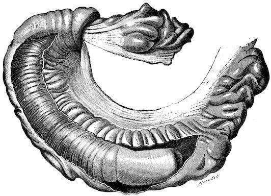
Invagination consists in the passage of one portion of the intestine into the next-following portion. When once the condition has been set up it tends to become aggravated, the invaginated part being
drawn further and further forwards. Invaginations therefore may vary in length between a few inches and sixteen to twenty inches.
Law states (Vol. II. p. 347) that in cattle and swine invagination of the large colon is almost impossible owing to the relation of the bowel with the layers of the mesentery. The anatomical arrangement is opposed to the formation of invagination, yet this accident is not uncommon in cattle and swine. The small intestine can be invaginated into the cæcum or into itself. The cæcum may become invaginated, or it may pass into the colon or rectum.
Cartwright, Veterinarian (1829), reports a case of invagination in a bull calf, and Youatt gives particulars of a similar case which was followed by sloughing and discharge per anum of the intussuscepted portion of bowel. (See also Möller and Dollar’s “Regional Surgery,” p. 328.)
Causation. This variety of colic is due to a number of somewhat obscure causes. In a general sense we may say that anything which increases intestinal peristalsis increases the risk of invagination. The accident may follow intestinal congestion, but is most frequent in animals suffering from intestinal worms, or in animals used for heavy work. Under the influence of violent tractive efforts the peristaltic movements are stimulated, and the intestine being in an oblique position on a plane inclined backwards, the contracted portion may slip into the dilated section behind it.
F��. 68. Invagination of the intestine in an ox (the constricting portion has been incised longitudinally).
Invagination may also occur without any apparent cause, even in animals standing in the stable.
Symptoms. The attack always occurs suddenly, develops rapidly, and is of an extremely grave character.
Colic comes on while the animal is working, moving about, or resting, according to circumstances, and at first resembles that due to congestion. It afterwards becomes very violent; the animals paw, stamp, show great uneasiness, throw themselves violently down, and rise suddenly, only to again lie down as before. The face expresses anxiety, suffering and depression; the tail is often kept lifted, and efforts are continually made to defæcate, mucus being passed. By passing the hand into the rectum the invagination may occasionally be discovered.