No part of this publication may be reproduced or transmitted in any form or by any means, electronic or mechanical, including photocopying, recording, or any information storage and retrieval system, without permission in writing from the publisher. Details on how to seek permission, further information about the Publisher’s permissions policies, and our arrangements with organizations such as the Copyright Clearance Center and the Copyright Licensing Agency can be found at our website: www.elsevier.com/permissions
This book and the individual contributions contained in it are protected under copyright by the Publisher (other than as may be noted herein).
Notice
Practitioners and researchers must always rely on their own experience and knowledge in evaluating and using any information, methods, compounds, or experiments described herein. Because of rapid advances in the medical sciences in particular, independent verification of diagnoses and drug dosages should be made. To the fullest extent of the law, no responsibility is assumed by Elsevier, authors, editors, or contributors for any injury and/or damage to persons or property as a matter of products liability, negligence or otherwise, or from any use or operation of any methods, products, instructions, or ideas contained in the material herein.
Senior Content Development Specialist: Joanie Milnes
Publishing Services Manager: Catherine Albright Jackson
Senior Project Manager: Doug Turner
Designer: Amy Buxton
Printed in China
Last digit is the print number: 9 8 7 6 5 4 3 2 1
CONTRIBUTORS
Megan E. Anderson, MD
Orthopedic Surgeon
Orthopedic Center
Boston Children’s Hospital
Assistant Professor of Orthopedic Surgery
Harvard Medical School
Boston, Massachusetts
Jane S. Chung, MD
Staff Sports Medicine Physician
Department of Orthopaedics
Texas Scottish Rite Hospital for Children
Assistant Professor
Department of Orthopaedic Surgery
University of Texas Southwestern Medical Center Dallas, Texas
Lawson A.B. Copley, MD, MBA
Professor
Departments of Orthopaedic Surgery and Pediatrics
University of Texas Southwestern Medical Center
Pediatric Orthopaedic Surgeon
Department of Orthopaedic Surgery
Children’s Medical Center of Dallas Dallas, Texas
Donald Cummings, CP, LP
Director of Prosthetics
Texas Scottish Rite Hospital for Children Dallas, Texas
Henry Bone Ellis, Jr., MD
Staff Sports Medicine Surgeon
Department of Orthopaedic Surgery
Texas Scottish Rite Hospital for Children
Associate Professor
Department of Orthopaedic Surgery
University of Texas Southwestern Medical Center
Staff Orthopaedic Surgeon
Department of Orthopaedics
Children’s Medical Center
Dallas, Texas
John A. Herring, MD
Chief of Staff Emeritus
Department of Orthopaedic Surgery
Texas Scottish Rite Hospital for Children
Professor
Department of Orthopaedic Surgery
University of Texas Southwestern Medical Center
Dallas, Texas
Christine A. Ho, MD
Staff Orthopaedist
Department of Orthopaedic Surgery
Texas Scottish Rite Hospital for Children
Division Director
Department of Pediatric Orthopaedics
Children’s Health Dallas
Professor
Department of Orthopaedic Surgery
University of Texas Southwestern Medical School
Dallas, Texas
Charles E. Johnston, MD
Chief of Staff Emeritus
Department of Orthopaedic Surgery
Texas Scottish Rite Hospital for Children
Professor
Department of Orthopaedic Surgery
University of Texas Southwestern Medical Center
Dallas, Texas
Lori A. Karol, MD
Chief
Division of Pediatric Orthopaedic Surgery
Rose Brown Chair of Pediatric Orthopaedics
Professor
Department of Orthopedics
University of Colorado School of Medicine
Aurora, Colorado
Harry K.W. Kim, MD
Director
Center for Excellence in Hip Disorders
Texas Scottish Rite Hospital for Children
Professor
Department of Orthopaedic Surgery
University of Texas Southwestern Medical Center
Dallas, Texas
Amy Lake, OTR, CHT
Occupational Therapist
Texas Scottish Rite Hospital for Children Dallas, Texas
Amy L. McIntosh, MD
Associate Professor
Department of Orthopaedic Surgery
Texas Scottish Rite Hospital for Children
Dallas, Texas
Scott Oishi, MD
Director of Hand Service
Texas Scottish Rite Hospital for Children Professor
Department of Orthopaedic and Plastic Surgery
University of Texas Southwestern Medical School Dallas, Texas
David Podeszwa, MD
Pediatric Orthopedic Surgeon
Co-director, Center for Excellence in Limb Lengthening
Texas Scottish Rite Hospital for Children Professor
Department of Orthopaedic Surgery
University of Texas Southwestern Medical Center Dallas, Texas
Marilyn Purano, MD
Professor of Pediatrics
University of Texas Southwestern Medical School Dallas, Texas
Brandon A. Ramo, MD
Staff Orthopaedist
Department of Orthopaedic Surgery
Texas Scottish Rite Hospital for Children Dallas, Texas
Karl E. Rathjen, MD
Assistant Chief of Staff
Department of Orthopaedic Surgery
Texas Scottish Rite Hospital for Children Professor
Department of Orthopaedic Surgery
University of Texas Southwestern Medical Center Dallas, Texas
Anthony I. Riccio, MD
Staff Orthopaedic Surgeon
Department of Orthopaedic Surgery
Texas Scottish Rite Hospital for Children
Professor
Department of Orthopaedic Surgery
University of Texas Southwestern Medical Center Dallas, Texas
B. Stephens Richards, MD
Chief Medical Officer
Texas Scottish Rite Hospital for Children Professor
Department of Orthopaedic Surgery
University of Texas Southwestern Medical Center Dallas, Texas
Mouin G. Seikaly, MD
Medical Director
Metabolic Bone Disease Clinic
Texas Scottish Rite Hospital for Children Professor
Department of Pediatric Nephrology
University of Texas Southwestern Medical Center Dallas, Texas
Chris Stutz, MD
Staff Hand Surgeon Hand Service
Texas Scottish Rite Hospital for Children
Assistant Professor
Department of Orthopaedic Surgery
University of Texas Southwestern Medical Center Dallas, Texas
Daniel J. Sucato, MD Chief of Staff
Department of Orthopaedic Surgery
Texas Scottish Rite Hospital for Children Professor
Department of Orthopaedic Surgery
University of Texas Southwestern Medical Center Dallas, Texas
Philip Wilson, MD
Assistant Chief of Staff
Department of Orthopaedic Surgery
Texas Scottish Rite Hospital for Children Professor
Department of Orthopaedic Surgery
University of Texas Southwestern Medical Center
Dallas, Texas, Assistant Chief of Staff Director, Pediatric Sports Medicine
Department of Sports Medicine and Orthopaedics
Texas Scottish Rite Hospital for Children North Frisco, Texas
Robert Lane Wimberly, MD
Medical Director of Movement Science
Texas Scottish Rite Hospital for Children
Associate Professor
Department of Orthopaedic Surgery
University of Texas Southwestern Medical Center Dallas, Texas
PREFACE
This edition of Tachdjian’s Pediatric Orthopaedics is the fourth that has been written and edited by the staff of the Texas Scottish Rite Hospital for Children. As we research and reevaluate each chapter, we are usually surprised to see how many things have changed between editions. For example, in recent years we have seen considerable growth in the knowledge and practice in the field of pediatric sports subspecialization. In recognition of this growth, we have added a new chapter dedicated to pediatric and adolescent sports conditions, which includes the most recent developments in the management of concussion. In the scoliosis chapter, exciting new information about the rapidly evolving management of early onset scoliosis has been added, including important nonoperative measures such as serial casting and bracing, as well as the use of tethering. The field of genetics is continually expanding, and genetics-related content, which appears in many of our chapters, has been updated to reflect current understanding.
Twenty years ago, we took on the challenge to build on the groundwork laid by Dr. Mirhan Tachdjian in his two editions. Our goal has been to produce a textbook that fully encompasses the broad field of pediatric orthopaedics. We have based our descriptions on the best available published knowledge. We have sought to present the most current evidence from the literature from level 1 to 5 in a succinct and readable format. We have augmented the discussions with recommendations based on personal experiences of a top-level clinical faculty. When presenting controversial topics, we prefer to give the reader the evidence from the different arguments so that the reader can make a reasoned decision after reviewing the conflicting evidence. We carefully avoid the “cookbook” approach in which one puts forth their preferred treatment as gospel.
We continue to insist that our text be comprehensive, even though a shorter text would be more convenient to handle. We fully present each topic, include the description of a disorder, and discuss appropriate history and physical exam, relevant studies, differential diagnosis, and details of treatment. We believe that it is important to present the important details of decision making, and we emphasize the complexity of overall patient care. Our surgical discussions stress proper preoperative planning and preparation, as well as description of operative details. We also provide the important postoperative protocols that are necessary to ensure the best results.
The authors of this book are experienced clinicians with expertise and training in pediatric orthopaedics, and most have subspeciality interests and expertise. They are leaders in their fields and base their discussions and recommendations on a very rich clinical experience in an academic practice. An important feature of our academic environment is the vigorous preoperative group discussion of surgical procedures. As academic leaders, they regularly present their research at national and international meetings. Their work is widely published and broadly respected.
Users of the text include students from all levels from medical school, residency, fellowship, new and established physicians, and non-physician practitioners, as well as established professors as they augment their publications. The text of this edition is fully produced in two print volumes. To lighten the load of the textbook, a comprehensive bibliography is available in the online version. This placement facilitates Internet access to other resources.
As in prior editions, our popular surgical videos are available online. These videos present the important steps of actual surgical cases and are narrated by the operating surgeons. We receive frequent positive comments from surgeons throughout the world who find these very useful for planning their surgical procedures. Other videos in the collection cover non-operative subjects such as cast application for scoliosis, club foot casting, and Pavlik harness application.
I am sincerely grateful to each of our authors and truly appreciate the effort involved in making the sixth edition a reality. We welcome two new authors, Dr. Jane Chung and Dr. Shane Miller, who are pediatric-trained practitioners with expertise in sports medicine. We continue to be grateful for the contribution of our Boston colleagues, Professor Mark Gebhardt and Dr. Megan Anderson, who are responsible for the chapter on malignant tumors. I especially want to thank my administrative assistant, Louise Hamilton, who had the huge task of putting the whole project together. She was able to devote the time needed for this edition because of coverage by the other administrative assistants, including Stacy Duckworth, Lisa Sherman, Rebecca Fuller, Amy Park, and our administrative director, Laura Griffiths. Again, our heartfelt appreciation goes to our families, who are vitally important in every facet of our lives; thanks for giving us your support and understanding.
John A. Herring, MD
CHAPTER 1
Growth and Development John A. Herring
Normal Growth and Development
Neonates are primarily reflexive, but they do exhibit some cognitive traits.8 These traits include showing more curiosity about facelike FIG.s than about other FIG.s of comparable brightness, as well as a preference for black-and-white tones rather than gray. Neonates should turn their eyes toward sound and be able to distinguish their mothers from other people.
This chapter on growth and development is presented first for several important reasons. One of the unique aspects of pediatric care is the dynamic evolution of each individual from neonate to adolescent. During this period, a remarkable process of growth and development takes place in gross and fine motor skills; intellectual, social, and verbal skills; body size; gait; and sexual characteristics.
Growth refers to an increase in an individual’s total body size or to an increase in the physical size of a particular organ or organ system.9,17 References to normal human growth parameters from the third trimester to adulthood are provided in Proceedings of the Greenwood Genetic Center: Growth References.10 This publication also provides parameters for growth patterns seen in specific diseases, such as achondroplasia, diastrophic dysplasia, Down syndrome, Marfan syndrome, and skeletal dysplasias (comparative curves). Growth standards are also available in Hensinger’s Standards in Pediatric Orthopedics.7
Development refers to the physical changes of maturation that occur as a child ages. The developmental process encompasses other aspects of differentiation of form, but it primarily involves changes in function that transform humans into increasingly more complex beings.9 Development is influenced by many interrelated factors, including genetics, physical trauma, nutrition, and socioeconomic status.17
The age at which children reach specific milestones of development depends on the maturation rate of their central nervous system (CNS), which varies from child to child. Ranges for variations in normal have been developed to assist in the assessment of the pediatric patient, and the most commonly used assessment tool is the revised Denver Developmental Screening Test (DDST)(Fig. 1.1).5-7 It is important to know when a child should normally achieve expected milestones of growth and development so that potentially abnormal situations are evident to the physician who is taking a patient’s history and performing a physical examination.
The significance of various findings must be related to the child’s particular stage of growth and development. Although no one should expect a 4-month-old infant to be walking, it is distinctly abnormal for an 18-month-old child not to be doing so. Similarly, a 12-month-old child is likely to have some degree of genu varum, whereas the presence of genu varum in a 3-year-old child should be cause for concern and a focus of further investigation.
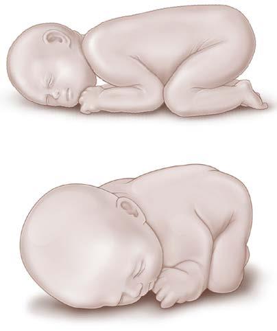
The normal neonate is born with a predominant flexor tone, and physiologic flexion contractures are typical (Fig. 1.2). At birth the newborn’s limbs are maintained in flexion posture, and passive movement of the extremities and neck elicits strong flexor tone. A normal neonate’s limbs move in an alternating fashion when they are stimulated.
Normal development progresses cephalocaudad; infants acquire the ability to control their head and hands before they are able to control their legs.8 During the first few months, gaining head control predominates. Hand control, such as the ability to grasp objects, follows. As development continues, the infant gains more and more control of the legs.
To determine whether an infant’s growth and development are progressing normally, the examiner needs to find out from the parents what developmental milestones the child has attained and when and then compare them with the norms. If the child appears to have developmental delays, referral to a physician who specializes in growth and development problems is recommended.
Because of the wide variations in the times at which developmental milestones are achieved and the numerous reasons for delays, the diagnosis of developmental delay can be difficult to make in the very young child. In addition, a child may exhibit delay in acquiring certain skills and unusual rapidity in acquiring others. When a delay is evident, the physician must determine the cause, which may be a neuromuscular condition. Factors suggesting a neurologic cause include failure of normal developmental responses to appear, prolonged retention of primitive infant reflexes, or a delay in achieving gross motor milestones within normal limits.
Disorders of Normal Growth and Development
Many pediatric orthopaedic problems result from disorders or conditions that adversely affect normal growth and development. The four major failures of normal growth and development are malformations, deformations, disruptions, and dysplasias.4,12
Malformations
Malformations are structural defects that result from interruption of normal organogenesis during the second month of
Name:
Birthdate: ID#:
FIG. 1.1 The revised Denver Developmental Screening Test showing the range of age when a child should achieve milestones in the development of gross motor skills, fine motor–adaptive skills, language, and personal-social skills. (Modified from Frankenburg WK, Dodds JB. The Denver Developmental Screening Test. J Pediatr. 1967;71:181; Hensinger RN. Standards in Pediatric Orthopedics. New York: Raven Press; 1986.)
gestation. Examples include myelomeningocele, syndactyly, preaxial polydactyly, Poland syndrome, and proximal focal femoral deficiency (congenital femoral deficiency).
Deformations
Deformations are defects in the form, shape, or site of body parts caused by mechanical stress. The mechanical stress, which may be intrinsic or extrinsic, alters or distorts tissues. Because the fetus grows considerably faster than the infant, fetuses are more vulnerable to deformations. Examples include supple metatarsus adductus, calcaneovalgus feet, congenital knee hyperextension, and physiologic bowing of the tibia.
Differentiating deformations from malformations is important. During a cursory examination, severe deformations may look like malformations.3 Careful assessment is essential if the child is to receive appropriate care for the condition. Malformations cannot be corrected directly, whereas deformations can often be reversed relatively easily
FIG. 1.2 Typical position of the neonate with vertex presentation. The hips and knees are flexed, the lower legs are rotated internally, and the feet are rotated further inward on the lower leg. The lower limbs are contracted into this position for a variable period after birth.
either by eliminating the deforming force or by counteracting the force with stretching, casting, or bracing.
Disruptions
Disruptions are morphologic abnormalities that result from an extrinsic interference with or breakdown of the normal growth and development process. Disruptions can be caused by drugs or toxic materials. These structural defects may affect organs or systems that were normal during organogenesis. A congenital constriction band in the limb is an example of a disruption.
Dysplasias
Dysplasias are structural defects caused by abnormal tissue differentiation as cells organize into tissues. Examples include osteogenesis imperfecta, achondroplasia, and spondyloepiphyseal dysplasia.
Evolution of Proportionate Body Size
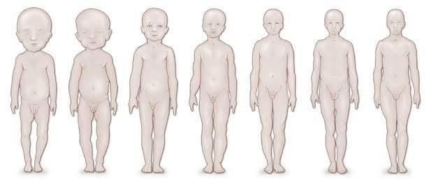
At birth, the neonate’s head is disproportionately large, comprising approximately one-fourth of the body’s total length. During the first year of infancy, the head continues to grow rapidly, and the head circumference usually is greater than the circumference of the infant’s chest. The evolution of body proportions is indicated by a change in the child’s upper to lower segment ratio (the relation of the center of gravity to body segments). This ratio is measured as the distance from the top of the head to the symphysis pubis, divided by the distance from the symphysis pubis to the bottom of the feet (Fig. 1.3).7 At birth, the ratio is approximately 1:7. At approximately 10 years of age, the upper and lower segments are almost equal in length (i.e., the ratio is ≈1.0). After 10 years of age, as individuals become adolescents and adults, the ratio normally becomes less than 1.0, as the upper segment becomes shorter than the lower segment.
Physical Growth
Head Circumference
During infancy it is essential to obtain individual or serial measurements of the patient’s head circumference to determine whether head growth is slower or faster than normal.
FIG. 1.3 Evolution of head-to-trunk proportion throughout growth. In the neonate the head is proportionately significantly larger relative to the trunk than it will be at skeletal maturity. (Reproduced from Hensinger RN. Standards in Pediatric Orthopedics. New York: Raven Press; 1986.)
Head circumference should be measured at every physical examination during the first 2 years and at least biennially thereafter. With the child supine, the examiner places a centimeter tape over the occipital, parietal, and frontal prominences of the head. The tape should be stretched and the reading noted at the point of greatest circumference. Possible conditions that can affect head circumference and growth include microcephaly, premature closure of the sutures, hydrocephalus, subdural hematoma, and brain tumor. Head circumference should be charted for age and percentile, as noted in Fig. 1.4
Height and Weight
A child’s growth, as demonstrated by an increase in body height and weight within predetermined normal limits, is one of the best indicators of health during infancy and childhood. The child’s height and weight should be plotted on a standard growth chart to verify that normal progress is being made. Numerous tables, charts, and graphs depicting pediatric growth standards are available in Hensinger’s Standards in Pediatric Orthopedics7 and in Proceedings of the Greenwood Genetic Center: Growth References.10 The World Health Organization published an extensive study of child growth standards for length and height for age, weight for age, weight for length, weight for height, and body mass index for age.18 Height and weight should be charted for age and percentile, as noted in Fig. 1.5. If growth measurements are lower than the 3rd percentile or higher than the 97th percentile, or if a recent deviation from previously stable percentile rankings is noted, further investigation is warranted.
Epiphyseal Growth and Closure
During normal growth and development, the pattern in the appearance of centers of ossification and fusion of epiphyses in the upper and lower limbs is orderly. This pattern varies among individuals and is different for boys and girls (Figs. 1.6–1.9). Thus the orthopaedist must understand the ranges of normal when treating the pediatric patient, particularly when interpreting radiographs. The percentage contribution of each epiphysis to longitudinal growth of the upper and lower extremity long bones is shown in Figs. 1.10 and 1.11
Tanner Stages of Development
The physical maturation of a child can also be compared with his or her chronologic age by using the pubertal stages of development as described by Tanner (Figs. 1.12 and 1.13).15,16 The Tanner stages of maturation are based on breast size in girls, genital size in boys, and pubic hair stages for both girls and boys. The onset of menstruation is also an important milestone in the physical maturation of girls.
Developmental Milestones
Gross Motor Skills
The development of gross motor skills depends on maturation of the CNS, which proceeds in a cephalocaudal direction.8
The approximate ages at which children should normally attain various gross motor skills are given in Table 1.1
By 3 months of age, infants should be able to hold their heads above the plane of the body when they are supported in a prone position. By 6 months of age, the head should not lag when infants are pulled from a supine to a sitting position. Normally, infants will begin to roll over between 4 and 6 months of age and can sit with minimal external support at 6 to 7 months. They should be able to pull up to a standing position by holding onto furniture at 9 to 12 months and stand without support by 14 months.
The average milestones of development of locomotion are as follows: the infant should be able to crawl by 7 to 9 months of age, cruise and walk with assistance at 12 months, walk forward without support by 12 to 16 months, and run at 18 months of age.1,2,11 Children should be able to ascend stairs with support by 18 months of age and without support by 2 years of age. They should be able to descend stairs with support at approximately 3 years of age and without support by 4 years.
On gross inspection the independent gait of the infant has a wide base, the hips and knees are hyperflexed, the arms are held in flexion, and the movements are abrupt. With maturation of the neuromuscular system, the width of the base gradually diminishes, the movements become smoother, reciprocal swing of the upper limbs begins, and step length and walking velocity increase.13 The adult pattern of gait develops between 3 and 5 years of age.14 A more complete description of normal pediatric gait patterns is provided in Chapter 5
Fine Motor Skills
The approximate ages at which children normally attain various fine motor skills are listed in Table 1.2. A child’s exploration of the environment by touch and the development of manual skills should emerge in an orderly and sequential manner. At 3 months of age, infants can apply lip pressure and coordinate sucking and swallowing during feeding (the sucking reflex is present at birth in all normal full-term neonates but usually disappears at 3–4 months of age). By 6 months of age, children are able to feed themselves from hand to mouth. By 9 months, children can feed themselves food such as cookies. By 12 months of age, children can pick up a spoon from the table, chew cookies or toast, and drink milk from a cup if assisted. Between 12 and 18 months, they are able to feed themselves (messily) with a spoon and drink from a cup by using one or two hands. By 24 months, they can feed themselves semisolid food with a spoon and drink holding the cup in one hand or using a straw.
Children should be able to purposefully grasp objects such as a bottle or toy rattle by 6 months of age. At 9 months of age, children use their fingers and thumb to grasp objects and are able to transfer objects from one hand to the other. By 12 months, children’s hand skills are such that they are able to hit two objects together, voluntarily release objects, manipulate and throw objects on the floor, and hold crayons and imitate scribbling. Between 18 and 24 months of age, their hand skills evolve to the point that they can build block towers, turn pages one at a time, and throw a ball (but inaccurately). Between 2 and 3 years of age, their writing skills evolve from imitating vertical, horizontal, and circular strokes to copying circles.
FIG. 1.4
the National Center for Health Statistics.)
FIG. 1.4, cont’d (B) Girls, birth to 36 months. NCHS, National Center for Health Statistics. (From
FIG. 1.5 Normal length and weight parameters for boys and girls from birth to 18 years. (A) Boys, birth to 36 months. Continued
FIG. 1.5, cont’d (B) Girls, birth to 36 months.
Boys: 2 to 18 years
FIG. 1.5, cont’d (C) Boys, 2 to 18 years.
Another random document with no related content on Scribd: