List of Contributors
The editors would like to acknowledge and offer grateful thanks for the input of all previous editions’ contributors, without whom this new edition would not have been possible.
Anthony Abdullah, BSc (Hons), MB, ChB (Hons), FRCP (London), DTM&H Consultant Dermatologist
The Birmingham Skin Centre and the Birmingham Regional Skin Laser Centre Sandwell and West Birmingham Hospitals NHS Trust Birmingham, UK
Mary Kathryn Abel, AB Medical Student
University of California, San Francisco School of Medicine San Francisco, CA, USA
Boya Abudu, MD, MPH Resident Physician Department of Dermatology University of California, Davis Sacramento, CA, USA
Mohannad Abu-Hilal, MD, MRCP Assistant Professor Division of Dermatology, Department of Medicine
McMaster University Hamilton, ON, Canada
Anwar Al Hammadi, MD, FRCPC, FAAD Consultant Dermatologist DermaMed Clinic
Adjunct Clinical Associate Professor of Dermatology
Mohammed Bin Rashid University of Medicine and Health Sciences Dubai, UAE
Ameen Al Awadhi, MD
International Dermatopathology Fellow Department of Dermatology
Mount Sinai Hospital New York New York, NY, USA
Andrew F. Alexis, MD, MPH Vice-Chair for Diversity and Inclusion Department of Dermatology
Weill Cornell Medicine New York, NY, USA
Khadija Aljefri, MBChB, MSc, MRCP(UK), CCT(UK)
Consultant Dermatologist
Dermatology Department DermaMed Clinic Dubai, UAE
Huda Al-Kutubi, MBBCh, MSc (Dist), MRCP Clinical Fellow Dermatology
The Birmingham Skin Centre
Sandwell and West Birmingham Hospitals
NHS Trust Birmingham, UK
Timothy H. Almazan, MD
Resident Physician
Department of Dermatology
Stanford University School of Medicine Stanford, CA, USA
Amer Ali Almohssen, MD, FAAD
Dermatologist & Dermatopathologist
Department of Dermatology
Dammam Medical Complex
Al Ahasa, Saudi Arabia
Erkan Alpsoy, MD, MPhil Professor
Department of Dermatology and Venereology
Akdeniz University School of Medicine Antalya, Turkey
Mahreen Ameen
Consultant Dermatologist
Department of Dermatology
Royal Free Hospital NHS Foundation Trust London, UK
Bryan E. Anderson, MD
Professor of Dermatology Department of Dermatology
Penn State Hershey Medical Center Hershey, PA, USA
Ellen Anshelevich, BA
Research Assistant Department of Dermatology University of Pennsylvania School of Medicine Philadelphia, PA, USA
Lindsay C. Bacik, MD Department of Dermatology
Penn State Hershey Medical Center Hershey, PA, USA
Donald J. Baker, MD
Clinical Associate Professor Division of Dermatology
Cooper Medical School of Rowan University Camden, NJ, USA
David Banach, MD, MPH
Associate Professor of Medicine
Division of Infectious Diseases
Department of Medicine
University of Connecticut School of Medicine
Farmington, CT, USA
Cedric C. Banfield, MB, FRCP
Consultant Dermatologist
Department of Dermatology
Peterborough City Hospital
North West Anglia NHS Foundation Trust Cambridgeshire, UK
Robert Baran, MD
Honorable Professor
Nail Disease Center
Private Practice Cannes, France
Benjamin Barankin, MD, FRCPC, FAAD
Dermatologist, Medical Director & Founder
Toronto Dermatology Centre
Toronto, ON, Canada
Melissa C. Barkham
Consultant Dermatologist
Dermatology Department
Ashford and St Peters NHS Foundation Trust
Surrey, UK
Johann W. Bauer, MD, MBA
Chairman
Department of Dermatology and Allergology
University Hospital of the Paracelsus
Medical University
Salzburg, Austria
Zoe Begun
Medical Student Department of Dermatology
Cooper University Hospital Camden, NJ, USA
Michael A. Bell, MSc
Dean’s Year Scholar-in-Residence Department of Dermatology
Johns Hopkins University School of Medicine Baltimore, MD, USA
Emma Benton, FRCP
Consultant Dermatologist
St. John’s Institute of Dermatology
Guy’s and St. Thomas’ NHS Trust London, UK
Wilma F. Bergfeld, MD, FAAD
Professor of Dermatology and Pathology
Senior Dermatologist & Emeritus Director
Dermatopathology
Director, Dermatopathology Fellowship
Departments of Dermatology and Pathology
Cleveland Clinic Cleveland, OH, USA
Brian Berman, MD, PhD
Professor Emeritus
Department of Dermatology and Cutaneous Surgery
University of Miami Miller School of Medicine
Miami, FL, USA
Co-Director
Center for Clinical and Cosmetic Research
Skin and Cancer Associates Aventura, FL, USA
John Berth-Jones, FRCP
Consultant Dermatologist
Department of Dermatology University Hospital Coventry, UK
Chinmoy Bhate, MD, FRCP (Edin)
Clinical Assistant Professor Dermatology, Pathology & Laboratory
Rutgers New Jersey Medical School Newark, NJ, USA
Clinical Assistant Professor Dermatology
University of Pittsburgh School of Medicine Pittsburgh, PA, USA
Bhavnit K. Bhatia, MD
Dermatologist
Department of Dermatology
The Permanente Medical Group Richmond, CA, USA
Asli Bilgiç, MD
Assistant Professor
Dermatology and Venereology Department University of Akdeniz, School of Medicine Antalya, Turkey
Avi Bitterman, MD
Clinical Research Fellow
Department of Dermatology
Icahn School of Medicine at Mount Sinai New York, NY, USA
Samantha Black, BS
Medical Student
Department of Dermatology
University of Texas Southwestern Medical Center Dallas, TX, USA
Jonathan E. Blume, MD
Clinical Instructor of Dermatology Department of Dermatology
Columbia University College of Physicians and Surgeons New York, NY, USA
Gérôme Bohelay, MD
Hospital Practitioner
Reference Center for Autoimmune Bullous Diseases
Groupe Hospitalier Paris Seine-Saint-Denis Bobigny, France
Nevianna Bordet, MB ChB, FRCP
Consultant Dermatologist
Department of Dermatology
Hinchingbrooke Hospital North West Anglia NHS Foundation Trust Huntingdon, UK
Robert T. Brodell, MD, FAAD
Tenured Professor and Chair, Department of Dermatology
Professor of Pathology
Billy S. Guyton Distinguished Professor University of Mississippi Medical Center Jackson, Mississippi, USA
Marc D. Brown, MD
Professor of Dermatology and Oncology
Director, Division of Mohs Surgery and Cutaneous Oncology, Department of Dermatology
University of Rochester Medical Center Rochester, NY, USA
Nicholas D. Brownstone, MD
Clinical Research Fellow
Department of Dermatology
Psoriasis and Skin Treatment Center
University of California, San Francisco San Francisco, CA, USA
Anne E. Burdick, MD, MPH
Professor Emeritus of Dermatology
University of Miami Miller School of Medicine Miami, FL, USA
Niraj Butala, MD
Associate Physician
Department of Dermatology Kaiser Permanente Lancaster, CA, USA
Jeffrey P. Callen, MD
Professor of Medicine (Dermatology); Chief, Division of Dermatology
Division of Dermatology, Department of Medicine
University of Louisville Louisville, KY, USA
Ivan D. Camacho, MD
Voluntary Assistant Professor
Dr. Phillip Frost Department of Dermatology and Cutaneous Surgery
University of Miami Miller School of Medicine Miami, FL, USA
Helena Camasmie, MD
Department of Dermatology
Federal University of the State of Rio de Janeiro (UNIRIO) Rio de Janeiro, Brazil
Daniel Caplivski, MD
Director, Travel Medicine Program
Professor, Division of Infectious Diseases
Icahn School of Medicine at Mount Sinai New York, NY, USA
Mitchell S. Cappell, MD, PhD Professor of Gastroenterology & Hepatology Division of Gastroenterology & Hepatology
William Beaumont Hospital at Royal Oak and Oakland University William Beaumont School of Medicine
Royal Oak, MI, USA
Genevieve A. Casey, MBBS, FACD
Consultant Dermatologist
Department of Paediatrics and Dermatology
Queensland Children’s Hospital South Brisbane, Queensland, Australia
Frédéric Caux, MD, PhD
Head of the Department of Dermatology
Reference Center for Autoimmune Bullous Diseases
Groupe Hospitalier Paris Seine-Saint-Denis
Bobigny, France
Cynthia X. Chan, BS
Department of Dermatology
Mount Sinai Hospital New York, NY, USA
Lawrence S. Chan, MD, MHA
Dr. Orville J. Stone Professor of Dermatology
Department of Dermatology
University of Illinois College of Medicine Chicago, IL, USA
Loi-Yuen Chan, MBBS, MRCPI, DIP Derm (London), FHKAM
Private Practice
Hong Kong, China
Stephanie Chan, BS
Clinical Research Assistant
Department of Dermatology
University of California, San Francisco San Francisco, CA, USA
Kevin Young Chao, MD
Dermatology Resident Department of Dermatology
University of California, San Diego San Diego, CA, USA
Jennifer K. Chen, MD
Clinical Assistant Professor, Dermatology
Stanford University School of Medicine Redwood City, CA, USA
Fiona J. Child, BSc, MD, FRCP
Consultant Dermatologist
St. John’s Institute of Dermatology
Guy’s and St. Thomas’ NHS Foundation Trust
London, UK
Keith Choate, MD, PhD
Professor
Departments of Dermatology, Genetics, and Pathology
Yale University School of Medicine
New Haven, CT, USA
Olivier Chosidow, MD, PhD
Professor
Department of Dermatology
University of Paris-est Créteil Créteil, France
Emmanouil Chousakos, MD, PhD(c) Research Fellow
Dermatology Service
Memorial Sloan Kettering Cancer Center, New York
New York, NY, USA
Antonio A.T. Chuh, MD, DSc, FRCP
Honorary Clinical Professor
Jockey Club School of Public Health and Primary Care
The Chinese University of Hong Kong and Prince of Wales Hospital Hong Kong
Ahuva D. Cices, MD Research Fellow Department of Dermatology Icahn School of Medicine at Mount Sinai New York, NY, USA
Gabriela A. Cobos, MD Assistant Program Director, DermatologyRheumatology Fellowship Director, Connective Tissue Diseases Clinic Department of Dermatology
Brigham and Women’s Hospital, Harvard Medical School Boston, MA, USA
Steven R. Cohen, MD, MPH Professor and Chief Division of Dermatology, Department of Medicine
Albert Einstein College of Medicine Bronx, NY, USA
Nicholas Collier, BMBS, MSc, MRCP (Derm), MMedEd
Consultant Dermatologist & Mohs Surgeon Department of Dermatology
Salford Royal Foundation Trust Manchester, UK
Elizabeth A. Cooper, BESc, HBSc Senior Research Assistant Clinical Trials Mediprobe Research Inc. London, ON, Canada
Ian H. Coulson, BSc, MB, FRCP Consultant Dermatologist East Lancashire NHS Trust Lancashire, UK
Edward W. Cowen, MD, MHSc Head, Dermatology Consultation Service Dermatology Branch
National Institute of Arthritis and Musculoskeletal and Skin Diseases
National Institutes of Health Bethesda, MD, USA
Brittany Craiglow, MD
Associate Professor Adjunct Department of Dermatology
Yale University School of Medicine New Haven, CT, USA
Ponciano D. Cruz Jr., MD
Endowed Chair in Honor of Paul R. Bergstresser MD
Distinguished Professor and Vice Chair Department of Dermatology
The University of Texas Southwestern Medical Center Dallas, TX, USA
Donna Cummins, MD, MRCPI, MRCP (Derm) Consultant Dermatologist
The Dermatology Centre
Salford Royal NHS Foundation Trust
Salford, Greater Manchester, UK
Carrie Ann R. Cusack, MD, FAAD Dermatologist/Dermatopathologist
Dermatology Associates of South Jersey
Mount Laurel, NJ, USA
Benjamin S. Daniel, MBBS, BA, BCom, MMed (Clin Epi), FACD Dermatologist
St George Hospital
University of New South Wales Sydney, Australia
Adam R.A. Daunton, MBChB, MRCP (Derm), BSc
Consultant Dermatologist
Salford Royal Hospital NHS Foundation Trust
Greater Manchester, UK
Mark D.P. Davis, MD
Professor & Chair
Department of Dermatology
Mayo Clinic Rochester Rochester, MN, USA
Robert S. Dawe, MBChB, MD, FRCPE Consultant Dermatologist and Honorary Reader
Ninewells Hospital and Medical School
NHS Tayside Dundee, UK
David P. D’Cruz, MD, FRCP Consultant Rheumatologist
The Louise Coote Lupus Unit Guy’s Hospital London, UK
David de Berker, MBBS, MRCP Consultant Dermatologist
Bristol Dermatology Centre
University Hospitals Bristol and Weston Bristol, UK
Marianne de Brito, BA, BM BCh, MRCP Dermatology Registrar Department of Dermatology
Burnley General Hospital Burnley, UK
Hannelore De Maeseneer, MD
Pediatrician Department of Pediatrics
O.L. Vrouw Hospital Aalst, Belgium
Ellen H. de Moll, MD
Fellow
Department of Dermatology and Dermatopathology
Icahn School of Medicine at Mount Sinai New York, NY, USA
Ashley Decker, MD
Assistant Professor of Medicine
Micrographic Surgery and Dermatologic Oncology
Cooper University Hospital – Cooper Medical School, Rowan University
Marlton, NJ, USA
Danielle M. DeHoratius, MD
Clinical Associate Professor of Dermatology
Department of Dermatology
Hospital of the University of Pennsylvania Philadelphia, PA, USA
James Q. Del Rosso, DO Research Director
JDR Dermatology Research
Las Vegas, NV, USA
Seemal R. Desai, MD, FAAD
Founder & Medical Director
Innovative Dermatology
Plano, TX, USA
Clinical Assistant Professor Department of Dermatology
The Univ of Texas Southwestern Medical Center Dallas, TX, USA
Gaurav K. Dhawan, BMBS, BMedSci, MRCP (Derm)
Dermatology Registrar Department of Dermatology
Salford Royal NHS Foundation Trust Manchester, UK
John J. DiGiovanna, MD
Senior Research Physician
DNA Repair Section
Laboratory of Cancer Biology and Genetics
Center for Cancer Research
National Cancer Institute
National Institutes of Health Bethesda, MD, USA
Roni P. Dodiuk-Gad, MD
Vice Head
Dermatology Department
Emek Medical Center
Afula, Israel
Clinical Assistant Professor
Bruce Rappaport Faculty of Medicine, Technion – Institute of Technology
Israel
Assistant Professor - status only
Department of Medicine University of Toronto Toronto, ON, Canada
Robert F. Duffy, MD
Dermatology Resident Division of Dermatology
Cooper University Health Care Camden, NJ, USA
Reinhard Dummer, MD
Professor and Vice Chair Department of Dermatology University Hospital Zurich Zurich, Switzerland
Annie Dunn, MD
Dermatopharmacology Fellow
Department of Dermatology
Icahn School of Medicine at Mount Sinai New York, NY, USA
Tu-Anh Duong, MD PhD
Dermatologist Attending Department of Dermatology
Hôpital Henri Mondor, AP-HP Créteil, France
Jan P. Dutz, MD, FRCPC
Head Department of Dermatology and Skin Science
University of British Columbia Vancouver, BC, Canada
Dawn Z. Eichenfield, MD, PhD
Assistant Clinical Professor Department of Dermatology
University of California, San Diego School of Medicine and Rady Children’s Hospital, San Diego San Diego, CA, USA
Lawrence F. Eichenfield, MD
Professor of Dermatology and Pediatrics Chief, Pediatric and Adolescent Dermatology
University of California, San Diego School of Medicine and Rady Children’s Hospital, San Diego San Diego, CA, USA
Drore Eisen, MD, DDS Dermatologists of Central States Cincinnati, OH, USA
Scott A. Elman, MD
Dermatology and Internal Medicine
Resident Harvard Combined Residency Program in Medicine and Dermatology Boston, MA, USA
Dirk M. Elston, MD
Professor and Chairman Department of Dermatology and Dermatologic Surgery
Medical University of South Carolina Charleston, SC, USA
Sarah Elyoussfi, BSc, MBBS, MRCP, PGDip Internal Medicine Trainee
Manchester University NHS Foundation Trust Manchester, UK
Patrick O.M. Emanuel, MB ChB FRCPA
Director
Division de Dermatopatologia
Clinica Ricardo Palma Lima, Peru
Clinton W. Enos, MD, MS
Eastern Virginia Medical School Norfolk, VA, USA
Maria Rosa Noliza Encarnacion, MD, MBA Clinical Fellow
Department of Dermatology
Icahn School of Medicine at Mount Sinai New York, NY, USA
Ervin H. Epstein Jr., MD Consultant
PellePharm
San Francisco, CA, USA
Aaron S. Farberg, MD
Dermatologist
Division of Dermatology
Baylor University Medical Center Dallas, TX, USA
Michael E. Farrugia, MD, MRCP (Derm), PgDip Consultant Dermatologist
Department of Dermatology
University of Edinburgh Scotland, UK
Hiva Fassihi, MA, MD, FRCP Consultant Dermatologist
Photodermatology Unit
St John’s Institute of Dermatology
Guy’s and St. Thomas’ Hospitals NHS Foundation Trust London, UK
Bilal Fawaz, MD
Dermatology Resident
Division of Dermatology
Cooper Medical School of Rowan University Camden, NJ, USA
Lawrence S. Feigenbaum, MD, FAAD Dermatologist
Mid-County Dermatology St. Louis, MO, USA
Claire Felmingham, MBBS (Hons), BMedSc (Hons)
Dermatology Research Fellow
Occupational Dermatology Research and Education Centre
Skin Health Institute, Carlton Melbourne, Victoria, Australia
Alisa Femia, MD
Associate Professor
Director of Autoimmune Connective Tissue Disease
Director of Inpatient Dermatology
The Ronald O. Perelman Dept. of Dermatology
NYU Grossman School of Medicine
New York, NY, USA
Justine Fenner, MD
Dermatology Resident
Department of Dermatology
Icahn School of Medicine at Mount Sinai New York, NY, USA
Felicity Ferguson, MBChB
Consultant Dermatologist
St. John’s Institute of Dermatology
Guy’s and St. Thomas’ NHS Foundation Trust London, UK
Nicole Fett, MD, MSCE Professor of Dermatology
Dermatology Residency Program Director
Oregon Health & Science University Portland, OR, USA
Andrew Y. Finlay, CBE, MB BS, FRCP Professor of Dermatology
Division of Infection and Immunity
Cardiff University School of Medicine Cardiff, UK
Alireza Firooz, MD
Prof. of Dermatology
Director, Center for Research & Training in Skin Diseases & Leprosy Director, Clinical Trial Center
Tehran University of Medical Sciences Tehran, Iran
Bahar F. Firoz, MD, MPH
Dermatologist
Dermatology Surgery Fellow
Derm Surgery Associates Houston, TX, USA
Amy E. Flischel, MD, FAAD
Associate Professor
Department of Dermatology
University of Mississippi Medical Center Jackson, MS, USA
Amy Forrestel, MD
Dermatology/Internal Medicine
University of Pennsylvania Philadelphia, PA, USA
Kelly A. Foley, PhD
Medical Research Associate
Dermatology
Mediprobe Research Inc. London, ON, Canada
Kayla Fourzali, MD, MS
Research Fellow
Miami Itch Center
Dr Phillip Frost Department of Dermatology & Cutaneous Surgery
University of Miami Miller School of Medicine Miami, FL, USA
Cindy P. Frare, MD
Dermatologist
Department of Dermatology
Hospital Aleman
Ciudad de Buenos Aires
Buenos Aires, Argentina
Derek Freedman, MD, FRCPI
Consultant Genitourinary Physician
Ranelagh Village Dublin, Ireland
Surina Frey, MD
Dermatology Resident Department of Dermatology and Allergy Ludwig Maximilian University Hospital Munich, Germany
Richard Fried, MD, PhD
Clinical Director
Dermatology
Yardley Dermatology and Yardley Clinical Research Associates Yardley, PA, USA
Adam J. Friedman, MD
Professor and Chair of Dermatology Residency Program Director Department of Dermatology
GW School of Medicine and Health Sciences Washington, DC, USA
L. Claire Fuller, MA, FRCP Consultant Dermatologist Department of Dermatology
Chelsea and Westminster NHS Foundation Trust London, UK
Colleen Gabel, MD Research Fellow Department of Dermatology Massachusetts General Hospital Boston, MA, USA
Joanna E. Gach, FRCP Consultant Dermatologist Department of Dermatology University Hospital Coventry, Warwickshire, UK
Jaya Ganesh, MD
Associate Professor Department of Genetics and Genomic Sciences
Icahn School of Medicine at Mount Sinai New York, NY, USA
Shikhar Ganjoo, MD, DNB, MRCP (SCE) Associate Professor Dermatology & STD
Shree Guru Gobind Singh Tricentenary University, Faculty of Medical & Health Sciences Gurugram, India
Jing Mia Gao, MA (Cantab), MBBS, MRCP (Derm)
Dermatology Specialist Registrar Department of Dermatology Royal Free London NHS Foundation Trust London, UK
Anthony A. Gaspari, MD Professor Department of Dermatology and Cutaneous Biology
Sidney Kimmel Medical College at Thomas Jefferson University Philadelphia, PA, USA
Katherine Glaser, MD
Dermatology Chief Resident Department of Dermatology
Cleveland Clinic Foundation Cleveland, OH, USA
Yamila Goenaga-Vázquez, MD
PGY-2 Dermatology Resident Department of Dermatology
University of Texas, Health Science Center at Houston Houston, TX, USA
Leonard H. Goldberg, MD, FRCP
Director and Chief of Surgery
DermSurgery Associates, P.A Greenpark One Medical Professional Building Houston, TX, USA
Johanna Goldfarb Staff Physician
Department of Infectious Diseases Cleveland Clinic Foundation Cleveland, OH, USA
Reina M. González Barreto, MD
Internal Medicine Senior Resident Department of Medicine
Mount Sinai Beth Israel New York, NY, USA
Peter K.C. Goon, PhD, FRCP (Lond)
Consultant Dermatologist and Clinical Lead Department of Dermatology
Peterborough City Hospital North West Anglia NHS Foundation Trust Cambridgeshire, UK
Alice B. Gottlieb, MD, PhD
Clinical Professor and Medical Director
Icahn School of Medicine at Mount Sinai
Mount Sinai Union Square
New York, NY, USA
Asha Gowda, MD, MS
Resident Physician
Department of Dermatology
Warren Alpert Medical School of Brown University, Rhode Island Hospital Providence, RI, USA
Ayman A. Grada, MD, MS
Dermatologist
Department of Dermatology
Boston University School of Medicine Boston, MA, USA
Donald J. Grande, MD, FACMS
Clinical Professor
Department of Dermatology
Boston University School of Medicine Boston, MA, USA
Matthew Grant, MD
Assistant Professor of Medicine
Department of Internal Medicine
Section of Infectious Diseases
Yale School of Medicine
New Haven, CT, USA
Clive E.H. Grattan, MA, MD, FRCP
Consultant Dermatologist
St John’s Institute of Dermatology
Guy’s Hospital London, UK
Justin J. Green, MD
Assistant Professor Department of Internal Medicine, Division of Dermatology
Cooper Medical School of Rowan University Camden, NJ, USA
Pearl E. Grimes, MD
Director
Vitiligo and Pigmentation Institute of Southern California
Clinical Professor
Division of Dermatology
David Geffen School of Medicine
University of California
Los Angeles, CA, USA
Charles A. Gropper, MD
Associate Clinical Professor of Dermatology
Icahn School of Medicine at Mount Sinai
Clinical Affiliate Professor of Medicine
CUNY School of Medicine
Chief of Dermatology Saint Barnabas Health System
New York, NY, USA
Anna L. Grossberg, MD
Assistant Professor
Departments of Dermatology and Pediatrics
The Johns Hopkins University School of Medicine Baltimore, MD, USA
Aditya K. Gupta, MD, PhD, MBA, FRCPC Professor of Dermatology
Faculty of Medicine
University of Toronto Toronto, ON, Canada
Emma Guttman-Yassky, MD, PhD
Waldman Professor and System Chair
The Kimberly and Eric J. Waldman
Department of Dermatology
Icahn School of Medicine at Mount Sinai New York, NY, USA
Anthony K. Guzman, MD
Chief Resident
Department of Internal Medicine, Division of Dermatology
Albert Einstein College of Medicine Bronx, NY, USA
Nazgol-Sadat Haddadi, MD-MPH
Visiting Scholar
Department: Dermatology
University of Massachusetts Medical School Worcester, MA, USA
Ali S. Hadi, MD
Chief Resident Department of Dermatology
Icahn School of Medicine at Mount Sinai New York, NY, USA
Suhail M. Hadi, MD, MPhil, FRCP, FAAD
Associate Professor
Department of Dermatology
Icahn School of Medicine at Mount Sinai New York, NY, USA
Iris A. Hagans, MD
Assistant Professor of Medicine Department of Medicine Cooper Medical School of Rowan University Camden, NJ, USA
Bethany R. Hairston, MD Dermatologist
Vitality Dermatology Columbus, MS, USA
Analisa Vincent Halpern, MD, FAAD
Associate Professor Department of Dermatology University of Pennsylvania Perelman School of Medicine Philadelphia, PA, USA
Caroline Halverstam, MD, FAAD
Associate Professor of Medicine (Dermatology)
Division of Dermatology Albert Einstein College of Medicine Bronx, NY, USA
Emily Haque, BSA
Medical Student
Department of Medical Education
Texas A&M University College of Medicine Dallas, TX, USA
Matthew Harries, MB ChB, PhD Consultant Dermatologist
The Dermatology Centre
Salford Royal NHS Foundation Trust
Salford, UK
Honorary Senior Lecturer Dermatological Sciences
The University of Manchester Salford, Greater Manchester, UK
John E. Harris, MD, PhD Professor and Chair, Department of Dermatology Director, Vitiligo Clinic and Research Center Director, Autoimmune Therapeutics Center
University of Massachusetts Medical School Worcester, MA, USA
William Haw, MBChB, MRCP
Dermatology Registrar
The Dermatology Centre
Salford Royal NHS Foundation Trust Manchester, UK
Ezra Hazan, MD
Clinical Instructor
Department of Dermatology
Icahn School of Medicine at Mount Sinai New York, NY, USA
Henry Heaton, MD
Mohs Surgeon/Procedural Dermatologist
The Dermatology Specialists New York, NY, USA
Adelaide A. Hebert, MD
Professor of Dermatology and Pediatrics
Chief of Pediatric Dermatology
UTHealth McGovern Medical School Houston, TX, USA
Matthew F. Helm, MD
Assistant Professor of Dermatology Department of Dermatology
Penn State Hershey Medical Center Hershey, PA, USA
Stephen E. Helms, MD
Professor
Department of Dermatology
University of Mississippi Medical Center Jackson, MS, USA
Camille L. Hexsel, MD
Dermatologic Surgeon
Madison Medical Affiliate Department of Mohs Surgery Milwaukee, WI, USA
Assistant Clinical Professor Medical College of Wisconsin Department of Dermatology Milwaukee, WI, USA
Doris M. Hexsel, MD
Dermatologist and Principal Investigator Hexsel Dermatologic Clinics
Brazilian Center for Studies in Dermatology Porto Alegre, RS, and Rio de Janeiro, RJ, Brazil
Warren R. Heymann, MD
Head, Division of Dermatology; Professor of Medicine and Pediatrics Medicine
Cooper Medical School of Rowan University, Camden NJ, USA
Clinical Professor of Dermatology
Dermatology
Perelman School of Medicine at the University of Pennsylvania Philadelphia, PA, USA
Whitney A. High, MD, JD, MEng Professor, Dermatology & Pathology Clinical Vice-Chairman Director of Dermatology Clinic Director of Dermatopathology (Dermatology)
University of Colorado School of Medicine Aurora, CO, USA
Melissa Hoffman, MD Clinical Research Fellow Department of Dermatology
University of Pennsylvania Perelman School of Medicine
Philadelphia, PA, USA
Marcelo G. Horenstein, MD
Adjunct Assistant Professor
Pathology Department
Weill Cornell Medicine
New York, NY, USA
Eva A. Hurst, MD, FAAD, FACMS
Director, Dermatologic Surgery
Distinctive Dermatology Fairview Heights, IL, USA
Walayat Hussain, BSc (Hons) MB ChB MRCP (UK) FRACP FACMS
Consultant Dermatological & Mohs
Micrographic Surgeon
Chapel Allerton Hospital Leeds, UK
Sally H. Ibbotson, BSc (Hons), MBChB (Hons), MD (with commendation), FRCP Professor of Photodermatology
Photobiology Unit, Dermatology Department
University of Dundee
Ninewells Hospital & Medical School Dundee, Scotland, UK
Sherrif F. Ibrahim, MD, PhD
Associate Professor
Department of Dermatology
University of Rochester Medical Center Rochester, NY, USA
Arun C. Inamadar, MD, FRCP(Edinburgh) Professor and Head
Department of Dermatology
Sri B M Patil Medical College BLDE Deemed University Vijayapura, Karnataka, India
Saskia Ingen-Housz-Oro, MD Medical Doctor
Department of Dermatology, Reference Center For Toxic Bullous Diseases and Severe Drug Reactions
AP-HP, Henri Mondor Hospital Créteil, France
Matilde Iorizzo, MD, PhD
Private Dermatology Practice
Bellinzona, Switzerland
Heidi Jacobe, MD, MSCS Professor
Department of Dermatology
University of Texas Southwestern Medical Center
Dallas, TX, USA
William D. James, MD
Paul R. Gross Professor Department of Dermatology
Hospital of the University of Pennsylvania Philadelphia, PA, USA
Gregor B.E. Jemec, MD, DMSc Professor, Chairman
Department of Dermatology
Zealand University Hospital Roskilde, Denmark
Tara Jennings, MD
Dermatology Resident PGY-2 Department or Dermatology
Cooper University Hospital Philadelphia, PA, USA
Micrographic Surgery and Dermatologic
Oncology Fellow
Vanderbilt University Medical Center Nashville, TN, USA
Atieh S. Jibbe, MD
Assistant Professor
Associate Director of Surgery Division of Dermatology University of Kansas Medical Center Kansas City, KS, USA
Micrographic Surgery and Dermatologic Oncology Fellow
Vanderbilt University Medical Center Nashville, TN, USA
Graham A. Johnston, MB ChB, FRCP, FRSPH Consultant and Honorary Associate Professor Department of Dermatology University Hospitals of Leicester NHS Trust Leicester, UK
Pascal Joly, MD, PhD Professor of Medicine and Head of Department Department of Dermatology
Rouen University Hospital, Normandie University Rouen, France
Adrienne Joseph, BS Medical Student
Department of Dermatology University of Texas at Southwestern Dallas, TX, USA
Antonios Kanelleas, MD, PhD Consultant Dermatologist-Venereologist
Academic Fellow, 2nd Department of Skin and Venereal Diseases
“Attikon” General University Hospital University of Athens School of Medicine Athens, Greece
Ayşe Serap Karadağ, MD
Associate Professor Dermatology and Venereology Memorial Health Group, Atasehir and Sisli Hospital Istanbul, Turkey
Ruwani P. Katugampola, MD, FRCP(UK) Consultant Dermatologist Welsh Institute of Dermatology University Hospital of Wales Cardiff, UK
Bruce E. Katz, MD
Clinical Professor
Icahn School of Medicine at Mount Sinai Director, Juva Skin & Laser Center Past Director, Cosmetic Surgery & Laser Clinic
Mount Sinai Hospital New York, NY, USA
Gina M. Kavanagh, MRCP Consultant Dermatologist Department of Dermatology University of Edinburgh Scotland, UK
Sidra S. Khan, MD, BSc (Hons), MBChB, DTM&H, MRCP
Dermatology Specialist Registrar The Dermatology Centre
Salford Royal NHS Foundation Trust Manchester, UK
Saakshi Khattri, MBBS, MD
Assistant Professor (Dermatology, Rheumatology, Internal Medicine)
Director, Dermatology Service to Treat Systemic Diseases
Kimberly and Eric J. Waldman Department of Dermatology
New York, NY, USA
Brendan Khong, BSci, MBChB
Internal Medicine Trainee
Department of Dermatology
Ashford and St Peter’s Hospital Chertsey, UK
Hooman Khorasani, MD, FACMS, FAACS, FACMS
Chief, Division of Dermatologic & Cosmetic Surgery Mount Sinai Health System, Program Director Mohs & Cosmetic Surgery Fellowships
Department of Dermatology
Ichan School of Medicine at Mount Sinai
New York, NY, USA
Ellen J. Kim, MD
Sandra J. Lazarus Professor in Dermatology Medical Director, Dermatology Clinic, Perelman Center for Advanced Medicine Department of Dermatology
Perelman School of Medicine at the University of Pennsylvania Philadelphia, PA, USA
Hee J. Kim, MD
Dermatopharmacology Fellow Department of Dermatology
Icahn School of Medicine at Mount Sinai New York, NY, USA
Youn H. Kim, MD
Joanne and Peter Haas Jr. Professor in Cutaneous Lymphoma
Director, Multidisciplinary Cutaneous Lymphoma Program
Department of Dermatology
Stanford University School of Medicine and Stanford Cancer Institute Stanford, CA, USA
Brett King, MD, PhD
Associate Professor of Dermatology
Department of Dermatology, Yale University School of Medicine
New Haven, CT, USA
Joslyn S. Kirby, MD, MS, MEd Associate Professor Department of Dermatology
Penn State Hershey Medical Center Hershey, PA, USA
Lisa Kirby, MBChB, MRCP(UK), FHEA
Clinical Research Fellow
Centre of Evidence Based Dermatology
School of Medicine, University of Nottingham Nottingham, UK
Robert S. Kirsner, MD, PhD
Chairman and Harvey Blank Professor
Dr. Phillip Frost Department of Dermatology & Cutaneous Surgery
Director, University of Miami Hospital and Clinics Wound Center
Miami, FL, USA
Lauren Ko, MD, MEd
Instructor
Harvard Medical School
Boston, MA, USA
Dimitra Koch, MD, MRCP (UK), CCT (Dermatology)
Consultant Dermatologist
Department of Dermatology
Dorset County Hospital NHS Foundation
Trust
Dorchester, Dorset, UK
Wei-Liang Koh, MBBS, MRCP
Consultant Dermatologist
Department of Dermatology
Changi General Hospital
Singapore
John Y.M. Koo, MD
Professor
Department of Dermatology
University of California San Francisco, San Francisco San Francisco, CA, USA
Sandra A. Kopp, MD
Consultant Dermatologist
Penn Medicine Princeton Health
Department of Medicine, Section of Dermatology Princeton, NJ, USA
Floriane Kouby
Doctor
Department of Dermatology
Henri Mondor Hospital Créteil, France
Carrie Kovarik, MD, FAAD
Professor
Departments of Dermatology and Medicine
Perelman School of Medicine at the University of Pennsylvania Philadelphia, PA, USA
Kenneth H. Kraemer, MD Chief, DNA Repair Section
Laboratory of Cancer Biology and Genetics Center for Cancer Research
National Cancer Institute
National Institutes of Health Bethesda, MD, USA
Firas C. Kreeshan, MBBS, MRCP(Derm) Dermatology Consultant
Department of Dermatology
Salford Royal Hospital NHS Foundation Trust
Salford, UK
Karthik Krishnamurthy, DO, FAOCD, FAAD
Chief & Program Director
Division of Dermatology
Mercer University/HCA Healthcare/Orange Park Medical Center
Associate Professor, VCOM Jacksonville, FL, USA
Daniela Kroshinsky, MD, MPH
Director of Inpatient Dermatology Director of Pediatric Dermatology
Massachusetts General Hospital Associate Professor of Dermatology Harvard Medical School Boston, MA, USA
Steven Krueger, MD
Resident Physician Department of Dermatology University of Massachusetts Medical School Worcester, MA, USA
Knut Kvernebo, MD, PhD
Professor Emeritus Department of Thoracic Surgery
Oslo University Hospital Oslo, Norway
Martin Laimer, MD, MSc
Lead Consultant and Deputy Head Department of Dermatology and Allergology University Hospital of the Paracelsus Medical University Salzburg, Austria
Leah Lalor, MD
Assistant Professor Department of Dermatology Medical College of Wisconsin Milwaukee, WI, USA
Charlene Lam, MD, MPH
Associate Professor Department of Dermatology Penn State University Hershey, PA, USA
Peter C. Lambert, MD
Internal Medicine Attending Department of Internal Medicine Oklahoma University Medical Center Oklahoma City, OK, USA
Claudia Lang, MD
Attending Dermatologist Department of Dermatology University Hospital of Zurich Zurich, Switzerland
Frances Lawlor, MB, Dch, DObst, RCOG, MD, FRCPI, FRCP (London)
Consultant Dermatologist
St. John’s Institute of Dermatology Department of Cutaneous Allergy Guy’s and St. Thomas’ NHS Trust London, UK
Naomi Lawrence, MD
Professor of Medicine Program Director Dermatology and Cutaneous Surgery
Cooper Medical School of Rowan University Marlton, NJ, USA
Heather Layher, DO, MBA, FAAD
Micrographic Surgery and Dermatologic Oncology Fellow
Department of Dermatology
Texas Tech University Health Sciences Center
Lubbock, TX, USA
Mark G. Lebwohl, MD
Chairman Emeritus
Kimberly and Eric J. Waldman Department of Dermatology
Dean for Clinical Therapeutics
Icahn School of Medicine at Mount Sinai New York, NY, USA
Oscar Lebwohl, MD
Richard and Rakia Hatch Professor of Medicine
Columbia University Medical Center New York, NY, USA
Helen Lee, MD
Resident Physician
Department of Dermatology and Cutaneous Surgery
Thomas Jefferson University Philadelphia, PA, USA
Julia S. Lehman, MD
Professor
Departments of Dermatology and Laboratory Medicine and Pathology Mayo Clinic Rochester, MN, USA
Alexandre Lemieux, MD
Resident and Research Fellow Department of Dermatology
Centre Hospitalier de l’Université de Montréal Montréal, QC, Canada
Tabi A. Leslie, BSc (Hons), MBBS, FRCP (London)
Consultant Dermatologist
Department of Dermatology
Royal Free Hospital London, UK
Alexander K.C. Leung, MBBS, FRCPC, FRCP (UK & Irel), FRCPCH, FAAP Clinical Professor
Department of Pediatrics
The University of Calgary Calgary, Alberta, Canada
Jacob O. Levitt, MD, FAAD Professor and Vice Chair
Department of Dermatology
Icahn School of Medicine at Mount Sinai New York, NY, USA
Daniel J. Lewis, MD
Resident Physician
Department of Dermatology
Perelman School of Medicine at the University of Pennsylvania Philadelphia, PA, USA
Fiona M. Lewis, MD, FRCP
Consultant Dermatologist
St. John’s Institute of Dermatology
Guy’s & St. Thomas’ NHS Foundation Trust
Great Maze Pond, London, UK
Maryam Liaqat, MD, FAAD
Assistant Clinical Professor
Division of Dermatology
University of California, Los Angeles School of Medicine
Los Angeles, CA, USA
Tsui Chin Ling, MD, FRCP
Consultant Photodermatologist and Honorary Senior Clinical Lecturer
Photobiology, Phototherapy and Photodynamic Therapy
Dermatology Centre
Salford Royal NHS Foundation Trust Manchester, UK
Graham H. Litchman, DO, MS
Physician
Department of Dermatology
St. John’s Episcopal Hospital Far Rockaway, NY, USA
Stephanie Liu, DO, MBA
Dermatopathology Fellow
Department of Dermatopathology
Icahn School of Medicine at Mount Sinai New York, NY, USA
Thomas A. Luger, MD
Senior Professor
Department of Dermatology
University of Münster Münster, Germany
Paula C. Luna, MD
Staff Dermatologist and Pediatric Dermatologist
Department of Dermatology
Hospital Alemán de Buenos Aires Buenos Aires, Argentina
Omar Lupi, MD, MSc, PhD
Chairman and Titular Professor
Policlinica Geral do Rio de Janeiro
Associate Professor
Department of Dermatology
Federal University of the State of Rio de Janeiro (UNIRIO)
Researcher & Physician
Federal University of Rio de Janeiro (UFRJ)
Rio de Janeiro, Brazil
Calum C. Lyon, MA, FRCP
Consultant Dermatologist
Department of Dermatology
York Hospital York, UK
Ismael Maatouk, MD, MPH
Dermatologist - STIs Specialist
Department of Dermatology
Clemenceau Medical Center affiliated with Johns Hopkins International Beirut, Lebanon
Margaret A. MacGibeny, MD, PhD
Resident Physician
Department of Medical Education
West Virginia University School of Medicine Morgantown, WV, USA
Spandana Maddukuri, BS
Predoctoral Dermatology Research Fellow Department of Dermatology
Perelman School of Medicine at the University of Pennsylvania Philadelphia, PA, USA
Bassel H. Mahmoud, MD, PhD, FAAD Associate Professor Department of Dermatology University of Massachusetts Worcester, MA, USA
Timothy Makkar, MD Department of Dermatology
Rutgers Robert Wood Johnson Medical School
New Brunswick, NJ, USA
Richard B. Mallett, MMD Consultant Dermatologist Department of Dermatology Peterborough City Hospital North West Anglia NHS Foundation Trust Cambridgeshire, UK
Kiana Malta, BS Medical Student
Cooper Medical School of Rowan University Camden, NJ, USA
Rachel Manci, BS Medical Student
Department of Dermatology
Cooper Medical School of Rowan University Camden, NJ, USA
Jennifer Mancuso, MD, FAAD Assistant Professor Department of Dermatology University of Michigan Ann Arbor, MI, USA
Steven Manders, MD Professor of Medicine and Pediatrics Division of Dermatology
Cooper Medical School of Rowan University Camden, NJ, USA
Jasmine Mann, MRCP, MBBS, BSc (Hons) Dermatology Registrar Department of Dermatology University of Derby and Burton NHS Foundation Trust Derby, UK
Ashfaq A. Marghoob, MD
Attending Physician Dermatology Service Memorial Sloan-Kettering Cancer Center Hauppauge, NY, USA
Alina Markova, MD
Director of Inpatient Consultative Dermatology
Assistant Attending Physician, Dermatology Service
Memorial Sloan Kettering Cancer Center New York, NY, USA
Orit Markowitz, MD
Founder and CEO of OptiSkin New York, NY, USA
Adjunct Professor of Dermatology
SUNY Downstate Health Sciences University Brooklyn, NY, USA
Director of Pigmented Lesions and Skin Cancer
Brooklyn Veterans Hospital New York, NY, USA
Daniela Martinez, MD, MSc Dermatologist
Department of Dermatology
Gaffrée & Guinle University Hospital – Rio de Janeiro
Rio de Janeiro, RJ, Brazil
Michal Martinka, MD, FRCPC Dermatologist
Division of Dermatology
University of Calgary Calgary, AB, Canada
Marcus Maurer, MD
Dermatological Allergology
Department of Dermatology and Allergy
Charité – Universitätsmedizin Berlin Berlin, Germany
Jean S. McGee, MD, PhD
Assistant Professor of Dermatology
Department of Dermatology
Beth Israel Deaconess Medical Center, Harvard Medical School Boston, MA, USA
R. Matthew McLarney, MD
Dermatology Resident Division of Dermatology
Cooper University Health Care Camden, NJ, USA
Nekma Meah, MBChB, MRCP (UK), MRCP (Derm), FACD
Consultant Dermatologist
Department of Dermatology
St. Helens and Knowsley Teaching Hospitals
NHS Trust Liverpool, UK
Martin Metz, MD
Professor of Dermatology
Department of Dermatology and Allergy
Charité – Universitätsmedizin Berlin, Germany
Giuseppe Micali, MD
Professor and Chairman Department of Dermatology
University of Catania Catania, Italy
Daniel Michalik, MD
Resident Physician
Department of Dermatology
Cleveland Clinic Foundation Cleveland, OH, USA
Robert G. Micheletti, MD
Associate Professor of Dermatology and Medicine
Perelman School of Medicine, University of Pennsylvania Philadelphia, PA, USA
Ginat W. Mirowski, DMD, MD
Clinical Professor Adjunct Department of Oral Pathology, Medicine and Radiology
Indiana University School of Dentistry, Indianapolis, IN, USA
Professor of Clinical Dermatology (Clinical Track)
Department of Dermatology
Indiana University School of Medicine Indianapolis, IN, USA
Shoko Mori, MD
Resident Physician
Department of Dermatology
New York Presbyterian Hospital/Weill Cornell Medical Center
New York, NY, USA
Cato Mørk, MD, PhD Dermatologist
Akershus Dermatology Center Lørenskog, Norway
Caroline R. Morris, MD
Fellow, Mohs Micrographic Surgery and Dermatologic Oncology
Internal Medicine, Division of Dermatology
Washington University in St. Louis St. Louis, MO, USA
Colin A. Morton, MBChB, MD, FRCP (UK)
Lead Consultant Dermatologist Department of Dermatology
NHS Forth Valley
Stirling Community Hospital Livilands, Scotland
Kiran Motaparthi, MD
Associate Professor
Department of Dermatology
University of Florida College of Medicine
Gainesville, FL, USA
Eavan G. Muldoon, MBBCh, MD, MPH, FRCPI Consultant in Infectious Diseases
Mater Misericordiae University Hospital
Adjunct Clinical Lecturer/Assistant Professor
School of Medicine, University College
Dublin Dublin, Ireland
Jenny E. Murase, MD
Associate Clinical Professor
Department of Dermatology
University of California, San Francisco and Palo Alto Foundation Medical Group Cupertino, CA, USA
Michele E. Murdoch
Consultant Dermatologist West Herts Hospitals NHS Trust Watford General Hospital Hertfordshire, UK
Dedee F. Murrell, MA (Cantab), BMBCh (Oxon), MD (UNSW), FACD, FRCP (Edin)
Professor and Chair of Dermatology Department of Dermatology St. George Hospital and the George Institute for Global Health University of New South Wales Sydney, Australia
Shareen Muthiah, MBBS Clinical Development Fellow Department of Dermatology NHS Forth Valley Stirling Community Hospital Livilands, Scotland
Bridget Myers, BS
Medical Student Researcher Department of Dermatology University of California San Francisco, San Francisco San Francisco, CA, USA
Zunaira Naeem, MD
Pathology PGY-2 Resident Department of Pathology Thomas Jefferson University Hospital Philadelphia, PA, USA
Mio Nakamura, MD, MS Chief Resident Department of Dermatology University of Michigan Ann Arbor, MI, USA
Rabindranath Nambi, MD, FRCP Consultant Dermatologist Department of Dermatology University Hospitals of Derby and Burton Derby, UK
Maria R. Nasca
Clinical Professor Department of Dermatology University of Catania Catania, Italy
Cristian Navarrete-Dechent, MD
Instructor and Attending Physician Department of Dermatology and Melanoma and Skin Cancer Unit Pontificia Universidad Católica de Chile Santiago, Chile
Glen R. Needham, PHD
Associate Professor Emeritus Department of Entomology The Ohio State University Columbus, OH, USA
Kishwer Nehal, MD
Attending Physician, Dermatology Service Memorial Sloan Kettering Cancer Center Professor of Dermatology, Weill Cornell Medical College New York, NY, USA
Glenn C. Newell, MD, FACP
Professor of Clinical Medicine
Department of Internal Medicine
Cooper University Hospital Camden, NJ, USA
Rosemary L. Nixon, BSc (Hons), MB BS, MPH, FACD, FAFOEM Director
Occupational Dermatology Research and Education Centre Skin Health Institute Melbourne, Australia
Mariana L. Noy, MBBS, BSc, MRCP (Derm) Dermatology Consultant Department of Dermatology Chelsea and Westminster Hospital London, UK
David Nunns, MD, FRCOG Consultant Gynaecological Oncologist Department of Gynaecology
Nottingham University Hospitals NHS Trust Nottingham, Nottinghamshire, UK
Suzanne M. Olbricht, MD, FAAD Associate Professor of Dermatology Department of Dermatology
Beth Israel Deaconess Medical Center, Harvard Medical School Boston, MA, USA
Reid Oldenburg, MD, PhD
Resident Physician
Department of Dermatology
University of California San Diego San Diego, CA, USA
Fikki Orekoya, MBChB, BSc (Hons) ST4 Registrar Department of Dermatology
Salford Royal NHS Foundation Trust Manchester, UK
Ruth A. Osborne, MS MD Candidate Indiana University School of Medicine Indianapolis, IN, USA
Cindy E. Owen, MD Associate Clinical Professor Division of Dermatology University of Louisville School of Medicine Louisville, KY, USA
Amit G. Pandya, MD Staff Dermatologist Department of Dermatology Palo Alto Foundation Medical Group Sunnyvale, CA, USA
Clinical Professor Department of Dermatology University of Texas Southwestern Medical Center Dallas, TX, USA
Vasileios Papaefthymiou, MD Consultant Dermatologist Private Practice Lamia, Greece
Taraneh Paravar, MD
Associate Professor
Department of Dermatology
University of California, San Diego San Diego, CA, USA
Jennifer L. Parish, MD
Clinical Assistant Professor
Dermatology and Cutaneous Biology
Sidney Kimmel Medical College at Thomas Jefferson University
Philadelphia, PA USA
Assistant Professor
Department of Dermatology
Tulane University School of Medicine
New Orleans, LA, USA
Lawrence Charles Parish, MD, MD (Hon)
Clinical Professor of Dermatology and Cutaneous Biology; Director of the Jefferson Center for International Dermatology
Sidney Kimmel Medical College at Thomas Jefferson University Philadelphia, PA, USA
Allison M. Perz, BS
Medical Student Division of Dermatology
Cooper Medical School of Rowan University Camden, NJ, USA
Danielle Peterson, MD
Post Doctoral Associate Department of Dermatology
Yale University School of Medicine
New Haven, CT, USA
Robert G. Phelps, MD, FAAD Director of Dermatopathology
Department of Dermatology
Icahn School of Medicine at Mount Sinai New York, NY, USA
Rebecca C. Philips, MD
Dermatologist and Dermatopathologist
Galveston Dermatology PA
DermoPath Laboratories LLC
Galveston, TX, USA
Clinical Assistant Professor
Department of Dermatology
University of Texas Medical Branch Galveston, TX, USA
Adjunct Assistant Professor of Pathology and Laboratory Medicine
Institute for Academic Medicine
Houston Methodist Hospital Houston, TX, USA
Tania J. Phillips, MD, FRCPC Professor
Department of Dermatology
Boston University School of Medicine Boston, MA, USA
Maureen B. Poh-Fitzpatrick, MD, FAAD
Professor Emerita and Special Lecturer
Department of Dermatology
Columbia University Vagelos, College of Physicians and Surgeons
New York, NY, USA
Maxim Polansky, MD
Mohs Micrographic Surgery and Cutaneous Oncology Fellow
Department of Dermatology
Memorial Sloan Kettering Cancer Center and Weill Cornell Medical Center
New York, NY, USA
Miriam Keltz Pomeranz, MD
Associate Professor of Dermatology
The Ronald O. Perelman Dept of Dermatology New York University Grossman School of Medicine New York, NY, USA
Elena Pope, MSc, FRCPC
Professor of Paediatrics, University of Toronto Section Head, Paediatric Dermatology
The Hospital for Sick Children Toronto, ON, Canada
Ashley N. Privalle, MD
Assistant Professor
Department of Dermatology
University of Wisconsin School of Medicine and Public Health Madison, WI, USA
Catherine Prost, MD, PhD Professor
Department of Dermatology University Paris 13 and Avicenne Hospital Bobigny, France
Syril Keena T. Que, MD, MPH, FAAD, FACMS Director of Dermatologic Surgery and Cutaneous Oncology, Carmel Associate Program Director of Micrographic Surgery and Dermatologic Oncology Fellowship
Assistant Professor of Clinical Dermatology Department of Dermatology
Indiana University School of Medicine Indianapolis, IN, USA
Koen D. Quint, MD, PhD Dermatologist
Department of Dermatology Leiden University Medical Center Leiden, The Netherlands
Sumera Qureshi, MB, BChir, MRCP (UK) Clinical Trials Fellow Department of Rheumatology
Guy’s and St. Thomas’ Hospital London, UK
Rabia S. Rashid, MBBS, BSc, MRCP (UK) Consultant Dermatologist
Department of Dermatology University Hospitals Coventry & Warwickshire NHS Trust Clifford Bridge Road, Coventry, UK
Mehdi Rashighi, MD
Assistant Professor of Dermatology and Medicine
Director, Connective Tissue Disease Clinic & Research Center
Department of Dermatology
University of Massachusetts Medical School Worcester, MA, USA
Ravi Ratnavel, DM (Oxon), FRCP (UK)
Dermatology Director
Cleveland Clinic London London, UK
Sarah Rawi, MD
Senior Internal Medicine Resident
Department of Internal Medicine
University of Connecticut School of Medicine
Farmington, CT, USA
Vidhatha Reddy, BA
Medical Student
Department of Dermatology
University of California, San Francisco School of Medicine
San Francisco, CA, USA
Michael A. Renzi Jr., MD
Micrographic Surgery and Dermatologic Oncology Fellow
Division of Dermatology
Cooper Medical School of Rowan University Camden, NJ, USA
Jean Revuz, MD, PhD Retired Dermatology
Paris, France
Gabriele Richard, MD, FACMG Medical Director GeneDx Inc. Gaithersburg, MD, USA
Darrell S. Rigel, MD, MS
Clinical Professor of Dermatology
New York University Medical Center New York, NY, USA
Sydney Rivera, MPH
MD Candidate
Indiana University School of Medicine Indianapolis, IN, USA
Wanda S. Robles, MD, PhD, FRCP
Honorary Consultant Dermatologist Emeritus
Department of Dermatology
Royal Free Hospitals NHS Foundation Trust London, UK
Ramiro Rodriguez Jr., BS
Medical Student
Department of Dermatology
University of Texas Southwestern Medical Center Dallas, TX, USA
Alain H. Rook, MD
Professor of Dermatology Director, Photopheresis Program Department of Dermatology
Perelman School of Medicine at the University of Pennsylvania Philadelphia, PA, USA
Ted Rosen, MD, FAAD Professor and Vice-Chair Department of Dermatology
Baylor College of Medicine Houston, TX, USA
Misha Rosenbach, MD
Associate Professor
Vice Chair, Education & Training Departments of Dermatology and Internal Medicine
Perelman School of Medicine at the University of Pennsylvania Philadelphia, PA, USA
Emma Rowland, BMBS
Internal Medicine Trainee
Dorset County Hospital Dorchester, Dorset, UK
Malcolm Rustin, BSc, MD, FRCP
Emeritus Consultant Dermatologist
Dermatology Department
The Royal Free Hospital London, UK
Muriel Sadlier, BMedSc, MB, MRCPI
Consultant Dermatologist
Department of Dermatology
Children’s Health Ireland
Dublin, Ireland
Carmen Maria Salavastru, MD, PhD
Professor of Dermatology
Pediatric Dermatology Discipline, Colentina
Clinical Hospital
‘Carol Davila’ University of Medicine and Pharmacy
Bucharest, Romania
Sara Samimi, MD
Physician
Department of Dermatology
University of Pennsylvania Philadelphia, PA, USA
Erika Romero Sandoval, MD, FACS
Dermatology Attending Department of Dermatology
Hospital Cayetano Heredia - Universidad
Peruana Cayetano Heredia Lima, Peru
Robert P.E. Sarkany, MD, FRCP
Consultant Dermatologist and Head of Photodermatology
St. John’s Institute of Dermatology
Guy’s and St. Thomas’ NHS Foundation Trust London, UK
Lawrence Scerri, MD, FRCP
Clinical Chairman & Senior Lecturer
Department of Dermatology and Venereology
Mater Dei Hospital, University of Malta
Malta
Joshua Schimmel, MD
Dermatology Resident Department of Dermatology
University of Colorado School of Medicine
Aurora, CO, USA
Another random document with no related content on Scribd:
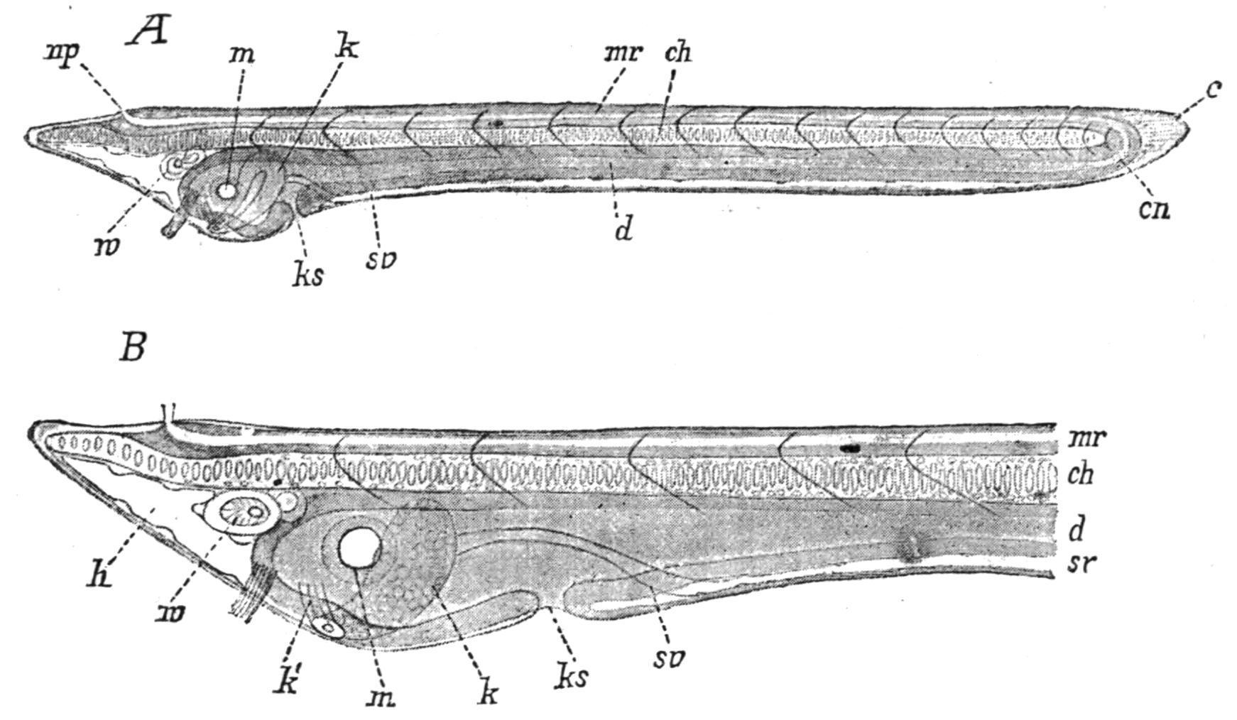
General Structure. The general plan of organisation of the body (see Fig. 71) is that a longitudinal skeletal axis, the notochord (nch), separates a dorsal nervous system (sp.cd) from a ventral reduced coelom (coel), in which lie the alimentary canal (int), the gonads (gon), and other organs. Thus a transverse section of the body (see Fig. 72) shows the typical Chordate arrangement of parts, and is comparable with a transverse section of a tadpole, a young fish, or a larval Ascidian. A peribranchial (atr) or atrial cavity (which is morphologically a part of the external world shut in) lies external to the coelom and body-wall around the pharynx and the greater part of the alimentary canal, and opens to the exterior by the atriopore. As in the Tunicata, the perforations (gill-slits) in the wall of the pharynx (br.cl) open into the atrial cavity and so indirectly to the exterior.
Musculature. The thick body-wall is largely formed by muscular tissue metamerically segmented into about 60 myotomes (Fig. 71, myom). These muscle-masses, which (as is usual in Vertebrata) are thickest dorsally at the sides of the notochord and spinal chord (Fig. 72, m), are so arranged as to present the appearance in a lateral view of the body of a series of shallow cones (<<) fitting into one another and with their apices directed forwards. The muscle fibres are striated, and run longitudinally along the body from the anterior to the posterior edge of each myotome, so as to be attached at their ends to the two septa of connective tissue which form the boundaries of the myotomes. These septa, the myocommas, are conspicuous features in the external appearance of the body (Fig. 70, B). They are not arranged so as to be opposite one another on the two sides, but the myotomes on the right and left sides alternate, as can be seen in a transverse section (Fig. 74, A, p. 121).

FIG. 72.—Branchiostoma lanceolatum. Diagrammatic transverse section of the pharyngeal region, passing on the right through a primary, on the left through a secondary branchial lamella. ao, Dorsal aorta; c, dermis; ec, endostylar portion of coelom; f, fascia, or investing layer of myotome; fh, compartment containing fin-ray; g, gonad; gl, glomerulus; k, branchial artery; kd, pharynx; ld, combined atrial and coelomic wall (ligamentum denticulatum); m, myotome; mt, transverse muscle; n, nephridium; n.ch, notochord; of, metapleural lymph space; p, atrium; sc, coelom; si, ventral aorta; sk, sheath of notochord and spinal cord (sp.cd); uf, spaces in ventral wall. (From Korschelt and Heider, after Boveri and Hatschek.)
It is by means of these lateral muscle-bundles that the rapid vibration or alternate bending of the body from side to side in swimming or burrowing can be performed. There are usually, on each side, 35 myotomes in front of the atriopore, 14 between the atriopore and the anus, and 11 postanal, making 60 in all: some species have only about 50 myotomes, and some as many as 85. (See Classification, p. 137, where a list of the species with the number of myotomes in each is given.)
There are also transverse muscles (Fig. 72, mt) extending across the ventral surface in the region of the body enclosed by the metapleural
folds, and serving to compress the atrial cavity, and so aid in the expulsion of its contents.
Outside the muscular layer of the body-wall the thin integument is formed of a dermal layer of soft connective tissue, covered by the epidermis, a single layer of columnar cells, many of which, especially on the oral cirri, have sensory bristles.
Skeleton.—The endoskeleton consists of the notochord and some tracts of modified connective tissue which support various parts of the body.
The notochord of this animal is noteworthy amongst Chordata for extending practically the entire length of the body, including the head, from snout to tip of tail (Fig. 71). It lies in the median plane, but nearer the dorsal than the ventral surface (Fig. 72), and has the myotomes at its sides, the nervous system above and the alimentary canal below. It is elliptical in section, and tapers to the two ends. The nuclei of the original notochordal cells are displaced to the dorsal and ventral edges, and the greater parts of the cells, in the adult, are occupied by large vacuoles filled with a fluid secretion, so as to form by their distended condition a stiff elastic structure. This state of the cells, and the appearance it gives rise to (Fig. 73), seen best in young specimens, is very characteristic of notochordal tissue. Around the notochord lies a sheath of connective tissue which is continuous with the similar sheath around the nervous system and with the septa between the myotomes.

FIG. 73.—Median sagittal section of notochord of an Amphioxus of 32 mm.
In addition to these skeletal layers of connective tissue there is a cartilage-like tract in the oral hood. This is jointed, or made up of separate rod-like pieces, one at the base of each cirrus, into which it sends a prolongation (Fig. 71, sk). The dorsal and ventral fins are supported by single and double rows respectively of what have been called "fin-rays." They are short rods of gelatinous connective tissue, each enclosed in a lymph space. Finally, the bars constituting the walls of the pharynx between the gill-slits contain slender skeletal rods which run obliquely dorso-ventrally, and are of a stiff, gelatinous nature (see Fig. 75, p. 122). This skeletal connective tissue consists in all cases of a fibrous deposit or matrix produced by the layer of epithelium (ectodermal, endodermal, or mesodermal) which adjoins the tissue.
Alimentary Canal.—This has, as its most noteworthy feature, the Chordate characteristic that the pharynx gives rise to the respiratory organ (see Figs. 71 and 74, A); and in size and prominence, both in side view and in sections, the modified pharynx of Amphioxus is fairly comparable with the branchial sac (pharynx) of many Tunicata (see Fig. 23, p. 51), and might be called by the same name.
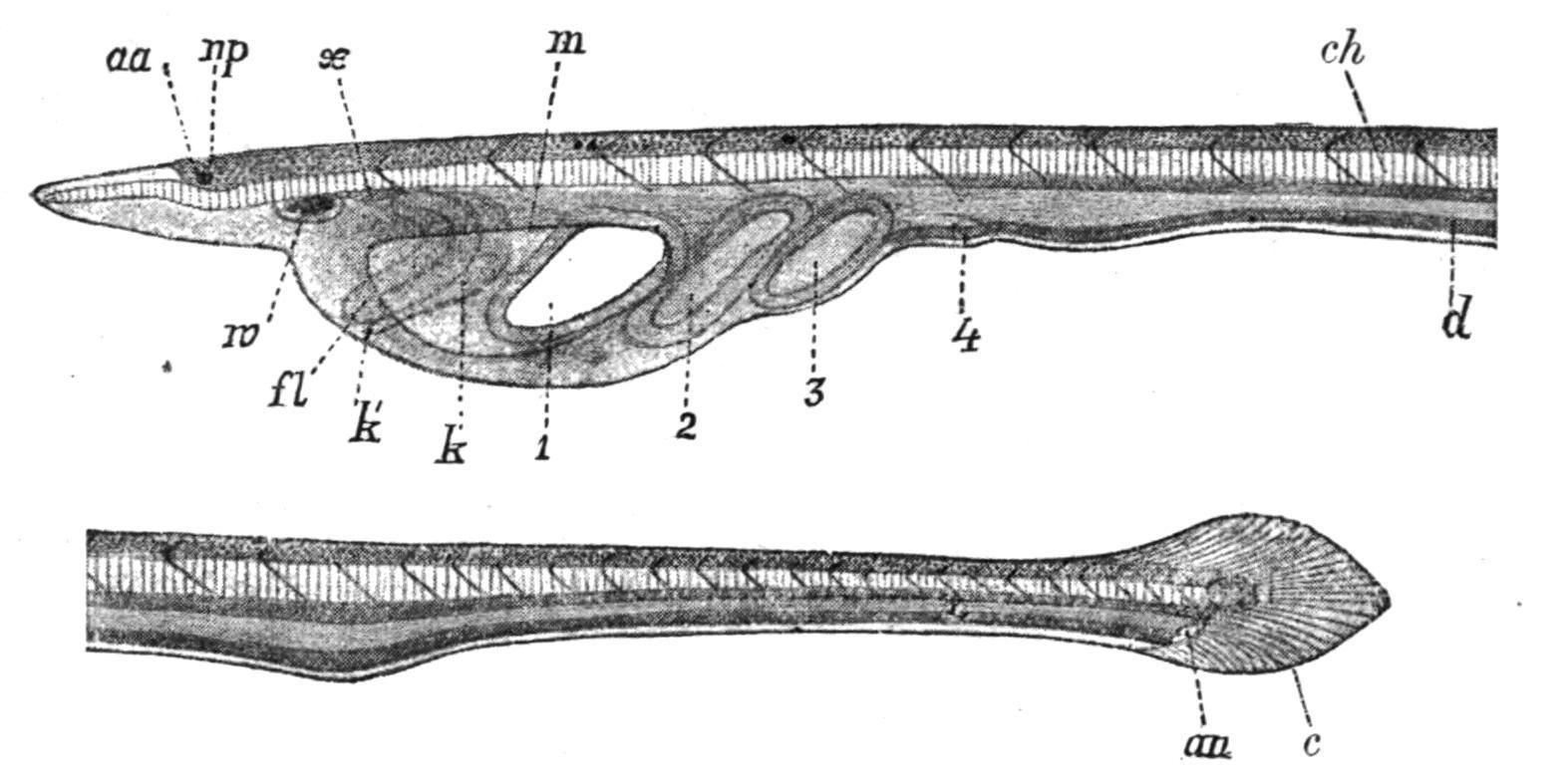
The small primitive mouth, at the bottom of the cavity bounded by the oral hood (stomodaeum), has a membranous border, the velum (Fig. 71, vl), the edges of which are prolonged into a circle of 10 or 12 (up to 16 in some species) simple oral tentacles turned inwards towards the pharynx (compare tentacles of Ascidians, p. 45).
The pharynx, by far the largest part of the alimentary canal, and extending nearly half-way along the body, is more important as a respiratory than as a nutritive organ. Its walls over nearly the whole extent are perforated by a large, and indefinite, number (100 or more on each side) of gill-slits which run on the whole dorsoventrally, but in the contracted condition seen in preserved specimens have their lower ends directed obliquely backwards, so
that a vertical transverse section may cut through a number of such slits and the intervening branchial bars (Fig. 74, A, kb). These bars, and therefore the slits between them, are of two orders, primary and secondary, the latter being developed later in larval life as downgrowths or "tongue-bars," one from the top of each primary gill-slit, so as to divide it into two secondaries. The primary and the secondary (or tongue-) bars can be distinguished from one another by their structure in the adult animal (Fig. 75, A and B).

FIG. 74. Branchiostoma lanceolatum. A, transverse section of the pharyngeal region. a, Dorsal aorta; b, atrium; c, notochord; co, coelom; e, endostyle; g, gonad (ovary); kb, branchial septa; kd, pharynx; l, liver; my, myotome; n, nephridium; r, spinal cord; sn, sn, dorsal and ventral spinal nerves. B, Transverse section of the intestinal region. atr, Atrium; coel, coelom; d.ao, dorsal aorta; int, intestine; myom, myotome; nch, notochord; neu, spinal cord; s.int.v, sub-intestinal vein. (From Parker and Haswell's Zoology. A, From Hertwig, after Lankester and Boveri; B, partly after Rolph.)
It must be remembered that these branchial bars, or septa between the gill-slits, are not merely portions of the wall of the pharynx, but are in a sense portions of the body-wall as well, and correspond in nature, though not in number, to the visceral arches in a Vertebrate lying between the visceral clefts which open on the exterior. In the adult Amphioxus the clefts in the wall of the pharynx do not open directly to the exterior, but into the peribranchial cavity or atrium, which, however, is only formed at a late larval period as an invagination or enclosure of ectoderm. Previous to that the first formed gill-slits opened to the exterior in Amphioxus (see larva, Fig.
86, p. 134), just as they do in a fish or a young tadpole. The atrial cavity is therefore, from its origin, lined by ectoderm, and the outer surface of a branchial bar is virtually a part of the outer surface of the body. It is only natural then to find that each bar contains a small section of the coelom in its interior, communicating dorsally and ventrally with other parts of that cavity (see Figs. 75 and 76). There are also blood-vessels which run in the branchial bars and their junctions. The greater part of the epithelium covering a branchial bar is pharyngeal epithelium or endoderm (Fig. 75, br.ep), but the external, wider, non-ciliated cells (Fig. 75, at.ep) are ectodermal cells lining the atrium. The gelatinous skeletal rods in the primary bars are forked ventrally, while those in the secondary bars are simple; and there are other points of detail in which the two kinds of bar differ. These bars are obviously more numerous in the adult than the myotomes, but in the young larva the first formed gillclefts are metamerically arranged, and then later they increase greatly in number. It is the cilia covering the pharyngeal epithelium on the branchial bars, possibly aided by the ciliated tracts of the oral hood, which cause the current of water already alluded to.

FIG. 75.—Transverse sections through primary (A) and secondary (B) branchial bars of Amphioxus. at.ep, Atrial epithelium; bl.s, blood spaces or "vessels"; br.ep, branchial epithelium; coel, coelomic cavity in primary bar; sk, skeletal rods. (From Willey, after Benham.)
Transverse branchial junctions (synapticula) run across the branchial bars, connecting them at frequent intervals, and these transverse
connexions, like the branchial bars, are supported by skeletal rods. Along the ventral median line of the pharynx runs a groove, the endostyle or hypopharyngeal groove, comparable with the similar structure in the branchial sac of Tunicata. This longitudinal groove (Fig. 76, gl) is lined by ciliated epithelium containing four tracts of gland cells (compare endostyle in Ascidians, Fig. 20, p. 46). There is reason to believe that this organ is the homologue of the thyroid gland of Vertebrata. As in the case of Tunicata the endostyle secretes mucus, which is carried forwards by the cilia to constitute a train with entangled food particles which pass back dorsally to the stomach. At the anterior end the ciliated lips of the endostyle diverge to the right and left to encircle the front of the pharynx as the peripharyngeal bands. These unite again dorsally to form the epipharyngeal (or hyperpharyngeal) groove which leads backwards, corresponding to the hypopharyngeal groove below (see Fig. 74, A), till the posterior end of the pharynx is reached.

FIG. 76.—Transverse section of the ventral part of the pharynx of Amphioxus. c, Coelom; e, endostyle; gl, endostylar glands; m.b.a, median branchial artery; p.b, primary bar; sk, endostylar and branchial rods and skeletal plates; t.b, tongue-bar. (After Lankester.)
The remainder of the simple alimentary canal is straight, and is scarcely differentiated into regions. A slight narrowing of the tube behind the pharynx has been called the oesophagus, and a slight enlargement which follows, the stomach. From this point the intestine tapers backwards to the anus (Fig. 71, p. 116). The ventral edge of the stomach gives off a blind pouch, the hepatic caecum or saccular liver, which runs forwards on the right-hand side of the
pharynx (Fig. 74, A, l). This is a digestive gland, is lined with glandular epithelium, and apparently corresponds with the liver of Vertebrata. There are no other digestive glands in connexion with the alimentary canal of Amphioxus.
Coelom. In the young larva there are at first (as in Balanoglossus) five coelomic spaces, a median anterior "head-cavity," a pair of antero-lateral "collar-cavities," and a pair of more posterior long lateral grooves from which arise, in the later larva, the segmented myotomes and ventrally a large coelomic space surrounding the alimentary canal and separating it from the body-wall. In the adult animal, however, the coelom has been so much displaced by the formation of the spacious atrium that in front of the atriopore it can only be recognised as a series of canals and crevices. The relations of coelom to atrium in the region of the intestine are seen in Fig. 74, B, and in the region of the pharynx in Fig. 74, A. Fig. 72 shows the distribution of the spaces more in detail (see also Fig. 71). Beginning anteriorly, along the dorsal surface of the pharynx and beneath the notochord run a pair of dorsal coelomic canals, one at each side of the epipharyngeal groove; these give off ventral diverticula which pass down the primary branchial bars of the pharyngeal wall and unite ventrally in a median tube, the endostylar coelom (see Fig. 72, ec). At the posterior end of the pharynx these dorsal and ventral canals unite in a narrow coelomic space encircling the stomach, inside the wall of the atrium, and sending an extension forwards around the liver (Fig. 74, A, l). In the region of the intestine, behind the atriopore, the coelom is allowed to expand to its primitive condition on the left-hand side (Fig. 74, B), but is still reduced on the right side, where there is a prolongation of the atrial cavity reaching nearly to the anus. All these coelomic spaces are lined by a coelomic epithelium.
The Blood System of Amphioxus, although as simple as that of a Chaetopod worm, is undoubtedly laid down on the Vertebrate plan— even though there is no distinct heart and the vessels are few and of
simple structure. Capillary networks are formed in some places, but the colourless blood also extends into many lacunae or lymph spaces, such as those around the fin-rays and in the metapleura. As in a typical lower Vertebrate, there is a contractile ventral vessel (the ventral or branchial aorta, Fig. 77, v.ao) running forwards under the alimentary canal to the pharynx, and giving off on each side afferent branchial vessels, which pass up the primary branchial bars and give off branches joining the vessels in the secondary bars. These latter do not communicate directly with the ventral aorta, but the vessels in all the branchial bars open dorsally by efferent branchial vessels into the paired dorsal aortae (Fig. 77, d.ao), which run backwards along the top of the pharynx, one at each side of the epipharyngeal groove. In the vessels of the branchial bars and their connectives the blood is aerated by the current of water passing through the gillslits, and so reaches the dorsal aortae in a purified condition. The right-hand dorsal aorta is continued forward further into the snout than its fellow of the other side, and is dilated at its extremity (Fig. 77). At the posterior end of the pharynx the paired dorsal aortae unite to form the median dorsal aorta which runs backwards, lying between notochord and alimentary canal. This vessel gives off branches to the wall of the intestine, and these break up into capillary networks (Fig. 77, cp), from which the blood is collected by the median sub-intestinal vein. This then flows forwards to pass by the hepatic portal vein to the ventral edge of the saccular liver, in the wall of which it is distributed in a capillary network. The blood is collected on the dorsal edge of the liver by the hepatic vein, which runs posteriorly and then turns downwards and forwards to become continuous with the posterior end of the ventral aorta or "heart."

FIG. 77. Diagram of the vascular system of Amphioxus. af.br.a, Afferent branchial arteries; af.br.a′, similar vessels of the secondary (tongue) bars; br.cl, gill-slits; cp, intestinal capillaries;
d.ao, paired dorsal aortae; d.ao′, median dorsal aorta; ef.br.a, efferent branchial arteries; hep.port.v, hepatic portal vein; hep.v, hepatic vein; int, intestine; lr, liver; ph, pharynx; s.int.v, subintestinal vein; v.ao, ventral aorta. (From Parker and Haswell.)
It is clear that this course of the circulation agrees with that of a typical lower Vertebrate in all essential points:—(1) in having the main artery a dorsal aorta in which the blood flows backwards; (2) in having a ventral vessel representing the heart, and sending impure blood forwards to the respiratory region of the alimentary canal to be aerated; and (3) in having a hepatic portal system consisting of the capillaries of the liver, through which the blood from the intestinal wall has to pass before reaching the ventral vessel (heart).
Renal Excretory functions have been attributed to various organs in Amphioxus, and it is quite possible that, in addition to the true nephridia which are now known, other tracts of tissue in the body may be able to eliminate nitrogenous waste matters. Such are certain clumps of columnar epithelial cells on the floor of the atrium, and the single pair of large brown atrio-coelomic funnels lying on the dorsal edge of the posterior end of the pharynx (Fig. 71, br.f). There are, however, a large number (about 100 pairs) of minute nephridia, discovered (1890) by Weiss and by Boveri independently, lying at the sides of the dorsal coelomic canals above the pharynx, which must be regarded as the chief functional renal organs. These are bent tubules, partly glandular and partly ciliated, each giving off several caecal knobs (at first supposed to be open nephrostomes, one shown at each end of the tubule and three along its upper surface in Fig. 78), which project into the coelom, and opening by one nephridiopore (on the lower surface, and opposite a tongue bar of the pharynx) into the atrial cavity. The knobs, or closed nephrostomes, are surrounded by peculiar, slender, club-shaped tubular and flagellated cells—which Goodrich[113] has shown to correspond to the "solenocytes" in the nephridia of Polychaete worms (see Fig. 79).

FIG. 78. Branchiostomalanceolatum. A nephridium of the left side with part of the wall of the pharynx, as seen alive, highly magnified. (From Willey, after Boveri.)
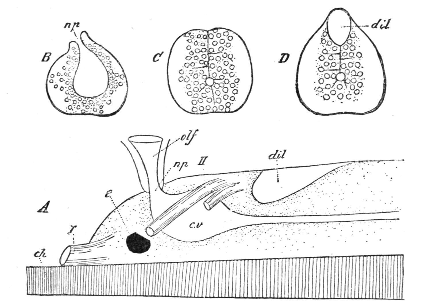
The Central Nervous System is dorsal and tubular as in Vertebrates, and lies in a connective-tissue sheath immediately above the notochord (Figs. 71, etc., and 80, A). Posteriorly it tapers to a fine point a little in front of the end of the notochord, but anteriorly it ends abruptly some distance behind the anterior extremity of the notochord. The central canal is connected with the dorsal surface by a median longitudinal cleft (Fig. 80, C), and at the anterior end it dilates to form the cerebral vesicle (c.v) with which two simple sense-organs, an eye-spot (e) and an olfactory pit (olf), are connected. A patch of ciliated epithelium in the floor of the vesicle has been described as an "infundibular-organ." There is also a surface dilatation of the dorsal cleft behind the cerebral vesicle (dil). The nervous system as far back as this point may be regarded as the brain, though scarcely distinguishable externally (Figs. 71 and 80, A) from the spinal chord behind. From this "brain" arise two pairs of "cranial" nerves, the first (I.) from the anterior end, and the second (II.) from the dorsal surface of the cerebral vesicle; both are in front of the first myotomes of the body, and supply the pre-oral snout with nerves.

FIG. 79. Nephridia. A, portion of a nephridium of Phyllodoce, a marine Polychaete, for comparison with B, portion of a nephridium of Amphioxus. These figures show the solenocytes with their flagella projecting through the long tubes into the lumen of the excretory organ, and demonstrate the essential similarity of the nephridia of Amphioxus with those of Polychaete worms (after Goodrich).
The spinal cord gives off a large number of spinal nerves segmentally arranged, but, like the myotomes, not opposite and symmetrical on the two sides, but placed alternately (Fig. 81). Moreover, the spinal nerves arise on each side at two levels, there being a more dorsal series each arising by a single root and supplying the integument as well as the transverse muscles, so as to be sensory as well as motor, and a ventral series arising each by a number of roots (Fig. 81) and wholly motor in function, as they supply only the myotomes. These two series may be compared to the dorsal and ventral roots which in the Vertebrata join to form a mixed spinal nerve.
FIG. 80. Branchiostoma lanceolatum. A, brain and cerebral nerves of a young specimen; B, transverse section through neuropore; C, behind cerebral vesicle; D through dorsal dilatation. ch, Notochord; cv, cerebral vesicle; dil, dorsal dilatation; e, eye-spot;
np, neuropore; olf, olfactory pit; I and II, cranial nerves. (From Willey, after Hatschek.)
In addition to ordinary small nerve cells the central nervous system contains certain large nerve cells with very long processes, the "giant fibres," which extend through the greater part of the length of the spinal cord. No trace of a sympathetic nervous system has been found.
The Sense-Organs connected with the nervous system are few and simple. There are sensory cells in the ectoderm, on the margin of the velum, on the velar tentacles, and especially in clumps on papillae of the cirri around the mouth, which are probably tactile. In the roof of the oral hood there is a sensory structure, the "groove of Hatschek," which is supposed to be an organ of taste. The olfactory pit alluded to above opens externally on the left-hand side of the snout. It is ciliated internally and leads to the so-called olfactory lobe, an antero-dorsal hollow outgrowth from the brain. In the young animal the olfactory pit opens by the neuropore into the central canal (Fig. 80, A), but that passage is closed in the adult. Possibly the olfactory pit is homologous with the hypophysis or pituitary body of Vertebrates, the homologue of which in Tunicata has a ciliated funnel. Finally, the median cerebral eye (Figs. 80 and 81) is a mere pigment spot in the anterior wall of the cerebral vesicle, and a series of somewhat similar pigment spots occurs along the floor of the central canal in the spinal cord.[114] There is no known auditory organ. On the under surface of the oral hood patches of ciliated epithelium drawn out into rounded lobes were called by Johannes Müller the "Räder-organ." This is probably of use in drawing water inwards to the pharynx, but it may also be a senseorgan.

FIG. 81. Branchiostomalanceolatum. Anterior portion of central nervous system from above, showing dorsal and ventral spinal nerves. (From Willey, after Schneider.)
The Gonads are segmentally arranged along the sides of the body, projecting into the atrial cavity at the sides of the pharynx and intestine. In some species the gonads are paired, but in others belonging to the genus Asymmetron (p. 137) only a single series, that of the right side, is present. In the common Amphioxus (Branchiostoma lanceolatum) there are about 26 pairs (Fig. 70, B), lying in somites 25 to 51; and ovaries and testes are found in separate individuals in all other respects. Each gonad is surrounded by a layer of coelomic epithelium. The gonad must therefore be regarded as having grown down from a myotome of the body-wall into a coelomic pouch, carrying before it the coelomic and then the atrial epithelium (Figs. 72, and 74, A, g). Eventually the gonads, when ripe, burst through the layers of epithelium, and the ova and sperms are shed into the atrium and escape to the exterior by the atriopore, or it may be in some cases by the mouth.
EMBRYOLOGY AND LIFE-HISTORY.
Development takes place in the sea-water where the egg is fertilised apparently always about sunset, the embryonic stages being
passed through during the night, and the larva hatched in the early morning.
FIG. 82. Stages in the segmentation of Amphioxus. A represents the eight-celled stage; B, the sixteen-celled; D, vertical section of C; F, vertical section of the blastosphere or blastula stage (E). (From Korschelt and Heider, after Hatschek.)
The egg is small (0.105 mm. in diameter when shed) and contains very little food-yolk. Segmentation is complete (Fig. 82, A), is nearly regular, and results in the formation of a hollow blastosphere (Fig. 82, E, F), the wall of which is one cell thick. The lower cells (Fig. 82, B, C, D) are slightly larger than the upper. Invagination of the lower cells then takes place (Fig. 83, A), resulting in the suppression of the blastocoele or segmentation cavity and the formation of an archenteron, at first shallow and opening widely to the exterior (Fig. 83, B), and then deeper and with the opening narrowed to a small posterior blastopore (Fig. 83, C). This "gastrula" stage differs from the blastosphere in having a mouth or blastopore, and in being two cell-layers thick—epiblast (ectoderm) on the outside and hypoblast (endoderm) within. It soon shows the future aspects of the body by its elongation and shape (Fig. 83, C), as the dorsal surface becomes flat and the ventral convex, while the blastopore is at the posterior end of the dorsal surface. The blastopore soon closes, and the mouth and anus are formed independently later.

FIG. 83. Three stages in the formation of the gastrula of Amphioxus. In A the nuclei of the endoderm have been omitted; C has the dorsal surface uppermost, and the posterior end to the right (From Korschelt and Heider, after Hatschek.)
The epiblast cells become ciliated all over the surface, so that the embryo rotates within the thin covering which still surrounds it. And now all the chief systems of the body begin to be marked out. The tubular nervous system develops from a depression of the epiblast (the medullary plate) in the middle line of the flattened dorsal surface (Fig. 84, A, mp). The edges of the depressed area grow inwards and unite over the deeper layer of epiblast, which becomes the wall of the neural canal or embryonic nervous system (Fig. 84, D, n); and further back these edges of the medullary plate join one another behind the blastopore, so that the latter comes to open into the floor of the neural canal, thus forming the neurenteric canal (Fig. 85, A, cn). Anteriorly the neural canal (n) opens to the exterior for some time by the neuropore.
The hypoblastic walls of the archenteron give off a long median dorsal groove which becomes the notochord (Fig. 84, C and D, ch); and also an anterior pouch and certain lateral pairs of diverticula which are the enterocoeles or coelomic pouches, and give rise to the mesoblastic somites (Fig. 84, B and C, mk). The notochord (Fig. 84, D, ch) is at first a longitudinal cellular ridge, which becomes segmented off from the hypoblast as a rod lying below the neural canal. It is seen in various stages of development in Figs. 84 and 86, leading to the vacuolated condition of the adult.
FIG. 84. Four stages in the development of the notochord, nervous system, and mesoderm of Amphioxus. ak, Ectoderm; ch, notochord; dh, cavity of archenteron; hb, ridge of ectoderm growing over medullary plate; ik, endoderm; lh, coelom; mk, coelomic pouch; mk1 , parietal layer of mesoderm; mk2 , visceral layer; mp, medullary plate; n, neural canal; ns, protovertebra. (From Korschelt and Heider, after Hatschek.)
The coelomic pouches are five in number—(1) one median, anterior, which gives rise to the two head cavities, the left-hand one of which opens to the exterior by means of the pre-oral pit; (2) a pair of small lateral pouches, placed anteriorly and dorsally, which do not divide but give rise to the first pair of myotomes only and their outgrowths which extend back into the metapleural folds, where, however, they are later replaced by lymph spaces; and (3) a second pair of diverticula, more posteriorly placed, which continue to grow back towards the blastopore, and have paired mesoblastic somites, the cavities in which are the beginnings of the coelom in the body, constricted off from them successively from before backwards (Fig. 85, A, ush) to form all the remaining myotomes.[115] This is the first sign of segmentation in the animal, and at this stage, when it has about five pairs of mesoblastic somites, it breaks out of its covering and becomes a free-swimming larva.

FIG. 85. Embryo of Amphioxus. A, in vertical section, slightly to the left of the middle line. B, in horizontal section. ak, Ectoderm; cn, neurenteric canal; dk and ud, archenteron; ik, endoderm; mk, mesodermal folds; n, medullary canal; us, first coelomic pouch; ush, coelomic cavity; V, anterior, H, posterior, end. (From Korschelt and Heider, after Hatschek.)
The mouth now appears, and soon grows to a large opening on the left side of the now pointed anterior end (Fig. 86, A, m), and the first gill-slit (ks) forms as a direct communication from the front of the mesenteron (pharynx) to the exterior. It is ventral at first, and then shifts over to the right side.
The anus forms posteriorly, and the neurenteric canal closes up. A depression on the floor of the enteron close to the mouth gives rise to the "club-shaped gland" (Fig. 86, B, k), which is probably a gillcleft in its nature.
FIG. 86.—A, young larva of Amphioxus. B, anterior end enlarged. c, Provisional tail-fin; ch, notochord; cn, neurenteric canal; d, enteron; h, coelom of snout; k, club-shaped gland; k′, its external
aperture; ks, first gill-slit; m, mouth; mr, nerve-tube; np, neuropore; sv, sub-intestinal vein; w, pre-oral pit. (After Hatschek.)
FIG. 87.—More advanced larva of Amphioxus. an, Anus; au, eye-spot; c, larval tail-fin; ch, notochord; d, enteron; fl, rudiment of endostyle; k, club-shaped gland; k′, its external aperture; m, mouth; np, neuropore; w, pre-oral pit; x, provisional nephridium; 1-4, gillslits. (From Korschelt and Heider, after Lankester and Willey.)
The walls of the coelomic pouches, which have been extending both dorsally and ventrally (Fig. 84, D), become the mesoderm, the outer the somatic and the inner the splanchnic layer; and the ventral parts of their cavities unite to form the coelom. The cells of the dorsal parts become muscle fibres, and constitute the myotomes internally and the connective tissue of the skin externally.
The larva (Fig. 87) is now long and narrow with many segments, pointed ends, and a caudal fin. The gill-slits all appear first in the mid-ventral line and then shift over to the right side (Fig. 87, 1-4): they are metamerically arranged. After fourteen have been so formed a series of eight appear dorsally to those on the right side, and then the first set, originally ventral, move over to the left side, and by the suppression of some they become equal in number and segmentally arranged on the two sides of the body. This is perhaps the stage at which Amphioxus shows the nearest approach to the typical embryo of a higher Vertebrate. The gill-slits are here seven to nine on each side, and the Vertebrate embryo has usually five to seven on each side. These first gill-slits in Amphioxus are later subdivided by the downgrowth of the tongue-bar from the dorsal edge.

FIG. 88. Ventral aspect of three larvae of Amphioxus, showing the metapleural folds and the formation of the atrium. ap, Atriopore; k, gill-slits; lf and rf, left and right metapleural folds; m, mouth; w, pre-oral pit. (From Korschelt and Heider, after Lankester and Willey.)
The atrium is an ingrowth of the external space between the two ventral metapleural or atrial folds (Figs. 88 and 89), paired lateral ridges of the body-wall, and so is lined by ectoderm. This ingrowth is shut off from the exterior by the growth towards each other of subatrial ridges on the inner sides of the metapleural folds (see Fig. 89, A, sl), and then becomes greatly enlarged by the increased relative growth of the ventro-lateral part of the body-wall (Fig. 89, B, C). The posterior opening between the metapleural folds remains as the atriopore (Fig. 88, C, ap); while the anterior end (Fig. 88) also remains open for some time, but eventually closes. As the metapleural folds lie outside the gill-slits (Fig. 88, A) when these folds close in (B and C), it comes about that the gill-slits which formerly opened freely to the exterior now open into the cavity of the atrium (compare Figs. 87 and 88).

FIG. 89.—Diagrammatic transverse sections of three larvae of Amphioxus to show the development of the atrium. ao, Aorta; c, dermis; ch, notochord; d, intestine; f, connective tissue; fh, cavity
of dorsal fin-ray; m, myotome; n, nerve-tube; p, atrium; sf, metapleural folds; sfh, lymph space in metapleural folds; si, subintestinal vein; sk, sheaths of notochord and nerve-tube; sl, subatrial ridge; sp, coelom. (From Korschelt and Heider, after Lankester and Willey.)
The mouth now becomes median and ventral, and is reduced in size, the oral hood (stomodaeum) is formed in front of it, the gill-slits become more numerous and vertically elongated, the endostyle forms along the floor of the pharynx, and the gonads grow as paired pouches from the body-wall. This brings the animal to the young adult condition, reached at a period of probably about three months after the fertilisation of the egg.
The development as a whole shows a very marked resemblance to that of the Tunicata (see p. 55), but lends no support to the view that Amphioxus has degenerated from a higher group of the Vertebrata.
Classification of the Cephalochordata.
The known species of Amphioxus may be classified as follows[116]:
FAMILY BRANCHIOSTOMATIDAE.
Genus 1. Branchiostoma(Costa, 1834).
Having biserial gonads and symmetrical metapleura.
B. lanceolatum (Pallas)—Myotomes 36 + 14 + 12, gonads 2329 pairs: Mediterranean, N.W. Europe, Ceylon, E. of United
States.
[B. belcheri, Gray—Myotomes 38 + 17 + 9: Torres Straits, Singapore, Borneo, Ceylon.
[B.nakagawae, Jord. and S.—Myotomes 37 + 16 + 11: Japan.
[B. caribbaeum, Sundevall—Myotomes 37 + 14 + 9: West Indies, Atlantic, N. and S. America.
B.capense, Gilchrist—Myotomes 47 + 19 + 9: S. Africa.
B. californiense, J. G. Cooper—Myotomes 45 + 17 + 9: California.
B. (Dolichorhynchus) indicum (Willey)—Myotomes 42 + 14 + 15: India and Ceylon.
(?) B.elongatum, Sundevall—Myotomes 49 + 18 + 12: Peru.
(?) B. pelagicum, Günther—Myotomes 36 + 16 + 15: Honolulu, Gulf of Manaar, South Indian Ocean. Genus 2. Asymmetron(Andrews, 1893).
With uniserial (right) gonads and asymmetrical metapleura.
A. lucayanum, Andrews—Myotomes 44 + 9 + 13: Bahamas, Maldives, Zanzibar.
A. caudatum (Willey)—Myotomes 40 + 9 + 11: Louisiade Archipelago.
A. (Heteropleuron) bassanum(Günther)—Myotomes 45 + 16 + 14: Bass Straits, Australia.
" cingalense (Kirkaldy)—Myotomes 39 + 16 + 8: Ceylon.
" cultellum (Peters)—Myotomes 32 + 10 + 10: Torres Straits, Australia, Ceylon.
" maldivense(F. Cooper)—Myotomes 45 + 16 + 12: Maldive Archipelago, Zanzibar.
" hectori (Benham)—Myotomes 53 + 19 + 12: New Zealand.
Thus sixteen species have been described, of which the three under Branchiostoma placed after square brackets, seem to be merely varieties of B. lanceolatum, and B. nakagawae is probably identical with B. belcheri; while it is a question whether Asymmetron caudatum is more than a variety of A. lucayanum, thus leaving eleven or twelve species that seem fairly well characterised. The exact positions of the two marked (?), viz. B. elongatum and B. pelagicum, cannot be determined in the absence of fuller descriptions of these species.
FIG. 90. Sketch-map showing geographical distribution of the Cephalochordata. + indicates Branchiostoma; o indicates Asymmetron.
The list above, and the map (Fig. 90), give some indication of the geographical distribution of the group, and show that, although the few species are widely distributed over the shallow waters of the globe, most of the records lie between 40° N. and 40° S. latitudes. In fact the group is mainly a tropical one, and is most abundant in the Indo-Pacific region. The crosses indicate records of species of Branchiostoma, and the circles those of Asymmetron (including Heteropleuron); the latter are confined to the Indo-Pacific seas, with the exception of A. lucayanum from the Bahamas—one of the numerous cases of interesting similarity between the marine faunas of the East and West Indies.