Another random document with no related content on Scribd:
ECHINODERMA.
Six table-cases contain the dried Echinoderms arranged in systematic order. The seventh is devoted to preparations, models, and figures illustrative of the structure and life history of various members of the group.
An inspection of that Case and the accompanying woodcuts will make clear the distinctive characters of the Echinoderma. Unlike that of a Crayfish or a Mussel, the body does not appear to be divided into two equal or symmetrical halves, though it really is; this is due to the possession of a number of rays, of which there are ordinarily five. The skin is strengthened by the deposition in it of carbonate of lime, which may be in the form of continuous plates or bars, or of separate scattered spicules. A series of tube-feet or suckers (podia) are generally developed along each ray, and these are supplied by a system of water-vessels peculiar to Starfish and their allies. These rays are often called “ambulacra.”
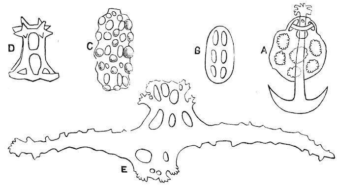
1.
A. Anchor and plate of Synapta. B, C. Tables of Holothuria impatiens; and D. Holothuria atra: from various aspects. E. Spicule from sucker of Stichopus variegatus, magnified about 200 times.
Fig.
Fig. 2.
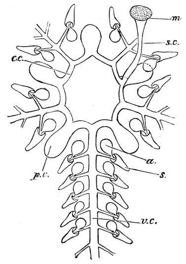
Diagram of Water-vessels.
c.c. Circular canal, with p.v., its Polian vesicles; from it a radial canal (v.c.) is given off along the lower surface of each arm; this supplies, by side branches, the suckers, s; connected with each sucker is a contractile swelling or ampulla (a). The circular canal is in connection with the exterior by s.c., the stone-canal, and opens to it by the madreporite (m).
Fig. 3.
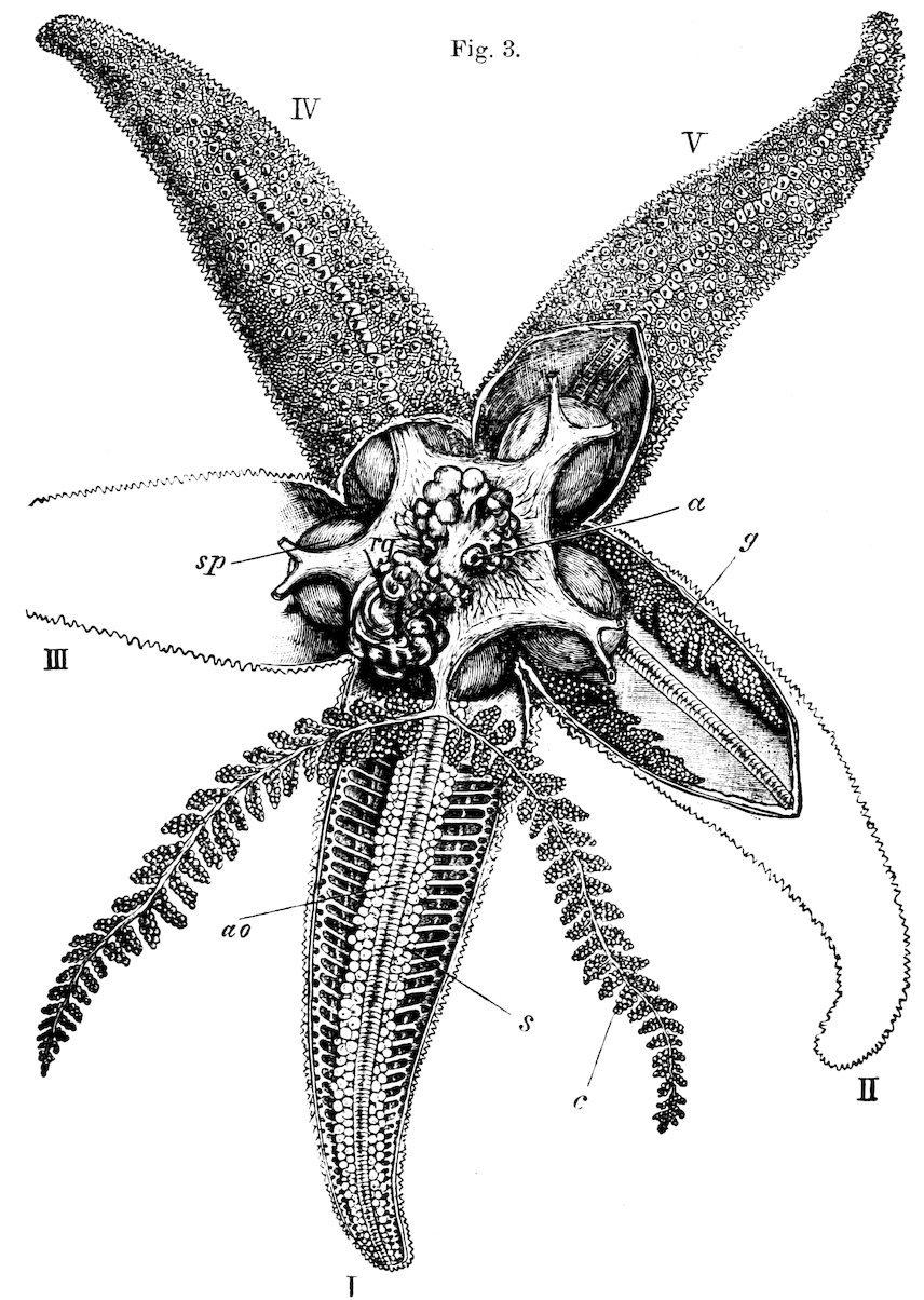
Figure of a Starfish (Asterias rubens).
In the ray marked I. the skin has been removed from the upper surface, and the ambulacral ossicles (ao) and the podia (s) are seen in situ; the blind outgrowths (c) from the central stomach (sp) have been dissected out. In II. the gonads (g) are exposed; and in the centre above the stomach the rectal glands (rg) are to be seen. The anus (a) is seen to be subcentral in position.
In the body of the Starfish (Fig. 3) the arms are seen to be continuous with the disk and to contain portions or prolongations of the chief organs. The middle of the arm is occupied by two rows of hard pieces (ambulacral ossicles), the fellows of which make an open angle with each other, and so form an open ambulacral groove; along this we find the suckers, the water-canal that supplies them, the blood-vessel of the arm, and a nerve-cord. At the centre of the disk is the mouth. The ossicles at the sides of the arms bear spines, which vary in different species; the surface of the back is supported by a network of hard pieces, and through the intervening spaces there project membranous pouches, which are respiratory in function. The modified plate on the upper surface opens into a tube by means of which the water-vessels communicate with the exterior; this plate is known as the madreporite (Fig. 2, m).
The organs for masticating the food are most highly developed in the regular Echinoids, where the complex apparatus known as the “Lantern of Aristotle” is found (Case 38) to consist of five sets of pieces; the tooth is strong and bevelled at its free end; it is supported by triangular jaws on either side, a pair uniting and having the form of an inverted pyramid; these alveoli are connected with their neighbours by oblong pieces (falces); above these there are elongated bars, which are hinged on to the inner end of the falces and have their outer ends free. The whole lantern is connected to the test by muscles which pass from its sides to the auricles or upstanding pillars which lie round the mouth; and, owing to this muscular apparatus, the teeth are capable of complicated and various movements.
In the Ophiuroids the edges of the mouth-slits are provided with short spinous processes, varying a good deal in arrangement, but
never having, apparently, any other function than that of a filteringapparatus; in the Starfishes the plates round the mouth have a supporting function only; in Crinoids and Holothurians the mouth is unarmed; the latter are often remarkable for a deposit of calcareous plates in the walls of the gullet, and in the former the grooves on the arms are the lines along which food comes to the mouth.
Echinoids live on seaweeds and the animals that are found on them; such as have no teeth, like Spatangus (Case 32), use their spout-like mouth to take up the sand and débris on which they move, and from which they extract some nutriment. Ophiuroids live on the smaller foraminifera; Asteroids on dead fishes (as line-fishermen well know), oysters, and other molluscs, and even on specimens of their own particular species; Holothurians on shell or coral débris and the minute organisms it contains; and Crinoids on small tests of foraminifera and on the adults of small and larvæ of larger crustacea.
In a number of Echinoids and Asteroids some of the spines are specially modified to act as seizing-organs—the free end being divided into two, three, or rarely four pieces, which are moved on one another by special muscles. These minute organs were regarded by earlier observers as parasites, and were named pedicellariæ; they may be movable, when they have a stalk, or the stalk may be absent and the valves sessile. Considerable difficulty attaches to the determination of the use that these organs may be to their possessors; but there is reason to suppose that they may act as cleansing-organs by removing minute particles of dirt, and as temporary organs of fixation, while M. Prouho has observed their use as organs of defence.
Echinoderms move but little; the unstalked Crinoids, if they cannot find stones or worm-tubes around which to attach themselves, swim by beating the water with their delicate arms, five being raised and five depressed alternately. The Echinoid or Asteroid is able to move by the aid of its podia or so-called ambulacral feet, which become erected by being filled with water, and are then contracted; by means of this contraction movement is effected; a similar kind of locomotion obtains with the pedate Holothurians; in the Ophiuroids the flexible arms either serve as the organs of movement, or act as an apparatus whereby the creature becomes coiled round the branches of corals (see Case 20).
Echinoderms are often of exceedingly bright colours, as is shown by the pictures on the wall, and are very conspicuous objects; this may, apparently, be associated with disagreeable tastes or odours; sometimes they cover themselves over with seaweed, and so hide their brilliancy; the spines of some forms are exceedingly painful to the touch, and the stout plates of some of the Goniasters must form admirable organs of protection. The power of restoring lost or injured parts is one of the most remarkable points in the Echinoderm organization (see Case 6).
Echinoderms are of great geological age, and were very abundant in earlier periods of the world’s history. Two groups (the Blastoids and Cystids) have completely disappeared, and the Stalked Crinoids (Lily-Encrinites) are far less common than they used to be. Echinoderms are now found in all seas, and extend to great depths of ocean; many of the species have exceedingly wide areas of distribution, and most are characterized by their gregarious habits, a large number of specimens of a single species being generally obtained by the dredge. They are most abundant in the tropical seas.
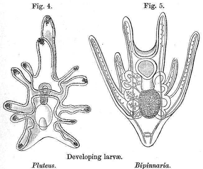
Most Echinoderms lay their eggs in the water, where the larvæ are developed and swim about freely; but in a few (Hemiaster, Ophiacantha vivipara, and others) the young do not pass through any metamorphosis, for the eggs are placed in special pouches of the body of the parent, in which they are hatched. The free-swimming larvæ of the other Echinoderms pass through a series of remarkable changes (Figs. 4 and 5); these are illustrated by the twelve models of various forms of larvæ exhibited in Case 36; in Case 35 is a set of models showing in detail the changes undergone by a single species (Asterina gibbosa). A portion only of the body of the larva is converted into the substance of the perfect animal; the rest is either absorbed by the growing animal, or shrivels up and disappears.
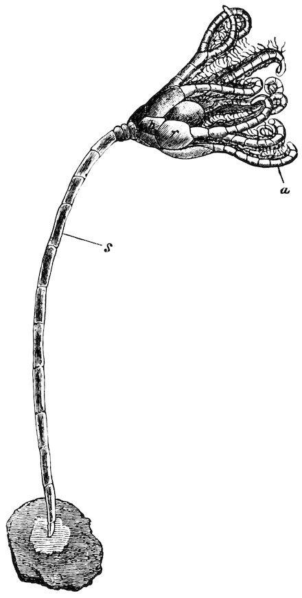
Below the twelve models in Case 36 may be seen a representation of three stages in the history of the Feather-star (Antedon bifida). The larvæ of this Echinoderm are not free, but are attached by a stalk (Fig. 6); in the common Feather-star and other Comatulidæ the stalk is found during larval stages only; in others, such as Pentacrinus, it persists throughout life.
The presence or absence of this stalk has been taken as the first character of importance in the classification of Echinoderma which may be divided into two groups:—
A. P���������,[24] or Echinoderms provided with a stalk throughout life or in the larval stages only. To this group belong the Crinoidea, and the extinct Blastoidea, and Cystidea.
b
B. E��������, or Echinoderms without stalks at any time of their existence. To this group belong the Asteroidea, Ophiuroidea,
Fig. 6.
Pentacrinoid stage of Antedon rosacea. a, arms;
, basals; r, radials; s, stalk.
Echinoidea, and Holothurioidea.
C��������.—This Order may be described as stalked, globular, or cup-shaped Echinoderms, in which the oral surface of the calyx or disk looks upwards, and in which five jointed and generally branched rays arise from the central disk. Their joints have jointed pinnules at their sides, and the sucking-feet have the form of tentacles.
The stalked representatives of this Order are placed on tables and brackets near the south door, and are worthy of being particularly noticed for their fine preservation, size, and beauty. The largest specimen of Pentacrinus decorus was taken on a telegraph-wire, to the covering of which the stalk of the Crinoid is still attached. Metacrinus is a more lately discovered genus, which appears to be confined to the eastern seas.
A few dried unstalked Crinoids are shown in Table-case 1; these show the leading modifications of structure in the two great genera Antedon and Actinometra.
Fig. 7.
Comet form of Linckia.
A���������.—This Order comprises Echinoderms with a depressed body of pentagonal or star-like shape, to the ventral surface of which the ambulacral feet are confined. The rays are more or less elongate movable arms, with skeletal structures, which consist of transversely arranged, paired, calcareous plates, articulated with each other like vertebræ, the series extending from the mouth to the end of the arms. The groove in which the ambulacral feet are arranged is uncovered.
Typical specimens of this Order are exhibited in Cases 2 & 3, in which the great variety of form in the genus Asterias and beautiful examples of Acanthaster are shown. Cases 6 & 7 contain specimens illustrating the curious habit of self-mutilation possessed by so many Echinoderms; among Starfishes, and notably in the genus Linckia,
the single arms separated from the disk are able to develop a fresh disk and arms, and so to multiply the species. Cases 9–11 contain fine series of Oreaster.
O����������, or “Brittle-stars.”—These Echinoderms appear to resemble the ordinary Starfish[25]; but they differ in having the organs of digestion, respiration, and reproduction confined to the disk, the arms having merely the function of locomotor organs. The arms therefore are more slender and cylindrical in form, and are sharply distinct from the disk; the separate joints consist of two central ossicles, which leave only a narrow canal between them, and these are covered above, below, and at the sides by specially developed investing plates; the lateral plates bear spines, which are always comparatively short and delicate, as compared with the spines found at the sides of the arm in starfishes.
The principal types of this Order are exhibited in Cases 17–22; the most exquisite of them are the forms whose arms are divided and subdivided till they end at last in the finest threads, as in Astrophyton, the so-called Basket-fish or Gorgon’s heads.
E���������, or “Sea-Urchins,” are Echinoderms in which the rays are not free, as in the Starfishes or Brittle-stars, but unite to form a compact, spherical, heart- or disk-shaped test; this test is covered with spines, which may attain to a great length, as is shown in the fine example of Diadema saxatile from the Andaman Islands; some of the tests are flexible and very fragile. Owing to the quantity of specimens that are sometimes dredged at one spot, the naturalist has been able to gain a better idea of the range of variation in the species of Echinoderms than in some other divisions of the Animal Kingdom; an instructive series, showing the variations of Echinometra lucunter, is shown in Case 28.
The genus Hemiaster offers an example of an Echinoderm in which the eggs are laid in special pouches; the hinder ambulacra are deepened to form pits, which are guarded by specially elongated spines (see Case 34); in these pits the young pass through all the stages of their development.
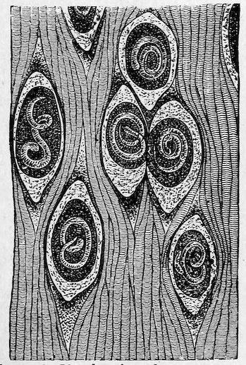
The minute structure of the spines of Sea-Urchins is illustrated by a series of figures on the wall.
The H�������������, or Sea-Cucumbers, form the last order of Echinoderms. Their body, as indicated by their English name, is elongate, subcylindrical, with a more or less flexible integument, according to the extent of the reduction of the calcareous skeleton; the mouth is at one end of the body and surrounded by tentacles, the vent at the opposite end.
As these animals cannot be shown in a dried state, some of them, preserved in spirit, are placed in Wall-Case IV. According as they have or have not the sucking-feet of the Echinoderma, they are ordinarily divided into the Pedata and the Apoda; the latter are represented by Synapta, which may attain to a great length, and by Chiridota; the Pedata are illustrated by the genera Cucumaria, Psolus, and Holothuria. Deep-sea investigations have revealed the existence of another group of specially modified Holothurians—the Elasipoda; these are remarkable for their well-marked bilateral symmetry and the distinctness between the dorsal and ventral portions of the body; the prominent processes on the dorsal surface are not contractile.
An exhibition of some interest is to be found in a Table-Case against the wall, in which there are various specimens of the edible Holothurians—trepang or bêche-de-mer; these were all bought in the market at Canton, and may be taken to be typical of the kinds offered for sale in various eastern countries.
WORMS.
By the name “Worms,” people commonly indicate a number of different forms whose relations with one another are by no means so close as those of a Holothurian and a Crinoid, or a Mussel and an Octopus. There are not, indeed, any common characters by the possession of which the worm-like animals can at once be distinguished from other animals. We take the divisions, examples of which are here represented, either by drawings, models, or specimens preserved in spirit separately.
The groups referred to may be enumerated as follows:—
Platyhelmia. Turbellaria. Trematoda. Cestoda.
Nemertinea. Nematoidea. Chætopoda.
P����������, or Flat-Worms.—These form the lowest and simplest division of the group.
The parasitic Platyhelmia—the Tapeworms (Cestoda) and the Flukes (Trematoda)—occupy Case I.; the life history of the common Tapeworm (Tænia solium) is shown by the aid of models and figures. A model of the anterior end of the common Tapeworm shows the four suckers and the crown of hooks; the unjointed neck is followed by the joints (proglottids), which increase in size the farther they are from the neck. Several entire specimens of Tænia follow, showing the size of the whole worm and the form of its joints. The structure of the body is shown in the models of two joints. The growth and development of the Tapeworm is dependent on a migration or a change of the hosts which it inhabits in the various stages of its life; and although the different kinds of Tapeworm differ from each other
somewhat in certain details of their migration and development, their life history exhibits, on the whole, the same events which we find in Tænia solium, a common Tapeworm of man in Northern Europe. This worm is matured in the intestines of man; its final joints consist merely of fertilized ova which have already passed through the earlier stages of development; when the joints are detached and discharged, their contents escape in the form of embryos contained in a thick chitinous shell. If these are now swallowed by a pig, the shell is digested by the gastric juices of the new host, and a rounded embryo, which is provided with three pairs of hooks, is set free; by means of these hooks the guest makes its way through the wall of the stomach or intestine, and finally settles down in the muscles of its host. The embryo now loses its hooks, and gradually acquires a bladder-like form, the central cavity of which is filled with fluid. This bladder-worm (Cysticercus) has its outer wall pushed inwards at the anterior end, and on this hooks and suckers become developed. We have now a narrow head and neck with an attached bladder, the head being at this time hollow. If during the long time that these bladder-worms remain alive, the pig is killed for food, its flesh is found to be “measly”; if it is afterwards insufficiently cooked and eaten, the worms are conveyed into the human stomach. Here the bladder-like termination becomes absorbed, and, the neck beginning to grow, we have the commencement of the form from which we started, and the completion of that “vicious circle” which is so curious a characteristic of many forms of parasitic life.
Fig. 8.
Tænia solium: showing the head (h) with its suckers (s′) and crown of hooks (s), the unjointed neck (n), and a few of the succeeding joints (j).
In other Tapeworms the cyst may be more complicated than that in the pig, as, for example, the form found in the sheep’s brain or the
liver of the horse.
Of the other Cestode parasites mention should specially be made of those of Fishes; the vulgar notion that the parasites of these animals are dangerous to man has been shown to be entirely erroneous.
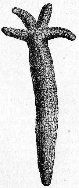
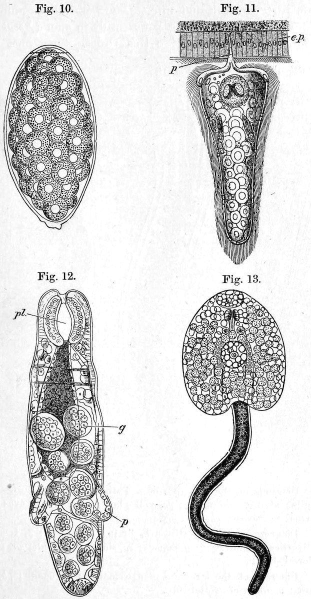
The Flukes infest animals of all kinds; that which is most dangerous to sheep, and the cause of much pecuniary loss (Distoma hepaticum), is selected here as a type; its structure is shown by a large model, and its life history by a series of diagrams (Figs. 10–13). Here, again, we have a creature which infests two hosts. If the larvæ which escape from the sheep fall on wet ground in or near a pool, they make their way to a small pond-snail (Limnæa truncatula, Fig. 9), into the lung-chamber of which they bore their way. On leaving them the larva may be, and is, too frequently, eaten by a sheep, and makes its way into the liver of that animal, where it causes the disease known as the “liver rot.”
The damage done by the liver-fluke may be imagined from the fact that in the winter of 1879–80 no less than three millions of sheep died of rot in the United Kingdom; this heavy loss is no doubt largely due to the immense number of eggs to which a single fluke may give rise. It has been estimated that every fluke may produce, during its life, several thousands of eggs; and in one case Prof. A. P. Thomas found as many as 7,400,000 eggs in the gall-bladder of a sheep
which was suffering from rot, and which, at that time, had in its liver about 200 flukes.
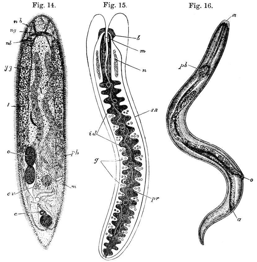
The non-parasitic Flat-worms are shown, magnified, in the upper parts of Cases I. & II. The Turbellaria proper, without any or with a simple or a branched intestine, but without a vent, are represented by Convoluta and Thysanozoon: the general structure is shown by a diagram in Case II., which is here reproduced (Fig. 14). Planaria, Thysanozoon, and Bipalium serve to illustrate the forms of members of this group.
The Nemertine Worms (Nemertinea), with a straight intestine, with a vent, and with a proboscis, may attain to a very considerable length; Carinella and Lineus are represented by large figures, and various species are shown in spirit. These forms, which used to be very unsatisfactory to exhibit, on account of the great difficulty of preserving them complete and uninjured, are now, with improved methods, very satisfactorily shown, as the specimens purchased from the Marine Biological Laboratory at Plymouth prove.
Stages in the life history of the Fluke.
Fig. 10. Egg of Fluke, showing the operculum and the contained yolk-spheres. Magnified 340 diams.
Fig. 11. An embryo forcing its way by its boring-papilla (p) into the wall of the lung of a Snail (e.p.) Magnified about 340 diams.
Fig. 12. A young Rédia (natural size, ½ millimetre or ¹⁄₅₀ inch): pl., pharynx; g, contained germs; p, characteristic posterior processes of the Rédia.
Fig. 13. Free-swimming Cercaria, before the commencement of the formation of the cyst. Magnified 100 diams.
Fig. 14. Diagram of the structure of a Turbellarian: ng, nerve- (cerebral) ganglia; nb, nerve-branches; yg, yolk-glands; t, testis; o, ova; ov, ovary; c, cirrus; m, mouth; ph, pharynx.
Fig. 15. Diagram of a Nemertine: b, brain; m, mouth; n, renal organs; id, diverticula of intestine; g, gonads; sn, side nerve-trunk; pr, proboscis in its dorsal sheath.
Fig. 16. Diagram of the structure of a Nematoid; m, mouth; ph, pharynx; a, anus; o, orifice of genital tube.
N�������� (Thread-Worms or Round-Worms).—These are for the most part parasitic, and infest plants as well as animals; the common Round-Worms living parasitically in man (Ascaris, Stronaylus, Trichocephalus) belong to this Order. Sometimes they are parasitic in their early stages and later live a free life—such are Gordius and Mermis. A specimen of a Mantid is exhibited from which half the body of the infesting Gordius has already protruded (Fig. 17). One of the most remarkable Gordii is the great elongated G. fulgur, or “Lightning Snake,” from Celebes. Another very large Nematode is the so-called Guinea-worm, or Dracunculus medinensis, which is found beneath the skin of the leg; it is very possible that this worm was the cause of the illness which afflicted the Israelites in their journey through the desert from Egypt to the Promised Land.
Fig. 17.
Gordius escaping from a Mantid.
Fig. 18.
Figure of Trichina spiralis, showing the worms encysted in muscle.
Of all Nematodes the most dangerous to man is the small worm which is known as Trichina spiralis (Fig. 18); a series of models are shown which give a good idea of the structure of the female and the smaller male. The young make their way through the walls of the stomach of their host, and encyst themselves among its muscles: a piece of a sternothyroid muscle is shown, taken from a man in whose body it was calculated there were forty millions of encysted Trichinæ.
Other Nematodes infesting man, such as Filaria sanguinis hominis, are too small for exhibition.
Plants are not free from the attacks of Nematodes, and examples are shown, accompanied by an illustrating figure, of the Ear-cockle gall of wheat; this gall is due to the injuries inflicted by a minute Thread-worm—Tylenchus tritici. Wheat is, of course, by no means
the only cultivated plant that is attacked by these minute worms; the history of most has, however, still to be made out.
Holding a somewhat uncertain position in relation to the Roundworms are the parasitic Acanthocephali (Thorn-headed Worms) and the free-swimming Chætognatha, or Bristle-jawed Worms; examples of both of these groups are shown, together with diagrams illustrative of their general structure.
A������� or Chætopoda.—So-called because consisting of a series of rings, and being provided with chætæ or bristles; they are to be associated with the Arthropoda, under the one head “Appendiculata,” a better name than “Articulata,” since Cuvier did not include worms in his group. The creatures that are most familiarly called worms are to be found in Case III.; here are a few examples of the numerous kinds of worms that are found living freely in the sea, of earth and freshwater Worms, and of Leeches. All these worms are distinctly characterized by the fact that they consist of a number of definite rings (somites), whence they have been called Annulata. The marine Worm and the Earthworm differ from the Leech in that these rings are provided with chætæ or bristles, of which there are a number in each bundle in the marine, and a few only in the terrestrial or freshwater form: hence the marine Worms are called Polychæta and the latter Oligochæta.
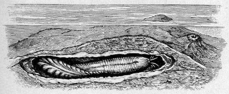
The former are divisible into two great groups. There are those that are free-swimming and are able to forage for themselves, such as the lovely Sea-mouse (Aphrodite aculeata), the large Eunice gigantea, the common Nereis pelagica, or the exquisitely coloured Chloeia flava. Others live a more retired life, dwelling in tubes, which they fashion for themselves; they lead either a solitary or a social life. Here we have examples of Sabella, Sabellaria, Serpula; a number of forms of worm-tubes, showing their great variety and beauty (see especially the delicate Filograna), are to be seen in the small Table-cases placed against the north wall of the Gallery. Attention should be especially directed to Mr. A. T. Watson’s beautiful preparations of Terebella littoralis. We give a figure (Fig. 19) after a drawing by that gentleman of the home of Panthalis oerstedi, the tube-forming habits of which have been carefully observed by him.
Fig. 19.
Home of Panthalis oerstedi.
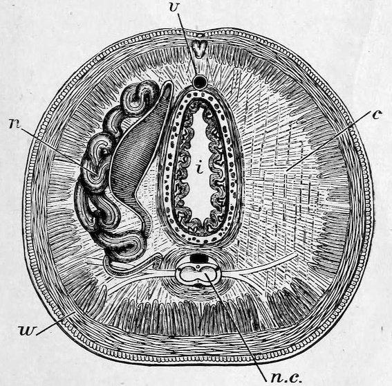
Fig. 20.
Section across the body of an earth-worm to show the disposition of the more important organs; the body wall (w) consists of dermis, circular, and longitudinal muscles; the body cavity is divided by membranes (c) into a series of chambers, in each of which opens the mouth of a coiled nephridium (n). The axis of the cavity is occupied by the intestine (i); above and below it is a longer blood-vessel (v), and below it is also the central nerve-cord (nc).
The Oligochæta are represented by the common Earthworm, the influence of which in the formation of mould and in the general ploughing of the soil was carefully investigated by Mr. Darwin; and by the little Tubifex rivulorum (Bloodworm), which owes both its red colour and its ability to dwell in mud, which is so poor in oxygen as to be unfit for respiration, to the same chemical compound as that which gives the red colour to our blood and carries the oxygen of respiration all over the body.
Acanthobdella: e, eyes; ch, chætæ; s, sucker.
The Hirudinea, or Leeches, are often said to be distinguished from the Chætopoda by the absence of bristles, but, as a fact, Acanthobdella (Figs. 21 and 22) has very well marked bristles. They always have a sucker at the hinder end of the body by which they are attached to their prey; they are found in fresh water (Piscicola), on sea-fishes (as Pontobdella), or in moist places, as the Leech (Hirudo). The last-named has three jaws, armed with as many as
ninety denticles. Trochetia subviridis (Land-Leech) is a species which is found rarely and sporadically in England.
The Myzostomaria form a division of Polychæta all the members of which live parasitically on Crinoids, and otherwise present great differences in their habits. Some move about freely on the Crinoids they infest, others are more sluggish and rarely move, others produce galls or cysts on their host, and yet others are internal parasites, and live in the alimentary canal. It is of interest to note that there are corresponding degrees of difference between the young and old specimens of the different groups of species.
The general organisation of Myzostomaria is shown in the accompanying figure (Fig. 23) in which the dorsal wall of the body is supposed to be transparent so as to allow of the chief internal organs being seen.
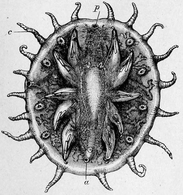
Fig. 23.
Diagram of Myzostomum to show the general form of the body and the marginal extensile cirri (c); within these and on the ventral surface are four pairs of suckers, and more internally five pairs of appendages each bearing two hooks; the proboscis (p), the digestive tract and its ramifications, and the reproductive organs are outlined as if seen through a transparent wall; a, anus.
The last group of Worms here represented is that of the Gephyrea; with the advance of our knowledge it is probable that they will be found to be more intimately allied to the Annulata than is now generally supposed; it will be seen indeed that Echiurus has bristles at its hinder end; Sipunculus is the best known representative of the unarmed Gephyrea; Bonellia is interesting both from the fact that it owes its green colour to a matter closely resembling the chlorophyll
of green plants, and from the possession by the female of a proboscis, which is protruded from the hole in the rock occupied by the worm: the male is very much smaller than the female, and is not nearly so well developed. Owing to the mode of lighting the Gallery, the visitor may have to shift his position several times before gaining a good view of the whole length of the proboscis.