Ocular Drug Delivery and Toxicology
FREDERICK T. FRAUNFELDER, MD
Drug delivery to the eye is a complex process. The eye is unique in the body in many ways that affect its pharmacology and toxicology. It includes several different cell types and functions basically as a self-contained system. The rate and efficacy of drug delivery differ in healthy and diseased eyes. Variables affecting delivery include age, genetic ancestry, and route of administration. The complexities of delivery, toxicology, or both are greatly influenced by patient compliance, especially in the management of glaucoma, which requires multiple topical ocular medications to be given at one sitting each day, often multiple times daily. Each time and method of drug delivery modify the therapeutic and toxicologic response.
Ocular toxicology is dependent on the concentration of the drug, frequency of application, speed of removal, and whether the drug reaches sensitive cells such as the corneal endothelium, lens epithelium, or macula in toxic concentrations. Of equal importance is the vehicle for delivery and the pH, buffering systems, and preservatives necessary for optimum drug delivery. Each adds its own potentially toxic effect to this complex picture. Originally, much of ocular pharmacology and toxicology was conducted by trial and error, often with local corner pharmacies compounding medications. Today, the ocular pharmaceutical industry is acutely aware of potential problems and is continuously researching and producing medications, usually with fewer side effects and delivered by better medications.
TOPICAL OCULAR ADMINISTRATION
This is by far the most commonly used method of drug delivery to the eye. Topically administered medications are convenient, easy to reapply, and relatively inexpensive. This method concentrates the pharmacologic activity of the drug on/in the eye while limiting systemic reactions. Local toxic responses are increased, however, especially with lifelong use, as with glaucoma medications. Unlike medication given orally, topical ocular medications reach systemic circulation
while avoiding the first-order pass effect through the liver. A drug absorbed through the nasal mucosa or conjunctiva “drains” to the right atrium and ventricle. The blood containing the drug is then pumped to the head before returning to the left atrium and ventricle. The second passage is through the liver, where the primary detoxification occurs before going to the right atrium. When medications are orally administered, the first pass includes absorption from the gut through the liver, where, depending on the drug, up to 90% of the agent is detoxified before going to the right atrium. Thus oral medications are metabolized during the first pass, whereas ocularly or nasally administered drugs are not metabolized until the second pass. This is the reason why therapeutic blood levels, and accompanying systemic side effects, may occur from topical ocular medications. Other factors include racial differences in metabolism, as with timolol. One percent of people with Japanese or Chinese genetic ancestry, 2.4% of African Americans, and 8% of those with European ancestry do not have the p450 enzyme CYP2D6 that is necessary to metabolize this drug. The lack of this enzyme significantly enhances systemic blood levels of timolol.1
BASIC PHARMACOLOGY AND TOXICOLOGY OF TOPICAL MEDICATIONS
Ocular toxicology is based on pharmacokinetics – how the drug is absorbed, including its distribution, metabolism, and elimination – as well as pharmacodynamics, the action of the drug on the body. This bioavailability is influenced by age, body weight, sex, and eye pigmentation. It is also affected by the disease process, interactions with other drugs, and mode of delivery. Only a small percentage of any topically applied drug enters the eye. At best, 1–10% of topical ocular solutions are absorbed by ocular tissues.2 This absorption is governed by ocular contact time, drug concentration, tissue permeability, and characteristics of the cornea and pericorneal tissue. Nearly all solutions will leave the conjunctival sac, or cul-de-sac, within 15–30 seconds
of application.3 The average volume of the cul-de-sac is 7 μL, with 1 additional μL in the precorneal tear film.4
The cul-de-sac may hold 25–30 μL of an eye drop; however, blinking will decrease this volume markedly and rapidly, so that, at most, only 10 μL remain for longer than a few seconds. The drop size of commercial drugs varies from 25 μL to more than 56 μL.4 In a healthy eye, one not affected by disease, lid manipulation to instill the drug will double or triple the normal basal tear flow exchange rate of 16% per minute, thereby decreasing ocular contact time via dilution.4
The cornea is the primary site of intraocular drug absorption from topical drug application. This is a complex process that favors small, moderately lipophilic drugs that are partially nonionized under physiologic conditions. Although the cornea is a five-layer structure, it has significant barriers to absorption into the eye. It can be visualized as three layers, like a sandwich, with a hydrophilic stroma flanked by lipophilic epithelium and endothelial layers.4
Topically administered drugs are also absorbed via the conjunctiva, sclera, and lacrimal system. The total surface area of the conjunctiva is 17 times the corneal surface area.4 The conjunctiva allows absorption of lipophilic agents to a lesser degree than the cornea, but it is relatively permeable to hydrophilic drugs. The sclera is porous via nerve and blood vessel tracts, but otherwise fairly resistant to penetration. Hydrophilic agents may pass through it 80 times faster than through the cornea; however, the lacrimal system can remove the drug 100 times faster than the cornea and conjunctiva can absorb it.4,5
Clearly, overflow from every administration of eye drops occurs not only over the eyelid but also in the lacrimal outflow system. Lynch et al showed that 2.5% phenylephrine topically applied to the eyes of newborn babies in 8-μL or 30-μL aliquots produced no difference in pupillary response.6 However, neonates who received the 30-μL dosage had double the plasma concentrations of phenylephrine of those who received 8 μL, increasing the potential for systemic complications.
INTRAOCULAR DISTRIBUTION
Once a drug reaches the inside of the eye, anatomic barriers play a major role in where it ends up. Drugs that enter primarily through the cornea seldom penetrate behind the lens. The pattern of aqueous humor flow and the physical barriers of the iris and ciliary body help keep the drug anterior. It is not uncommon for a drug to be more concentrated in the ciliary body than in the aqueous humor due to scleral absorption
directly into the ciliary body, with less fluid exchange than in the aqueous humor. In addition, pigmented tissue reacts differently to different drugs. For example, lipid-soluble mydriatics that are more slowly absorbed by pigmented cells will dilate dark pupils more slowly, resulting in longer duration but a decrease in maximum dilation.7
Drug distribution is markedly affected by eye inflammation. Tissue permeability is increased, allowing greater drug availability. However, as Mikkelson et al have demonstrated, protein binding may decrease drug availability 75–100% in inflamed eyes.8 The protein–drug complex decreases bioavailability. Increases in aqueous or tear protein, such as mucus, are also factors in bioavailability, as is the increased tearing that may wash away a drug before it can be absorbed.8
PRESERVATIVES
Preservatives are important parts of topical ocular medications, not only to prolong shelf life but also to disrupt the corneal and conjunctival epithelium to allow greater drug penetration.
Preservatives such as benzalkonium have been shown to have antibacterial properties almost as great as those of topical ocular antibiotics. Even in exceedingly low concentrations, benzalkonium causes significant cell damage by emulsification of the cell-wall lipids. De Saint Jean et al report cell-growth arrest and death at concentrations as low as 0.0001%.9 Shortterm use seldom causes clinically significant damage to healthy corneas and conjunctiva other than superficial epithelial changes. However, with long-term use, e.g. in patients with glaucoma and dry eye, preservatives in topical eye medication may cause adverse effects. Hong et al have shown induction of squamous metaplasia by chronic application of glaucoma medications containing preservatives.10 This may progress to more severe side effects, as shown in Table 2.1
VEHICLES FOR TOPICAL OCULAR MEDICATION DELIVERY
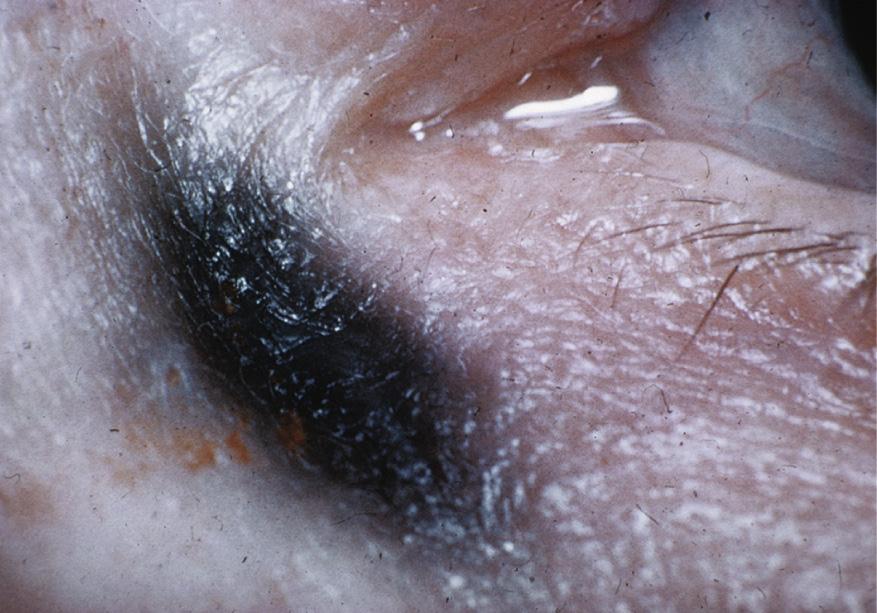
Aqueous solutions: With aqueous solutions, ingredients are fully dissolved within a solution. Benefits include easy application and few visual side effects. The main drawback is a short ocular contact time, which leads to poor absorption and limited bioavailability. Nevertheless, this is still the most commonly used means of delivering topical ocular medications. Solutions may congregate in the lacrimal sac (Fig. 2.1).
TABLE 2.1
Preservative Ocular Side Effects
Eyelids and Conjunctiva Cornea
Allergic reactions Punctate keratitis
Hyperemia Edema
Erythema Pseudomembrane formation
Blepharitis Decreased epithelial microvilli
Conjunctiva, papillary Vascularization
Edema Scarring
Pemphigoid lesion with Delayed wound-healing symblepharon
Squamous metaplasia Increased transcorneal permeability
Contact allergies Decreased stability of tear film Squamous metaplasia
Suspensions: With this vehicle, the active ingredient is in a fine particulate form suspended in a saturated solution of the same medication. This method allows for longer contact time with greater bioavailability. Its drawbacks include the necessity of vigorously shaking the container before application and a possible increase in foreign-body sensation after application because of the deposition of particles in the corneal tear film.
Ointments: These consist of semisolid lipoid preparations containing lipid-soluble drugs. They are designed to melt at body temperature and are dispersed by the shearing action of blinking. Ointments are frequently entrapped in lashes, fornices, and canthal areas, which are capable of acting as reservoirs. They can also become entrapped in corneal defects (Fig. 2.2); e.g. ointment at the base of the lashes comes in contact with the skin. Because ointment will melt when it comes in contact with the skin, the ointment at the base of the lashes reaches the eye in a continuous process of becoming entrapped in the lashes and remelting into the eye. Ointments have high bioavailability and require less frequent dosing than other methods but suffer by being difficult to administer. Other problems include variable dosing (it is difficult to control the amount applied) and possible unacceptability to patients due to blurred vision and cosmetic disfigurement.
Pledgets: Pledgets (small absorbent pads saturated with medication) may be used to deliver high concentrations of drugs directly to the ocular surface for relatively prolonged periods. This method of drug delivery
to the eye is not approved by the US Food and Drug Administration. Pledgets of vasoconstrictors to limit bleeding in keratorefractive surgery have been shown to cause significant systemic reactions, including hypertension, cardiac arrest, subarachnoid hemorrhage, convulsions, and death.11
Injections: Subconjunctival injections allow medication to be concentrated locally, with high bioavailability and limited systemic side effects. Wine et al suggested that the mechanism of drug delivery may be in part simple leakage of the drug through the needlepuncture site with subsequent absorption through the cornea.12 McCartney et al showed that subconjunctival injections of hydrocortisone did penetrate the overlying sclera and that the injection site should be located directly over the area of pathology.13
Intracameral injections are administered directly to the anterior chamber of the eye and are most frequently used to place viscoelastics. Although small amounts of antibiotics may also be administered, some of these drugs pose risks to the corneal endothelium, and cataracts, corneal opacities, anterior uveitis, and neovascularization are possible.
Intravitreal injections have become increasingly popular due to their efficacy against macular degeneration, bacterial and fungal endophthalmitis, and viral retinitis. Each drug has its own toxicity profile; however, these injections are so commonly done that the volumes, concentrations, and vehicles are well tested, and complications are within an acceptable risk–benefit ratio.
Other delivery devices: Ocuserts (small plastic membranes impregnated with medication); collagen corneal shields (biodegradable contact-lens–shaped clear
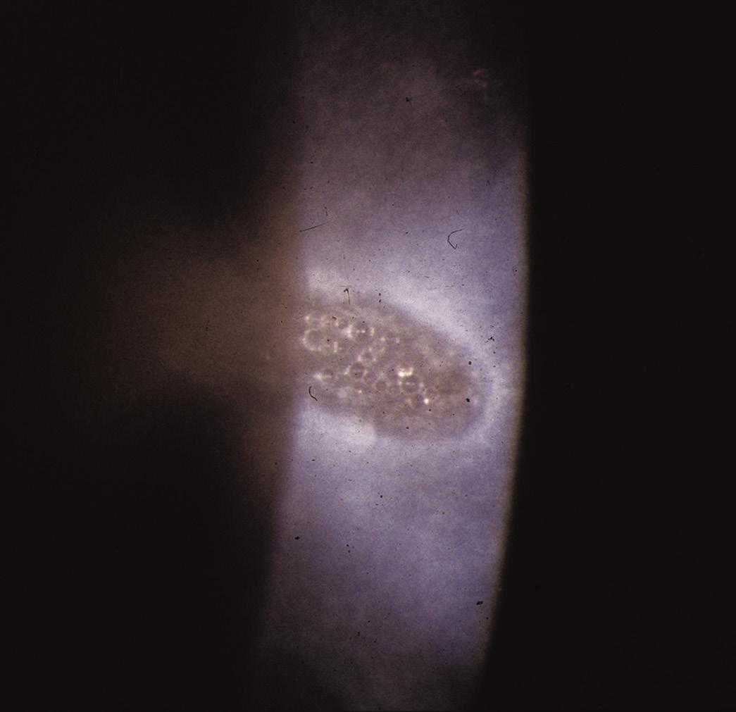
FIG. 2.1 Chronic use of silver nitrate solutions causes staining of the lacrimal sac and surrounding tissue18.
films made to dissolve within 12–72 hours); contact lenses; and various other delivery systems, including nanoparticles, liposomes, emulsions, and gels, have either made it to market with limited success or are still in the research pipeline.
TOXICITY RESPONSES
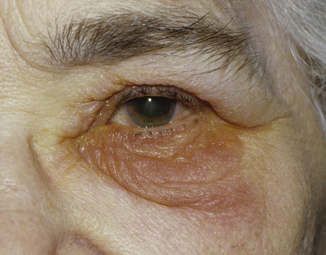
Anterior segment: Toxicity produces an inflammatory response without prior exposure to the host, whereas hypersensitivity responses require prior exposure. In general, allergic reactions involve repeated exposure to the antigen and sufficiently elapsed time to allow the immune system to react. Depending on the potency of the sensitizing agent or the strength of the immune system, this may vary from a few days to years.14 The clinical diagnosis of a toxic response is usually presumptive, whereas in allergic reactions conjunctival scraping may reveal eosinophils or basophils. One of the most common signs of ocular toxicity from topical medication is hyperemia. This reaction includes burning and irritation, usually without itching, occurring after starting an offending agent, with classic symptoms of intracanthal eyelid edema and erythema (Fig. 2.3). There are no definitive confirmatory tests. In more severe cases, a papillary hyperemia with a watery mucoid type of
discharge is evident. If the cornea is involved, this may present as a superficial punctate keratitis, usually more severe inferiorly or inferior nasally. Occasionally, intraepithelial microcysts may be seen, although these are more commonly seen with chemical toxicity. If the reaction is severe enough or goes unrecognized, it may become full blown with corneal ulceration, limbal neovascularization, anterior uveitis, cataracts, and damage to the lacrimal outflow system. The diagnosis is confirmed if clearing occurs after stopping the offending drug and the eye and adnexa improve markedly.
Drugs can induce a condition such as ocular pemphigoid, a syndrome of nonprogressive toxic reactions, which are self-limiting once the drug is discontinued. This condition is clinically and histologically identical to idiopathic ocular pemphigoid and includes a conjunctival cicatricial process with scarring of the fornix and tarsal conjunctiva, corneal and conjunctival keratinization, corneal vascularization, and lacrimal outflow scarring with occlusion.
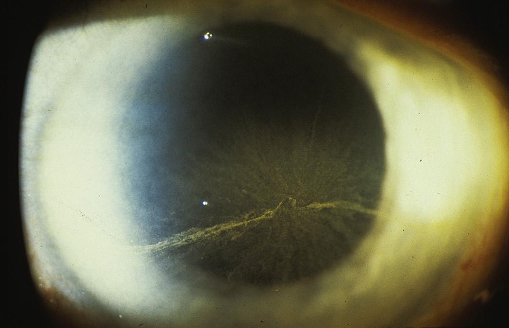
Almost any type of pathology can be seen as a result of a toxic response in the anterior segment. Systemic medications affect the anterior segment and occur via secretion of the drug into the tears with secondary changes due to the drug or its metabolites on ocular structures (Fig. 2.4). If the drug is secreted in the tears
FIG. 2.2 Corneal defects may entrap ointment on the surface, creating ointment globules18.
and deposited in the conjunctiva or cornea, it may produce changes in color vision or visual changes. The key to recognizing a toxic response is a high degree of suspicion that the pattern of symptoms and signs is not characteristic for the clinician’s differential diagnosis. A toxic effect is due to a pharmacologic effect from a drug that damages a structure or disturbs its function. An irritation is an inflammatory effect unrelated to sensitization or cellular immunity.
Ciliary body: Ciliary body ultrasound has shown bilateral choroidal effusions caused by various systemic drugs that may cause bilateral narrow-angle glaucoma.
Lens: It is difficult to identify which drugs are weak cataractogenic agents because these studies often require large numbers of patients. Findings are also difficult to confirm because instrumentation or classification systems are often cumbersome and costly. Some drugs used in the past, such as MER-29 (triparanol), caused acute lens changes, but cataractogenic drugs in current use are slow to cause lens changes, which may take many years to develop. In general, a drug-induced lens change is fairly specific for that drug. For example, both topical and systemic corticosteroid medications produce posterior subcapsular
FIG. 2.3 Allergic reaction18.
FIG. 2.4 Amiodarone keratopathy secondary to the drug being secreted in the tears18
opacities. Early recognition may in some cases reverse these changes, but this is rare for almost all druginduced cataracts.
Posterior segment : As newer classes of drugs are introduced, we are seeing more adverse retinal and optic nerve abnormalities. Whereas in the past visual acuity, color vision testing, and ophthalmoscopy were our primary tools for investigating retinal and optic nerve changes, electrophysiology testing is now being used with improved instrumentation and better standardization of methodology. Drugs can cause blood vessels to narrow, dilate, leak, swell, and hemorrhage. They can also cause pigmentary changes, photoreceptor damage, or inflammation. There can be deposition of the drug or its metabolites into the retina, as well as lipidosis. A drug can cause edema of the choroid, exudative detachment, or retinal detachment ( Fig. 2.5 ).
Elevation of intraocular pressure: Adverse ocular effects may cause acute glaucoma by dilation of the pupil or ciliary body effusions, by vasodilatation, by affecting the mucopolysaccharides in the trabecula (secondary to uveitis), or by means of a substance that interferes with aqueous outflow. Drugs or preservatives may, on chronic exposure, deposit in the ocular outflow system causing ocular pressure elevation.
Neurologic disorders: Multiple drugs can affect the extraocular muscles, causing weakness or paralysis, which in turn leads to ptosis, nystagmus, oculogyric crisis, or lid retraction. Direct neurotoxicity to the retina or optic nerve can occur, as can secondary optic nerve edema from benign intracranial hypertension.
Miscellaneous: Eyelash, eyebrow, and orbital disturbance reactions such as poliosis, madarosis, and exophthalmos or enophthalmos can also occur. Newer methods of delivery and new drugs have brought on side effects and toxicities not seen or recognized previously. The various metabolic pathways of patients and multiple variables such as drug, food, or disease interactions make recognition more difficult. Also, the basic incidence is often small, which makes an association difficult to prove.
HOW TO APPLY TOPICAL OCULAR MEDICATION
Applying Medication to Someone Else15
1. Tilt the person’s head back so he or she is looking up toward the ceiling. Grasp the lower eyelid below the lashes and gently pull it away from the eye (Fig. 2.6A).
2. Apply one drop of solution or a match-head–sized amount of ointment into the pocket between the lid and the eye (Fig. 2.6B). The external eye holds only about one quarter to one half of a drop, so don’t waste medicine by applying two drops.
3. As the person looks down, gently lift the lower eyelid to make contact with the upper lid (Fig. 2.6C). The person should keep their eyelid closed for 3 minutes.
Applying Your Own Medication15
1. Tilt your head back. Rest your hand on your cheek and grasp your lower eyelid below the lashes. Gently lift the lid away from your eye. Next, hold the
FIG. 2.5 Canthaxanthine perimacular deposition18
Another random document with no related content on Scribd:
Staph. alb. Staph. alb.
alb. Staph. alb. from heart’s blood.
B. coli
coli
coli Staph. alb. Staph. alb. Staph. alb.
Strep. viridans
viridans Strep. viridans
viridans from heart’s blood.
Group VI
N����� R.S.V. L.S.V. Prostate Cowper’s R. Testis R. Epidid. L. Testis L. Epidid. R. Scrotal Sac. L. Scrotal Sac.
1 Strep. vir. Staph. alb.
Staph. alb.
2 Strep. Strep. vir.
Strep. hem Strep. vir. Staph. alb. Strep. could
Staph. alb. Staph. alb. not grow.
Staph. alb. Strep. vir. Staph. alb. Strep. vir. (neutral) Staph. alb.
3 Strep. vir. Strep. vir.
Staph. alb. Staph. alb.
Strep. vir. Strep. vir. Strep. vir. Staph. alb. Staph. alb.
4 Staph. alb. Strep. vir. Strep. hem.
5 B. coli. B. coli. Staph. alb. B. coli. Staph. alb. Staph. alb. Staph. alb.
6 Strep. vir. Strep. vir. Staph. alb.
7 8 Ps. pyoca n. Ps. pyoc an. Strep. hem. Strep. hem.
9 Staph. alb. Staph. alb. Staph. alb. Strep. vir. Unident. Unident. Strep. hem. rod. rod.
10 Staph. alb. Staph. alb.
11 Staph. alb.
Staph. alb. Strep. hem. Strep. vir. Strep. Staph. alb. (neutral)
12 Staph. alb. Strep. vir. Staph. alb. Strep. vir.
13 Staph. alb. Staph. alb. Staph. alb. Strep. vir. B. coli. B. coli.
14 Staph. alb. Staph. alb. Strep. (neutr al) Strep. vir. Strep. vir.
15 Strep. hem. Strep. hem.
16 Staph. alb. Strep. vir.
Staph. alb. Strep. vir. Strep. hem.
R. Sem. Vesicle L. Sem. Vesicle Ductus Deferens R. Testis R. Epididymis L. Testis L. Epididymis
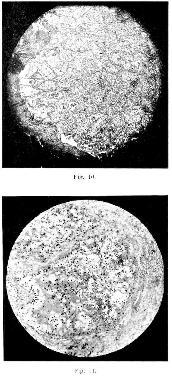
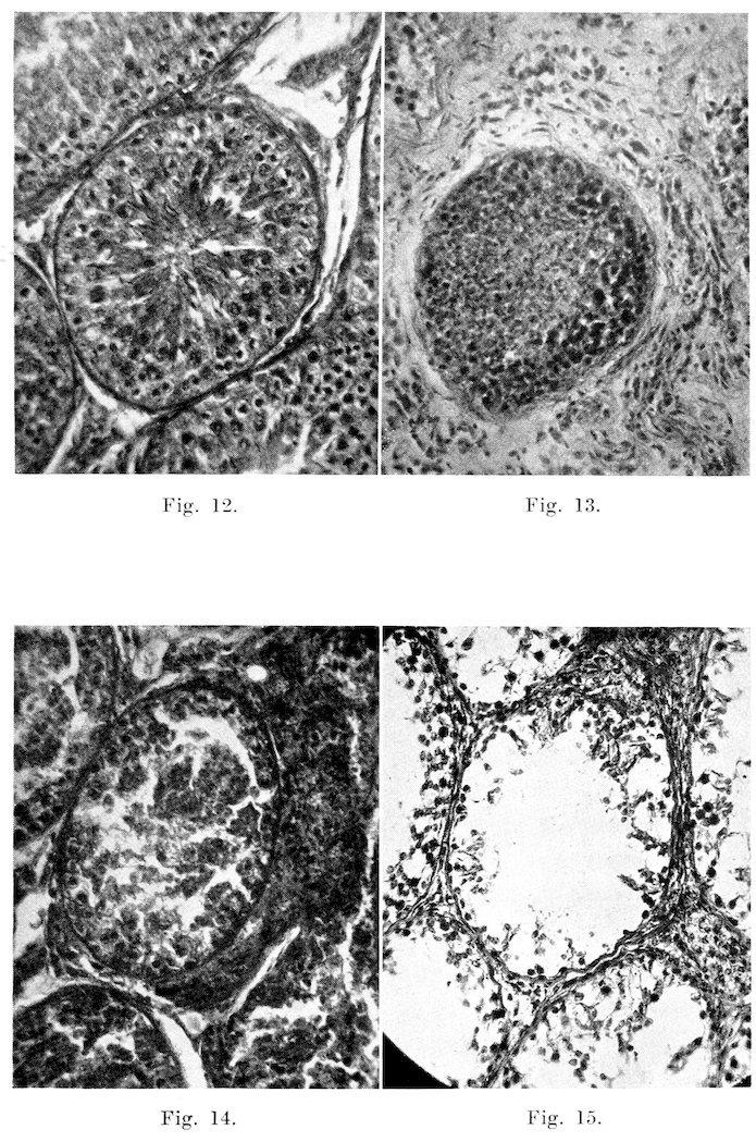
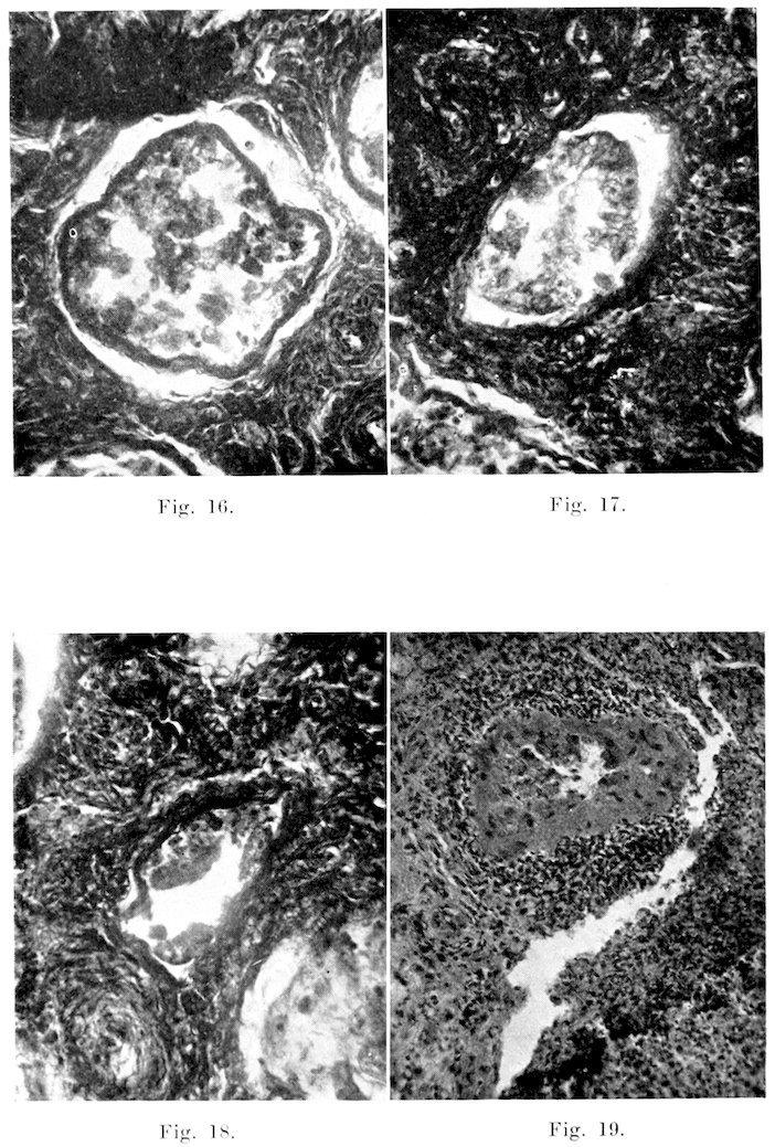
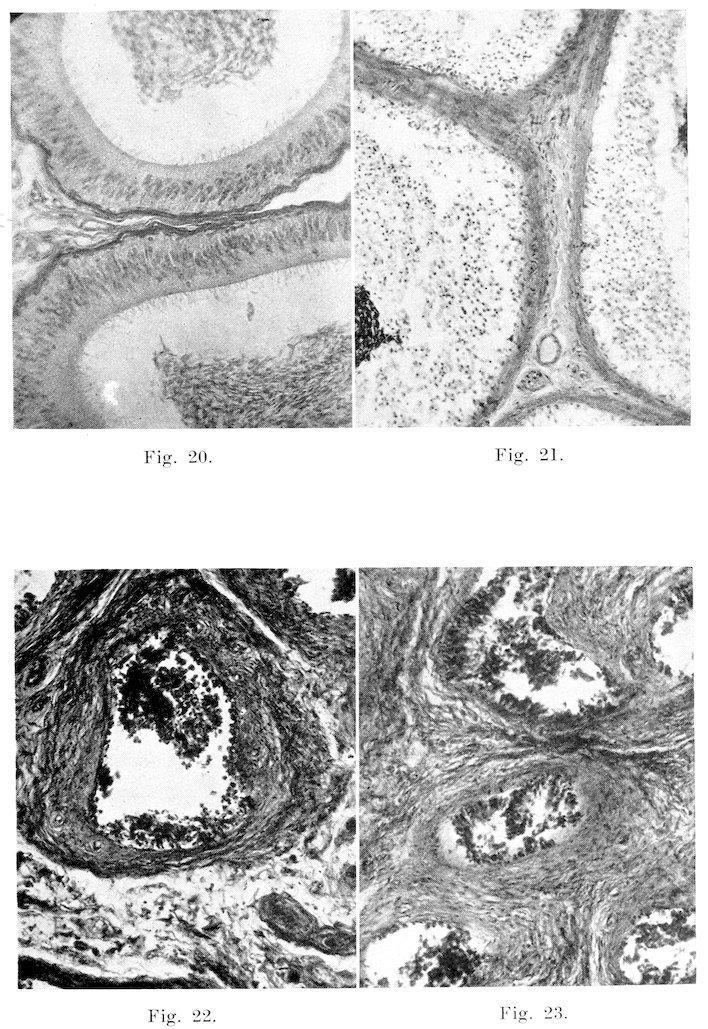
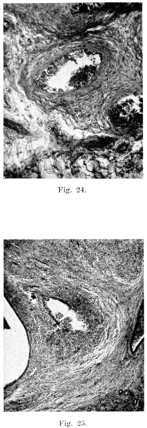
1 Desquamation and degeneration of lining membrane.
Same as right. Both apparently normal.
Apparently normal. Acute desquamati on and degeneratio n in some tubules.
Apparently normal. Degeneration and desquamatio n in some tubules.
Debris and exudates in lumen. Others atrophied due to a chronic interstitial inflammation .
2 Most vesicles degenerating. Same as right. Right apparently normal.
Interstitial cellular infiltration.
Necrosis in one large area.
Left shows degeneratio n of the lining membrane.
3 Entire gland degenerated. Same as right. Slight desquamati on and degeneratio n of mucosa of right ductus.
Coagulation necrosis of interstitial tissue. Debris and exudates in the vesicles.
4 Same as No. 3. Same as No. 3. Epithelium entirely denuded in both ducts.
Most tubules apparently normal. Degeneration and desquamati on of epithelium of the tubules.
Some show degeneratio n of the seminal epithelium.
Some tubules normal. Others show degeneratio n of the seminal epithelium.
Much of the epithelium denuded.
Most tubules becoming atrophied due to a chronic inflammation . Same as right, but some tubules are becoming atrophied due to a chronic inflammation .
Much degeneratio n and desquamati on of the seminal epithelium.
Debris and exudates in the lumen.
Same as right. Same as right.
Desquamation of most of liningmembrane.
Same as right. Same as right.
R. Sem. Vesicle L. Sem. Vesicle Ductus Deferens R. Testis R. Epididymis L. Testis L. Epididymis
5 Epithelium becoming degenerated.
Vesicles filled with debris and exudates.
6 Acutely inflamed.
7 Some degeneration of lining membrane.
Same as right. Both apparently normal. Degeneration of many tubules.
All epithelium denuded.
Debris in lumen.
Same as right. Nearly normal.
Same as right. Slight degeneration of lining membrane of both. Degeneration of mucosa in some tubules. Apparently normal. Degeneration in some tubules. Apparently normal.
No mitosis in some.
Same as right. Both apparently normal. Apparently normal. Apparently normal. Apparently normal. Apparently normal.
8 Necrosis in areas. Same as right. Both apparently normal. Apparently normal. Apparently normal. Apparently normal. Apparently normal. Infiltration of interstitial tissue. Degeneration of lining membrane.
R. Sem. Vesicle L. Sem. Vesicle Ductus Deferens R. Testis R. Epididymis L. Testis L. Epididymis
9 Apparently normal. Apparently normal. Both apparently normal. Apparently normal. Apparently normal. Apparently normal. Apparently normal.
10 Slight desquamatio n and degeneration of the lining membrane.
Same as right. Both apparently normal. Apparently normal. Degeneration of lining membrane. Apparently normal. Same as right.
11 Apparently normal. Apparently normal. Both apparently normal. Some tubules show no mitosis, others are degenerating. Degeneration and desquamatio n of lining membrane. Same as right. Degeneration and desquamatio n of lining membrane.
12 Degeneration and desquamatio n of epithelium of some vesicles. Same as right. Both apparently normal. Apparently normal. Degeneration of epithelium of all tubules. Apparently normal. Apparently normal.
Debris and exudates in lumen. Some increase in connective tissue.
R. Sem. Vesicle L. Sem. Vesicle Ductus Deferens R. Testis R. Epididymis L. Testis L. Epididymis
13 Degeneration of most of gland.
Same as right. Slight catarrhal inflammati on of mucosa of both. Apparently normal.
Degeneration and desquamati on of lining membrane of most tubules. Apparently normal.
Same as right.
Cellular infiltration of interstitial connective tissue.
Debris and exudates in lumen.
14 Apparently normal. Apparently normal. Apparently normal. Some degeneratio n of seminal epithelium of some tubules. Apparently normal. Apparently normal. Acute inflammatio n. Some desquamatio n of lining membrane.
15 Slight desquamatio n and degeneration of lining membrane.
16 Chronic interstitial inflammatio n.
Acute catarrhal inflammatio n. Both apparently normal.
Debris and exudates in lumen.
More acute than right. Both apparently normal.
Degeneration of seminal epithelium in some tubules.
Desquamation of lining membrane. Apparently normal. Apparently normal.
Debris in lumen.
Degeneration in some tubules. Apparently normal.
Atrophy of many tubules.
Same as right. Acute inflammatio n. Some desquamatio n and degeneration of lining membrane.
Some show poor mitosis.
B�����������
B��� B. Die Aetiologie des seuchenhaften (infectiösen) Verwerfens. Zeit Med. Band I, S. 241, 1897. Das seuchenhaften Verwerfen der Rinder. Arch. wiss. u. prakt. Thierheilk., Band 33, S. 312, 1907.
L��, J����. Contagious Abortion of Cows. Circular No. 5, Agr. Experiment Sta. University of California, June, 1903.
Report of English Commission of Epizootic Abortion. Appendix to Part I, p. 17, 1909.
H�����, F. B., and L����, H. The Bull as a Disseminator of Contagious Abortion. Jour. Amer. Vet. Med. Assoc., L, 1916–17, p. 143.
H�����, F. B. Contagious Abortion Questions Answered. Bulletin No. 296, Oct. 1921, Agr. Experiment Sta., University of Wisconsin.
B���, J. M., C�����, G. T., and L�����, H. H. Bacterium Abortus Infection of Bulls (Preliminary Report). Jour. Agr. Research, August, 1919.
S��������, E. C., and C�����, W. E. The Bull as a Factor in Abortion Disease.
C�����, W. E. Proceedings of A. V. M. A., p. 851, 1913.
C��������, C. M. Report of the New York State Veterinary College, Cornell University, 1920–21.
R������, L. F., and W����, G. C. Infectious Abortion in Cattle. Storrs Agr. Exp. Sta. Bulletin No. 93. January, 1916.
M�F������, S������� and M�����. Researches Regarding Epizootic Abortion. Jour. of Comp. Path. and Therap., XXVI,
142, 1913.
S��������, E. C. Bureau of Animal Industry Investigations of Bovine Infections Abortion. Jour. Amer. Vet. Med. Assoc., LX, p. 542. February, 1922.
B�����, J. D. Recent Studies on the Pathology of the Seminal Vesicles. Bost. Med. and Surg. Jour., CLXXI, 1914, 59.
W�������, W. L. The Diseases of Bulls. Cornell Veterinarian. X, 94. April, 1920.
W�������, W. L. Report of the New York State Veterinary College, Cornell University, 1920–21.
W�������. W. W. Technique of Collecting Semen for Laboratory Examination with Review of Several Diseased Bulls. Cornell Vet. X, 87, April, 1920.
W�������, W. W. Diseases of the Bull Interfering With Reproduction. Jour. of Amer. Vet. Med. Assoc., LVIII, 29, October, 1920.
H�����, E. B. Herd Efficiency from the Standpoint of the Veterinarian. North Amer. Vet., III, 71, February, 1922.
W�������, W. W. Observations on Reproduction in a Purebred Dairy Herd. Cornell Veterinarian, XII, 19, January, 1922.
W�����, K. M. The Diagnosis and Treatment of Sterility in the Male. Lancet, CCI, 228. July 30, 1921.
P����. Endocrinoglia-Pathologia E Clinica. Abstract in Endocrinology, II, 42.
F�������, J. S. Normal Histology and Microscopical Anatomy.
E����������. Vergl. mikroskop. Anatomie der Haustiere. II.
C�������, M. R. On the Existence of a Secretion of the Epididymis of the Hibernating Bat and Its Significance. C. R. Soc. de Biol. (Paris), 1920, LXXXIII, 67–69.
S������, R. Abstract in Jour. Phys. et Path. Gen. (2) CLXXI, 273, September, 1918.
27. 28. 29. 30. 31. 32. 33. 34. 35. 36. 37. 38.
D����������. Ansfuhrapparat und Anhangsdrusen der männlichen Geschlechtsorgane. Oppel’s Lehrbuch der vergleichenden mikr. Anat. der Wirbeltiere, IV, Jena, 1904.
R�����, A. A. The Verumontanum, With Special Reference to the Sinus Pocularis: Its Histology, Anatomy, and Physiology. Jour. Urology, I, 1917, 231.
F���, P. A. The Spermatic Secretion and Its Ultraparticles. Cornell Veterinarian, October, 1921.
M�������, F. H. A. The Physiology of Reproduction.
K��������, B. F. Professor of Histology and Embryology, Cornell University, Ithaca, New York.
W�����. Arch. f. Anat. u. Entwicklungsgesch., 1899, und Arch. f. Anat. u. Physiol. 1899.
B��������, W. On the Significance of the Secretions of the Male Accessory Genital Organs. Münch. med. Wchnschr. 1920, 67, 1, p. 44.
E��������. Jour. of Amer. Med. Assoc., LXXVII, No. 1, 42, July 2, 1921.
R�������, E. Fertility and Sterility. Jour. of Amer. Med. Assoc., LXVII, 1193–1199, October 21, 1916.
C���, W. H. Examination of Semen With Special Reference to Its Gynecological Aspects. Amer. Jour. of Obstetrics and Diseases of Women and Children. LXXIV, No. 4, 1916.
W���, C. G. L. The Survival of Motility in Mammalian Spermatozoa. Jour. of Physiol. IV, 246, August 3, 1921.
B����, J. H. The Use of Blood Agar for the Study of Streptococci. Monograph of the Rockefeller Institute for Med. Res., 1919, IX.
G������, J. B., and others. The Surgical Treatment of Sterility due to Obstruction at the Epididymis Together with a Study of the Morphology of Human Spermatozoa. Univ. of Penn. Med. Bulletin, March, 1902.
39. 40. 41. 42. 43. 44. 45. 46. 47. 48.
R�������, E. and M�������, D. Defective Diet as a Cause of Sterility. Jour. of Amer. Med. Assoc., LXXVII, 169, July 16, 1921.
N������, P. A. Tissues of the Testicle and Antaminosis. Gazetta degli Ospedali (Milano), 1920, XLI, 424.
W�������, W. L. Observations on Reproduction in a Pure Bred Beef Herd. Cornell Veterinarian. January, 1922.
B���, W. B. Correlation of Function: With Special Reference to the Organs of Internal Secretion and the Reproductive System. Brit. Med. Jour., 1920, 1, 787.
B����, W. L. The Principles of Internal Secretion. Brit. Med. Jour, 1920, II, 687–691.
J���. Discussion in Penn. Med. Jour., XXV, 81.
B����, A. The Internal Secretory Organs.
H����, E. E. The Effect of Thymus Feeding on the Activity of the Reproductive Organs of the Rat. Jour. of Physiology, XLVII, 1913–1914, 479.
L����-J����, O. and H���, F. A. The Influence of Excessive Sexual Activity of Male Rabbits. 1. On the Properties of the Seminal Discharge. Jour. of Exper. Zoology, 1918, XXV, 463.
W�������, W. L. Improvement of the Reproductive Efficiency of Cattle. North Amer. Veterinarian, III, May, 1922.
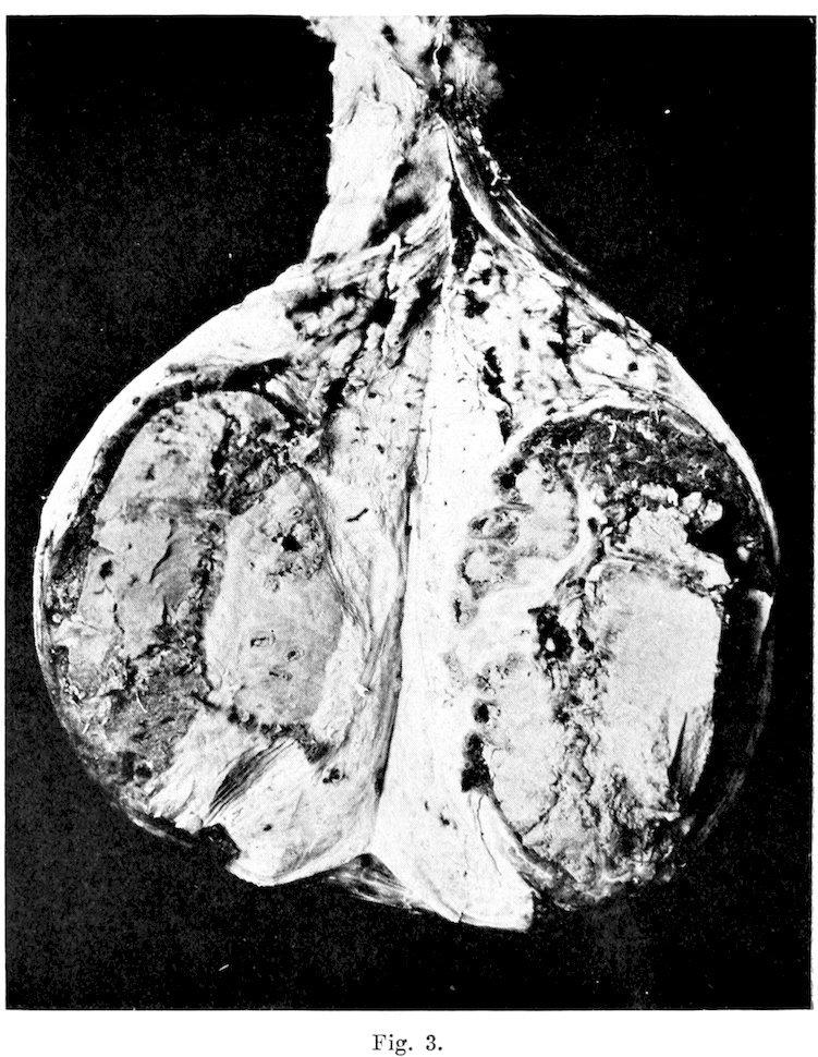
P���� I.
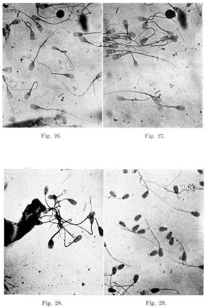
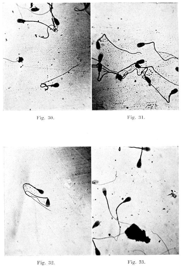
Fig. 1.
Fig. 2.