Oxford Textbook of Cancer Biology
Francesco

Francesco
Visit to download the full and correct content document: https://ebookmass.com/product/oxford-textbook-of-cancer-biology-francesco-pezzella /
More products digital (pdf, epub, mobi) instant download maybe you interests ...

Oxford Textbook of Cancer Biology Francesco Pezzella (Editor)
https://ebookmass.com/product/oxford-textbook-of-cancer-biologyfrancesco-pezzella-editor/
Principles of Cancer Biology 1st Edition
https://ebookmass.com/product/principles-of-cancer-biology-1stedition/

Oxford Textbook of Migrant Psychiatry Bhugra
https://ebookmass.com/product/oxford-textbook-of-migrantpsychiatry-bhugra/
Cell biology : translational impact in cancer biology and bioinformatics 1st Edition Mitchell
https://ebookmass.com/product/cell-biology-translational-impactin-cancer-biology-and-bioinformatics-1st-edition-mitchell/
Oxford Textbook of Palliative Medicine 5th Edition
https://ebookmass.com/product/oxford-textbook-of-palliativemedicine-5th-edition/

Oxford Textbook of Neurological Surgery 1st Edition Ramez Kirollos
https://ebookmass.com/product/oxford-textbook-of-neurologicalsurgery-1st-edition-ramez-kirollos/

Oxford textbook of palliative nursing Fifth Edition Ferrell
https://ebookmass.com/product/oxford-textbook-of-palliativenursing-fifth-edition-ferrell/
Oxford Textbook of Interventional Cardiology 2nd Edition Redwood
https://ebookmass.com/product/oxford-textbook-of-interventionalcardiology-2nd-edition-redwood/

eTextbook 978-0199360574 Oxford Textbook of Correctional Psychiatry (Oxford Textbooks in Psychiatry)
https://ebookmass.com/product/etextbook-978-0199360574-oxfordtextbook-of-correctional-psychiatry-oxford-textbooks-inpsychiatry/

EDITED BY Francesco Pezzella
Nuffield Division of Clinical Laboratory Sciences, Radcliffe Department of Medicine, University of Oxford, Oxford, UK
Mahvash Tavassoli
Department Mucosal and Salivary Biology, King’s College London, London, UK
Nuffield Division of Clinical Laboratory Sciences, Radcliffe Department of Medicine, University of Oxford, Oxford, UK; Weill Cornell College of Medicine, New York, USA
Great Clarendon Street, Oxford, OX2 6DP, United Kingdom
Oxford University Press is a department of the University of Oxford. It furthers the University’s objective of excellence in research, scholarship, and education by publishing worldwide. Oxford is a registered trade mark of Oxford University Press in the UK and in certain other countries © Oxford University Press 2019
The moral rights of the authors have been asserted
First Edition published in 2019
Impression: 1
All rights reserved. No part of this publication may be reproduced, stored in a retrieval system, or transmitted, in any form or by any means, without the prior permission in writing of Oxford University Press, or as expressly permitted by law, by licence or under terms agreed with the appropriate reprographics rights organization. Enquiries concerning reproduction outside the scope of the above should be sent to the Rights Department, Oxford University Press, at the address above
You must not circulate this work in any other form and you must impose this same condition on any acquirer
Published in the United States of America by Oxford University Press 198 Madison Avenue, New York, NY 10016, United States of America
British Library Cataloguing in Publication Data
Data available
Library of Congress Control Number: 2018960018
ISBN 978–0–19–877945–2
Printed in Great Britain by Bell & Bain Ltd., Glasgow
Oxford University Press makes no representation, express or implied, that the drug dosages in this book are correct. Readers must therefore always check the product information and clinical procedures with the most up-to-date published product information and data sheets provided by the manufacturers and the most recent codes of conduct and safety regulations. The authors and the publishers do not accept responsibility or legal liability for any errors in the text or for the misuse or misapplication of material in this work. Except where otherwise stated, drug dosages and recommendations are for the non-pregnant adult who is not breast-feeding
Links to third party websites are provided by Oxford in good faith and for information only. Oxford disclaims any responsibility for the materials contained in any third party website referenced in this work.
The textbook is dead. Long live the textbook! With increased output of rapidly published new data and availability of teaching material on the web, it has often been predicted that the textbook will become extinct. However, in our experience, it has also become increasingly difficult to find a comprehensive text which enables us to catch up with the current state of art in multiple fields, within a wider contextual framework. While the high number of research and review papers provide a continuous update on increasingly narrow and specialized topics in cancer biology, we think there will be always a need for concise, coherent descriptions of the fundamentals on areas like cell cycle or cell death. This is particularly important for students who require a platform of basic information before venturing more deeply into the literature. We have assembled a fantastic cast of authors, each of whom are outstanding in their field, and have attempted, when relevant, to make the translational link to the application of cancer biology for patient benefit. We must
understand that without novel basic science and the generation of new knowledge, there cannot be sustainable innovations in cancer diagnosis and therapy. We have structured this book logically and trust that the inquisitive reader will select which chapters to explore in greater depth.
There is a difference between the textbooks of today and yesterday: before, publication was the terminus or end of the work for its authors; now, because of the integration between the printed book and online resources, this is no longer the case. This will allow us to annually review, revise, and update the chapters on the online version of the book to reflect recent developments in the field.
Finally, as this is a cancer textbook, we would like to remember our parents, relatives, friends and, of course, patients whose lives have been affected and in many cases, ended too soon by this disease. We hope this book is another small step forward in the right direction.
We would like to thank our friends Sandor Paku, Balazs Dome, and Andrew Reynolds for granting us permission to use the picture on the cover of the book, illustrating a non-angiogenic tumour growing in a mouse model.
We would also like to acknowledge the help and support by Oxford University Press staff that guided us through the process
of creating this book: Andrea, Caroline, Janine, Sree, and Anya. We also would like to thank all the authors for their work and their willingness and commitment to write.
Abbreviations xi
Contributors xv
SECTION I
The multicellular organism
1. The multicellular organism and cancer 3
Francesco Pezzella, David J. Kerr, and Mahvash Tavassoli
2. DNA repair and genome integrity 13
Giacomo Buscemi
3. Evolution and cancer 33
Tom Donnem, Kingsley Micklem, and Francesco Pezzella
SECTION II
The aetiology of cancer
4. Genetics and genetic instability in cancer 43
Mark A. Glaire and David N. Church
5. Epigenetics 56
Edward Hookway, Nicholas Athanasou, and Udo Oppermann
6. Viral carcinogenesis—an overview 71
Dirk P. Dittmer and Blossom Damania
7. Chemical carcinogens 79
David H. Phillips
8. Radiation as a carcinogen 91
Yan-Qun Xiang and Chao-Nan Qian
How the cancer cell works
9. Growth factors and associated signalling pathways in tumour progression and in cancer treatment 105
Nadège Gaborit and Yosef Yarden
10. Hormones and cancer 123
Balkees Abderrahman and V. Craig Jordan
11. Oncogenesis and tumour suppression 136
Mahvash Tavassoli and Francesco Pezzella
12. The signalling pathways in cancer 155
Jiangting Hu and Francesco Pezzella
13. Cell cycle control 178
Simon Carr and Nicholas La Thangue
14. Cancer and cell death 196
Jessica Bullenkamp and Mahvash Tavassoli
15. Telomerase and immortalization 209
Laura Collopy and Kazunori Tomita
16. Cancer metabolism 221
Almut Schulze, Karim Bensaad, and Adrian L. Harris
17. Chaperones and protein quality control in the neoplastic process 239
Andrea Rasola
18. Oxygen and cancer: The response to hypoxia 255
Adrian L. Harris and Margaret Ashcroft
19. Invasion, metastasis, and tumour dormancy 270
Andrey Ugolkov and Andrew P. Mazar
20. Cancer stem cells 283
Connor Sweeney, Lynn Quek, Betty Gration, and Paresh Vyas
SECTION IV
Cancer microenvironment
21. Cancer-associated stroma 303
Wilma Mesker and Rob Tollenaar
22. Blood vessels and cancer 314
Francesco Pezzella and Robert Kerbel
23. Cancer immunology 330
Herman Waldmann
SECTION V
Global vision of cancer
24. Molecular profiling in cancer research and personalized medicine 347 Pieter-Jan van Dam and Steven Van Laere
25. Proteomics and metabolomics applications in cancer biology 363
Pedro Cutillas and Benedikt M. Kessler
26. Cancer systems biology: From molecular profiles to pathways, signalling networks, and therapeutic vulnerabilities 375
Lieven Verbeke and Steven Van Laere
27. Cancer biology through immunohistology 394
Karen Pulford and Kevin Gatter
The biology of cancer treatment
28. Principles of chemotherapy 413
David J. Kerr, Daniel Haller, and Jaap Verweij
29. Immunotherapy and tumour resistance to immune-mediated control and elimination 423
Gwennaëlle C. Monnot and Pedro Romero
30. Biological effect of radiotherapy on cancer cells 438
Anna Dubrovska, Mechthild Krause, and Michael Baumann
Conclusions
31. Benign tumours: The forgotten neoplasms 453 Francesco Pezzella, Adrian L. Harris, and Mahvash Tavassoli
32. Conclusions: Cancer biology, a moveable feast 463
David J. Kerr, Francesco Pezzella, and Mahvash Tavassoli
Index 469
AID activation-induced deaminase
AIDS acquired immune deficiency syndrome
ALCL anaplastic large cell lymphoma
ALK anaplastic lymphoma kinase
ALK+DLBCL anaplastic lymphoma kinase-positive diffuse large B cell lymphoma
ALO17 lymphoma oligomersation parter on chromosome 17
ALT adult T-cell lymphoma
Alt-NHEJ alternative non-homologous end-joining
AML acute myeloid leukaemia
AMPK adenosine-monophosphate-activated protein kinase
AP alkaline phosphatase
AP1 Activator protein 1
APAAP alkaline phosphatase-anti-alkaline phosphatase
APB ALT-associated PML body
APE1 apurinic/apyrimidinic endonuclease 1
AT ataxia telangiectasia
Atg autophagy-related protein
ATIC 5-aminoimidazole-4-carboxamide ribonucleotide formyl transferase/IMP cyclohydrolase
ATM ataxia telangiectasia mutated protein kinase
ATP adenosine triphosphate
ATR ataxia telangiectasia and rad-3-related protein kinase
ATRIP ATR-interacting protein
B biotin
β-CAT- β-catenin
B-CLL B-cell chronic lymphocytic leukaemia
BCL2 B-cell leukemia/lymphoma 2
BCL6 B-cell leukemia/lymphoma 6
BCR-ABL breakpoint cluster region - Abelson
BER base excision repair
BL Burkitt lymphoma
BLM bloom syndrome protein
BM bone marrow
BRCA1, 2 breast cancer type 1 and 2
CAH IX congenital adrenal hyperplasia IX
CAK CDK-activating kinase
CAM cell adhesion molecules
CARs cysteinyl-tRNA synthetase
cART combination antiretroviral therapy
CBP CREB-binding protein
CDC25A, C cell division cycle 25A and 25C
CDK cyclin-dependent kinase
CDKI cyclin-dependent kinase inhibitor
CHK1, 2 checkpoint protein kinase 1 and 2
CIN chromosomal instability
CK7 cytokeratin 7
CK8/18 cytokeratin 8/18
CKI CDK-inhibitory
CLTC clathrin heavy chain
CMIP CpG island methylator phenotype
c-RAF RAF proto-oncogene serine/threonine-protein kinase
CRE cyclic AMP response element
CRKL CRK avian sarcoma virus CT-10 homologue-like
CS Cockayne syndrome
CSC cancer stem cell
CSR class switch recombination
CtIP CtBP-interacting protein
C3G-CRKL protooncogene c-CRK
D-loop displacement loop
DAPK death-associated protein kinase
DDR DNA damage response
Deptor DEP domain-containing mTOR-interacting protein
DNA-PKcs DNA-dependent protein kinase catalytic subunit
DNA Pol DNA polymerase
DR5 death receptor 5
DRAM damage-regulated autophagy modulator
DSB double-strand break
E-cad E-cadherin
E2F1 E2F transcription factor 1
EBV Epstein–Barr virus
EGF epidermal growth factor
EGFR epidermal growth factor receptor
EME1 essential meiotic endonuclease 1
EML4 echinoderm microtubule-associated protein like 4
ER endoplasmic reticulum
ER oestrogen receptor
ERCC1, 5 excision repair cross-complementation group 1 and 5
ERK extracellular-signal-regulated kinase
EXO1 exonuclease 1
FA Fanconi anaemia
FANCA, B, C, Fanconi anaemia complementation
D2, M, I, P group A, B, C, D2, M, I, P
FGFR fibroblast growth factor receptor
FITC fluorescein isothiocyanate
FOXM1 forkhead box M1
FOXO3A forkhead box protein O3
FOXP1 forkhead box protein 1
FOXP3 forkhead box protein 3
FRS2 fibroblast growth factor receptor substrate 2
GAB2 GRB2 associated binding protein
GADD45 growth arrest and DNA damage 45
GATA4 GATA-binding protein 4
GC germinal centre
GF growth factor
GFR growth factor receptors
GG-NER global genome nucleotide excision repair
GGR global genome repair
GRB2 growth factor-receptor-bound protein 2
HAV hepatitis A virus
HBV hepatitis B virus
HCV hepatitis C virus
HDAC histone deacetylase
HER2 human epidermal growth factor receptor 2
HIV Human Immunodeficiency Virus
HIF-1 hypoxia-inducible factor
HNSCC head and neck squamous cell carcinoma
HPV human papilloma virus
HR homologous recombination
HRP horseradish peroxidase
HSC haematopoietic stem cells
HTLV human T-lymphotropic retrovirus
HVS herpesvirus saimiri
IARC International Agency for Research on Cancer
ICL interstrand DNA crosslink
IDL insertion and deletion loop
IMT inflammatory myofibroblastic tumours
IR ionizing radiation
IR Insulin receptor
IRF-4 interferon regulatory factor 4
IRS-1 insulin receptor substrate 1
JAK3 Janus kinase 3
JNK-C Jun N-terminal kinase
KAP-1 KRAB-associated protein-1
KEGG Kyoto Encyclopedia of Genes and Genomes
KIF5B kinesin/family member 5B
KRAS Kirsten rat sarcoma viral oncogene homologue
KS Kaposi’s sarcoma
KSHV Kaposi’s sarcoma-associated herpesvirus
LC3 microtubule-associated protein 1A/1B-light chain 3
LFS Li-Fraumeni syndrome
LUCA last unknown common ancestor
MAP MUTYH-associated polyposis
MAPK mitogen-activated protein kinase
MCD multicentric Castleman disease
MCL mantle cell lymphoma
MCPV Merkel cell polyomavirus
MDC1 mediator of DNA-damage checkpoint 1
MDM2 mouse double minute 2 homologue
MIN microsatellite instability
MLH1 MutL homologue 1
MMR mismatch repair
MMP matrix metalloproteinase
MRN Mre11/Rad50/Nbs1 complex
MSH2 MutS homologue 2
MSN moesin
mTOR mammalian target of rapamycin
MYC MYC proto-oncogene
MYCN MYCN protooncogene, neuroblastoma derived
MYH9 non-muscle heavy chain
NAD nicotinamide adenine dinucleotide
NBS Nijmegen breakage syndrome
NER nucleotide excision repair
NF-kB nuclear factor kappa-light-chain-enhancer of activated B cells
NHEJ non-homologous end-joining
NPC nasopharyngeal carcinoma
NPM nucleophosmin
NPM-ALK nucleophosmin-anaplastic lymphoma kinase
NSCLC non-small cell lung cancer
p53BP1 p53 binding protein 1
p130CAS breast cancer anti-estrogen resistance 1
p130Cas breast cancer anti-oestrogen resistance protein 1
pAMPK phosphorylated 5’ adenosine monophosphateactivated protein kinase
PAR poly-ADP-ribose
PARG poly (ADP-ribose) glycohydrolase
PARP poly (ADP-ribose) polymerase
PCAF P300/CBP-associated factor
PCNA proliferating cell nuclear antigen
PDGF platelet-derived growth factor
PI3K phosphoinositide 3-kinase
PKB protein kinase B
PLC-g phospholipase C-gamma
PLK1 polo kinase 1
PLWHA people living with HIV/AIDS
PML promyelocytic leukaemia
PR progesterone receptor
PTEN phosphatase and tensin homologue
p16 cyclin-dependent kinase inhibitor 2A
p53 TP53 or tumour protein
p56 phosphoglycerate kinase
P53R2 p53-inducible ribonucleotide reductase small subunit 2-like protein
RAD51, radiation repair 51 and 52
RAD52
RANBP2 Ran-binding protein 2
KRAS rat sarcoma viral oncogene homologue
Rb retinoblastoma protein
Redd1 regulated in development and DNA damage response 1
RISC RNA-induced silencing complex
RNA ribonucleic acid
ROS reactive oxygen species
RPA replication protein A
RSV rouse sarcoma virus
RTK receptor tyrosine kinase
SAC spindle assembly checkpoint
SASP senescence-associated secretory phenotype
SEC31L1 SEC31 homologue A
SH2 src homology 2
SHM somatic hypermutation
SHP2 protein-tyrosine phosphatase 1D or proteintyrosine phosphatase 2C
SIRT1 sirtuin 1
SOS Son of Sevenless
SRC non-receptor protein kinase sarcoma
ssDNA single-strand DNA
SSB single-strand break
SSBR single strand break repair
STAT5 signal transducer and activator of transcription 5
STORM stochastic optical reconstruction microscopy
T-loop telomere loop
T-SCE telomere-sister chromatid exchange
T-stump telomere stump
TAL-1 T-cell acute lyphocytic leukaemia protein 1
TC-NER transcription-coupled nucleotide excision repair
TCP tumour control probability
TCR transcription-coupled repair
TEN telomerase N-terminal
TFG TRK-fused gene
TFIIH transcription factor IIH
TLS translesion DNA synthesis
TNFSF10 tumour necrosis factor superfamily member 10
TopBP1 topoisomerase (DNA) II binding protein 1
TPM tropomyosin
TrkA tropomyosin receptor kinase A
TSC2 tuberous sclerosis 2
TTD trichothiodystrophy
ULK1 Unc-51-like kinase 1
VEGF vascular endothelial growth factor
VEGFR vascular endothelial growth factor receptor
VIM vimentin
Wip1 wild-type P53-induced phosphatase 1
XLF XRCC4-like factor
XP xeroderma pigmentosum
XPA, B, C, xeroderma pigmentosum complementation group
D, F, G A, B, D, F, G
XRCC1, 4 X-ray cross-complementing protein 1 and 4
ZAP70 zeta-chain (TCR) associated protein kinase 70 kD
Balkees Abderrahman, Department of Breast Medical Oncology, University of Texas, MD Anderson Cancer Center, Houston, USA
Margaret Ashcroft, Department of Medicine, University of Cambridge, Cambridge, UK
Nicholas Athanasou, Nuffield Department of Orthopaedics, Rheumatology, and Musculoskeletal Science, University of Oxford, Oxford, UK
Michael Baumann, German Cancer Research Center (DKFZ); and Department of Radiotherapy and Radiation Oncology, Faculty of Medicine and University Hospital Carl Gustav Carus, Technische Universität Dresden, Germany
Karim Bensaad, Department of Oncology, University of Oxford, Oxford, UK
Jessica Bullenkamp, Molecular and Clinical Sciences Research Institute, St. George’s University London, London, UK
Giacomo Buscemi, Department of Biosciences, University of Milan, Milan, Italy
Simon Carr, Department of Oncology, University of Oxford, Oxford, UK
David N. Church, Cancer Genomics and Immunology Group and NIHR Comprehensive Biomedical Research Centre, The Wellcome Centre for Human Genetics, University of Oxford, Oxford, UK
Laura C. Collopy, Cancer Institute, Faculty of Medical Sciences, University College London, London, UK
Pedro Cutillas, Cell Signalling and Proteomics Group, Barts Cancer Institute (CRUK Centre), Queen Mary University of London, London, UK
Blossom Damania, Lineberger Comprehensive Cancer Center and Department of Microbiology and Immunology, School of Medicine, University of North Carolina, Chapel Hill, USA
Dirk P. Dittmer, Lineberger Comprehensive Cancer Center and Department of Microbiology and Immunology, School of Medicine, University of North Carolina, Chapel Hill, USA
Tom Donnem, Department of Oncology, University Hospital of North Norway and the Arctic University of Norway, Tromso, Norway
Anna Dubrovska , OncoRay-National Center for Radiation Research in Oncology, Faculty of Medicine and University Hospital Carl Gustav
Carus, Technische Universität Dresden; and Helmholtz-Zentrum Dresden-Rossendorf, Institute of Radiooncology-OncoRay; Cancer Consortium (DKTK), partner site Dresden, and German Cancer Research Center (DKFZ), Germany
Nadège Gaborit, Institut de Recherche en Cancérologie de Montpellier, INSERM U1194, Université de Montpellier, Institut régional du Cancer de Montpellier, Montpellier, France
Kevin Gatter†, Nuffield Division of Clinical Laboratory Sciences, Radcliffe Department of Medicine, University of Oxford, Oxford, UK
Mark A. Glaire, Cancer Genomics and Immunology Group and NIHR Comprehensive Biomedical Research Centre, The Wellcome Centre for Human Genetics, University of Oxford, Oxford, UK
Betty Gration, MRC Molecular Haematology Unit, Radcliffe Department of Medicine, Weatherall Institute of Molecular Medicine, University of Oxford, Oxford, UK
Daniel Haller, Department of Medicine, Perelman School of Medicine, University of Pennsylvania, Philadelphia, USA
Adrian L. Harris, Department of Oncology, University of Oxford, Oxford, UK
Edward Hookway, Nuffield Department of Orthopaedics, Rheumatology and Musculoskeletal Sciences, University of Oxford, Oxford, UK
Jiangting Hu, Radcliffe Department of Medicine, University of Oxford, Oxford, UK
V. Craig Jordan, Department of Breast Medical Oncology, University of Texas MD Anderson Cancer Center, Houston, USA
Robert Kerbel, Biological Sciences Platform, Sunnybrook Research Institute, Department of Medical Biophysics, University of Toronto, Toronto, Canada
David J. Kerr, Nuffield Division of Clinical Laboratory Sciences, Radcliffe Department of Medicine, University of Oxford, Oxford, UK; and Weill Cornell College of Medicine, New York, USA
Benedikt M. Kessler, Target Discovery Institute, Nuffield Department of Medicine, University of Oxford, Oxford, UK
† It is with regret we report the death of Kevin Gatter during the preparation of this textbook.
Mechthild Krause, Department of Radiotherapy and Radiation Oncology, Faculty of Medicine and University Hospital Carl Gustav Carus, Technische Universität Dresden; German Cancer Consortium (DKTK), partner site Dresden, and German Cancer Research Center (DKFZ); OncoRay – National Center for Radiation Research in Oncology, Faculty of Medicine and University Hospital Carl Gustav Carus, Technische Universität Dresden, Helmholtz-Zentrum Dresden - Rossendorf; Helmholtz-Zentrum Dresden - Rossendorf, Institute of Radiooncology – OncoRay, Dresden, Germany; National Center for Tumor Diseases (NCT), Partner Site Dresden; German Cancer Research Center (DKFZ); Faculty of Medicine and University Hospital Carl Gustav Carus, Technische Universität Dresden; and Helmholtz Association / Helmholtz-Zentrum Dresden - Rossendorf (HZDR), Germany
Nicholas La Thangue, Department of Oncology, University of Oxford, Oxford, UK
Andrew P. Mazar, Monopar Therapeutics, Wilmette, USA
Wilma Mesker, Department of Surgery, Leiden University Medical Center, Leiden, the Netherlands Kingsley Micklem, Nuffield Division Clinical Laboratory Science, Radcliffe Department of Medicine, University of Oxford, Oxford, UK
Gwennaëlle C. Monnot, Ludwig Cancer Research Center, Department of Fundamental Oncology, Faculty of Biology and Medicine, University of Lausanne, Lausanne, Switzerland
Udo Oppermann, Nuffield Department of Orthopaedics, Rheumatology, and Musculoskeletal Science, University of Oxford, Oxford, UK
Francesco Pezzella, Nuffield Division of Clinical Laboratory Sciences, Radcliffe Department of Medicine, University of Oxford; and Cellular Pathology Clinical Service Unit, Oxford University Hospitals, Oxford, UK
David H. Phillips, Department of Analytical, Environmental and Forensic Sciences, School of Population Health and Environmental Sciences, King’s College London, London, UK
Karen Pulford, Emeritus Reader in Immunodiagnostics, Nuffield Division of Clinical Laboratory Sciences, Radcliffe Department of Medicine, University of Oxford, Oxford, UK
Chao-Nan Qian, Department of Nasopharyngeal Carcinoma, State Key Laboratory of Oncology South China, Sun Yat-Sen University Cancer Center, Guangzhou, China
Lynn Quek, MRC Molecular Haematology Unit, Radcliffe Department of Medicine, Weatherall Institute of Molecular Medicine, University of Oxford, Oxford, UK
Andrea Rasola, Department of Biomedical Sciences, University of Padova, Padova, Italy
Pedro Romero, Ludwig Cancer Research Center, Department of Fundamental Oncology, Faculty of Biology and Medicine, University of Lausanne, Lausanne, Switzerland
Almut Schulze, Department of Biochemistry and Molecular Biology, Biocenter, University of Würzburg, Würzburg, Germany
Connor Sweeney, MRC Molecular Haematology Unit, Radcliffe Department of Medicine,
Weatherall Institute of Molecular Medicine, University of Oxford, Oxford, UK
Mahvash Tavassoli, Department Mucosal and Salivary Biology, King’s College London, London, UK
Rob Tollenaar, Department of Surgery, Leiden University Medical Center, Leiden, the Netherlands
Kazunori Tomita, Cancer Institute, Faculty of Medical Sciences, University College London, London, UK
Andrey Ugolkov, Division of Hematology and Oncology, Feinberg School of Medicine, Northwestern University, Chicago, USA
Pieter-Jan van Dam, Faculty of Medicine and Health Sciences, University Antwerp, Antwerp, Belgium
Steven Van Laere, HistoGeneX NV, Antwerp, Belgium
Lieven Verbeke, Department of Information Technology, Ghent University, Ghent, Belgium
Jaap Verweij, Department of Medical Oncology, Erasmus University Medical Centre, Rotterdam, the Netherlands
Paresh Vyas, MRC Molecular Haematology Unit, Radcliffe Department of Medicine, Weatherall Institute of Molecular Medicine, University of Oxford, Oxford, UK
Herman Waldmann, Sir William Dunn School of Pathology, University of Oxford, Oxford, UK
Yan-Qun Xiang, Department of Nasopharyngeal Carcinoma, Sun Yat-Sen University Cancer Center, Guangzhou, China
Yosef Yarden, Department of Biological Regulation, Weizmann Institute of Science, Rehovot, Israel
1. The multicellular organism and cancer 3
2. DNA repair and genome integrity 13
3. Evolution and cancer 33
Francesco Pezzella, David J. Kerr, and Mahvash Tavassoli
Cancer has a lot to do with the way life has developed on our planet and the successful evolution of multicellular organisms. Cells are the smallest unit containing all the features necessary and sufficient to life, the viruses occupying a special place. In 1863, the German pathologist Rudolf Virchow introduced the concept of cellular pathology (Virchow, 1863) stating that diseases are due to the occurrence of a pathological process at cellular level. This is very much the case with cancer that is definitively a disease of a cell belonging to a multicellular organism.
A brief history of the cell: Eubacteria (Bacteria), Archaea, Eukaryotes, and the last unknown common ancestors
The defining moment for the appearance of the cell has been the formation of what we now call the cell membrane. This is a complex structure able to form vesicles allowing the segregation inside of genetic material (the Genotype) plus the molecular machinery (the Phenotype) needed for this new structure to grow and reproduce copies of itself, through the cell cycle.
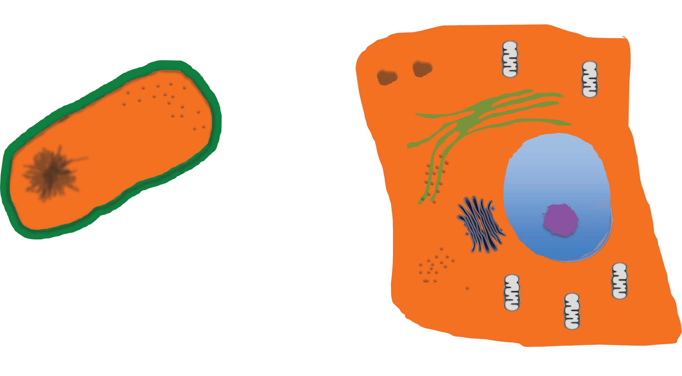
Cells are divided into two taxons, Prokaryota and Eukaryota, a taxon being formed by organisms included in a particular entity (e.g. in a family or in a genus; (Thain and Hickman, 2004). This distinction is based on the structure and organization of the cell: in the Eukaryotes (Composite), cell membranes are present also inside the cells delimiting discrete internal structures such as, for example, nucleus and mitochondria, while no such division can be found in the prokaryotes (non-composite; Fig. 1.1). All the cells share a set of common features: they contain their genetic information, replicate throughout the cell cycle, their activity is governed through cell signalling and can produce energy through a metabolic apparatus. Approximately 200 gene families are common to the two taxons.
The introduction of genomic studies, as a tool to investigate the evolutionary correlations between organisms, has unveiled within the prokaryotes two distinct groups or domains, the Bacteria and the Archaea, as distant from one other as they are from the Eukaryotes. It has therefore been proposed that, above the division into animal
Kingdoms, exists a division into three domains: the Eubacteria (or Bacteria), the Archaea, and the Eukaryotes (Woese et al., 1990), each domain comprising a variety of kingdoms (Fig. 1.2). Molecular studies have demonstrated that the two domains Bacteria and Archaea derive from the last unknown common ancestors (LUCA), while the Eukaryotes evolved from the Archaea (Fig. 1.2). LUCA is defined as the last organism preceding, in the evolutionary tree, the division into the two domains of Bacteria and Archaea and it is assumed to be the living organism from which all present living organisms descend. It is estimated that LUCA lived between some 3.5 to 3.8 billion years ago (Fig. 1.3).
The genetic division of cells into these three domains is reflected by their biological characteristics, some of which are summarized in Table 1.1. The mechanism of transcription, translation, and splicing in the Archaea is close to that of the Eukarya and both differ from the one found in the Prokaryota. Crucially, although the Archaea do not have a nucleus, they have histone proteins that bind to DNA double strand, compacting it into nucleosome-related structures, and Archaea RNA polymerases have the multisubunit complexity of Eukarya RNA polymerases. On the other side, the metabolism of the Archaea is more similar to Eubacteria than to Eukarya (Olsen and Woese, 1997). Despite the closer similarity in metabolic functions of Eubacteria to Archaea, there is one exception: the use of photosynthesis that can be found both in Eubacteria and Eukarya but is absent in Archaea. This is due to the fact that, although genetic evidences show that the Eukarya evolved from the Archaea, horizontal transfer of genes has happened between Eubacteria and Eukarya (Hedges, 2002).
In the cytological classification dividing the prokaryote from the eukaryotic cell the latter is distinguished as it is composed by several organelles, some possibly reminding a more primitive cell, which have learned to live in symbiosis. Each of these organelles contributes to specific need(s) of the eukaryotic cell. It is now believed that the crucial moment to the transition from a simpler cell to the more complex eukaryote was when different cells started to live inside others. Crucial to all this was the formation of the nucleus and the appearance of mitochondria. The main anatomical
Cell wall, external (green)
Cell membrane internal (black)
Coiled
DNA
Ribosomes
Endoplasmic reticulum
Rough, with ribosomes
Lysosomes
Endoplasmic reticulum
Smooth
Nuclear membrane black
NUCLEUS
Golgi
Cytoplasm
Fig. 1.1 The prokaryotic and the eukaryotic cells. (A) The prokaryotic cell is defined by the cell membrane. Inside the space delimitated by this membrane is the cytoplasm in which all the molecules are contained in one unique space. (B) The eukaryotic cell is also defined by the cell membrane, however the cell membrane is also present inside the cells where defines different organelles. The most prominent is the nucleus, in which the genetic material, the DNA, is segregated. Other cell membrane-defined organelles are the mitochondria, the Golgi apparatus, lysosomes, and the endoplasmic reticulum (ER). The former is divided into the ER rough, when ribosomes are attached to its membrane, and smooth, when ribosomes are not present.
LUCA
Last common unknown ancestor
Bacteria
Aquifex
Cyanobacteria
Gram-positive bacteria
Euryarchaeota
Archea
Diplomonodas
Crenarchaeota
Amoebe
Thaumarchaeota
Microsporidia
Flagellate
Slime molds
Purple bacteria
Animals
Fungi
Plants
Fig. 1.2 The three domains: Bacteria, Archaea, and Eukaryota. The last unknown common ancestor (LUCA) evolved into the first two domains, Bacteria and Archaea, which, as far as the anatomical structure is concerned, are prokaryotic cells. Subsequently from the Archaea, the third domain evolved: the Eukaryota. Each of these three domains evolved into several kingdoms. Adapted from https://commons.wikimedia.org/wiki/File:Phylogenic_Tree-en.svg © Conquistador / Wikimedia Commons / CC BY-SA 3.0
4500 mya Earth formation 3800–3600 mya Last Unknown Common Ancestor
3500 mya First form of life: fossil bacteria
1200–2100 mya First multicellular eukaryotic organism: Red algae. These algae have Cancer-like lesions
665 mya Early invertebrate, know to have Cancer
3465 mya first multicellular prokariote: Filamentous Cyanobacteria, able of “Cheating” behaviour
650 mya Sponges
200–220 mya Mammals
Tertiary Quaternary
6 mya Last Common Ancestor to Humans and Chimpanzee
5.8-6 mya Oldest hominids: Orrorin tugenensis, Ardipithecus ramidus kadabba and Sahelanthropus tchadensis
characteristics of the eukaryotic cells, when not dividing, are represented in Fig. 1.1.
The cell membrane
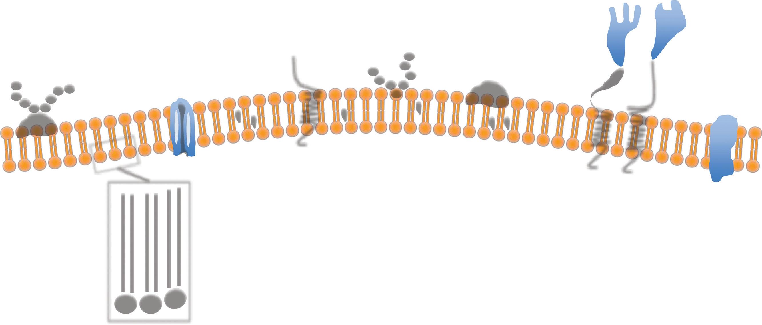
The cell membrane is a bilayer of phospholipids, each with a hydrophilic head and a hydrophobic tail. In aqueous environments, the phospholipids spontaneously organize themselves as a double layer with the hydrophobic tails inside and the head outside, so originating the cell bio membrane (Fig. 1.4). In a cell, the membranous network is divided into two main components: the cell surface membrane, the plasma membrane delimiting the actual cell and representing the border with the extracellular world, and those membranes delimiting the internal cellular compartments. In the plasma membrane within the scaffolding formed by the phospholipid bilayer are numerous different structures. These are made by proteins, 500 (five hundred) types of lipid
1.98 mya Oldest benign tumour known in an hominid (Australopithecus Sediba)
1.7 mya Oldest malignant tumour known in an hominid (Homo genus or Paranthrapus).
0.2 mya Homo Sapiens
molecules and 10,000 (ten thousand) proteins are involved in the making of the cell membrane. The main structures formed by intramembranous proteins are channels (e.g. ion pumps) and receptors (e.g. epidermal growth factor receptor). Some molecules however, like oxygen, can diffuse through the membrane without needing a specific pump.
The plasma membrane is a highly dynamic fluid structure and all the protein complexes are ‘floating’ wtihin it and also the very same lipid molecules are continuing moving within the membrane. Groups of lipids can also form units called ‘rafts’, which move among the other lipids. This dynamic nature of the plasma membrane was firstly described in 1972 as the fluid mosaic model (Singer and Nicolson, 1972; Edidin, 2003). The external cell membrane is in continuity with the internal membranes that not only defines the internal organelles of the cells, but also provides a framework for countless biochemical reactions and trafficking of molecules.
Table 1.1 Comparison of the main biological characteristics of Eubacteria, Archaea, and Eukaryota
Cell
Cytoskeleton
Organelles
Multicellularity
Transcription
tail Fatty acids
Hydrophilic head Glycero-phosphate group
Fig. 1.4 The cell membrane. The cell membrane is made up by phospholipid molecules with a hydrophilic head (orange) and a hydrophobic tail (yellow). Within the membrane are several different structures that can ‘float’ across the membrane, which has fluid property. Transmembrane proteins span all thickness of the membrane and the main types are the protein channels, the integral protein, and the alpha-helix proteins. Glycoproteins and carbohydrates are present on the external surface. Reproduced with permission from Saikat R. / socratic.org / CC BY-NC-SA 4.0. Available from: https://socratic.org/questions/in- the- cell-membrane-plasma-membrane-phospholipid-bilayer-what-do- the-peripheral
The two major compartments inside the cells are the nucleus and the cytoplasm; the latter includes all the intracellular volume which is not nucleus.
The cytoplasm is occupied by cytosol, an aqueous medium rich in proteins and salts accounting for approximately 50% of the cytoplasm. The main site of protein synthesis and degradation and of intermediate metabolism, forms the cytosol that permeates all the organelles. The main reason for different organelles is the need for keeping apart different biochemical reactions. A single membrane delimits all the organelles, with the only exceptions being the mitochondria and the nucleus, which have an outer and one inner membrane.
Mitochondria are organelles formed by an external and one internal membrane filled with matrix. It is where the oxidative phosphorylation (i.e. respiration), occurs and where adenosine triphosphate (ATP) is produced. ATP is the source of energy for all cellular functions: such an energy is liberated when an ATP molecule is hydrolysed producing adenosine diphosphate (ADP), phosphate, and energy. The endoplasmic reticulum, or ER, forms the major cytoplasmic network, most of which is Rough ER where ribosomes are located and protein synthesis occurs. The remaining one is called the Smooth ER.
Another prominent function of the ER is lipid synthesis. The Golgi apparatus is another system of cisterns where simpler molecules are packaged into more complex ones: it is also where lysosomes are built. The lysosomes are spherical membrane vesicles containing a wide range of hydrolytic enzymes able to degrade many molecules and their purpose is to eliminate any damaged or unwanted molecules. Such molecules are transported to the lysosome by specialized vesicles called endosomes. Peroxisomes are instead involved in several metabolic and catabolic functions. The most important are catabolism of very long chain fatty acids into branched chain fatty acids, D-amino acids, and polyamines with reduction of reactive oxygen species. They are also the place where phospholipids are synthesized and the pentose phosphate pathway, critical for the energy metabolism, is located. Finally, free ribosomes are also present within the cytoplasmic matrix.
The nucleus is the largest organelle. A double bilayer membrane, the nuclear membrane or nuclear envelope, in which numerous pores (nuclear pore complexes) are present, thus allowing communication with the cytoplasm, which delimits it. Within the nucleus is the genome (with only the exception of mitochondrial DNA) and its transcriptional machinery. It can be divided into two main structures: the nuclear membrane and the nuclear interior (Lammerding, 2011).
Between the two layers of the nuclear membrane is the perinuclear space. Under the nuclear membrane is the nuclear lamina, mainly made up by laminin filaments. This membrane is perforated by the nuclear pore complexes which cause the inner and outer membranes to fuse. Nuclear pore are large complexes made of approximately 50 nucleoporins and regulate the trafficking between nucleus and cytoplasm. Nuclear pore complexes are not the only protein structure within the nuclear membrane, with some spanning the whole thickness (Lammerding, 2011).
When not dividing, approximately half of the nuclear volume is occupied by chromatin made of the unfolded DNA packed around histone proteins. There are two types of chromatin: heterochromatin, more packed and less transcriptionally active, and euchromatin, which is not so condensed and in which most of the transcription occurs (Lammerding, 2011). The aggregates of DNA and histones form structures called nucleosomes: packaged DNA from different chromosomes occupy distinct areas in the non-dividing nucleus. Nucleoli are discrete bodies formed by proteins and nucleic acids and are the production site of the ribosome. Cajal bodies are located in the proximity of the nucleoli and contain different formations: for example, the snurposome and spliceosome are involved in the processing of the recently transcribed mRNA. Finally, there is the nucleoskeleton, a protein scaffolding supporting the different nuclear components. All these structures are immersed in the nucleoplasm, a very protein rich aqueous medium equivalent to the cytosol.
All the unicellular organisms tend to grow without limitation according to the availability of resources. The life cycle of each of these organisms therefore coincides with the time required to duplicate (i.e. to complete the cell cycle), the cell cycle being a complex of events that brings one cell to divide into two. Prokaryotes do not age: their life cycle is very simple. They die when conditions became adverse and food is scanty, although this is not always the case: in determinate conditions, some cells can become ‘dormant’ and resume growth when the environment becomes permissive again. In most eukaryotic cells ageing does appear: their lifespan is regulated by an internal clock made up by telomeres and telomerase. The main physiological events in the life cycle of a single cell are reproduction through mitosis, response to damage, cell death, and movement. Cells divide through mitosis, a process in which the genetic code is duplicated and then the cells divide into equal new cells. As cells are exposed to external insults, either chemical or physical, repair mechanisms are present that can block the further division until the necessary corrections are made. Should the repairs fail, the cells can trigger their own death through apoptosis. Apoptosis does not only follow damage within a multicellular organism but can also be triggered at appropriate moments during the organism’s development or life.
During the evolution of life, multicellularity has appeared independently at several different times, exploiting different strategies (Kaiser, 2001). There are therefore several mechanisms leading to the formation of multicellular organisms. For example, while plants relied on the formation of a rigid cell wall which brings different cells into one organism, the animal cells, which do not have cell walls, had to rely on membrane proteins called adhesion molecules to provide a mechanism allowing the cells to stick to each other (Bonner, 1998).
When confronted by an aggregate of cells, the first issue is how to differentiate a multicellular organism from a colony. The most commonly used, and the broadest criteria, is the existence of a spatial division of work in multicellular organisms, compared
to the colony in which each unicellular member performs the same tasks. According to this definition, the oldest known unambiguous multicellular organisms belong to the Bacteria domain and are the filamentous cyanobacteria. These emerged, as suggested by fossil dating, 3,465 million years ago (mya), approximately 1,000 million years after the Earth’s formation, which is estimated at 4,500 mya ( Fig. 1.3).
Cyanobacteria were the first organisms to develop photosynthesis and release oxygen. However, these bacteria also rely on the enzyme nitrogenase to convert nitrogen gas into ammonia, necessary to build their proteins and other essential structural components when combined nitrogen (i.e. reactive molecules containing nitrogen), like nitrate, nitrite, ammonium, urea, and amino acids, are not available (Fig. 1.5). However, nitrogenase is irreversibly destroyed in the
presence of oxygen and therefore cyanobacteria had to find a way to be able to perform oxygen-producing photosynthesis and, at the same time, to maintain nitrogenase function. This problem has been solved by multicellularity: formation of filaments, made up by a line of cyanobacteria in which two differently evolved cyanobacteria can be found. Those containing chlorophyll, performing photosynthesis, and releasing oxygen, are more numerous. These differentiated into a form called heterocysts, in which nitrogenase function is maintained in the absence of photosynthesis, as they do not contain chlorophyll (Fig. 1.3). Therefore, a simple prokaryotic multicellular organism containing two types of cells was formed (Bonner, 1998; Adams, 2000; Flores and Herrero, 2010).
The time of the emergence of eukaryotic multicellular organisms as assessed today is still a broad estimate, possibly sometime between 2,100 (Donoghue and Antcliffe, 2010) and 1,200 (Rokas, 2008) mya. Red algae are so far considered as the first eukaryotic multicellular organisms, appearing 1,200 mya (Fig. 1.3). The main strategies employed by eukaryotic cells to build a multicellular entity include: lack of cell separation after mitosis; mostly found in aquatic organisms; and aggregation of single cells prevalent in terrestrial creatures. A final fundamental characteristic in the classification of multicellular organisms is complexity. The easiest and most practical approach to ‘measure’ complexity is the number of cells making up the organism (Rokas, 2008).
Hallmarks of multicellularity
Multicellularity required the acquisition of functions not present or diversely utilized in single cell organisms. Up to seven hallmarks of multicellularity have been described (Rokas, 2008; Srivastava et al., 2010; Aktipis et al., 2015).
Regulation and control of the cell cycle
Fig. 1.5 The filamentous cyanobacteria. Cyanobacteria exists more commonly as vegetative form but can differentiate into three further forms: heterocyst, akinetes, and hormogonial cells. In absence of ‘combined nitrogen’, like nitrate, nitrite, ammonium, urea, and amino acids which easily react and combine with other molecules and can be used for protein production, cyanobacteria needs to ‘fixate’ the poorly reactive nitrogen and transform it into the more reactive ammonia according to the following reaction N2 + 8 H+ + 8 e → 2 NH3 + H2, catalysed by a nitrogenase enzyme. Vegetative cells cannot do that as they produce oxygen which is toxic for the nitrogenase enzyme. Therefore, some vegetative cells differentiate into heterocysts, cells that do not produce oxygen, but are able to fixate N2 into ammonia in response to deprivation of combined nitrogen. Subsequently, the filamentous cyanobacteria acquires its new structure, characterized by a number of vegetative cells regularly interrupted by one heterocyst. When nutrients and energy are scanty some vegetative cells differentiate into akinetes, which can than start to proliferate again and produce vegetative cells when nutrients became available again. Vegetative cells from some filamentous cyanobacteria can also differentiate into hormogonia, which are than dispersed and can subsequently originate new filamentous cyanobacteria growing in symbiosis with plants.
Adapted by permission from Macmillan Publishers Ltd: Springer Nature, Nature Reviews Microbiology, 'Compartmentalized function through cell differentiation in filamentous cyanobacteria’, Flores E and Herrero A. Copyright © 2010 Macmillan Publishers Limited, part of Springer Nature. All Rights Reserved.
A strict control of proliferation is essential to the development and survival of a multicellular organism. For the organism to maintain itself, proliferation can occur only in well-defined circumstances and is regulated by a series of positive signals, inducing it, and suppressive signals, blocking it. Furthermore, these control rules are different from tissue to tissue (e.g. the neurons do not enter proliferation ever), while, on the other extreme, bone marrow stem cells are continuously proliferating to provide new blood cells, which have a very high turnover. To guarantee this strict control, redundant mechanisms are present.
Apoptosis (programmed cell death)
While unicellular organisms just proliferate, within multicellular organisms remodelling takes place, mostly during development when some embryonic structures are temporary and need to be eliminated as the fetus develops. Apoptosis is also required in adulthood (e.g. after immune stimulations, only some of the immune cells specifically responding will survive; the others, responding in a non-specific way, will undergo apoptosis and disappear). This is possible thanks to the appearance of apoptosis, or programmed cell death, which causes, when necessary, the death of selected cells according to the organism’s blueprint.
The interaction with the extracellular environment
The extracellular matrix is essential for cells to maintain their physiological functions. Furthermore, it is where cells come into
contact with the immune system. Multicellular entities need to protect themselves from the intrusion of external pathogenic organisms and, at the same time, to maintain tolerance against ‘self’ antigens.
Specialization of cell types and division of work
As discussed before, the need for specialized cells cooperating is the very reason for which multicellularity developed. Different tissues need to perform a huge variety of different tasks, allowing the multicellular organisms to development degrees of complexity well outside the reach of single cell living forms. Again, this requires a strict control as each different tissue needs to differentiate in a very precise way according to its designated function.
Resources transport and allocation
According to the work each cell needs to do, different resources are required. While smaller organisms can rely on diffusion, the larger ones have different approaches with some relying on body cavities providing the required transport system, while the most complex have developed a branching vascular network. This is of course the case in humans, where the vascular and the lymphatic systems carry out this function.
Cell–cell and cell–matrix adhesion
As already discussed, different strategies for creation of multicellular organisms have emerged across the history of life. The cardinal function, which allows multicellularity in Metazoa, is that of adhesion (i.e. the creating stable mechanical contacts between cell and cell or cell and extracellular matrix). This is obtained by a variety of molecules like intercellular junctions, adhesion molecules, and adaption of cytoskeleton proteins.
Signalling and gene regulation
To develop and maintain a multicellular organism it is fundamental to have an efficient signalling system, both between and inside cells. This system is responsible for having each cell acting in synchrony with the other according to the organism’s blueprint. This is realized by a high regulation of gene transcription leading to the synthesis of proteins making up appropriate signalling pathways.
Hallmarks of multicellularity and cancer
Disruption of these functions has been found to be closely linked to the development of cancer (Hanahan and Weinberg, 2011; Aktipis et al., 2015).
Regulation and control of cell cycle
Uncontrolled proliferation due to either an excess of proliferative stimuli, classic oncogenes, or the loss of inhibitory functions, loss of tumour suppressor genes are covered in more detail in Chapters 11 and 13.
Apoptosis (programmed cell death)
Resistance to programmed cell death or to ageing leads to abnormal neoplastic cell accumulation, covered in Chapters 14 and 15.
The interaction with the extracellular environment
This includes avoiding immune destruction and cross-talk between tumours, their supportive stroma, and the immune system. Alteration of these functions leads to neoplastic cells to escape
potential anticancer activity of the immune system (see Chapters 23 and 29).
Specialization of cell types and division of work
Cancers develop from specific tissues but the ability to reproduce the structure and specialization of the cells seen in the normal tissue is lost to a variable degree across different malignancies (see Chapter 20).
Resources transport and allocation
Disruption of the normal blood supply is followed by the establishment of a new relationship between the neoplastic cells and the blood vessels. Cancer needs a resource transport system, which can be achieved in a variety of ways (Chapter 22). Also, a metabolic reprogramming of the cancer cell follows these changes (see Chapters 16 and 18).
Cell–cell and cell–matrix adhesion
Disruption or inhibition of these functions leads to the formation of abnormal neoplastic organ-like structures and to metastatic dissemination throughout the body (see Chapter 19).
Signalling and gene regulation
The normal network maintaining coordination gets disrupted following alterations at different levels along the way causing a pathological resetting of behaviour (Chapters 9, 10, and 12).
Cancer is a disease of multicellular organisms in which the emergence of changes in the DNA disrupts the instructions controlling the growth and physiology of some of the organism’s cells. Eventually one cell acquires enough changes to be able to abnormally grow outside the organism blueprint into a clone (i.e. a group of cells deriving from the same ancestor cell) of neoplastic cells. It is therefore an ‘information’ disease caused by alteration in the information blueprint (i.e. the genome). As DNA codes such instructions, any damage can lead to two main effects on the single host organism. The first is that the damage has actually no effect, if the area of changed genetic code is silent or redundant or not active at the time in which the damage occurs. The second leads to a change in the instruction blueprint, which is followed by pathological events of various nature. If the event is cellular death, some type of disease other than cancer can occur (e.g. degenerative diseases). Cancer is one of the pathological situations that can follow a genetic injury. It is characterized by a cellular growth that follow a reset of growth instructions that varies from one type of cancer to another and, indeed, from patient to patient causing the cancer cells to replace and destroy the normal body structures in an apparently chaotic way.
The instruction for changes leading to cancer are fundamentally those governing the set of functions necessary to ‘make’ a multicellular organism. As the cancer-linked pathways and cellular functions are associated with the appearance of multicellular organisms, it is not surprising that cancer, characterized by invasion and metastasis, or cancer-like phenomenon, characterized by abnormal proliferation and differentiation of ‘cheating’ cells (Aktipis et al., 2015), have been found across the spectrum of multicellular organisms
(Schlumberger and Lucke, 1948; Scharrer and Lochhead, 1950; Leroi et al., 2003; Aktipis et al., 2015).
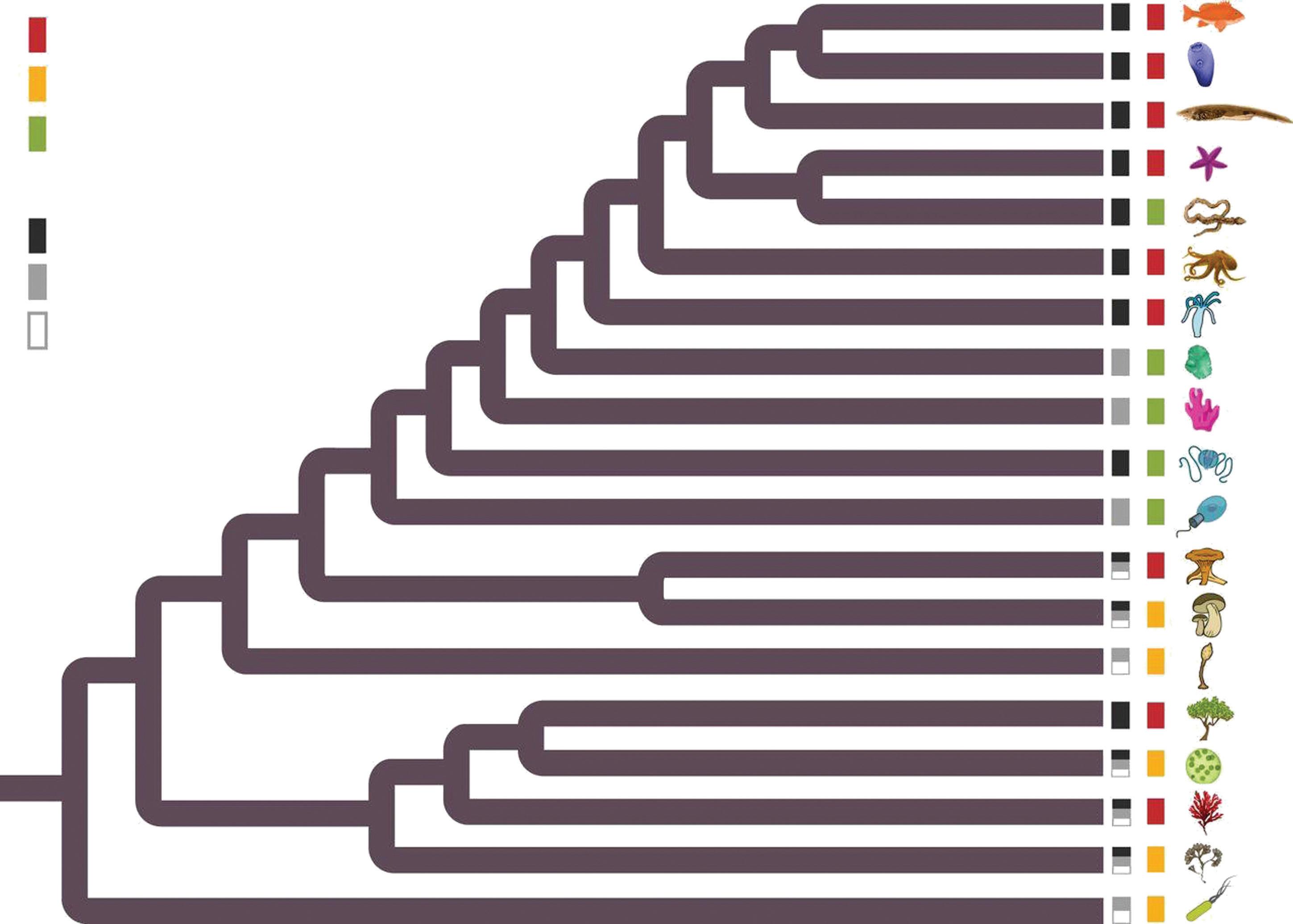
As shown in Fig. 1.6, ‘cheating’ of multicellular cooperation has been observed already in multicellular bacterial organisms, like overgrowing due to loss of proliferative inhibition. ‘Cheating’ describes ‘the breaking of shared rules, including genetically encoded phenotypes or behaviour, that leads to a fitness advantage for the cheater’ (Aktipis et al., 2015). Cheating has been described as early as bacteria multicellular organisms (Fig. 1.6), involves mostly the functions of proliferation and/or apoptosis, and leads to forming a ‘mass’ of cells. Cancer is instead defined as a primary mass causing metastases and is mostly occurring in Metazoan but not only, as some plants have cancer-like lesions. Actually, the simplest organism in which lesions appear, sharing many characters of what we call cancer, is red algae. In these plants, primary tumours due to loss of proliferation and apoptotic control occur. These lesions can ulcerate and propagate in a metastasislike fashion.
Sponges are the oldest surviving metazoans and appeared approximately 650 million years ago (Srivastava et al., 2010) and already contain all the pathways characterizing both metazoans and cancer (Domazet-Loso and Tautz, 2010; Srivastava et al., 2010; Aktipis et al., 2015). Sponges do not have distinct organs but have specialized structures like pores, canals, ostia, chambers, and a rudimentary immune system. No cancer has been observed in sponges.
Cancer-like lesions and/or cheating, a clear distinction between the two being sometime difficult, has been seen in other early Metazoa such as hydra and corals, where fast-growing, destructive lesions with loss of architecture grow. Proper malignant tumours such as lymphoid, epithelial, neuronal, and those from gonad cells are instead commonly seen in protostomes, invertebrates, and occur in all the more complex types of Metazoa (Aktipis et al., 2015).
However it must be noted that as complexity, dimensions, and lifespan increases, not all the species are susceptible to cancer (Aktipis et al., 2015): the so-called Peto’s paradox (Peto et al., 1975; Caulin and Maley, 2011; Roche et al., 2013).
Cancer reported
Cancer-like phenomena reported
No cancer-like phenomena reported
Complex multicellularity
Simple or aggregative multicellularity
Unicellular
Urochordata (e.g. tunicates)
Cephalochordata (e.g. lanceletes)
Echinodermata (e.g. starfish)
Hemichordata (e.g. acorn worm)
Protostomia (e.g. molluscs)
Cnidaria (e.g. hydra)
Placozoa (i.e Trichoplax)
Porifera (e.g. sponges)
Ctenophora (e.g. comb jellies)
Choanoflagellata (e.g. collared flagellates)
Ascomycota (e.g. sac fungi)
Basidiomycota (e.g. fruiting body fungi)
Amoebozoa (e.g. slime moulds)
Embryophyta (e.g. plants)
Chlorophyta (e.g. Volvox)
Rhodophyta (e.g. red algae)
Stramenopila (e.g. brown algae)
Bacteria (e.g. Pseudomonas)
Fig. 1.6 Cancer across the tree of life. Black, grey, or white boxes at branch tip indicates the cellularity status as unicellular (white), aggregative multicellularity (grey), or complex multicellularity (black). Red, yellow, or green boxes represent whether a cancer phenotype (invasion or metastasis) was reported or cancer-like/‘cheating’- type lesion was observed (abnormal proliferation or differentiation) such as callus or galls (yellow box). If no cancer or cancer-like/cheating lesions were reported, there is a green box.
Reproduced with permission from Aktipis C et al., ‘Cancer across the tree of life: cooperation and cheating in multicellularity’, Philosophical Transaction Royal Society London B, Volume 370, Issue 1673, 20140219, Copyright © 2015 The Authors. Published by the Royal Society under the terms of the Creative Commons Attribution License http://creativecommons.org/licenses/by/4.0
The Peto paradox
Every time that a cell proliferates, there is risk of an error at the time when the DNA is copied. Different types of mistakes can happen, like single mutations, duplication, and/or redistribution of the genetic material among the daughter cells. Consequently, as Metazoa increased in complexity and size and the lifespan got longer and longer, the risk of cancer was expected to grow in direct proportion; the larger and long-lived the animal, the higher the number of mitosis occurring in its body, and therefore the higher the chance of DNA damage to occur. However, this turned out not to be the case as large dimensions and longer life do not necessarily means increased risk of cancer (Aktipis et al., 2015): this is the ‘Peto’s paradox’, which get its name from a study published in 1995 by Richard Peto et al. (Peto et al., 1975). In this experiment a large cohort of mice of different ages were exposed to topical application of the carcinogen. The rate of appearance by epithelial tumours was related to the duration of exposure to the chemical but not to the mouse’s age: it was the time of exposure to the carcinogen dictating the risk of developing cancer and not the age of the exposed mouse—and neither the span of survival after the exposure. This study demonstrated that, against the then current wisdom, increased lifespan per se can be irrelevant as far as increase in cancer risk is concerned. More broadly, ‘Peto’s paradox’ is now a term to indicate a counter-intuitive event.
As far as cancer is concerned, two classic examples are those of the blue whales and the elephants. Blue whales are approximately six million times larger than mice; however, variation in cancer in nonlaboratory animals varies in average for no more than a factor of two, independently of the mass. In the whale’s case, the paradox is even more remarkable as only very rarely do they die of cancer (Leroi et al., 2003). Calabrese and Shibate (Calabrese and Shibata, 2010), using a mathematical model, have been able to correctly approximate the risk of colorectal cancers in humans: their equation resulted in an overall risk of approximately 2.5% by the age of 90 years while the risk actually observed is 5 (Caulin and Maley, 2011). However, using the same model they predicted a risk for the blue whales of 100% of getting this cancer by the age of 80 years while they can live beyond 100 years and cancer of any type is a rare event.
The question raised by Peto’s paradox is therefore how some large long-lived animals manage to have such a low rate of cancer.
According to natural selection, this is because the mechanisms giving protection from cancer must have been selected in order to allow large animals to exist and to have a long life span. However, the nature of these mechanisms remains unclear. While no studies are available for blue whales, some have been carried out on other animals and the main mechanisms involved in low susceptibility to cancer are concerned with telomerases replicative senescence and cell proliferation control, tumour suppressor activity, and genome stability.
One example is the elephant, which is notoriously a large mammal but can live in the wilderness to 60–70 years with a low cancer incidence. In this pachyderm, the protective mechanism is suggested to be redundancy of tumour suppressor function as both the African and the Asian species have 20 copies of the tumour suppressor TP53 gene (Abegglen et al., 2015). Gain and loss of genes involved in DNA repair, cell–cycle regulation, and ageing could be responsible for the lack of malignancies in the bowhead whale, possibly the longest-lived mammal, which is estimated to live in excess of 200 years (Keane et al., 2015). The largest wealth of data is available for rodents. To
this order belong animals with a great variability of longevity, body mass, and cancer incidences. Naked mole rats are the longest living, in excess of 30 years, while mice and rats live approximately 3 or 4 years. The difference in cancer incidence is striking: mice, among the smallest rodents, are prone to cancer and in some strains the incidence goes up to 95% while the larger naked and the blind mole rats are virtually cancer free.
Systemic studies on rodents have started to unravel the mechanisms behind these differences. Small but long-lived rodents appear to have cells which proliferate more slowly than small short-lived animals, while larger long-lived rodents are protected by shorter telomerases and therefore enhanced replicative senescence. Longlived animals also show higher levels of expression of DNA repair genes, raising the hypothesis of a more efficient DNA repair activity (Gorbunova et al., 2014).
Resistance to cancer evolved several times independently as demonstrated by the comparison of two long-lived cancer-resistant rodents: the Naked mole rat (Heterocephalus glaber) and the blind mole rat (Spalax ehrenbergi). These two species are phylogenetically distant from each other. In naked mole rats, there are large levels of high molecular mass hyaluronan polysaccharides, five times longer than those of humans. These longer forms bind to CD44 triggering cell cycle arrest, while the low molecular mass hyaluronans promote cell cycle. When the Has2 gene responsible for hyaluronan synthesis is knocked down or when the hyaluronoglucosaminidase 2 (Hyal2) gene, responsible for breaking down hyaluronan, is overexpressed, naked mole rat cells start forming tumours. Furthermore, in these rodents the 28S rRNA is cleaved in two, increasing the fidelity of translation. The mechanisms in the blind mole rat are different; one is the secretion of interferon by premalignant cells, which causes a massive necrosis in the surrounding tissue eliminating the premalignant cells. The second is again linked to hyaluronan, but this time the hyaluronan present is not able to block mitosis but is rather a powerful antioxidant (Gorbunova et al., 2014).
Cancer is a disease due to the malfunctioning of the biological functions necessary for cell growth and for the formation and maintenance of the multicellular organisms. Because of the complexity of large multicellular animals, this means that many different types of errors and damages can lead to what is known as cancer leading to malignant lesions with varied and complex biology and clinical behaviour. Cancer must be regarded from a practical point of view as many different diseases, each to be individually unravelled to fully understand it and produce an effective treatment.
• Cancer is a disease due to the malfunctioning of the pathways necessary to the life of a multicellular organism.
• It is a disease of information, as genetic damage alters the information blueprint of the organism: the DNA.
• Evidence of cancer-like behaviour has been found even in the simplest prokaryotic multicellular organisms.
• To be prone to cancer is not inevitable for a multicellular organism as some are very resistant to the disease, as per the Peto paradox.
• Open questions about the cancer are numerous! They are scattered through the book.
Alberts, B., Johnson, A., Lewis, J., et al. (2015). Molecular Biology of the Cell, 6th edition. New York: Garland Science. Allen, T. & Cowling, G. (2011). The Cell: A Very Short Introduction. Oxford: Oxford University Press. Benton, M. J. (2008). The History of Life: A Very Short Introduction Oxford: Oxford University Press. Dawkins, R. & Wong, Y. (2016). The Ancestor’s Tale, 2nd edition. London; Weidenfeld & Nicolson.
Diamond, J. C. (1998). Because Cowards Get Cancer Too. London: Vermilion.
Schiffman, J., Maley, C. C., Nunney, L., Hochberg, M., & Breen, M. (eds.) (2015). Theme issue ‘Cancer across life: Peto’s paradox and the promise of comparative oncology’. Philosophical Transactions of the Royal Society B, 370 (1673), DOI: 10.1098/rstb.2015.0198. Weinberg, R. A. (1998). One Renegade Cell. New York: Basic Books.
Abegglen, L. M., Caulin, A. F., Chan, A., et al. (2015). Potential mechanisms for cancer resistance in elephants and comparative cellular response to DNA damage in humans. JAMA, 314, 1850–60. Adams, D. G. (2000). Heterocyst formation in cyanobacteria. Curr Opin Microbiol, 3, 618–24.
Aktipis, C. A., Boddy, A. M., Jansen, G., et al. (2015). Cancer across the tree of life: cooperation and cheating in multicellularity. Philos Trans R Soc Lond B Biol Sci, 370, pii: 20140219.
Bonner, J. T. (1998). The origins of multicellularity. Integrative Biology Issues News and Reviews, 1, 27–36.
Calabrese, P. & Shibata, D. (2010). A simple algebraic cancer equation: calculating how cancers may arise with normal mutation rates. BMC Cancer, 10, 3.
Caulin, A. F. & Maley, C. C. (2011). Peto’s paradox: evolution’s prescription for cancer prevention. Trends Ecol Evol, 26, 175–82.
Domazet-Loso, T. & Tautz, D. (2010). Phylostratigraphic tracking of cancer genes suggests a link to the emergence of multicellularity in metazoa. BMC Biol, 8, 66.
Donoghue, P. C. & Antcliffe, J. B. (2010). Early life: origins of multicellularity. Nature, 466, 41–2.
Edidin, M. (2003). Lipids on the frontier: a century of cell-membrane bilayers. Nat Rev Mol Cell Biol, 4, 414–18.
Flores, E. & Herrero, A. (2010). Compartmentalized function through cell differentiation in filamentous cyanobacteria. Nat Rev Microbiol, 8, 39–50.
Gorbunova, V., Seluanov, A., Zhang, Z., Gladyshev, V. N., & Vijg, J. (2014). Comparative genetics of longevity and cancer: insights from long-lived rodents. Nat Rev Genet, 15, 531–40.
Hanahan, D. & Weinberg, R. A. (2011). Hallmarks of cancer: the next generation. Cell, 144, 646–74.
Hedges, S. B. (2002). The origin and evolution of model organisms. Nat Rev Genet, 3, 838–49.
Kaiser, D. (2001). Building a multicellular organism. Annu Rev Genet, 35, 103–23.
Keane, M., Semeiks, J., Webb, A. E., et al. (2015). Insights into the evolution of longevity from the bowhead whale genome. Cell Rep, 10, 112–22.
Lammerding, J. (2011). Mechanics of the nucleus. Compr Physiol, 1, 783–807.
Leroi, A. M., Koufopanou, V., & Burt, A. (2003). Cancer selection. Nat Rev Cancer, 3, 226–31.
Olsen, G. J. & Woese, C. R. (1997). Archaeal genomics: an overview. Cell, 89, 991–4.
Peto, R., Roe, F. J., Lee, P. N., Levy, L., & Clack, J. (1975). Cancer and ageing in mice and men. Br J Cancer, 32, 411–26.
Roche, B., Sprouffske, K., Hbid, H., Misse, D., & Thomas, F. (2013). Peto’s paradox revisited: theoretical evolutionary dynamics of cancer in wild populations. Evol Appl, 6, 109–16.
Rokas, A. (2008). The molecular origins of multicellular transitions. Current Opinion in Genetics & Development, 18, 472–8.
Scharrer, B. & Lochhead, M. S. (1950). Tumors in the invertebrates: a review. Cancer Res, 10, 403–19.
Schlumberger, H. G. & Lucke, B. H. (1948). Tumors of fishes, amphibians, and reptiles. Cancer Res, 8, 657–753.
Singer, S. J. & Nicolson, G. L. (1972). The fluid mosaic model of the structure of cell membranes. Science, 175, 720–31.
Srivastava, M., Simakov, O., Chapman, J., et al. (2010). The Amphimedon queenslandica genome and the evolution of animal complexity. Nature, 466, 720–6.
Thain, M. & Hickman, M. (2004). Dictionary of Biology. London: Penguin Books.
Virchow, R. K. (1863). Cellular Pathology as Based Upon Physiological and Pathological Histology. Philadelphia, PA: J. B. Lippincott and Co. Woese, C. R., Kandler, O., & Wheelis, M. L. (1990). Towards a natural system of organisms: proposal for the domains Archaea, Bacteria, and Eucarya. Proc Natl Acad Sci U S A, 87, 4576–9.
Giacomo Buscemi
DNA damage and repair studies started in the late 1930s by physicists’ experiments on recovery of cells inadvertently exposed to longwave light. Nowadays, it is estimated that mammalian cells suffer ~2 × 10⁵/day DNA lesions induced by normal metabolism products or environmental agents (Barnes and Lindahl, 2004). This number is further increased by the genotoxic effects of air pollution, cigarette smoking, food additives, toxins, and nuclear plant disasters. In the early 1960s, the discovery that the carcinogenicity of polycyclic aromatic hydrocarbons, the classic components of tobacco smoke, is directly dependent on their ability to form DNA adducts provided an unambiguous link between tumorigenesis and chemical DNA modifications. Now, we are aware that most carcinogens operate by generating DNA damage and causing mutations. Similar data were also obtained about radiation-induced mutations through the analysis of atomic bomb survivors, although the evidence that Xray exposure causes an increased risk of malignancies was already accepted in 1895, soon after their discovery. Moreover, the role of DNA repair in cancer was further supported by the disclosure, in the last 15 years, of rare syndromes harbouring mutations in DNA repair genes characterized by a predisposition to cancer, starting from xeroderma pigmentosum or Lynch syndrome.
The necessity of defects in DNA repair as an early or late event during cancer development is still debated. However, the consciousness that endogenous and exogenous DNA damage induces mutations potentially leading to carcinogenesis, and that efficient DNA repair mechanisms are required to protect organisms from cancer, are key concepts in cancer aetiology. More recently emerged the notion that general DNA damage response (DDR) mechanisms, more than repair pathways per se, are essential to prevent cancer. Indeed, the global cellular response to DDR is more complex than the simple activation of a specific DNA repair mechanism, since it is formed by network of pathways involving hundreds of proteins affecting cell cycle progression, cell survival, metabolism, and ageing. Alterations of DDR are essential to express some of those features that characterize a cancer cell, like uncontrolled replication or resistance to cell death. Notably, DDR signalling defects frequently have also a greater impact on chromosomal stability upon damage than DNA repair pathways, which mostly influence cell survival. Current models of
tumorigenesis (Fig. 2.1) indicate that single or multiple initiating events, often caused by mutations, lead to hyper-replication and replicative stress. Replication stress promotes cancer development by inducing breaks, particularly at common fragile sites, specific genomic regions showing increased fragility when DNA replication is perturbed. These events, coupled with pre-existing genetic alterations or acquired mutations that downregulate DDR mechanisms, enable replicative immortality and resistance to cell death, finally enhancing the possibility of misrepaired lesions and genome instability (Fig. 2.1). These features are essential for the rapid adaptation of a cancer cell to its ever-changing microenvironment and for malignancy progression.
In the 1940s the DNA duplex, which contains essentially all genetic information, was initially perceived as a highly stable macromolecule. Therefore, it was a surprise to find that DNA is subjected to incessant damage. Spontaneous DNA alterations include deamination, hydrolysis, non-enzymatic methylation, and oxidation of DNA bases (Lindahl and Barnes, 2000). Some of them are generated indirectly by normal cellular processes: base oxidations, for example, are induced by reactive oxygen species (ROS), that are continuously produced in living cells as toxic by-products of oxygen metabolism. In addition, the frequency of DNA lesion is further increased by exogenous sources including ionizing (IR) and ultraviolet (UV) radiation, and various chemicals agents.
To prevent the accumulation of nuclear DNA lesions, all organisms have evolved a complex signalling cascade to repair damage and eliminate cells that are beyond repair. Named DDR (Ciccia and Elledge, 2010), this cascade involves, in eukaryotes, hundreds of proteins that control the outcome of DNA repair at different levels (Fig. 2.2). Indeed, if a cell suffering DNA damage survives and continues growing with a restored unaltered genome, it will depend on the ability of DDR:
• To tightly regulate the activity of a multitude of DNA repair enzymes and regulators, with the final goal to optimize repair;
• To recruit chromatin remodelling proteins around the injured region thus improving the access of repair factors;