CENTRAL NERVOUS SYSTEM
Robert Brown
Q u E stions
1. A 71-year-old man with a history of heart failure, poorly controlled diabetes, and chronic kidney disease is brought to the emergency department (ED) by his family with altered mental status. He is found to have a serum creatinine of 5.3 mg/dL, blood urea nitrogen (BUN) of 81 mg/dL, glucose of 313 mg/dL, and pH of 7.17. On arrival to the ED, he does not open his eyes but swats your hand away with both arms when you squeeze his trapezius muscles. He moans but does not speak or follow commands. The cranial nerves are intact; however, you notice an irregular breathing pattern of alternating periods of hyperventilation and apnea in a crescendo-decrescendo repeating pattern. Which of the following MOST likely describes this respiratory pattern?
A. Brainstem injury
B. Metabolic derangement
C. Stroke
D. Seizure
2. An 88-year-old man with a history of heart failure and mild dementia is intubated with a diagnosis of community-acquired pneumonia. He takes donepezil daily. His daughter informs you that he was hospitalized several years ago following a hip fracture and required four-point restraints due to hallucinations, agitation, and injuring a staff member. Which of the following would MOST reduce the risk for a similar issue during this hospitalization?
A. Avoidance of central alpha agonists
B. Scheduled haloperidol
C. Utilization of the ABCDEF bundle
D. Increased sedation
3. A 67-year-old man is admitted to the intensive care unit (ICU) following a pulseless electrical activity (PEA) arrest with restoration of spontaneous circulation (ROSC) after 17 minutes of cardiopulmonary resuscitation (CPR). Initial examination reveals 3-mm pupils with absent pupillary light reflex bilaterally, absent corneal reflex, and no motor response to pain. He is breathing over the ventilator. He undergoes targeted temperature management with a temperature target of 33° C and is rewarmed slowly after 24 hours. During rewarming, he has diffuse jerking involving spontaneous eye opening and twitching of all four limbs, which are not suppressed with levetiracetam and valproic acid. Which of the following would MOST support a poor prognosis in this patient?
A. Absent pupillary light reflex before cooling
B. Bilateral absence of N2O response with somatosensory evoked potentials (SSEPs)
C. Early status myoclonus
4. During morning rounds, a 37-year-old patient admitted with severe traumatic brain injury (TBI) now has absent cranial nerve reflexes. On examination, pupils are 5 mm and nonreactive; corneal nerve reflexes, oculocephalic reflex, gag, cough, and motor response to painful stimulation are all absent. The patient’s respiratory rate is the same as the prescribed rate on the ventilator. You have reviewed your hospital’s brain death policy and confirm that all prerequisites have been met. Which of the following statements is NOT required to declare brain death?
A. Cerebral blood flow study
B. Oculovestibular reflex testing (cold calorics)
C. Normothermia
D. Lack of vasopressors
5. A 24-year-old female with past medical history significant for polysubstance abuse presents after a witnessed seizure. En route to the hospital, the patient has another seizure and is given 10 mg intramuscular (IM) midazolam. In the ED, she has two more seizures and is given 2 mg intravenous (IV) lorazepam each time, ultimately resulting in intubation with etomidate and rocuronium. What is the next MOST appropriate drug to administer?
A. Propofol
B. Levetiracetam
C. Lacosamide
D. Phenytoin
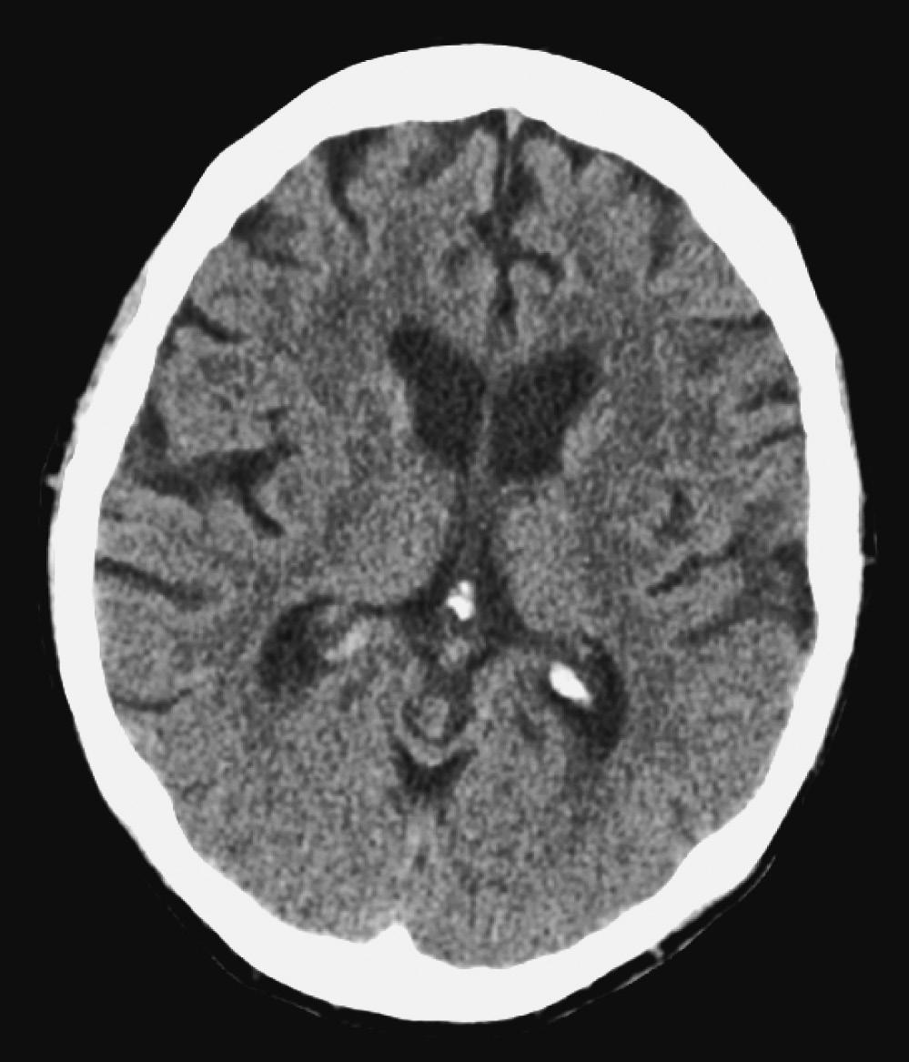
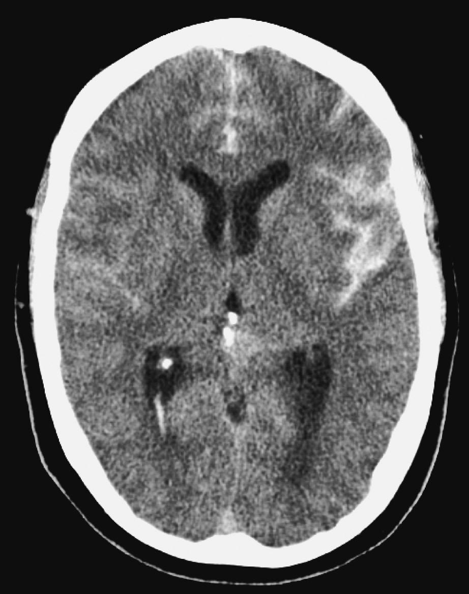
6. A patient presents with status epilepticus refractory to lorazepam and is intubated in the ED. Convulsive activity stops after treatment with a loading dose of phenytoin and a midazolam infusion. An electroencephalogram (EEG) is obtained and shows frequent epileptiform discharges without seizures. The patient is noted to have a temperature of 38.7° C, and lab results reveal a white blood cell (WBC) count of 18 (80% polymorphonuclear neutrophils [PMNs]). A computed tomography (CT) scan of the head is shown here. What is the next MOST appropriate step?
A. Obtain a lumbar puncture
B. Titrate midazolam to obtain a suppression-burst pattern
C. Magnetic resonance imaging (MRI) of the brain with or without contrast
D. Administer vancomycin, ceftriaxone, or acyclovir
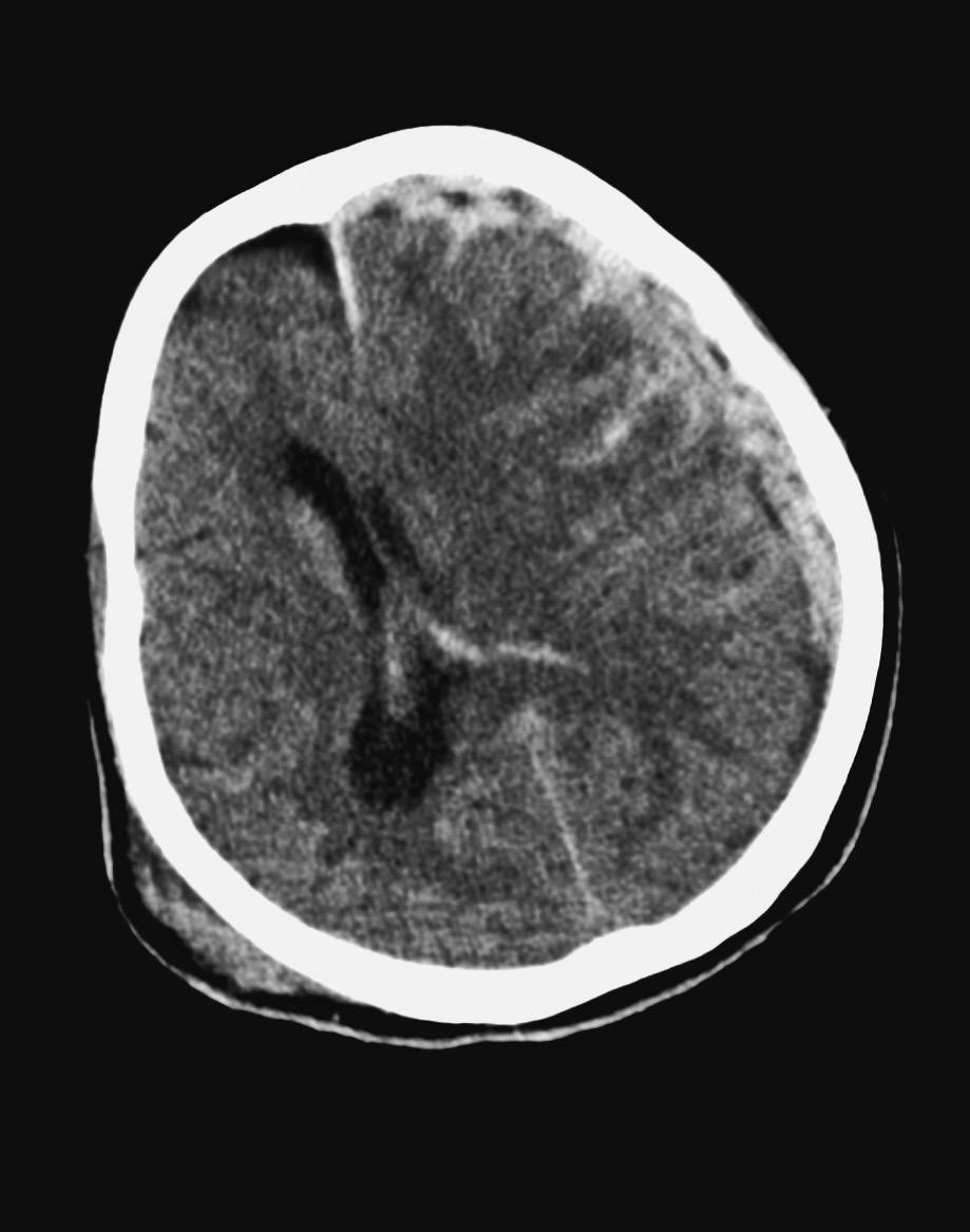
7. A 42-year-old male is admitted for sudden onset of severe headache associated with nausea, vomiting, and neck pain. He is otherwise neurologically intact. A CT scan of the brain is completed and shows the following image. What is the next best step in management?
A. Administer levetiracetam
B. Administer nimodipine
C. Ventriculostomy placement
D. CT angiogram of the brain
8. A 62-year-old patient with past medical history of hypertension, hyperlipidemia, type 2 diabetes, and coronary artery disease presents at 5 a.m. with acute onset of confusion. On exam, he has a left gaze preference, has dense right hemiplegia, and is globally aphasic. He was found by his family this morning and was last seen normal at 10 p.m. the previous night. A CT scan of the brain is completed and is remarkable for a dense left middle cerebral artery (MCA) sign with an Alberta Stroke Program Early Computed Tomography (ASPECT) score of 8. What is your next MOST immediate step in management?
A. IV thrombolytics
B. Diffusion-weighted MRI of the brain
C. CT angiogram of the brain with perfusion
D. Aspirin
9. Which of the following is FALSE regarding traumatic intracranial epidural hemorrhage?
A. Good prognosis with early evacuation
B. Biconvex shape
C. Always due to arterial bleeds
D. Most commonly in the temporoparietal region
10. A 53-year-old 70-kg female with a history of myasthenia gravis presents to the ED with dyspnea and worsening dysphagia over the past week. She takes pyridostigmine 90 mg four times daily. On examination, there is weakness of the neck flexors and proximal muscles of the upper extremity, significant dysarthria, and use of accessory muscles of respiration. Forced vital capacity (FVC) is 1 L and negative inspiratory force (NIF) is –17 cm H2O. Arterial blood gas (ABG) analysis reveals:
pH: 7.42
PaCO2: 37 mm Hg
PaO2: 90 mm Hg
Hemoglobin saturation: 97% on room air
Which of the following options is the MOST appropriate next step?
A. Close clinical monitoring
B. Endotracheal intubation
C. Prednisone
D. Pyridostigmine
11. A patient with myasthenia gravis on pyridostigmine is admitted with shortness of breath. She is started on biphasic positive airway pressure (BiPAP) with an inspiratory pressure of 12 cm H2O and expiratory pressure of 5 cm H2O, with stabilization in her symptoms. She is afebrile and demonstrates no clinical signs of infection. Which of the following is the MOST appropriate next step in this patient’s management?
A. High-dose IV methylprednisolone
B. Increased pyridostigmine
C. IV immunoglobulin
D. Levofloxacin
12. A 65-year-old male presents with a 5-day history of progressively worsening mental status and fever. A lumbar puncture is performed, followed by IV vancomycin and ceftriaxone administration. A 20-minute EEG shows periodic lateralized epileptiform discharges (PLEDs). Which diagnostic test would be MOST appropriate to evaluate this patient’s condition?
A. CT of the brain with contrast
B. MRI of the brain with contrast
C. Cerebrospinal fluid (CSF) polymerase chain reaction (PCR) testing
D. Continuous EEG
13. A 45-year- old male presents to the ED with progressive lower extremity weakness. The symptoms started 4 days ago with vague tingling in his toes. He denies respiratory symptoms. On exam, his lower extremity strength is 1/5 bilaterally, while his upper extremities are 2/5 distally and 4/5 proximally. Deep tendon reflexes are absent, sensory exam shows partial numbness over all distal extremities without a spinal level, and cranial nerve function is intact. He reports having an upper respiratory illness 1 month ago. He is admitted to the Neurology ICU, where a nerve conduction study/electromyography (NCS/EMG) is performed. What is one of the earliest and MOST sensitive electrophysiological findings in patients with this diagnosis?
A. Absent tibial nerve H reflex
B. Reduced F-wave latency
C. Abnormal sural sensory nerve action potential (SNAP)
D. Absent temporal dispersion of proximal compound muscle action potential (CMAP) responses
14. Which among these are NOT included in the differential diagnosis for a patient presenting with acute flaccid paralysis?
A. Acute poliomyelitis
B. West Nile virus (WNV) meningoencephalitis
C. Critical illness myopathy
D. Charcot-Marie-Tooth syndrome
15. A 37-year- old male suffers a severe asthma attack that results in cardiac arrest. ROSC is achieved in the field, and hypothermia is initiated in the ED. The patient undergoes controlled rewarming on day 2; however, overnight he becomes hypotensive, and norepinephrine is started to maintain mean arterial pressure (MAP) above 60 mm Hg. Over the course of the night, the patient’s vasopressor requirement increases significantly, and loss of cranial nerve reflexes is noted. Serum sodium increased from 142 mEq/ L the day before to 159 mEq/ L currently. Urine output has been 300 to 500mL/ hour for the past several hours. Which of the following is the next BEST step?
A. D5W infusion
B. Isotonic fluid bolus
C. Urine osmolality
D. Brain death testing
16. A 42-year-old female with a history of IV drug abuse presents to the ED complaining of severe back pain. She is reluctant to participate in the neurological examination because of shooting pain with movement; however, she does not seem to have weakness or sensory loss. She denies bowel or bladder dysfunction and is afebrile.
Laboratory findings include:
WBC count: 11.2 × 10³
Hemoglobin: 12.6 g/dL
Hematocrit: 37%
Platelets: 214 × 109/L
Which of the following is the MOST appropriate next step in management?
A. Urgent MRI of the cervical, thoracic, and lumbar spine
B. No further workup because she is likely drug-seeking
C. Cervical radiograph
D. Lumbar puncture
17. A 57-year-old is involved in a high-speed motor vehicle collision. In the ED, the patient is quadriplegic, breath sounds are shallow, and O2 saturation is 88% on a non-rebreather mask. The patient undergoes endotracheal intubation, and a CT scan of the spine shows bilateral facet dislocations at C5– 6. The patient is taken emergently to the operating room for spinal stabilization and is now admitted to the ICU for further management. On arrival, the patient is awake and alert but has no movement or sensation below the deltoids. Which of the following is FALSE?
A. MAP should be maintained above 85 mm Hg
B. The patient can be safely extubated
C. IV glucocorticoids are not recommended
D. The long-term prognosis for ambulation is poor
18. A 32-year-old male is admitted with severe TBI. Admission Glasgow Coma Scale (GCS) score is 6T; admission CT scan is shown here. A ventriculostomy is in place for intracranial pressure (ICP) monitoring. He is sedated with propofol and fentanyl with an ICP of 15 mm Hg and MAP of 66 mm Hg. He is intubated and mechanically ventilated on assist control mode with a respiratory rate of 20 breaths/minute and tidal volume of 500 mL. The patient’s ABG reveals a pH of 7.49, PaCO2 of 29 mm Hg, and PaO2 of 113 mm Hg. The patient’s brain tissue oxygen monitor reads 13 mm Hg; hemoglobin is 7.2 g/dL. Which of the following is LEAST likely to improve the patient’s brain tissue oxygenation?
A. Decreasing sedation
B. Reducing the patient’s minute ventilation
C. Cerebral perfusion pressure (CPP) augmentation with norepinephrine
D. Red blood cell (RBC) transfusion
19. In a patient with severe TBI undergoing multimodality monitoring, which of the following MOST suggests an increased risk for further neuronal injury?
A. EEG reactivity to painful stimuli
B. Decreased jugular venous bulb O2 saturation
C. Decreased cerebral lactate-to-pyruvate ratio
D. ICP of 25 mm Hg
20. A 62-year-old female with a history of hypertension, dyslipidemia, and type 2 diabetes mellitus is undergoing a diagnostic cardiac catheterization. She receives 2 mg of IV midazolam before the procedure. Twenty minutes into the procedure, the patient is noted to be unresponsive, although she is breathing well. A closer examination shows that the patient does not open her eyes to stimulation, her pupils are 1.5 mm and reactive, and the left eye is lower than the right eye. The left upper extremity weakly withdraws to noxious stimulation, and there is no motor response on the right. What is the MOST likely cause of the patient’s clinical symptoms?
A. Left frontal lobe intracerebral hemorrhage (ICH)
B. Brainstem infarction
C. Left lacunar infarction
D. Oversedation
21. A 50-year-old female admitted with moderate TBI and small bifrontal contusions is found acutely unresponsive by a nurse. Her vital signs are as follows:
Heart rate: 93 bpm
Blood pressure: 162/82 mm Hg
Respiratory rate: 18 breaths/minute (on mechanical ventilation)
Temperature: 99.1° F
The patient localizes bilaterally, opens eyes to sternal rub, has equal and reactive pupils, and has intact gag/ cough and corneal reflexes. The patient then has a sudden clinical deterioration manifested by extension in all extremities with a 5-mm nonreactive left pupil. After emergent intubation, what is the next MOST appropriate management?
A. Mannitol
B. Levetiracetam
C. Dexamethasone
D. Hypertonic saline 23.4%
22. A 27-year-old female, G1P1001 at 34 weeks’ gestational age, is brought to the ED after she had a witnessed generalized tonic-clonic convulsion at home. She has no prior medical history. Her blood pressure is 163/102 mm Hg. She slowly regains consciousness but has no focal neurological deficits. What would be the MOST appropriate next step in management of this patient?
A. Diazepam
B. Fosphenytoin
C. Magnesium sulfate
D. Levetiracetam
23. A 57-year-old female with no known medical history presents after being found in a confused state by family members. On examination, she is afebrile and appears cachectic and with poor dentition. Neurological examination demonstrates aphasia and a right facial droop. Cardiac examination reveals normal S1 and S2 without murmurs. A CT scan shows an ovoid lesion with surrounding vasogenic edema in the left parietal lobe, which is concerning for cerebral metastasis versus abscess. Which of the following features MOST favor cerebral abscess over metastasis?
A. Rim enhancement
B. Multiple lesions
C. Restricted diffusion on diffusion-weighted imaging (DWI)
D. Vasogenic edema that spares the cortex
24. A 34-year-old woman with a history of a prolactinoma presents to the ED with thunderclap headache. The CT scan of the brain is read as normal. She is drowsy but follows commands and has no focal neurological deficits. Shortly after admission to the ICU, she complains of lightheadedness and is noted to have a blood pressure of 77/40 mm Hg. Serum sodium is 132 mEq/L, and urine output is 80 to 100 mL/hour since admission. Which of the following is the BEST initial treatment?
A. Vasopressin
B. Hydrocortisone
C. Hypertonic saline 3%
D. Norepinephrine infusion
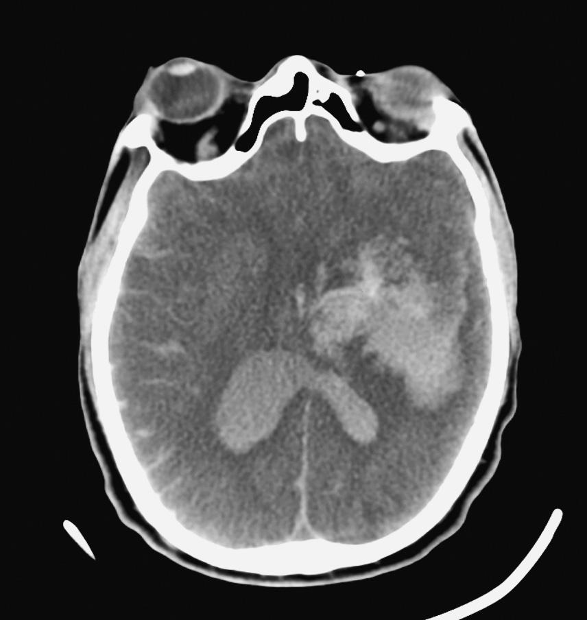
25. A 67-year-old man with a history of hypertension is “found down” by his neighbors. A CT scan of the brain shows ICH in the left basal ganglia (see the following image). His blood pressure is 210/120 mm Hg. On neurological examination, the patient is alert, follows simple commands, and answers simple questions. He extends his right arm and strongly follows with the left arm. The patient’s home medications include aspirin and lisinopril. Which of the following is the next BEST step in management?
A. Emergent craniotomy and hematoma evacuation
B. Nicardipine infusion
C. Factor VIIa
D. Emergent cerebral angiogram
26. On your rounds, you visit a 49-year-old male who is post-bleed day 6 from an aneurysmal subarachnoid hemorrhage. The patient had an external ventricular drain (EVD) placed on admission for hydrocephalus and then
underwent coiling of an anterior communicating artery aneurysm. The clinical course has been unremarkable; however, your resident tells you that the patient’s transcranial Doppler ultrasound exams from this morning were abnormal. The results include:
Left MCA velocity: 156 cm/second
Right MCA velocity: 207 cm/second
On examination, you now note a right-sided facial droop and new pronator drift. Which of the following is the MOST appropriate management at this time?
A. Maintenance fluids
B. Vasopressors
C. MRI
D. Brain CT with angiography
27. A 57-year-old female with a history of smoking and hypertension presents with thunderclap headache. On arrival, she does not open her eyes to pain, moans, and localizes bilaterally. A CT scan shows diffuse cisternal subarachnoid hemorrhage with mild hydrocephalus. She is intubated for airway protection, and an external ventriculostomy drain is placed. A CT angiogram is performed that shows a left posterior communicating artery aneurysm. She arrives to the ICU in the evening and is scheduled for a cerebral angiogram in the morning. Which of the following treatments is MOST indicated?
A. Vasopressors
B. Avoidance of antifibrinolytics
C. Phenytoin
D. EVD drainage
1. ANSWER: B
An often overlooked component of the neurological examination is the patient’s respiratory pattern. This is largely because the patient’s natural respiratory pattern can be obscured by mechanical ventilation. However, two patterns are commonly seen in the ICU but are often misunderstood. These patterns are post-hyperventilation apnea and Cheyne-Stokes respirations. Cheyne-Stokes respirations can be quite dramatic when observed, and if not familiar with the pattern, healthcare providers usually impute this to a severe structural brain abnormality. However, this is almost never the case. To understand Cheyne-Stokes respiration requires an understanding of post-hyperventilation apnea. While the medullary respiratory center is responsible for controlling the respiratory drive, the frontal lobes have hegemony over the process (this is why one can hold one’s breath under water but may hyperventilate when nervous). When a patient is hypocapnic, rather than an apneic period until CO2 is normalized, the respiratory rate is simple slowed, and tidal volumes are decreased to allow CO2 to return to normal more gradually. If the frontal lobes are damaged or malfunctioning owing to a systemic process causing encephalopathy (much more common), apnea is commonly seen following periods of hypocapnia. This can be observed by asking an encephalopathic patient to take several deep breaths and observing for apnea; however, in day-to-day practice, apnea is typically encountered in anxious or agitated patients who are causing apnea alarms on a ventilator during spontaneous breathing trials. Instead of returning to an assisted mode of ventilation and delaying extubation, the amount of pressure support can be reduced to avoid large tidal volumes, and the patient can be directed to relax, or other measures to control agitation can be employed. The apnea alarm on the ventilator can also be extended. However, the best way to solve this issue is to extubate the patient. Cheyne-Stokes is a more extreme version of post-hyperventilation apnea and results from a delay between alveolar CO2 and the partial pressure of CO2 in the medullary interstitium such that the brain is always trying to “catch up” to the alveolar CO2 . Low-output states can exacerbate this condition because changes in alveolar CO2 take much longer to reach the medullary respiratory centers. Many clinicians, after observing a patient with this respiratory pattern, assume a severe structural or brainstem injury when in fact the pattern demonstrates intact brainstem reflexes, as is the case with this patient. Respiratory patterns that are reflective of brainstem pathology include apneustic breathing (inspiratory pauses, seen with pontine injuries), ataxic breathing (irregular pattern, seen with rostral medullary lesions), and apnea (seen with caudal medullary lesions). These are typically not observed (with the exception of apnea) owing to early intubation
and mechanical ventilation in patients with these brain injuries. This patient’s active heart failure with signs of endorgan dysfunction is more likely the cause of this patient’s presentation.
Keywords: Central nervous system (CNS) diagnosis; Altered mental status (coma)
r EFE r E nc E
Posner JB, Saper, CB, Schiff ND, Plum F. Plum and Posner’s diagnosis of stupor and coma, 4th ed. New York: Oxford University Press; 2007, pp. 46–52.
2. ANSWER: C
Delirium is associated with increased mortality, prolonged ICU and hospital length of stay, and post-ICU cognitive impairment. A multifaceted approach should be implemented for its prevention, detection, and treatment. One method is the ABCDEF bundle. This is an acronym for pain a ssessment and utilization of a nalgesics, daily awakening and spontaneous breathing trials, choosing an appropriate sedation strategy, employing a delirium detection method, early mobilization, and family engagement. Daily awakening periods with interruption of sedatives and spontaneous breathing trials (SBTs) have been shown to decrease delirium incidence and ventilator and ICU lengths of stay. Indeed, a randomized controlled trial of daily sedation interruptions (frequently called “holidays”) with SBTs demonstrated reductions in ICU and hospital lengths of stay of 3.8 and 4.3 days, respectively. Benzodiazepines may lead to delirium and prolonged ventilator and ICU lengths of stay, while sedation with dexmedetomidine may have the opposite effect. Depth of sedation should be monitored with an appropriate sedation scale (e.g., Richmond Agitation and Sedation Scale), and sedatives should be regularly titrated to achieve a lighter level of sedation. Additionally, CAM-ICU is a simple assessment tool used to detect delirium and has been shown to have a high sensitivity and specificity. Once delirium has been detected, one can then review potential causes and address them accordingly. Results of trials investigating antipsychotic medication have not been conclusive, although the recent REDUCE trial demonstrated that prophylactic haloperidol (1 or 2 mg) is not effective in preventing delirium. Typical antipsychotics such as haloperidol are not recommended, while atypical antipsychotics should be used only when other measured have failed. Early mobilization has been shown to reduce delirium as well as improve functional status at discharge, likely by reducing the incidence of ICU-acquired weakness. Mobilization and exercise with physical and occupational therapy can
be paired with the patient’s daily sedation holiday and breathing trial. Restraints, while often necessary to avoid self-extubation or removal of other life-sustaining medical devices (e.g., ventriculostomies), can agitate patients and exacerbate delirium and their use should be minimized.
Keywords: CNS diagnoses; Altered mental status (delirium, hallucinations)
r EFE r E nc E s
Barr J, Fraser GL, Puntillo K et al. Clinical practice guidelines for the management of pain, agitation, and delirium in adult patients in the intensive care unit. Crit Care Med 2013;41:263–306.
Girard TD, Kress JP, Fuchs, BD et al. Efficacy and safety of a paired sedation and ventilator weaning protocol for mechanically ventilated patients in intensive care (Awakening and Breathing Controlled trial): a randomized controlled trial. Lancet 2008;12:126–134.
Morandi A, Brummel NE, Ely EW. Sedation, delirium and mechanical ventilation: the “ABCDE” approach. Curr Opin Crit Care 2011;17:43– 49.
van den Boogaard M, Slooter AJC, Brüggemann RJM et al. Effect of haloperidol on survival among critically ill adults with a high risk of delirium: The REDUCE Randomized Clinical Trial. JAMA 2018;319(7):680– 690.
3. ANSWER: B
Prognostication following cardiac arrest is complex and is most accurate when utilizing a multimodal approach. Some features have been consistently associated with a poor prognosis and can thus be used with a high degree of confidence. These include absent pupillary light reflexes at 72 hours and bilateral absence of N2O response during SSEP testing; loss of N2O is defined as no negative deflection 20 seconds after stimulation on both sides. Of note, automated methods of assessing pupillary light reflex have been developed and tested and may be expected to be seen more often in the coming years. Other features may be strongly associated but have lower specificity and should therefore not be used as the sole indictor of poor prognosis. These include early (within the first 48 hours) status myoclonus, neuroimaging features (other than frank herniation), and malignant EEG findings. Neuroimaging features associated with poor prognosis include loss of gray-white differentiation and sulcal effacement on CT imaging, and restricted diffusion on MRI. Of note, patchy or regional abnormalities, including isolated basal ganglia abnormalities, do not preclude neurological recovery. Malignant EEG findings that augur poor prognosis include suppression or a suppression-burst pattern, absent reactivity, and low voltage. Hypothermia and sedatives can confound the EEG; thus, the patient should be normothermic and off sedation. Serum biomarkers have also been used, with neuron-specific enolase being the most studied. However, studies have demonstrated a range of
thresholds that predict poor prognosis with high specificity, which has limited their clinical utility.
Keywords: CNS diagnosis; AMS; Other (hypoxic/metabolic encephalopathy); CNS; Diagnostic modalities; Evoked potential; CNS; Diagnostic modalities; EEG
r EFE r E nc E s
Oddo M, Rossetti AO. Early multimodal outcome prediction after cardiac arrest in patients treated with hypothermia. Crit Care Med 2014;42:1340–1347.
Sandroni C, Cariou A, Cavallaro F et al. Prognostication in comatose survivors of cardiac arrest: an advisory statement from the European Resuscitation Council and the European Society of Intensive Care Medicine. Resuscitation 2014;85:1779–1789.
Solari D, Rossetti AO, Carteron L et al. Early prediction of coma recovery after cardiac arrest with blinded pupillometry. Ann Neurol 2017;81(6):804– 810.
4. ANSWER: D
While guidelines have been published for brain death declaration, some details are left to the discretion of individual hospitals. In general, however, the first step in declaring brain death is confirming that all prerequisites have been met and that there are no confounding factors. This involves knowing the cause of injury and having that be compatible with causing brain death. For example, severe Guillain-Barré syndrome (GBS) has mimicked brain death, including absent cranial nerve reflexes, absent motor response, and failed apnea testing. A brain death examination would incorrectly diagnose brain death, yet cerebral function might be normal. A patient who is “found down” with an unknown cause of injury should be approached cautiously. Severe metabolic derangements should be excluded because these may confound the examination, although what defines a severe derangement is mostly left to the provider. The patient should be adequately resuscitated, normothermic, and normotensive (although vasopressors to achieve normotension do not preclude the ability to declare brain death). Brain imaging compatible with brain death is not mandatory but is performed almost universally. For example, in a patient with anoxic brain injury, imaging demonstrating herniation and diffuse loss of gray-white differentiation is not required and often not present because CT imaging may be performed before the development of cerebral edema; however, a CT scan obtained on day 4 without evidence of anoxic injury would not be consistent with brain death. Medications that may confound the examination, including benzodiazepines, barbiturates, and opiates, should be held, and testing should be delayed until these medications have been fully metabolized. The brain death examination should demonstrate lack of response to
simulation, absent pupillary light reflex, corneal reflex, oculocephalic reflex, oculovestibular reflex, gag reflex, cough reflex, and motor response. Finally, a formal apnea test should be performed to demonstrate no respiratory effort despite an arterial PaCO2 higher than 60 mm Hg (or 20 mm Hg above baseline in patients with chronic CO2 retention). This is typically performed by disconnecting the patient from the ventilator and providing continuous oxygen through a catheter inserted into the endotracheal tube (passive oxygenation). Patients may become hypotensive during apnea testing as acidosis develops; however, this can be managed with vasopressor titration and does not usually prevent one from completing the test. Because brain death is a clinical diagnosis, ancillary tests are not required unless part of the clinical examination cannot be performed. It is also for this reason that one should use the term “ancillary test” rather than “confirmatory test” to avoid the misconception that the brain death examination requires confirmation. Acceptable ancillary tests include EEG, transcranial Doppler ultrasound, catheter angiography, and cerebral blood flow testing using single photon emission computed tomography (SPECT). The latter is typically the most commonly used test because of its availability and ease of performance. Importantly, while brain death is considered “whole brain death,” certain features such as absence of diabetes insipidus and blood pressure variability do not preclude the diagnosis.
Keywords: CNS; Diagnoses; Brain death; Basic physiology; CNS; Brain death; CNS; Diagnostic modalities; Nuclear medicine studies
r EFE r E nc E s
Bonetti MG, Ciritella P, Valle G, Perrone E. 99mTc HM-PAO brain perfusion SPECT in brain death. Neuroradiology 1995;37(5):365. Wijdicks EF, Varelas PN, Gronseth GS et al. Evidence-based guideline update: determining brain death in adults. A report of the Quality Standards Subcommittee of the American Academy of Neurology. Neurology 2010;74:1911–1918.
hydrolyzed to phenytoin by serum phosphatases with a conversion half-time of 15 minutes; the loading dose is 20 mg/ kg (considered phenytoin equivalents) and is infused at a rate of 100 to 150 mg/minute. It should be noted that while hypotension is less likely to be seen with phosphenytoin, it may still be occur, and hemodynamic monitoring remains indicated.
Levetiracetam can be considered in this case, but evidence for its efficacy in aborting status epilepticus is unclear. An appropriate dose per guidelines is 20 to 60 mg/kg, up to a maximum of 4500 mg. The mechanism of action for levetiracetam is not known, although it is hypothesized to modulate GABA receptors indirectly and bind to protein SV2A (which has been linked to seizures in animal models). Since levetiracetam does not induce CYP, it has minimal interactions with other drugs such as immunosuppressants, and IV to oral dosing and bioequivalence are identical.
Lacosamide works by helping inactivate sodium channels, thereby preventing repetitive firing of neurons, and it can be used to treat focal-onset seizures; unfortunately, lacosamide has no role in treating generalized status epilepticus. If the patient does not respond to phenytoin, while second-line alternatives or third-line agents such as valproic acid and phenobarbital are acceptable, an infusion of midazolam or propofol has a higher likelihood of aborting the status epilepticus.
Keywords: CNS; Diagnoses; Seizures and status epilepticus; CNS; Management strategies; Anticonvulsants
r EFE r E nc E s
Chokshi R, Openshaw J, Mehta N, Mohler E. Purple glove syndrome following intravenous phenytoin administration. Vasc Med 2007;12:29–31.
Glauser T, Shinnar S, Gloss D et al. Evidence-based guideline: treatment of convulsive status epilepticus in children and adults: a report of the guideline committee of the American epilepsy society. Epilepsy Currents 2016;16:48– 61.
5. ANSWER: D
Status epilepticus is defined as a seizure duration of greater than 5 minutes or recurrent seizures without return to baseline. Benzodiazepines, including midazolam (IV or IM) and lorazepam (IV), have the highest efficacy and are thus first-line agents to arrest a seizure. Phenytoin is an appropriate second-line agent. Phenytoin can cause hypotension if infused too quickly and also has the risk for skin necrosis if the infusion infiltrates, a condition known as purpleglove syndrome. For these reasons, a recent guideline has recommended fosphenytoin, a prodrug of phenytoin, if available because it can be infused rapidly. Fosphenytoin is
6. ANSWER: D
Antibiotics to cover both bacterial meningitis and herpes encephalitis are indicated. Ceftriaxone would cover the most likely bacterial agents (Streptococcus pneumoniae, Haemophilus influenzae , and Neisseria meningitidis), while vancomycin would cover beta-lactam-resistant pneumococcus. Acyclovir would cover herpes encephalitis. Ampicillin would be added in immunocompromised or elderly patients or to cover Listeria monocytogenes . Additionally, it is appropriate to include dexamethasone until pneumococcal meningitis is ruled out because this may improve mortality and reduce neurological sequalae.
Glucocorticoids should be administered shortly before (or at the same time as) antibiotics because this approach has been shown to reduce unfavorable outcomes. It is hypothesized that glucocorticoid administration (usually dexamethasone) reduces cerebral edema as well as cytokine levels in the CSF. A lumbar puncture should be performed ideally within 6 hours of starting treatment in order to improve yield from cultures. A diagnosis on the basis of the CSF profile can still be made if the lumbar puncture is delayed more than 6 hours but may yield less useful information, especially in cases of meningococcal meningitis. While a CT scan was appropriate in this patient, it is not necessary in most patients and if performed often leads to an unnecessary delay in treatment, as would waiting for results of the lumbar puncture. Continuous EEG monitoring should be applied because the patient could be having subclinical events; however, starting treatment for the likely infection is of utmost importance. The advantage of titrating sedation to a suppression-burst pattern if the patient is no longer seizing is unclear and thus would not be the most appropriate next step in this patient. The head CT shown is normal, and while an MRI would likely assist with diagnosis and prognosis, it would be the last step in the management of this patient.
Keywords: CNS; Diagnoses; Infectious; Encephalitis/ meningitis; CNS; Diagnostic modalities; EEG; CNS; Diagnostic modalities; Lumbar puncture; CNS; Management strategies; Antimicrobials
r EFE r E nc E
raphy of the head before lumbar puncture in adults with suspected meningitis. N Engl J Med 2001;345:1727.
7. ANSWER: B
The patient has spontaneous subarachnoid hemorrhage (SAH), which is most commonly caused by a ruptured aneurysm. SAH affects nearly 30,000 patients in the United States each year and has a mortality rate of around 45%. CT angiography of the brain will assist both in accurate diagnosis and surgical planning if an aneurysm is found. The yield of CT angiography in aneurysmal detection is approximately 95% when compared with digital subtraction angiography. This makes it a quick and important step in the workup before surgical interventions. Although seizure prophylaxis is commonly used for patients with SAH, there are no studies suggesting that it improves outcomes. Furthermore, there is evidence demonstrating that long-term treatment with
anticonvulsants may lead to worse cognitive outcomes, although these studies were performed with phenytoin. It should be noted that generalized status epilepticus has an incidence of 0.2%, but nonconvulsive status epilepticus occurs 31% of the time when SAH patients have stupor or coma; such seizures are associated with poor neurological outcome. Nimodipine may improve neurological outcomes and reduce the incidence of cerebral infarctions in patients with aneurysmal SAH but does not reduce the actual rate of vasospasm. Nimodipine is normally administered in doses of 60 mg every 4 hours and can be given less frequently in smaller doses when hypotension is seen after administration. The most important first step in a patient with SAH is to prevent aneurysmal rebleeding. This is done by surgical clipping or endovascular coiling. There is no clear evidence that dexamethasone is beneficial.
Keywords: CNS; Diagnoses; Stroke; Hemorrhagic (SAH); Diagnostic modalities; Other imaging (e.g., CTA)
r EFE r E nc E s
Diringer MN, Bleck TP, Claude Hemphill J 3rd et al. Critical care management of patients following aneurysmal subarachnoid hemorrhage: recommendations from the Neurocritical Care Society’s Multidisciplinary Consensus Conference. Neurocrit Care 2011;15:211–240.
Dorhout Mees S, Rinkel GJE, Feigin VL et al. Calcium antagonists for aneurysmal subarachnoid haemorrhage. Cochrane Database Syst Rev 2007;(3):CD000277.
Feigin VL, Anderson N, Rinkel GJE et al. Corticosteroids for aneurysmal subarachnoid haemorrhage and primary intracerebral haemorrhage. Cochrane Database Syst Rev 2005;(3):CD004583.
Kelliny M, Maeder P, Binaghi S et al. Cerebral aneurysm exclusion by CT angiography based on subarachnoid hemorrhage pattern: a retrospective study. BMC Neurol 2018;11: 8.
8. ANSWER: C
The ASPECTS score was used to predict outcome with thrombolytic use and demonstrated improved outcomes in patients with scores of 8 or higher. Patients with “wake-up stroke” such as this one often present outside of the acceptable window for administration of IV thrombolytics (up to 4.5 hours from last seen normal). This does not, however, exclude them from receiving treatment for their stroke by endovascular modalities, and the ASPECT score is one modality that has been used in selecting patients who may have favorable outcome with mechanical thrombectomy. A recent study known as the DAWN trial demonstrated that mechanical thrombectomy improves outcome in patients presenting 6 to 24 hours from when they were last seen normal who still have a mismatch between clinical deficits and infarct volume. In this case, the patient’s
Hasbun R, Abrahams J, Jekel J, Quagliarelllo VJ. Computed tomog-
neurological symptoms and dense MCA sign (hyperdensity of the MCA) suggest a proximal occlusion of the MCA. CT angiography of the brain would confirm this, while a CT perfusion study would assess for a “mismatch” between infarcted, and hence unsalvageable tissue (the core) and ischemic but not yet infarcted tissue (penumbra). While MRI of the brain is indicated to diagnose the location and size of the infarct, revascularization strategies are paramount and can usually be accomplished faster with the CT perfusion route over an MRI. Although aspirin is indicated for patients not receiving IV tissue plasminogen activator, revascularization has primacy in the management of acute ischemic stroke.
Keywords: CNS; Diagnoses; Stroke; Embolic/thrombotic and ischemic
r
EFE r E nc E s
Aviv RI, Mandelcorn J, Chakraborty S et al. Alberta Stroke Program Early CT scoring of CT perfusion in early stroke visualization and assessment. Am J Neuroradiol 2007;28:1975–1980.
Hill MD, Rowley HA, Adler F et al. ASPECTS score to select patients for endovascular treatment: the IMS-III trial. Stroke 2014;45(2):444– 449.
Nogueira RG, Jadhav AP, Haussen DC et al. Thrombectomy 6 to 24 hours after stroke with a mismatch between deficit and infarct. N Engl J Med 2018;378:11–21.
Ryu CW, Shin HS, Park S et al. Alberta Stroke Program Early CT score in the prognostication after endovascular treatment for ischemic stroke: a meta-analysis. Neurointervention 2017;12.1:20–30.
Bullock MR, Chesnut R, Ghajar J et al. Surgical management of acute epidural hematomas. Surgical Management of Traumatic Brain Injury Author Group. Neurosurgery 2006;58(3 Suppl):S7.
10. ANSWER: B
Patients in myasthenic crisis should be admitted urgently to an ICU because they can decompensate rapidly. Frequent respiratory assessments with FVC and NIF are essential. Indications for intubation in patients with myasthenic crisis include FVC ≤15 mL/kg (normal, ≥60 mL/kg), NIF ≤20 cm H 2 O (normal, ≥70 cm H 2 O), and peak expiratory flow (PEF) 40 cm H 2 O (normal, ≥100 cm H 2 O). ABG is not a reliable guide to determine need for intubation because this can remain normal just before respiratory collapse. Endotracheal intubation is best performed electively before this. Several studies have shown BiPAP to be effective in patients with myasthenic crises because it can reduce rates of intubation and decrease lengths of stay. However, BiPAP should be used on a case-by- case basis. Hypercapnia predicts BiPAP failure and should warrant immediate intubation. Severe bulbar dysfunction and difficulty managing secretions are a contraindication to BiPAP.
9. ANSWER: C
Although epidural hemorrhages are commonly arterial in nature and secondary to shearing of meningeal arteries, occasionally an epidural hemorrhage can form as a result venous bleeding such as a torn venous sinus with an associated skull fracture. About 15% of the time, an injury to one of the dural sinuses is the source be of the hemorrhage. More than 95% of epidural hemorrhages are supratentorial, and about 85% are associated with the presence of a skull fracture. The temporoparietal region is the most common location, and most result from tearing of the middle meningeal artery. Although patients can decompensate rapidly, prognosis is good if surgical evacuation is carried out promptly, and most patients make full neurological recovery with no residual deficits. In contrast to subdural hematomas, epidural hematomas typically do not cross suture lines owing to the tight adherence of the dura to the skull at these points. Additionally, from a test-taking strategy perspective, the word “always” may be a warning that the answer is a trap.
Keywords: CNS; Diagnoses; Epidural hemorrhage
Pyridostigmine has a rapid onset of action with peak action at 2 hours, but because of the bulbar signs of weakness exhibited by this patient, this would not be the immediate optimal management strategy given information presented in this vignette. High-dose glucocorticoids can be helpful in management of myasthenia gravis in general; however, the clinical effect can take 2 to 3 weeks to become optimal, and nearly 50% of patients can experience deterioration about a week after glucocorticoids are initiated. Therefore, steroids do not have an immediate role when respiratory insufficiency is being urgently addressed.
Keywords: CNS; Management strategies; Plasmapheresis; CNS; Diagnoses; Neuromuscular disease; Myasthenia gravis
r EFE r E nc E s
Jani-Acsadi A, Lisak RP. Myasthenic crisis: guidelines for prevention and treatment. J Neurol Sci 2007;261(1–2):127–133.
Seneviratne J, Mandrekar J, Wijdicks EF, Rabinstein AA. Noninvasive ventilation in myasthenic crisis. Arch Neurol 2008;65(1):54–58.
11. ANSWER: C
About 20% of patient with a diagnosis of myasthenia gravis present at some point with myasthenic crisis.
Myasthenic crisis can be associated with acute respiratory failure from fatigue of the diaphragm. Clinical examination of face for eyelid weakness, bulbar weakness (i.e., difficulty swallowing, fatigue with chewing), tachypnea with a paradoxical respiratory pattern, and limb weakness are clinical signs consistent with myasthenic crisis. Monitoring of vital capacity and negative inspiratory force can help establish a threshold for preemptive intubation. The threshold for intubation in myasthenic crisis is generally agreed to be an FVC of <15 mL/kg or an NIF of <20 cm H 2 O.
IV immunoglobulin and plasmapheresis are both treatments of choice in patients with myasthenic crisis because studies have not demonstrated clear superiority of one over the other. Plasmapheresis is likely to work more quickly, whereas IV immunoglobulin may have a lower incidence of serious side effects. In addition to myasthenic crisis, plasma exchange also has a role before and after thymectomy and at the start of immunosuppressive drug therapy. The volume and number of plasma exchanges indicated for the treatment of myasthenic crisis are not rigid, but treatment is generally planned for several exchanges each of 2 to 3.5 L, with a goal amount of around 125 mL/ kg; these exchanges are commonly performed over a week. In general, a 2-L exchange will remove 80% of circulating antibodies, although there is not a tight correlation between a reduction in antibodies and the degree of clinical improvement. Because the sensitivity to anticholinesterase drugs may be increased after plasma exchange, they should be held and their dosage adjusted as the patient recovers respiratory function.
Steroids, if used by themselves in myasthenic crisis, can cause transient worsening of symptoms. If the patient is already intubated and on mechanical ventilation, there is little downside to starting steroids; however, in patients who are not intubated, steroids should be started at a low dose and titrated slowly as needed. Pyridostigmine has little role in the management of myasthenic crisis and can increase airway secretions and are thus often held initially.
Keywords: CNS; Diagnoses; Neuromuscular disease; Myasthenia gravis; CNS; Management strategies; Steroids
r EFE r E nc E s
Barth D, Nabavi Nouri M, Ng E et al. Comparison of IVIg and PLEX in patients with myasthenia gravis. Neurology 2011;76(23):2017–2023. Bershad EM, Feen ES, Suarez JI. Myasthenia gravis crisis. South Med J 2008;101(1):63– 69.
Jani-Acsadi A, Lisak RP. Myasthenic crisis: guidelines for prevention and treatment. J Neurol Sci 2007;261(1-2):127–133.
Ropper AH, Samuels MA, Klein JP. Myasthenia gravis and related disorders of the neuromuscular junction. In: Ropper AH, Samuels MA, Klein JP, eds. Adams and Victor’s principles of neurology, 10th ed. New York: McGraw-Hill; 2014.
12. ANSWER: C
This patient likely has herpes simplex virus encephalitis (HSE) secondary to herpes simplex virus type 1 (HSV 1). Morbidity and mortality are high especially if untreated or if treatment is delayed. EEG is usually abnormal, with focal slowing being the most frequent finding, but epileptiform discharges, including PLEDs, are commonly encountered. CSF PCR study is the diagnostic test of choice for HSV and has a sensitivity and specificity of 98% and 94%, respectively. CT and MRI are commonly abnormal, with asymmetrical involvement of the temporal lobes being the most common. Treatment of HSE requires early initiation of acyclovir to reduce the risk for severe neurological sequelae.
Keywords: CNS; Management strategies; Antimicrobials; CNS; Diagnostic modalities; EEG; CNS; Diagnosis; Infectious; Encephalitis
r EFE r E nc E s
Boivin G. Diagnosis of herpesvirus infections of the central nervous system. Herpes 2004;11(Suppl 2):48A. Whitley RJ. Herpes simplex encephalitis: adolescents and adults. Antiviral Res 2006;71(2–3):141–148.
13. ANSWER: A
This patient likely has GBS or acute inflammatory demyelinating polyneuropathy (AIDP). While NCS/EMG studies very early in the course of AIDP can be normal, absent H reflex is one of the earliest and most common findings, which clinically is essentially a confirmation of loss of ankle reflexes. F-wave latencies are often prolonged and are another early neurophysiological marker for AIDP. Increased temporal dispersion is often seen in distal (early) as well as proximal (late) CMAPs during the course of the disease. Sural SNAPs are often normal.
Electrodynamic studies, as well as CSF examination, are accepted parts of the diagnostic paradigm for AIDP. CSF is usually under normal pressure and acellular, although occasionally some lymphocytes are seen. In the acute setting where the patient clinically appears consistent with AIDP and NCS/EMG studies are available, a CSF analysis is not needed.
Paresthesias in the toes and fingers are the earliest symptoms of AIDP. The major clinical manifestation of AIDP is weakness in a symmetrical pattern evolving over several days to 1 to 2 weeks. The proximal and distal muscles of the limbs are involved; usually the lower extremities are symptomatic before the upper extremities. Ten percent of patients will have dysautonomia resulting in hypotension requiring volume administration and vasopressors,
although usually briefly. Five percent of AIDP patients will progress to have complete neuromuscular failure, with extreme cases involving the oculomotor nerve; even the pupils may become unreactive.
Keywords: CNS; Diagnoses; Neuromuscular disorders; GBS; CNS; Diagnoses; Neuromuscular disorders; Critical illness polyneuropathy, myopathy, other; CNS; Diagnostic studies; NCS/EMG
14. ANSWER: D
With the exception of Charcot-Marie-Tooth syndrome, all of the options presented can cause an acute flaccid paralysis and can mimic GBS/AIDP. Polio, although extremely rare in developed countries, is resurfacing owing to resistance to vaccination in many societies. WNV is an important pathogen to be aware of; fever and rash are commonly present, and concurrent symptoms of meningoencephalitis may be noted. GBS/AIDP can be distinguished from both WNV and poliomyelitis by the absence of CSF pleocytosis. This “albuminocytologic dissociation” is seen in more than 75% of patients, although CSF pleocytosis can be seen in GBS patients with human immunodeficiency virus (HIV). Neuromuscular weakness is common in patients with critical illness, and critical illness myopathy (CIM) is the most common form. CSF studies are negative, and EMG typically shows coexisting myopathic changes. Serum creatine kinase is frequently elevated but may be normal in patients receiving steroids. Critical illness polyneuropathy (CIP) can be clinically indistinguishable from CIM. NCS/EMG shows decreased sensory and motor nerve amplitudes, normal Fwaves, and fibrillation potentials. Both CIM and CIP can cause failure to wean from mechanical ventilation. Treatment is supportive and often requires months to reverse.
Charcot-Marie-Tooth syndrome is an inherited disorder that most commonly presents with athletic imprecision in youth and a gradual decline in neuromuscular function in the distal extremities (i.e., foot drop, hand weakness).
Keywords: CNS; Diagnoses; Infectious; Encephalitis
after brain death owing to the loss in sympathetic tone. However, brain death per se rarely results in profound hypotension requiring high doses of vasopressors. If a brain-dead patient requires high vasopressor support, a concomitant etiology should be looked for. This is often due to underresuscitation following trauma, diuresis following mannitol administration, cardiac dysfunction, or in this case, diabetes insipidus (DI). Arterial waveform pulse contour analysis is of particular value in this clinical situation.
After DI is diagnosed, a common tendency is to focus on correcting the abnormal serum sodium (especially if markedly elevated). However, the most important first step is volume resuscitation using isotonic fluids. Correction of the patient’s water deficit can occur after the patient is euvolemic. Hormonal replacement should occur concomitantly with volume resuscitation and can be accomplished using arginine vasopressin (AVP) or desmopressin (DDAVP).
Of note, in brain-dead donors, the use of AVP has been associated with an increased rate of successful organ procurement and is an excellent option in patients with both hypotension and DI. It is also reasonable in hypotensive patients without DI. If the patient already has DI, a bolus of 5 to 10 units is necessary before starting the infusion. Alternatively, especially in patients without hypotension, desmopressin can be administered. We typically administer 4 mcg IV for DI in the setting of brain death. The tendency to underbolus AVP or DDAVP should be avoided given the detrimental effects of ongoing DI on organ procurement success. Oliguria following DDAVP administration is NOT secondary to overdosing the medication, but rather to inadequate replacement of fluids lost before treatment of the DI. It is important to note that the DDAVP dose administered for platelet dysfunction is much higher (0.3– 0.4 mcg/kg).
While DI is diagnosed by the triad of polyuria, hypernatremia, and low urine osmolality, in this case, it would be incorrect to delay fluid resuscitation in order to confirm the diagnosis because the likelihood of DI is nearly certain. It would also be incorrect to proceed to formal brain death testing without first treating the patient for reasons discussed previously.
r EFE r E nc E s
Wijdicks EF, Klein CJ. Guillain-Barré syndrome. Mayo Clin Proc 2017;92:467– 479.
Zhou C, Wu L, Ni F et al. Critical illness polyneuropathy and myopathy: a systematic review. Neural Regen Res 2014;9:101–110.
15. ANSWER: B
The patient has likely progressed to brain death given the lack of cranial nerve findings. Hypotension is common
Keywords: CNS; Management strategies; Vasoactive drugs; CNS; Basic pathophysiology; Brain death
r EFE r E nc E s
Callahan DS, Neville A, Bricker S et al. The effect of arginine vasopressin on organ donor procurement and lung function. J Surg Res 2014;186:452.
Plurad DS, Bricker S, Neville A et al. Arginine vasopressin significantly increases the rate of successful organ procurement in potential donors. Am J Surg 2012;204:856– 860.
16. ANSWER: A
While the classic triad for spinal epidural abscess (SEA) is fever, neurological deficits, and back pain, all three are rarely present. While back pain is essentially universal, neurological deficits are unlikely early in the course, and fever may be present in less than 50% of patients. For these reasons, the diagnosis is commonly delayed, and most patients have had prior visits with similar complaints. This can have severe consequences because neurological deficits may not be reversible. While SEA should be strongly considered in patients with more than one feature of the classic triad, the diagnosis should also be considered in patients with spinal pain who have risk factors for SEA. These include IV drug use, hemodialysis, recent bacteremia, diabetes mellitus, alcoholism, and HIV. About two thirds of cases are caused by Staphylococcus aureus. MRI is the diagnostic test of choice; and given the potential for more than one lesion, an MRI of the entire spine is preferred.
Keywords: Diagnoses; Infectious; Abscess; Diagnoses; Spinal cord injury
r EFE r E nc E s
Chen WC, Wang JL, Want JT et al. Spinal epidural abscess due to Staphylococcus aureus: clinical manifestations and outcomes. J Microbiol Immunol Infect 2008;41:215.
Darouiche RO. Spinal epidural abscess. N Engl J Med 2006;355:2012.
at 1 year, which prompted the NASCIS 3 study in which all patients received a steroid bolus within 8 hours of injury. Following this bolus, patients were randomized into three groups: a 24-hour infusion, a 48-hour infusion, or the lipid peroxidation inhibitor tirilazad mesylate. Again, there were no differences overall; however, in the subgroup of patients treated between 3 and 8 hours, the longer steroid infusion led to a greater motor recovery compared with the other treatments. While the controversy surrounding these studies is beyond the scope of this discussion, since the studies were overall negative and because sepsis and pneumonia were more common in patients receiving the 48hour steroid infusion, steroids are no longer recommended.
The need for mechanical intubation is common in patients with cervical spinal cord injury. While the diaphragm receives its innervation from the C3–5 segments, it is important to remember that accessory muscles receive innervation from C2–7 (scalenes) as well as T1– 6 (intercostals) and that the cough reflex involves these segments as well as T5–L1 (rectus abdominus). The implications are that patients with lower cervical cord injury and even thoracic injury are not without risk for respiratory failure in settings requiring accessory muscle use (pulmonary edema, pneumonia) or a strong cough (thick secretions, mucous plugging). These should all be considered before attempting extubation.
Of note, the American Spinal Cord Injury Association (ASIA) Impairment Scale is a common method used to grade spinal cord injury. This patient is likely an ASIA A, in which rates of ambulation at 1 year are typically poor.
17. ANSWER: B
Proper management of patients with acute traumatic spinal cord injury requires a multidisciplinary approach. Despite the lack of strong evidence, current guidelines recommend maintaining MAP of at least 85 mg Hg to optimize spinal cord perfusion. In patients with cervical cord injury, impaired sympathetic tone (sympathetic nerves arise from cells in the intermediolateral nuclei in the thoracolumbar region) may result in both arterial hypotension and bradycardia such that this goal often requires vasopressors and volume administration to achieve. Despite their promise from animal studies, current guidelines do not recommend glucocorticoids. The efficacy of IV glucocorticoids was evaluated in the National Acute Spinal Cord Injury Studies 1–3. NASCIS 1 compared a low dose (100 mg) to a higher dose (1000 mg) of methylprednisolone and did not show a difference in neurological outcome at 1 year. NASCIS 2 compared a 30-mg/kg methylprednisolone bolus followed by 5.4 mg/kg per hour over 23 hours to placebo and also did not show a difference in neurological outcome. However, patients treated with steroids within 8 hours of injury were noted to have a slight improvement in motor scores
Keywords: CNS; Basic pathophysiology; CNS; Spinal cord injury; CNS; Management strategies; Steroids; CNS; Management strategies; Neuroprotectants
r EFE r E nc E s
Bracken MB, Shepard JM, Collins WF et al. Methylprednisolone or naloxone treatment after acute spinal cord injury: 1 year follow up data. Results of the second National Acute Spinal Cord Injury Study. J Neurosurg 1992;76:23.
Bracken MB, Shepard JM, Holford TR et al. Methylprednisolone or tirilazad mesylate administration after acute spinal cord injury: 1 year follow up data. Results of the third National Acute Spinal Cord Injury Study. J Neurosurg 1998;89:699.
Ryken TC, Hurlbert RJ, Hadley MN et al. The acute cardiopulmonary management of patients with cervical spinal cord injuries. Neurosurgery 2013;72:84–92.
18. Answer: A
Part of a multimodality monitoring strategy in the management of severe brain injury includes monitoring brain tissues oxygen. One strategy is to directly measure brain tissue oxygen
tension with a probe inserted into the patient’s white matter. Normal levels are typically 25–50 mm Hg with critical brain hypoxia defined as <15mm Hg. While low brain oxygen measured in this manner has been associated with worse outcome after TBI, the threshold at which treatment should be initiated is not well-established but is typically done between 15–20 mm Hg. Therapies that improve cerebral oxygenation either increase cerebral oxygen delivery (e.g. augmenting cerebral blood flow or increasing arterial oxygen content) or decrease oxygen utilization (e.g. sedation). Therefore, decreasing the patient’s sedation may increase cerebral metabolic activity and increase oxygen requirements, which might exacerbate the imbalance between delivery and demand. RBC transfusion may increase oxygen delivery by increasing blood oxygen content. CPP can be increased by blood pressure augmentation or ICP reduction, which could improve cerebral blood flow. While hyperventilation is effective at lowering ICP, it may decrease CBF by vasoconstriction. In this case, the patient is mildly hyperventilated. Assuming ICP is maintained, reducing this patient’s minute ventilation to a CO2 of 35 to 40 mm Hg may have a salutary effect on cerebral oxygenation.
Keywords: CNS; Basic pathophysiology; Cerebral blood flow; CNS; Diagnoses; Head injury
oxygen delivery and may precede ischemia. One step closer to actual cellular function is the cerebral microdialysis catheter, in which elevations in the lactate-to-pyruvate ratio occur during ischemia. Additionally, increased velocity on transcranial Doppler may be a harbinger of ischemia. While less common than in patients with aneurysmal subarachnoid hemorrhage, post-traumatic vasospasm does occur. Based on the formula that flow is equal to velocity times area, assuming cerebral blood flow is unchanged, an increase in blood flow velocity implies a reduction in area (cerebral vasospasm). Finally, EEG reactivity to stimuli is a normal finding and, in the setting of severe brain injury, suggests a favorable prognosis.
Keywords: CNS; Diagnostic modalities; ICP measurement; CNS; Diagnostic modalities; Jugular venous saturation; CNS; Diagnostic modalities; Transcranial Doppler; CNS; Diagnostic modalities; Microdialysis
De Georgia MA. Brain tissue oxygen monitoring in neurocritical care. J Intensive Care Med 2015;30:473– 483.
19. ANSWER: B
Patients with severe brain injury are at significant risk for secondary neuronal injury. While ICP monitoring is considered the sine qua non of TBI management, a number of devices are currently being used that may provide more specific information regarding cellular function. These include jugular venous O2 saturation monitoring, brain tissue oxygen tension monitoring, cerebral microdialysis, intraparenchymal blood flow monitors, near infrared spectroscopy, and electroencephalography. A full review of these devices is beyond the scope of this chapter. In brief, various ICP thresholds ranging typically between 20 and 25 mm Hg have been associated with worse outcomes after TBI. While potentially a surrogate for a more severe initial brain injury, elevations in ICP can decrease cerebral blood flow, strain axonal tracts, or precipitate herniation. Measuring O2 saturation from a catheter inserted into the jugular vein with its tip at the skull base provides information synonymous to central and mixed venous O2 saturation. That is, a reduction in jugular venous saturation from increased cerebral oxygen extraction suggests inadequate
Kirkman MA, Smith M. Multimodality neuromonitoring. Anesthesiol Clin 2016;34:511–523.
Le Roux PD, Levine J, Kofke A. Monitoring in neurocritical care. Philadelphia: Elsevier; 2013.
20. ANSWER: B
Stroke after cardiac catheterization has been reported in up to 0.6% of patients. ICH may occur given the use of anticoagulants and antiplatelet medications, although most cases are related to ischemic strokes from the procedure itself. This typically occurs from the catheter dislodging an atheromatous plaque in the aortic arch or proximal cervical vessels, although de novo thrombus may form on the catheter and subsequently embolize. Although this patient received midazolam, it is unlikely to account for her severe depression in arousal and focal findings on examination. The decreased arousal and weakness of both extremities are very concerning for a posterior circulation event involving the brainstem. This is essentially confirmed by the presence of a skew deviation. A skew deviation is a vertical strabismus of the eyes that most commonly occurs after injury to the brainstem. In the setting of stroke, vertebral or basilar thrombosis should be assumed.
Keywords: Basic pathophysiology; CNS; Stroke; Embolic/ thrombotic; Basic pathophysiology; CNS; Stroke; Ischemic
r EFE r E nc E
Keane JR. Ocular skew deviation: analysis of 100 cases. Arch Neurol 1975;32:185–190.
r EFE r E nc E
r EFE r E nc E s