Instant Access to Principles of development 6th edition lewis wolpert ebook Full Chapters

Page 1


Principles of Development 6th Edition Lewis Wolpert

Visit to download the full and correct content document: https://ebookmass.com/product/principles-of-development-6th-edition-lewis-wolpert/

More products digital (pdf, epub, mobi) instant download maybe you interests ...

Principles of Information Security 6th Edition Whitman

https://ebookmass.com/product/principles-of-informationsecurity-6th-edition-whitman/

Rossi's Principles of Transfusion Medicine, 6th Edition

Toby L. Simon

https://ebookmass.com/product/rossis-principles-of-transfusionmedicine-6th-edition-toby-l-simon/

Theories of development : concepts and applications 6th Edition

William Crain

https://ebookmass.com/product/theories-of-development-conceptsand-applications-6th-edition-william-crain/

Corporate Financial Management 6th New edition Edition

Glen Arnold / Deborah Lewis

https://ebookmass.com/product/corporate-financial-management-6thnew-edition-edition-glen-arnold-deborah-lewis/

Principles of Criminal Law 6th Edition, (Ebook PDF)

https://ebookmass.com/product/principles-of-criminal-law-6thedition-ebook-pdf/

Principles of Human Physiology 6th Edition, (Ebook PDF)

https://ebookmass.com/product/principles-of-human-physiology-6thedition-ebook-pdf/

The Power of Critical Thinking: Effective Reasoning about Ordinary and Extraordinary Claims (6th Ed.) 6th Edition Lewis Vaughn

https://ebookmass.com/product/the-power-of-critical-thinkingeffective-reasoning-about-ordinary-and-extraordinary-claims-6thed-6th-edition-lewis-vaughn/

Bioethics: Principles, Issues, and Cases (5th Ed.) 5th Edition Lewis Vaughn

https://ebookmass.com/product/bioethics-principles-issues-andcases-5th-ed-5th-edition-lewis-vaughn/

Theories of Development: Concepts and Applications 6th Edition, (Ebook PDF)

https://ebookmass.com/product/theories-of-development-conceptsand-applications-6th-edition-ebook-pdf/

Principles of Development

Principles of Development

Sixth Edition

Lewis Wolpert | Cheryll Tickle | Alfonso Martinez Arias
Peter Lawrence
James Locke

Great Clarendon Street, Oxford, OX2 6DP, United Kingdom

Oxford University Press is a department of the University of Oxford. It furthers the University’s objective of excellence in research, scholarship, and education by publishing worldwide. Oxford is a registered trade mark of Oxford University Press in the UK and in certain other countries © Oxford University Press 2019

The moral rights of the authors have been asserted

Third edition 2006

Fourth edition 2011

Fifth edition 2015

Impression: 1

All rights reserved. No part of this publication may be reproduced, stored in a retrieval system, or transmitted, in any form or by any means, without the prior permission in writing of Oxford University Press, or as expressly permitted by law, by licence or under terms agreed with the appropriate reprographics rights organization. Enquiries concerning reproduction outside the scope of the above should be sent to the Rights Department, Oxford University Press, at the address above

You must not circulate this work in any other form and you must impose this same condition on any acquirer

Published in the United States of America by Oxford University Press 198 Madison Avenue, New York, NY 10016, United States of America

British Library Cataloguing in Publication Data Data available

Library of Congress Control Number: 2018959364

ISBN 978–0–19–252081–4

Printed in Great Britain by Bell & Bain Ltd., Glasgow

Links to third party websites are provided by Oxford in good faith and for information only. Oxford disclaims any responsibility for the materials contained in any third party website referenced in this work.

Preface

The fundamental questions about how an embryo develops into a new individual were posed long ago by the ancient Greek scientists and philosophers. Over the centuries, advances in experimental embryology and, more recently, the application of genetics, cell biology, and advances in imaging have revealed many similarities between the development of different organisms, particularly during the early stages, suggesting the existence of general principles underlying the process. Developmental biology is now a mature discipline, and this is the foundation on which this book, focused on principles, was built. We have not included in this sixth edition every new detail that has emerged since the previous edition, which are many and wonderful, but instead, where appropriate, have provided up-to-date examples to illustrate the general principles.

Principles of Development is designed for undergraduates, and we have tried to make these principles as clear as possible and to provide numerous summaries, in both words and pictures. As we understand the principles better, the book should become shorter, but we have not yet achieved this!

We concentrate on the development of vertebrates and Drosophila but include other organisms, such as the nematode, the sea urchin, Hydra, planarians, and crustaceans, where they best illustrate a concept. Chapter 1 provides a brief history of embryology and an introduction to some of the main general principles and processes involved. Chapter 2 considers the process of pattern formation in laying down the body plan in Drosophila (Chapter 2). This small fly has played, and still plays, a central role in elucidating developmental mechanisms. Chapter 3 describes the embryology and genetics of our vertebrate model organisms—Xenopus, zebrafish, chick, and mouse—together with some of the main methods used to study them, with coverage in this edition of new methods of gene editing such as CRISPR-Cas9. Although the fundamental principles of development are still largely illuminated by studies on the embryos of model organisms, there is an emerging focus on human embryonic development, and we have expanded our coverage of the topic to include advances in studying early stages, an outline of the development of the human placenta, and a box on human twinning. The mechanisms involved in pattern formation in the early development of our vertebrate model organisms are considered in Chapters 4 and 5. These chapters are organized as in the previous edition, with the process of laying down the early body plan being first described in its entirety in Xenopus (Chapter 4), the vertebrate in which the general principles were discovered. This is followed by comparisons with the process in the zebrafish (Chapter 4), and in chick and mouse (Chapter 5). Chapter 5 also considers how the body plan is completed, which mainly rests on studies in chick and mouse embryos. Chapter 6 focuses on pattern formation in two invertebrate model organisms, the nematode and the sea urchin, with an online section on ascidian development. Chapters 7 and 8 focus on the fundamental processes of morphogenesis and differentiation, respectively, and have been extensively revised, with particular reference to the role of planar cell polarity and convergent extension in Chapter 7, and the impact of single-cell transcriptomics in dissecting cell-fate decisions, with the blood cell lineages as an example, in Chapter 8. Chapter 8 also includes more extensive coverage of the role of epigenetics in development and cell differentiation, and some new boxes, including one describing organoids—three-dimensional organ cultures that mimic tissues and organs—and their potential clinical uses. Chapter 9 deals with germ cells and

fertilization. Organogenesis (Chapter 10) and the development of the nervous system (Chapter 11) are huge topics, so we have had to be very selective in our coverage, but have included a new box in Chapter 10 on mammary gland development and its relevance to understanding breast cancer. Sections of the chapter on the development of the Drosophila tracheal system and the mammalian vascular system have been moved to Chapter 7, and other sections from previous editions have been placed online. Growth and regeneration are considered together in the same chapter (Chapter 12). Chapter 13 deals with plant development. This chapter has been updated and includes a new section on vernalization. The last chapter (Chapter 14) deals with development in relation to evolution and has been reorganized and updated throughout, highlighting the impact of genomics and the many insights it has brought to this aspect of developmental biology. Evolution and development are inextricably linked in ways we are only now beginning to understand, with the convergence of genomics and cell biology on embryos. Together with the new technologies for gene editing, genomics is rapidly expanding the range of species accessible to evolutionary studies.

We have assumed that students have some familiarity with basic cell and molecular biology and genetics, but all key concepts, such as the control of gene activity, are explained in the text. There is an extensive Glossary, which means that the book is self-contained. The illustrations are a special feature and have been carefully designed and chosen to illuminate both experiments and mechanisms. New diagrams and photographs are included throughout the book, together with information about their sources. In providing further reading, our prime concern has been to guide the student to particularly helpful papers and reviews, rather than to give credit to all the scientists who have made major contributions: to those whom we have neglected, we apologize.

The main authors for this new edition are, as for the last edition, Lewis Wolpert, Cheryll Tickle, and Alfonso Martinez Arias. We have bid farewell to co-authors of previous editions, Jim Smith, Elliot Meyerowitz, Elizabeth Robertson, and Andrew Lumsden, and thank them for their pivotal contributions over several editions. The remaining long-standing co-author, Peter Lawrence, has been joined by James Locke for this edition, who has revised the chapter on plants. Each chapter has also been reviewed by a number of experts (see page xxv), to whom we are very grateful. The authors made the initial revisions, which were then deciphered, edited, and incorporated by our editor, Eleanor Lawrence, who has been helped for this edition by Amanda Tromans. We thank them both. Eleanor’s involvement has been absolutely essential in the preparation of this edition and her expertise and influence pervades the book. Her input has also been invaluable in ensuring that the information in the book is readily accessible to students. The new illustrations were brilliantly drawn or adapted by Matthew McClements, who created the illustrations for the first and subsequent editions.

We are indebted to Roseanna Levermore and Jonathan Crowe at Oxford University Press for their help and suggestions throughout the preparation of this new edition.

Learning from this book

Principles of Development includes a number of features to help make it easy to use, and to make your learning as effective as possible.

Experimental boxes discuss both classic and current experimental research, demonstrating ‘how we know what we know.’

During development, di erentiating cells progress from a multi potent state towards terminally di erentiated states, in which they express a speci c set of genes that are associated with the specialized structure and function of the fully di erentiated cells. This progression is generally envisaged as a sequence o

Cell Biology boxes equip you with a robust conceptual framework on which to add further detail from the vast amount of scientific information available to us today.

The important developmental signaling pathway initiated by the Wnt family of intercellular signaling proteins (pronounced ‘Wints’) is named after the proteins

Medical boxes illustrate the direct relevance of developmental biology to medicine and health-related issues.

The brain works through the release of neurotransmitter mol ecules at synapses, which interconnect neurons in highly com plex networks. Correct synapse formation and re nement during development is therefore of crucial importance to cognitive func tion. Autism is a common (1 in around 120 live births) and per

Special interest boxes highlight topics of interest such as ‘The development of the neural circuit for the knee-jerk reflex’ in Chapter 11 and the ‘Origins of morphological diversity in dogs’ in Chapter 14.

Summary boxes provide a brief overview of each main section, which we hope you will find particularly useful when revising for examinations. Summary boxes are augmented by a bullet-point review of the chapter’s major concepts at the end of each chapter.

Transcriptional control is a key feature of cell di erentiation. Tis sion of a eukaryotic gene depends on control elements located regions anking the gene. These elements comprise both the adjacent to the start site of transcription, which bind RNA po distant elements such as enhancers that control tissue-speci stage speci c gene expression The combination of di erent

MEDICA

Online resources

The online resources that accompany this text contain additional teaching and learning resources for both lecturers and students. Visit the site at www.oup.com/uk/wolpert6e

For students

Flashcard glossary

Flashcards, which can be downloaded your mobile phone, help you to test your recall of key terminology.

Multiple-choice questions

Use the extensive bank of multiple-choice questions to check your understanding of concepts introduced in the book, and get instant feedback on your progress.

Answer guidance

The authors have written answer guidance to the long-answer questions found at the end of each chapter, so you can check that you have considered all the appropriate points when responding to each question.

Web links and web activities

Links to websites, with notes to explain how each site relates to concepts featured in the book, are provided to help you explore topics in the book in more detail. Complete the associated activities to get to grips with the material in a hands-on way.

In silico practicals

In the textbook, we set out the current understanding of developmental processes and provide some examples of ‘how we know what we know’ in experimental boxes. However, it is impossible to present the raw data that provide the evidence on which our knowledge is based. The purpose of the practicals is to give you the opportunity to examine raw data and appreciate the way in which results are interpreted and lead to advances in understanding. The in silico practicals include questions to help you think more deeply about the material you have learned.

Movies from real research

The movies show key developmental processes occurring in real embryos, to help you visualize the processes of developmental biology as they unfold in three dimensions.

Signaling pathway animations

Custom-made animations of key signaling pathways break down these complex processes into stages, making them easier to understand and remember.

Online extracts

Online extracts provide additional information on a range of extra topics, including:

● P-element-mediated transformation

● Genetic mosaics and mitotic recombination

● Kidney development

● Development of the Drosophila compound eye

● Reaction–diffusion mechanisms (extended version)

● How the bird wing evolved

● Ascidians

● Directed dilation

● Chromosome capture techniques

For registered adopters

Electronic artwork

Figures from the book are available to download, for use in lecture slides.

Journal clubs

Journal clubs consist of discussion questions focused around primary literature articles that relate to topics featured in the book. Use these as an additional learning tool to help your students become more adept at assimilating knowledge from the research literature.

Test bank

A test bank of questions is available for you to use when assessing your students.

About the authors

Lewis Wolpert is Emeritus Professor of Biology as Applied to Medicine, in the Department of Anatomy and Developmental Biology, University College London, London, UK. He is the author of The Triumph of the Embryo, A Passion for Science, The Unnatural Nature of Science, and Six Impossible Things Before Breakfast.

Cheryll Tickle is Emeritus Professor in the Department of Biology and Biochemistry, University of Bath, Bath, UK

Alfonso Martinez Arias is Professor of Developmental Mechanick at the University of Cambridge, UK.

Peter Lawrence is in the Department of Zoology, University of Cambridge, UK, and Emeritus member of the Medical Research Council Laboratory of Molecular Biology, Cambridge, UK. He is the author of The Making of a Fly.

James Locke is Royal Society University Research Fellow and Research Group Leader in the Sainsbury Laboratory, University of Cambridge, UK, where he examines noise in gene regulation.

Eleanor Lawrence is a freelance science writer and editor.

Matthew McClements is an illustrator who specializes in design for scientific, technical, and medical communication.

Summary of contents

Chapter 1

Chapter 2

Chapter 3 Vertebrate development I: life cycles and experimental techniques

Chapter 4 Vertebrate development II: Xenopus and zebrafish

Chapter 5 Vertebrate development III: chick and mouse—completing the body plan

Chapter 6 Development of nematodes and sea urchins

Chapter 7 Morphogenesis: change in form in the early embryo

Chapter 8

Chapter 9

Chapter

Chapter 11

Chapter 12

of the nervous system

post-embryonic development, and regeneration

1.2 Cell theory changed how people thought about embryonic development and heredity

1.3 Two main types of development were originally proposed

1.4 The discovery of induction showed that one group of cells could determine the development of neighboring cells

Lateral inhibition can generate spacing patterns

Localization of cytoplasmic determinants and asymmetric cell division can make daughter cells different from each other

The embryo contains a generative rather than a descriptive program

The reliability of development is achieved by various means

The complexity of embryonic development is due to the complexity of cells themselves

2.3 After hatching, the Drosophila larva develops through several larval stages, pupates, and then undergoes metamorphosis to become an adult

2.4 Many developmental genes were identified in Drosophila through large-scale genetic screening for induced mutations

Experimental Box 2A Mutagenesis and genetic screening strategy for identifying

The body axes are set up

the Drosophila embryo is still

2.6 Maternal factors set up the body axes and direct the early stage of Drosophila development 46

2.7 Three classes of maternal genes specify the antero-posterior axis 47

2.8 Bicoid protein provides an antero-posterior gradient of a morphogen 48

2.9 The posterior pattern is controlled by the gradients of Nanos and Caudal proteins 50

2.10 The anterior and posterior extremities of the embryo are specified by activation of a cell-surface receptor 51

2.11 The dorso-ventral polarity of the embryo is specified by localization of maternal proteins in the egg vitelline envelope 52

2.12 Positional information along the dorso-ventral axis is provided by the Dorsal protein 53

■ Cell Biology Box 2B The Toll signaling pathway: a multifunctional pathway 54

Summary 54

Localization of maternal determinants during oogenesis 55

2.13 The antero-posterior axis of the Drosophila egg is specified by signals from the preceding egg chamber and by interactions of the oocyte with follicle cells 56

■ Cell Biology Box 2C The JAK–STAT signaling pathway 58

2.14 Localization of maternal mRNAs to either end of the egg depends on the reorganization of the oocyte cytoskeleton 58

2.15 The dorso-ventral axis of the egg is specified by movement of the oocyte nucleus followed by signaling between oocyte and follicle cells

2.16 The expression of zygotic genes along the dorso-ventral axis is controlled by Dorsal protein 62

2.17 The Decapentaplegic protein acts as a morphogen to pattern the dorsal region

2.18 The antero-posterior axis is divided up into broad regions by gap gene expression

2.19 Bicoid protein provides a positional signal for the anterior expression of zygotic hunchback

2.20 The gradient in Hunchback protein activates and represses other gap genes

■ Experimental Box 2D

of the pair-rule genes and the establishment of parasegments

2.21 Parasegments are delimited by

2.22 Gap gene activity positions stripes of

2.23 Expression of the engrailed gene defines the boundary of a parasegment, which is also a boundary of cell lineage restriction 76

2.24 Segmentation genes stabilize parasegment boundaries

2.25 Signals generated at the parasegment boundary delimit and pattern the future segments 78

■ Cell Biology Box 2E The Hedgehog signaling pathway 80

■ Experimental Box 2F Mutants in denticle pattern provided clues to the logic of segment patterning

2.26 Segment identity in Drosophila is specified by Hox genes

2.27 Homeotic selector genes of the bithorax complex are responsible for diversification of the posterior segments

2.28 The Antennapedia complex controls specification of anterior regions

2.29 The order of Hox gene expression corresponds to the order of genes along the chromosome

2.30 The Drosophila head region is specified by genes other than the Hox genes

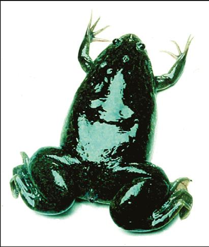
3.1 The frog Xenopus laevis is the model amphibian for studying development of the body plan

3.2 The zebrafish embryo develops around a large mass of yolk

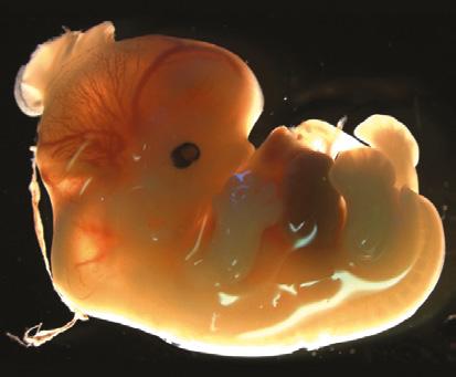
3.3 Birds and mammals resemble each other and differ from Xenopus in some important features of early development

3.4 The early chicken embryo develops as a flat disc of cells overlying a massive yolk

3.5 The mouse egg has no yolk and early development involves the allocation of cells to form the placenta and extra-embryonic membranes

3.6 Gene expression in embryos can be mapped by in situ nucleic acid hybridization

■ Experimental Box 3A Gene-expression profiling by DNA microarrays and RNA seq

3.7 Fate mapping and lineage tracing reveal which cells in which parts of the early embryo give rise to particular adult structures

3.8 Not all techniques are equally applicable to all vertebrates 120

3.9 Developmental genes can be identified by spontaneous mutation and by large-scale mutagenesis screens 121

■ Experimental Box 3B Large-scale mutagenesis screens for recessive mutations in zebrafish 123

3.10 Transgenic techniques enable animals to be produced with mutations in specific genes 124

■ Experimental Box 3C The Cre/loxP system: a strategy for making gene knock-outs in mice 127

■ Experimental Box 3D The CRISPR-Cas9 genome-editing system 128

3.11 Gene function can also be tested by transient transgenesis and gene silencing 130

Human embryonic development

3.12 The early development of a human embryo is similar to that of the mouse 131

■ Medical Box 3E Preimplantation genetic diagnosis 134

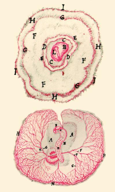
3.13 The timing of formation and the anatomy of the human placenta differs from that in the mouse 135

3.14 Some studies of human development are possible but are subject to strict laws 136

■ Box 3F Identical twins

Summary to Chapter 3

Chapter 4 Vertebrate development II: Xenopus and zebrafish

Setting up the body axes

4.1 The animal–vegetal axis is maternally determined in Xenopus 143

■ Cell Biology Box 4A Intercellular protein signals in vertebrate development 145

■ Cell Biology Box 4B The Wnt/β-catenin signaling pathway 146

4.2 Local activation of Wnt/β-catenin signaling specifies the future dorsal side of the embryo 147

4.3 Signaling centers develop on the dorsal side of the blastula 149 Summary 150 The origin and specification of the germ layers 150

4.4 The fate map of the Xenopus blastula makes clear the function of gastrulation 151

4.5 Cells of the early Xenopus embryo do not yet have their fates determined and regulation is possible 152

4.6 Endoderm and ectoderm are specified by maternal factors, whereas mesoderm is induced from ectoderm by signals from the vegetal region 152

■ Cell Biology Box 4C Signaling by members of the TGF-β family of growth factors 155

4.7 Mesoderm induction occurs during a limited period in the blastula stage 155

4.8 Zygotic gene expression is turned on at the mid-blastula transition 156

4.9 Mesoderm-inducing and patterning signals are produced by the vegetal region, the organizer, and the ventral mesoderm 157

4.10 Members of the TGF-β family have been identified as mesoderm inducers 158

■ Experimental Box 4D Investigating receptor function using dominant-negative proteins 159

4.11 The zygotic expression of mesoderm-inducing and patterning signals is activated by the combined actions of maternal VegT and Wnt signaling 159

4.12 Threshold responses to gradients of signaling proteins are likely to pattern the mesoderm 161

Summary

162

The Spemann organizer and neural induction 163

■ Cell Biology Box 4E The fibroblast growth factor signaling pathway 163

4.13 Signals from the organizer pattern the mesoderm dorso-ventrally by antagonizing the effects of ventral signals 164

4.14 The antero-posterior axis of the embryo emerges during gastrulation

165

4.15 The neural plate is induced in the ectoderm 168

4.16 The nervous system is patterned along the antero-posterior axis by signals from the mesoderm 170

4.17 The final body plan emerges by the end of gastrulation and neurulation 171

Summary 172

Development of the body plan in zebrafish 172

4.18 The body axes in zebrafish are established by maternal determinants 173

4.19 The germ layers are specified in the zebrafish blastoderm by similar signals to those in Xenopus 173

4.20 The shield in zebrafish is the embryonic organizer 176

■ Box 4F A zebrafish gene regulatory network 176

Summary to Chapter 4 178

Chapter 5 Vertebrate development III: chick and mouse—completing the body plan 183

Development of the body plan in chick and mouse and generation of the spinal cord 184

5.1 The antero-posterior polarity of the chick blastoderm is related to the primitive streak 184

5.2 Early stages in mouse development establish separate cell lineages for the embryo and the extra-embryonic structures

186

5.3 Movement of the anterior visceral endoderm indicates the definitive antero-posterior axis in the mouse embryo 190

5.4 The fate maps of vertebrate embryos are variations on a basic plan 192

■ Cell Biology Box 5A Fine-tuning Nodal signaling 193

5.5 Mesoderm induction and patterning in the chick and mouse occurs during primitive streak formation 195

5.6 The node that develops at the anterior end of the streak in chick and mouse embryos is equivalent to the Spemann organizer in Xenopus 196

5.7 Neural induction in chick and mouse is initiated by FGF signaling with inhibition of BMP signaling being required in a later step 198

■ Cell Biology Box 5B Chromatin-remodeling complexes 201

5.8 Axial structures in chick and mouse are generated from self-renewing cell populations 202

Summary 204

Somite formation and antero-posterior patterning 205

■ Cell Biology Box 5C Retinoic acid: a small-molecule intercellular signal 206

5.9 Somites are formed in a well-defined order along the antero-posterior axis 206

■ Cell Biology Box 5D The Notch signaling pathway 211

5.10 Identity of somites along the antero-posterior axis is specified by Hox gene expression 213

■ Box 5E The Hox genes 214

5.11 Deletion or overexpression of Hox genes causes changes in axial patterning 217

5.12 Hox gene expression is activated in an anterior to posterior pattern 219

5.13 The fate of somite cells is determined by signals from the adjacent tissues 221

Summary 223

The origin and patterning of neural crest 223

5.14 Neural crest cells arise from the borders of the neural plate and migrate to give rise to a wide range of different cell types 223

5.15 Neural crest cells migrate from the hindbrain to populate the branchial arches 225

Summary 226

Determination of left–right asymmetry 227

5.16 The bilateral symmetry of the early embryo is broken to produce left–right asymmetry of internal organs 227

5.17 Left–right symmetry breaking may be initiated within cells of the early embryo 229

Summary to Chapter 5 230

Chapter 6 Development of nematodes and sea urchins

Nematodes

■ Cell Biology Box 6A Apoptotic pathways in nematodes, Drosophila, and mammals

6.1 The cell lineage of Caenorhabditis elegans is largely invariant

6.2 The antero-posterior axis in Caenorhabditis elegans is determined by asymmetric cell division 239

■ Experimental Box 6B Gene silencing by antisense RNA and RNA interference

6.3 The dorso-ventral axis in Caenorhabditis elegans is determined by cell–cell interactions

6.4 Both asymmetric divisions and cell–cell interactions specify cell fate in the early nematode embryo

6.5 Cell differentiation in the nematode is closely linked to the pattern of cell division

6.6 Hox genes specify positional identity along the antero-posterior axis in Caenorhabditis elegans 247

6.7 The timing of events in nematode development is under genetic control that involves microRNAs 248

■ Box 6C Gene silencing by microRNAs

6.8 Vulval development is initiated through the induction of a small number of cells by short-range signals from a single inducing cell

6.9 The sea urchin embryo develops into a free-swimming

6.10 The sea urchin egg is polarized along the animal–vegetal axis

6.11 The sea urchin fate map is finely specified, yet considerable regulation is possible

6.12 The vegetal region of the sea urchin embryo acts as an organizer

6.13 The sea urchin vegetal region is demarcated by the nuclear accumulation of β-catenin

6.14 The animal–vegetal axis and the oral–aboral axis can be considered to correspond to the antero-posterior and dorso-ventral axes of other deuterostomes

6.15 The pluteus skeleton develops from the primary mesenchyme

6.16 The oral–aboral axis in sea urchins is related to the plane of the first cleavage

6.17 The oral ectoderm acts as an organizing region for the oral–aboral axis

■ Experimental Box 6D The gene regulatory network for sea urchin endomesoderm specification 265

Summary 266

Summary to Chapter 6 267

Chapter 7 Morphogenesis: change in form in the early embryo 271

Cell adhesion 273

■ Cell Biology Box 7A Cell-adhesion molecules and cell junctions 274

7.1 Sorting out of dissociated cells demonstrates differences in cell adhesiveness in different tissues 275

7.2 Cadherins can provide adhesive specificity 276

7.3 The activity of the cytoskeleton regulates the mechanical properties of cells and their interactions with each other 277

■ Cell Biology Box 7B The cytoskeleton, cell-shape change, and cell movement 278

7.4 Transitions of tissues from an epithelial to a mesenchymal state, and vice versa, involve changes in adhesive junctions 279

Summary 280

Cleavage and formation of the blastula 280

7.5 The orientation of the mitotic spindle determines the plane of cleavage at cell division 281

7.6 The positioning of the spindle within the cell also determines whether daughter cells will be the same or different sizes 283

7.7 Cells become polarized in the sea urchin blastula and the mouse morula 285

7.8 Fluid accumulation as a result of tight-junction formation and ion transport forms the blastocoel of the mammalian blastocyst 287

Summary 288

Gastrulation movements 289

7.9 Gastrulation in the sea urchin involves an epithelial-tomesenchymal transition, cell migration, and invagination of the blastula wall 289

7.10 Mesoderm invagination in Drosophila is due to changes in cell shape controlled by genes that pattern the dorso-ventral axis 293

7.11 Germ-band extension in Drosophila involves myosindependent remodeling of cell junctions and cell intercalation 295

7.12 Planar cell polarity confers directionality on a tissue 296

7.13 Gastrulation in amphibians and fish involves involution, epiboly, and convergent extension 299

■ Box 7C Convergent extension 302

7.14 Xenopus notochord development illustrates the dependence of medio-lateral cell elongation and cell intercalation on a pre-existing antero-posterior polarity 305

7.15 Gastrulation in chick and mouse embryos involves the separation of individual cells from the epiblast and their ingression through the primitive streak

tube formation

7.16 Neural tube formation is driven by changes in cell shape and convergent extension

■ Cell Biology Box 7D Eph receptors and their ephrin ligands

■ Medical Box 7E Neural tube defects

of tubes and branching morphogenesis

7.17 The Drosophila tracheal system is a prime example of branching morphogenesis

7.18 The vertebrate vascular system develops by vasculogenesis followed by sprouting angiogenesis

7.19 New blood vessels are formed from pre-existing vessels in angiogenesis

7.20 Embryonic neural crest gives rise to a wide range of different cell types

7.21 Neural crest migration is controlled by environmental cues

7.22 The formation of the lateral-line primordium in fishes is an example of collective cell migration

7.23 Body wall closure occurs in Drosophila, Caenorhabditis, mammals, and chick

Chapter 8 Cell differentiation and stem cells

■ Box 8A Conrad Waddington’s ‘epigenetic landscape’ provides a framework for thinking about how cells differentiate

The control of gene expression

8.1 Control of transcription involves both general and tissue-specific transcriptional regulators

8.2 Gene expression is also controlled by epigenetic chemical modifications to DNA and histone proteins that alter chromatin structure

■ Cell Biology Box 8B Epigenetic control of gene expression by chromatin modification

8.3 Patterns of gene activity can be inherited by persistence of gene-regulatory proteins or by maintenance of chromatin modifications

8.4 Changes in patterns of gene activity during differentiation can be triggered by extracellular signals

Cell differentiation and stem cells 350

8.5 Muscle differentiation is determined by the MyoD family of transcription factors 350

8.6 The differentiation of muscle cells involves withdrawal from the cell cycle, but is reversible 352

8.7 All blood cells are derived from multipotent stem cells 354

8.8 Intrinsic and extrinsic changes control differentiation of the hematopoietic lineages 357

■ Experimental Box 8C Single-cell analysis of cell-fate decisions 358

8.9 Developmentally regulated globin gene expression is controlled by control regions far distant from the coding regions 361

8.10 The epidermis of adult mammalian skin is continually being replaced by derivatives of stem cells 363

■ Medical Box 8D Treatment of junctional epidermolysis bullosa with skin grown from genetically corrected stem cells 366

8.11 Stem cells use different modes of division to maintain tissues 367

8.12 The lining of the gut is another epithelial tissue that requires continuous renewal 368

8.13 Skeletal muscle and neural cells can be renewed from stem cells in adults 370

8.14 Embryonic stem cells can proliferate and differentiate into many cell types in culture and contribute to normal development in vivo 372

■ Experimental Box 8E The derivation and culture of mouse embryonic stem cells 374 Summary 375

The plasticity of the differentiated state 376

8.15 Nuclei of differentiated cells can support development 376

8.16 Patterns of gene activity in differentiated cells can be changed by cell fusion 378

8.17 The differentiated state of a cell can change by transdifferentiation

8.18 Adult differentiated cells can be reprogrammed to form pluripotent stem cells 381

■ Experimental Box 8F Induced pluripotent stem cells 382

8.19 Stem cells could be a key to regenerative medicine 382

■ Experimental Box 8G Stem cells can be cultured in vitro to produce ‘organoids’—structures that mimic tissues and organs 386

8.20 Various approaches can be used to generate differentiated cells for cell-replacement therapies

Chapter 9 Germ cells, fertilization, and sex

The development of germ cells

9.1 Germ cell fate is specified in some embryos by a distinct germplasm in the egg

9.2 In mammals germ cells are induced by cell–cell interactions during development

9.3 Germ cells migrate from their site of origin to the gonad

9.4 Germ cells are guided to their destination by chemical signals

9.5 Germ cell differentiation involves a halving of chromosome number by meiosis

■ Box 9A Polar bodies

9.6 Oocyte development can involve gene amplification and contributions from other cells

9.7 Factors in the cytoplasm maintain the totipotency of the egg

9.8 In mammals some genes controlling embryonic growth are ‘imprinted’

9.9 Fertilization involves cell-surface interactions between egg and sperm

9.10 Changes in the egg plasma membrane and enveloping layers at fertilization block polyspermy

9.11 Sperm–egg fusion causes a calcium wave that results in egg activation

9.12 The primary sex-determining gene in mammals is on the Y chromosome

9.13 Mammalian sexual phenotype is regulated by gonadal hormones

9.14 The primary sex-determining factor in Drosophila is the number of X chromosomes and is cell autonomous 422

9.15 Somatic sexual development in Caenorhabditis is determined by the number of X chromosomes

9.16 Determination of germ cell sex depends on both genetic constitution and intercellular signals

9.17 Various strategies are used for dosage compensation of X-linked genes

10.1 Imaginal discs arise from the ectoderm in the early Drosophila

10.2 Imaginal discs arise across parasegment boundaries and are patterned by signaling at compartment boundaries 438

10.3 The adult wing emerges at metamorphosis after folding and evagination of the wing imaginal disc 439

10.4 A signaling center at the boundary between anterior and posterior compartments patterns the Drosophila wing disc along the antero-posterior axis 440

■ Box 10A Positional information and morphogen gradients 443

10.5 A signaling center at the boundary between dorsal and ventral compartments patterns the Drosophila wing along the dorso-ventral axis 445

10.6 Vestigial is a key regulator of wing development that acts to specify wing identity and control wing growth 445

10.7 The Drosophila wing disc is also patterned along the proximo-distal axis 447

10.8 The leg disc is patterned in a similar manner to the wing disc, except for the proximo-distal axis 448

10.9 Different imaginal discs can have the same positional values 450

Summary 450

The vertebrate limb 452

10.10 The vertebrate limb develops from a limb bud and its development illustrates general principles 452

10.11 Genes expressed in the lateral plate mesoderm are involved in specifying limb position, polarity, and identity 454

10.12 The apical ectodermal ridge is required for limb-bud outgrowth and the formation of structures along the proximodistal axis of the limb 457

10.13 Formation and outgrowth of the limb bud involves oriented cell behavior 458

10.14 Positional value along the proximo-distal axis of the limb bud is specified by a combination of graded signaling and a timing mechanism 460

10.15 The polarizing region specifies position along the limb’s antero-posterior axis 462

10.16 Sonic hedgehog is the polarizing region morphogen 464

■ Medical Box 10B Too many fingers: mutations that affect antero-posterior patterning can cause polydactyly 465

■ Cell Biology Box 10C Sonic hedgehog signaling and the primary cilium 466

10.17 The dorso-ventral axis of the limb is controlled by the ectoderm 468

■ Medical Box 10D Teratogens and the consequences of damage to the developing embryo 470

10.18 Development of the limb is integrated by interactions between signaling centers 470

10.19 Hox genes have multiple inputs into the patterning of the limbs 472

10.20 Self-organization may be involved in the development of the limb 475

■ Box 10E Reaction–diffusion mechanisms 476

10.21 Limb muscle is patterned by the connective tissue 477

10.22 The initial development of cartilage, muscles, and tendons is autonomous 478

10.23 Joint formation involves secreted signals and mechanical stimuli 478

10.24 Separation of the digits is the result of programmed cell death

10.25 Tooth development involves epithelial–mesenchymal interactions and a homeobox gene code specifies tooth identity 482

Summary 484

Vertebrate lungs 484

10.26 The vertebrate lung develops from a bud of endoderm 484

■ Medical Box 10F What developmental biology can teach us about breast cancer 486

10.27 Morphogenesis of the lung involves three modes of branching 488

heart 489

10.28 The development of the vertebrate heart involves morphogenesis and patterning of a mesodermal tube 489

The vertebrate eye 492

10.29 Development of the vertebrate eye involves interactions between an extension of the forebrain and the ectoderm of the head 493 Summary 497

Summary to Chapter 10 497

Chapter 11 Development of the nervous system 505

Specification of cell identity in the nervous system 507

11.1 Initial regionalization of the vertebrate brain involves signals from local organizers 507

11.2 Local signaling centers pattern the brain along the antero-posterior axis 508

11.3 The cerebral cortex is patterned by signals from the anterior neural ridge 509

11.4 The hindbrain is segmented into rhombomeres by boundaries of cell-lineage restriction 509

11.5 Hox genes provide positional information in the developing hindbrain 512

11.6 The pattern of differentiation of cells along the dorso-ventral axis of the spinal cord depends on ventral and dorsal signals 513

11.7 Neuronal subtypes in the ventral spinal cord are specified by the ventral to dorsal gradient of Shh 515

11.8 Spinal cord motor neurons at different dorso-ventral positions project to different trunk and limb muscles 516

11.9 Antero-posterior pattern in the spinal cord is determined in response to secreted signals from the node and adjacent mesoderm 517

Summary 518

The formation and migration of neurons 518

11.10 Neurons in Drosophila arise from proneural clusters 519

11.11 The development of neurons in Drosophila involves asymmetric cell divisions and timed changes in gene expression 521

11.12 The production of vertebrate neurons involves lateral inhibition, as in Drosophila 522

■ Box 11A Specification of the sensory organs of adult Drosophila 523

11.13 Neurons are formed in the proliferative zone of the vertebrate neural tube and migrate outwards 524

■ Experimental Box 11B Timing the birth of cortical neurons 526

11.14 Many cortical interneurons migrate tangentially 528

Summary 528

Axon navigation 529

11.15 The growth cone controls the path taken by a growing axon 530

■ Box 11C The development of the neural circuit for the knee-jerk reflex 532

11.16 Motor neuron axons in the chick limb are guided by ephrin–Eph interactions 533

11.17 Axons crossing the midline are both attracted and repelled 534

11.18 Neurons from the retina make ordered connections with visual centers in the brain 535

Summary 538

Synapse formation and refinement 539

11.19 Synapse formation involves reciprocal interactions 539

11.20 Many motor neurons die during normal development 542

■ Medical Box 11D Autism: a developmental disorder that involves synapse dysfunction 543

11.21 Neuronal cell death and survival involve both intrinsic and extrinsic factors 544

11.22 The map from eye to brain is refined by neural activity 545

Summary 546

Summary to Chapter 11 547 Chapter 12 Growth, post-embryonic

12.1 Tissues can grow by cell proliferation, cell enlargement, or accretion 555

12.2 Cell proliferation is controlled by regulating entry into the cell cycle 556

12.3 Cell division in early development can be controlled by an intrinsic developmental program 557

12.4 Extrinsic signals coordinate cell division, cell growth, and cell death in the developing Drosophila wing 558

■ Cell Biology Box 12A The core Hippo signaling pathways in Drosophila and mammals 559

12.5 Cancer can result from mutations in genes that control cell proliferation 560

12.6 The relative contributions of intrinsic and extrinsic factors in controlling size differ in different mammalian organs 562

12.7 Overall body size depends on the extent and the duration of growth 564

12.8 Hormones and growth factors coordinate the growth of different tissues and organs and contribute to determining overall body size 565

12.9 Elongation of the long bones illustrates how growth can be determined by a combination of an intrinsic growth program and extracellular factors 566

■ Box 12B Digit length ratio is determined in the embryo 568

12.10 The amount of nourishment an embryo receives can have profound effects in later life

12.11 Arthropods have to molt in order to grow

12.12 Insect body size is determined by the rate and duration of larval growth

12.13 Metamorphosis in amphibians is under hormonal

12.14 Regeneration involves repatterning of existing tissues and/or growth of new tissues

12.15 Amphibian limb regeneration involves cell dedifferentiation and new growth 578

■ Box 12C Regeneration in Hydra 580

■ Box 12D Planarian regeneration 582

12.16 Limb regeneration in amphibians depends on the presence of nerves 586

12.17 The limb blastema gives rise to structures with positional values distal to the site of amputation 587

12.18 Retinoic acid can change proximo-distal positional values in regenerating limbs 589

12.19 Mammals can regenerate the tips of the digits 590

12.20 Insect limbs intercalate positional values by both proximo-distal and circumferential growth 591

■ Box 12E Why can’t we regenerate our limbs? 592

12.21 Heart regeneration in zebrafish involves the resumption of cell division by cardiomyocytes 594

Summary 596

Aging and senescence 597

12.22 Genes can alter the timing of senescence 598

12.23 Cell senescence blocks cell proliferation 600

12.24 Elimination of senescent cells in adult salamanders explains why regenerative ability does not diminish with age 601

Summary 602

Summary to Chapter 12 602

Chapter 13 Plant development 609

13.1 The model plant Arabidopsis thaliana has a short life cycle and a small diploid genome 611

Embryonic development 612

13.2 Plant embryos develop through several distinct stages 612

■ Box 13A Angiosperm embryogenesis 614

13.3 Gradients of the signal molecule auxin establish the embryonic apical–basal axis 616

13.4 Plant somatic cells can give rise to embryos and seedlings 617

13.5 Cell enlargement is a major process in plant growth and morphogenesis 619

■ Experimental Box 13B Plant transformation and genome editing 620

Summary 621

Meristems 622

13.6 A meristem contains a small, central zone of self-renewing stem cells 623

13.7 The size of the stem cell area in the meristem is kept constant by a feedback loop to the organizing center 623

13.8 The fate of cells from different meristem layers can be changed by changing their position 624

13.9 A fate map for the embryonic shoot meristem can be deduced using clonal analysis 626

13.10 Meristem development is dependent on signals from other parts of the plant 627

13.11 Gene activity patterns the proximo-distal and adaxial–abaxial axes of leaves developing from the shoot meristem 628

13.12 The regular arrangement of leaves on a stem is generated by regulated auxin transport 629

13.13 The outgrowth of secondary shoots is under hormonal control 630

13.14 Root tissues are produced from Arabidopsis root apical meristems by a highly stereotyped pattern of cell divisions 633

13.15 Root hairs are specified by a combination of positional information and lateral inhibition 635 Summary 636

Flower development and control of flowering 636

13.16 Homeotic genes control organ identity in the flower 637

■ Box 13C The basic model for the patterning of the Arabidopsis flower 639

13.17 The Antirrhinum flower is patterned dorso-ventrally, as well as radially 640

13.18 The internal meristem layer can specify floral meristem patterning 641

13.19 The transition of a shoot meristem to a floral meristem is under environmental and genetic control 642

■ Box 13D The circadian clock coordinates plant growth and development 643

13.20 Vernalization reflects the epigenetic memory of winter 643

13.21 Most flowering plants are hermaphrodites, but some produce unisexual flowers 645

Chapter 14 Evolution and development

■ Box 14A Darwin’s finches

The evolution of development

14.1 Multicellular organisms evolved from single-celled ancestors

14.2 Genomic evidence is throwing light on the evolution of animals 657

■ Box 14B The metazoan family tree 658

14.3 How gastrulation evolved is not known 659

14.4 More general characteristics of the body plan develop earlier than specializations 660

14.5 Embryonic structures have acquired new functions during evolution 661

14.6 Evolution of different types of eyes in different animal groups is an example of parallel evolution 663

The diversification of body plans 665

14.7 Hox gene complexes have evolved through gene duplication 665

14.8 Differences in Hox gene expression determine the variation in position and type of paired appendages in arthropods 667

14.9 Changes in Hox gene expression and their target genes contributed to the evolution of the vertebrate axial skeleton 671

14.10 The basic body plan of arthropods and vertebrates is similar, but the dorso-ventral axis is inverted 672

Summary 673

The evolutionary modification of specialized characters 674

14.11 Limbs evolved from fins 674

14.12 Limbs have evolved to fulfill different specialized functions 678

14.13 The evolution of limblessness in snakes is associated with changes in axial gene expression and mutations in a limb-specific enhancer 679

14.14 Butterfly wing markings have evolved by redeployment of genes previously used for other functions 680

■ Experimental Box 14C Using CRISPR-Cas9 genome-editing techniques to test the functioning of the snake ZRS 681

14.15 Adaptive evolution within the same species provides a way of studying the developmental basis for evolutionary change

■ Experimental Box 14D Pelvic reduction in sticklebacks is based on mutations in a gene control region 686

Changes in the timing of developmental processes 687

14.16 Changes in growth can modify the basic body plan 687

■ Box 14E Origins of morphological diversity in dogs 689

14.17 Evolution can be due to changes in the timing of developmental events 690

14.18 The evolution of life histories has implications for development 692

■ Box 14F Long- and short-germ development in insects 693

to Chapter 14 696

List of Boxes

Special Interest Boxes

Box 1A Basic stages of Xenopus laevis development 4

Box 3F Identical twins 137

Box 4F A zebrafish gene regulatory network 176

Box 5E The Hox genes 214

Box 6C Gene silencing by microRNAs 250

Box 7C Convergent extension 302

Box 8A Conrad Waddington’s ‘epigenetic landscape’ provides a framework for thinking about how cells differentiate 335

Box 9A Polar bodies 405

Box 10A Positional information and morphogen gradients 443

Box 10E Reaction–diffusion mechanisms 476

Box 11A Specification of the sensory organs of adult Drosophila 523

Box 11C The development of the neural circuit for the knee-jerk reflex 532

Box 12B Digit length ratio is determined in the embryo 568

Box 12C Regeneration in Hydra 580

Box 12D Planarian regeneration 582

Box 12E Why can’t we regenerate our limbs? 592

Box 13A Angiosperm embryogenesis 614

Box 13C The basic model for the patterning of the Arabidopsis flower 639

Box 13D The circadian clock coordinates plant growth and development 643

Box 14A Darwin’s finches 654

Box 14B The metazoan family tree 658

Box 14E Origins of morphological diversity in dogs 689

Box 14F Long- and short-germ development in insects 693

Cell Biology Boxes

Box 1B The mitotic cell cycle 7

Box 1C Germ layers 15

Box 1E Signal transduction and intracellular signaling pathways 26

Box 2B The Toll signaling pathway: a multifunctional pathway 54

Box 2C The JAK–STAT signaling pathway 58

Box 2E The Hedgehog signaling pathway 80

Box 4A Intercellular protein signals in vertebrate development 145

Box 4B The Wnt/β-catenin signaling pathway 146

Box 4C Signaling by members of the TGF-β family of growth factors 155

Box 4E The fibroblast growth factor signaling pathway 163

Box 5A Fine-tuning Nodal signaling 193

Box 5B Chromatin-remodeling complexes 201

Box 5C Retinoic acid: a small-molecule intercellular signal 206

Box 5D The Notch signaling pathway 211

Box 6A Apoptotic pathways in nematodes, Drosophila, and mammals 238

Box 7A Cell-adhesion molecules and cell junctions 274

Box 7B The cytoskeleton, cell-shape change, and cell movement 278

Box 7D Eph receptors and their ephrin ligands 313

Box 8B Epigenetic control of gene expression by chromatin modification 344

Box 10C Sonic hedgehog signaling and the primary cilium

Box 12A The core Hippo signaling pathways in Drosophila and mammals

Experimental Boxes

Box 1D Visualizing gene expression in embryos

466

559

20

Box 2A Mutagenesis and genetic screening strategy for identifying developmental mutants in Drosophila 43

Box 2D Targeted gene expression and misexpression screening 69

Box 2F Mutants in denticle pattern provided clues to the logic of segment patterning 81

Box 3A Gene-expression profiling by DNA microarrays and RNA seq 117

Box 3B Large-scale mutagenesis screens for recessive mutations in zebrafish 123

Box 3C The Cre/loxP system: a strategy for making gene knock-outs in mice

127

Box 3D The CRISPR-Cas9 genome-editing system 128

Box 4D Investigating receptor function using dominant-negative proteins 159

Box 6B Gene silencing by antisense RNA and RNA interference 241

Box 6D The gene regulatory network for sea urchin endomesoderm specification 265

Box 8C Single-cell analysis of cell-fate decisions

Box 8E The derivation and culture of mouse embryonic stem cells

358

374

Box 8F Induced pluripotent stem cells 382

Box 8G Stem cells can be cultured in vitro to produce ‘organoids’—structures that mimic tissues and organs

386

Box 11B Timing the birth of cortical neurons 526

Box 13B Plant transformation and genome editing 620

Box 14C Using CRISPR-Cas9 genome-editing techniques to test the functioning of the snake ZRS 681

Box 14D Pelvic reduction in sticklebacks is based on mutations in a gene control region

Medical Boxes

Box 1F When development goes awry

Box 3E Preimplantation genetic diagnosis

Box 7E Neural tube defects

Box 8D Treatment of junctional epidermolysis bullosa with skin grown from genetically corrected stem cells

686

28

134

314

366

Box 10B Too many fingers: mutations that affect antero-posterior patterning can cause polydactyly

Box 10D Teratogens and the consequences of damage to the developing embryo

Box 10F What developmental biology can teach us about breast cancer

Box 11D Autism: a developmental disorder that involves synapse dysfunction

Reviewer acknowledgments

We would like to reiterate our thanks to the people who contributed to the first five editions. Many people gave their advice based on the fifth edition, and others reviewed the draft chapters for the sixth edition as they emerged. We wish to express our gratitude to the following colleagues.

Katie Abley, University of Cambridge

Mike Akam, University of Cambridge

Willy M. Baarends, University Medical Centre, Rotterdam

Hegias Mira Bontenbal, University Medical Centre, Rotterdam

Thomas Butts, University of Liverpool

Sinéad Drea, University of Leicester

Sander van den Driesche, University of Edinburgh

Anna-Pavlina Haramis, Leiden University

Brigitte Galliot, University of Geneva

Erik Griffin, Dartmouth College

Jane Kenney-Hunt, Westminster College, Fulton, Missouri

Jane Langdale, University of Oxford

Tony Perry, University of Bath

Mary Elizabeth Pownall, University of York

Aida Rajic, University of Suffolk

Joanna Richardson, University of Sussex

Guojun Sheng, Kumamoto University

Kate Storey, University of Dundee

Shahragim Tajbakhsh, Institut Pasteur, Paris

Michael Taylor, Cardiff University

Adri Thomas, Utrecht University

Fiona Wardle, King’s College London

Deneen Wellik, University of Michigan Medical School, Ann Arbor

Larissa Williams, Bates College, Lewiston, Maine

We would also like to thank our colleagues who wrote the online Journal Clubs to accompany this edition.

Katie Abley, University of Cambridge

Zoe Burke, University of Bath

Andrew Economou, The Francis Crick Institute

Caitlin McQueen, University of Sheffield

Adriana Amorim Torres, AAT Assessoria Cientìfica

History and basic concepts

● The origins of developmental biology

A conceptual tool kit

The aim of this chapter is to provide a conceptual framework for the study of development. We start with a brief history of the study of embryonic development, which illustrates how some of the key questions in developmental biology were first formulated, and continue with some of the essential principles of development. The big question is: How does a single cell—the fertilized egg—give rise to a multicellular organism, in which a multiplicity of different cell types are organized into tissues and organs to make up a three-dimensional body? This question can be studied from many different viewpoints, all of which have to be fitted together to obtain a complete picture of development: which genes are expressed, and when and where; how cells communicate with each other; how a cell’s developmental fate is determined; how cells proliferate and differentiate into specialized cell types; and how major changes in body shape are produced. All the information for embryonic development is contained within the fertilized egg. We shall see that an organism’s development is ultimately driven by the regulated expression of its genes in space and time, determining which proteins are present in which cells and when. In turn, proteins largely determine how a cell behaves. The genes provide a generative program for development, not a blueprint, as their actions are translated into developmental outcomes through cellular behavior such as intercellular signaling, cell proliferation, cell differentiation, and cell movement.

The development of a multicellular organism from a single cell—the fertilized egg—is a brilliant triumph of evolution. The fertilized egg divides to give rise to many millions of cells, which form structures as complex and varied as eyes, arms, heart, and brain. This amazing achievement raises a multitude of questions. How do the cells arising from division of the fertilized egg become different from each other? How do they become organized into structures such as limbs and brains? What controls the behavior of individual cells so that such highly organized patterns emerge? How are the organizing principles of development embedded within the egg, and, in particular, within the genetic material, DNA? Much of the excitement in developmental biology today comes from our growing understanding of how genes direct these developmental processes, and genetic control is one of the main themes of this book. Thousands of genes are involved in controlling development, but we will focus only on those that have key roles, and illustrate general principles.

Fig. 1.1 Human fertilized egg and embryo.

(a) Human fertilized egg. The nuclear membranes of the sperm and egg nuclei (pronuclei) have not yet broken down to allow the parental chromosomes to mingle. (b) Human embryo at around 51 days’ gestation (Carnegie stage 20), which is equivalent to a mouse embryo at 13.5 days post-fertilization. A human embryo at this stage is about 21–23 mm long.

(a) Courtesy of A. Doshi, CRGH, London. (b) Reproduced courtesy of the MRC/ Wellcome-funded Human Developmental Biology Resource.

Understanding how embryos develop is a huge intellectual challenge, and one of the ultimate aims of the science of developmental biology is to understand how we humans develop (Fig. 1.1). We need to understand human development for several reasons. We need to properly understand why it sometimes goes wrong and why a fetus may fail to be born or a baby be born with congenital abnormalities. The link here with genetic control of development is very close, as mutations in genes can lead to abnormal development; environmental factors, such as drugs and infections, can affect it too. Another area of medical research related to developmental biology is regenerative medicine—finding out how to use cells to repair damaged tissues and organs. The focus of regenerative medicine is currently on stem cells. Stem cells that can proliferate and give rise to all the different tissues of the body are present in embryos. These, and the stem cells with more limited developmental potential that are found in adult tissues, are discussed in Chapter 8. Cancer cells also display some properties of embryonic cells, such as the ability to divide indefinitely, and so the study of embryonic cells and their behaviour could lead to new and better treatments for cancer, as many of the same genes are involved.

The development of an embryo from the fertilized egg is known as embryogenesis. One of the first tasks that cells have in an embryo is to lay down the overall body plan of the organism, and we shall see that different organisms solve this fundamental problem in several ways. The focus of this book is mainly on animal development, in particular that of vertebrates—frogs, birds, fish, and mammals—whose early development is discussed in Chapters 3–5. We also look at selected invertebrates, particularly the fruit fly and the nematode worm, and also the sea urchin. Our understanding of the genetic control of development is founded on work with fruit flies and nematodes, where it is also most advanced, and the main features of their early development are considered in Chapters 2 and 6, respectively. The fruit fly is also used throughout the book to illustrate particular aspects of development. In Chapter 13 we look briefly at some aspects of plant development, which differs in many respects from that of animals but involves similar basic principles.

Morphogenesis, or the development of form, is discussed in Chapter 7. In Chapter 9 we look at how sex is determined and how germ cells develop. The differentiation of unspecialized cells into cells that carry out particular functions, such as muscle cells and blood cells, is considered in Chapter 8. Structures such as the vertebrate limb, and organs such as insect and vertebrate eyes, the heart and the nervous system, illustrate the problems of multicellular organization and tissue differentiation in embryogenesis, and we consider some of these systems in detail in Chapters 10 and 11. The study of developmental biology, however, goes well beyond the development of the embryo. Post-embryonic growth and aging, how some animals undergo metamorphosis, and how animals can regenerate lost organs is discussed in Chapter 12. Taking a longer view, we shall consider in Chapter 14 how developmental mechanisms have evolved and how they constrain the very process of evolution itself.

One might ask whether it is necessary to cover so many different organisms in order to understand the basic features of development. The answer is yes. Developmental biologists do, indeed, believe that there are general principles of development that apply to all animals, but life is too wonderfully diverse to find all the answers in a single organism. As it is, developmental biologists have tended to focus their efforts on a relatively small number of animals, chosen because they were convenient to study and amenable to experimental manipulation or genetic analysis. This is why some creatures, such as the frog Xenopus laevis (Fig. 1.2) and the fruit fly Drosophila melanogaster, have such a dominant place in developmental biology. Similarly, work with the thale cress, Arabidopsis thaliana, has uncovered many features of plant development.

One of the most exciting and satisfying aspects of developmental biology is that understanding a developmental process in one organism can help to illuminate similar processes elsewhere—for example, giving insights into how humans develop. Nothing illustrates this more dramatically than the influence that our understanding of Drosophila development, and especially of its genetic basis, has had throughout

Fig. 1.2 The South African claw-toed frog, Xenopus laevis. Scale bar = 1 cm.
Photograph courtesy of J. Smith.

developmental biology. The identification of genes controlling early embryogenesis in Drosophila has led to the discovery of related genes being used in similar ways in the development of mammals and other vertebrates. Such discoveries encourage us to believe in the existence of general developmental principles.

Amphibians have long been favorite organisms for studying early development because their eggs are large and their embryos are easy to grow in a simple culture medium and relatively easy to experiment on. Embryogenesis in the South African frog Xenopus (Box 1A) illustrates some of the basic stages of development in all animals.

In the rest of this chapter we first look briefly at the history of embryology—as the study of developmental biology used to be called. The term developmental biology itself is of much more recent origin and reflects the appreciation that development is not restricted to the embryo alone. Traditionally, embryology described experimental results in terms of morphology and cell fate, but we now understand development in terms of molecular genetics and cell biology as well. In the second part of the chapter we will introduce some key concepts that are used over and over again in studying and understanding development.

The origins of developmental biology

Many questions in embryology were first posed hundreds, and in some cases thousands, of years ago. Appreciating the history of these ideas helps us to understand why we approach developmental problems in the way that we do today.

1.1 Aristotle first defined the problem of epigenesis versus preformation

A scientific approach to explaining development started with Hippocrates in Greece in the fifth century bc. Using the ideas current at the time, he tried to explain development in terms of the principles of heat, wetness, and solidification. About a century later the study of embryology advanced when the Greek philosopher Aristotle formulated a question that was to dominate much thinking about development until the end of the nineteenth century. Aristotle addressed the problem of how the different parts of the embryo were formed. He considered two possibilities: one was that everything in the embryo was preformed from the very beginning and simply got bigger during development; the other was that new structures arose progressively, a process he termed epigenesis (which means ‘upon formation’) and that he likened metaphorically to the ‘knitting of a net’. Aristotle favored epigenesis and his conjecture was correct. Aristotle’s influence on European thought was enormous and his ideas remained dominant well into the seventeenth century. The contrary view to epigenesis, namely that the embryo was preformed from the beginning, was championed anew in the late seventeenth century. Many could not believe that physical or chemical forces could mold a living entity such as the embryo. Along with the contemporaneous background of belief in the divine creation of the world and all living things, was the belief that all embryos had existed from the beginning of the world, and that the first embryo of a species must contain all future embryos.

Even the brilliant seventeenth-century Italian embryologist Marcello Malpighi could not free himself from preformationist ideas. While he provided a remarkably accurate description of the development of the chick embryo, he remained convinced, against the evidence of his own observations, that the fully formed embryo was present from the beginning (Fig. 1.3). He argued that at very early stages the parts were so small that they could not be seen, even with his best microscope. Other preformationists believed that the sperm contained the embryo, and some even claimed to see a tiny human—an homunculus—in the head of each human sperm (Fig. 1.4).

The preformation/epigenesis issue was vigorously debated throughout the eighteenth century. But the problem could not be resolved until one of the great advances in biology had taken place—the recognition that living things, including embryos, were composed of cells.

Fig. 1.3 Malpighi’s description of the chick embryo. The figure shows Malpighi’s drawings, made in 1673, depicting the early embryo (top) and at 2 days’ incubation (bottom). His drawings accurately illustrate the shape and blood supply of the embryo. Reprinted by permission of the President and Council of the Royal Society.

Fig. 1.4 Some preformationists believed that an homunculus was curled up in the head of each sperm.

An imaginative drawing, after N. Harspeler (1694).

Turn static files into dynamic content formats.

Create a flipbook
Issuu converts static files into: digital portfolios, online yearbooks, online catalogs, digital photo albums and more. Sign up and create your flipbook.