Great Clarendon Street, Oxford, OX2 6DP, United Kingdom
Oxford University Press is a department of the University of Oxford It furthers the University’s objective of excellence in research, scholarship, and education by publishing worldwide Oxford is a registered trade mark of Oxford University Press in the UK and in certain other countries
The moral rights of the authors have been asserted
First published in 2019
ISBN 978 0 19 842363 8
All rights reserved No part of this publication may be reproduced, stored in a retrieval system or transmitted in any form or by any means without the prior permission in writing of Oxford University Press, or as expressly permitted by law, by licence or under terms agreed with the appropriate reprographics rights organization Enquiries concerning reproduction outside the scope of the above should be sent to the Rights Department, Oxford University Press, at the address above
You must not circulate this work in any other form and you must impose this same condition on any acquirer
Printed in Great Britain by Bell and Bain Ltd Glasgow
Acknowledgements
Photo credits:
Cover image: James Paterson/PhotoPlus Magazine via Getty Images; p3: Peter Hermes Furian/Shutterstock; p3: Jeenson; p3: Paramjeet; p5: MEDIMAGE/SCIENCE PHOTO LIBRARY; p9: Sebastian Kaulitzki; p9: D PHILLIPS / SCIENCE PHOTO LIBRARY; p9: DR STANLEY FLEGLER/VISUALS UNLIMITED, INC /SCIENCE PHOTO LIBRARY; p13: PR G GIMENEZMARTIN / SCIENCE PHOTO LIBRARY; p14: Leonid Andronov/Shutterstock; p33: BIOPHOTO ASSOCIATES/SCIENCE PHOTO LIBRARY; p33: BIOPHOTO ASSOCIATES/SCIENCE PHOTO LIBRARY; p40: Vladimir Melnik/ Shutterstock; p50: Zens/Shutterstock; p59: Jurgen Ziewe/Shutterstock; p60: STEVE GSCHMEISSNER / SCIENCE PHOTO LIBRARY; p63: Ttsz/ Istockphoto; p70: Steve Cymro/Shutterstock; p79: The Biochemist Artist/ Shutterstock; p90: Volodymyr Krasyuk/Shutterstock; p102: Dimarion/ Shutterstock; p102: Dimarion/Shutterstock; p104: Gallagher, Aisling; p118: DR KEITH WHEELER/SCIENCE PHOTO LIBRARY; p118: PRARTHAK; p134: Jose Luis Calvo/Shutterstock; p136: R BICK, B POINDEXTER, UT MEDICAL SCHOOL/SCIENCE PHOTO LIBRARY; p143: Wearset Ltd and David Russell Illustration; p146: Michal Kowalski/Shutterstock; p147: DR JEREMY BURGESS / SCIENCE PHOTO LIBRARY; p154: DR GOPAL MURTI/ SCIENCE PHOTO LIBRARY; p155: SINCLAIR STAMMERS/SCIENCE PHOTO LIBRARY; p170: Mark Herreid/Shutterstock; p171: Toeytoey/Shutterstock; p185: Cathy Keifer/Shutterstock; p201: Ase/Shutterstock; p222: Beata Aldridge/Shutterstock; p234: Bildagentur Zoonar GmbH/Shutterstock; p234: AlessandroZocc/Shutterstock; p234: Denis Tabler/Shutterstock; p234: TB studio/Shutterstock; p235: MARTYN F CHILLMAID / SCIENCE PHOTO LIBRARY
Artwork by Aptara Corp and OUP
The publisher would like to thank the International Baccalaureate for their kind permission to adapt questions from past examinations and content from the subject guide The questions adapted for this book, and the corresponding past paper references, are summarized here:
This book provides full coverage of the IB diploma syllabus in biology and offers support to students preparing for their examinations. The book will help you revise the study material, learn the essential terms and concepts, strengthen your problem-solving skills and improve your approach to IB examinations The book is packed with worked examples and exam tips that demonstrate best practices and warn against common errors. All topics are illustrated by annotated student answers to questions from past examinations, which explain why marks may be scored or missed
A separate section is dedicated to data-based and practical questions, which are the most distinctive feature of the syllabus (first assessment in 2016) Numerous examples show how to tackle unfamiliar situations, interpret and analyse experimental data, and suggest improvements to experimental procedures Practice problems and a complete set of IB-style examination papers provide further opportunities to check your knowledge and skills, boost your confidence and monitor the progress of your studies. Full solutions to all problems and examination papers are given online at www.oxfordsecondary.com/ib-prepared-support.
As any study guide, this book is not intended to replace your course materials, such as textbooks, laboratory manuals, past papers and markschemes, the IB Biology syllabus and your own notes To succeed in the examination, you will need to use a broad range of resources, many of which are available online This book will navigate you through this critical part of your studies, making your preparation for the exam less stressful and more efficient.
DP Biology assessment
All standard level (SL) and higher level (HL) students must complete the internal assessment and take three papers as part of their external assessment Papers 1 and 2 are usually sat on one day and Paper 3 a day or two later. The internal and external assessment marks are combined as shown in the table at the top of page V to give your overall DP Biology grade, from 1 (lowest) to 7 (highest)
Overview of the book structure
The book is divided into several sections that cover the internal assessment, core SL and additional higher level (AHL) topics, data-based and practical questions, the four options (A–D) and a complete set of practice examination papers
The largest section of the book, core topics, follows the structure of the IB diploma biology syllabus (for first assessment 2016) and covers all understandings and applications and skills assessment statements. Topics 1–6 contain common material for SL and HL students while topics 7–11 are intended for HL students only. The nature of science concepts are also discussed where applicable.
The data-based and practical questions section (chapter 12) provides a detailed analysis of problems and laboratory experiments that often appear in section A of paper 3 Similar to core topics, the discussion is illustrated by worked examples and sample scripts, followed by IB-style practice problems
The options section reviews the material assessed in the second part of paper 3. Each of the four options is presented as a series of SL and AHL subtopics
The internal assessment section outlines the nature of the investigation that you will have to carry out and explains how to select a suitable topic, collect and process experimental data, draw conclusions and present your report in a suitable format to satisfy the marking criteria and achieve the highest grade.
The final section contains IB-style practice examination papers 1, 2 and 3, written exclusively for this book. These papers will give you an opportunity to test yourself before the actual exam and at the same time provide additional practice problems for every topic of core and options material
The answers and solutions to all practice problems and examination papers are given online at www.oxfordsecondary.com/ib-prepared-support. Blank answer sheets for examination papers are also available at the same address
Assessment overview
Assessment Description Topics
Internal Experimental work with a written report
Paper 1 Multiple-choice questions
The final IB diploma score is calculated by combining grades for six subjects with up to three additional points from theory of knowledge and extended essay components
Command terms
Command terms are pre-defined words and phrases used in all IB Biology questions and problems. Each command term specifies the type and depth of the response expected from you in a particular question For example, the command terms state, outline, explain and discuss require answers with increasingly higher levels of detail, from a single word, short sentence or numerical value (“state”) to comprehensive analysis (“discuss”), as shown in the next table.
Question
State the eect of increasing temperature on the reaction rate
Outline how an increase in temperature aects the reaction rate
Explain why an increase in temperature increases the reaction rate
Discuss the eects of increasing temperature and the presence of an enzyme on the reaction rate
Rate increases
Possible answer
For most reactions, the rate approximately doubles when temperature increases by 10 degrees
As temperature increases, the average speed and thus kinetic energy of particles also increase. The particles collide with one another more frequently and with a greater force. As a result, the frequency of successful collisions increases, so the rate increases.
Both factors increase the rate by increasing the frequency of successful collisions
However, an increase in temperature increases the frequency and intensity of all collisions (successful and unsuccessful) but has no eect on the activation energy
In contrast, an enzyme has no eect on the frequency or intensity of collisions but lowers the activation energy by providing an alternative reaction pathway and thus allowing slow-moving particles to collide successfully Thus, the same macroscopic eect is achieved by dierent microscopic changes
A list of commonly used command terms in biology examination questions is given in the following table. Understanding the exact meaning of frequently used command terms is essential for your success in the examination. Therefore, you should explore this table and use it regularly as a reference when answering questions in this book
Command term
Analyse
Annotate
Calculate
Comment
Compare
Compare and contrast
Denition
Break down in order to bring out the essential elements or structure
Add brief notes to a diagram or graph
Obtain a numerical answer showing your working
Give a judgment based on a given statement or result of a calculation
Give an account of the similarities between two or more items
Give an account of similarities and dierences between two or more items
Construct Present information in a diagrammatic or logical form
Deduce Reach a conclusion from the information given
Dene
Describe
Design
Determine
Give the precise meaning of a word, phrase, concept or physical quantity
Give a detailed account
Produce a plan, simulation or model
Obtain the only possible answer
Discuss Oer a considered and balanced review that includes a range of arguments, factors or hypotheses
Command term Denition
Distinguish
Draw
Make clear the dierences between two or more items
Represent by a labelled, accurate diagram or graph, drawn to scale, with plotted points (if appropriate) joined in a straight line or smooth curve
Estimate Obtain an approximate value
Explain
Identify
Label
List
Outline
Predict
Sketch
Give a detailed account including reasons or causes
Provide an answer from a number of possibilities
Add labels to a diagram
Give a sequence of brief answers with no explanation
Give a brief account or summary
Give an expected result
Represent by means of a diagram or graph (labelled as appropriate), giving a general idea of the required shape or relationship
State Give a specic name, value or other brief answer without explanation
Suggest Propose a solution, hypothesis or other possible answer
A complete list of command terms is available in the subject guide
Preparation
and exam strategies
In addition to the above suggestions, there are some simple rules you should follow during your preparation study and the exam itself
1. Get ready for study. Have enough sleep, eat well, drink plenty of water and reduce your stress by positive thinking and physical exercise. A good night’s sleep is particularly important before the exam day, as it can improve your score
2. Organize your study environment. Find a comfortable place with adequate lighting, temperature and ventilation Avoid distractions Keep your papers and computer files organized. Bookmark useful online and offline material
3. Plan your studies. Make a list of your tasks and arrange them by importance. Break up large tasks into smaller, easily manageable parts Create an agenda for your studying time and make sure that you can complete each task before the deadline.
4. Use this book as your first point of reference. Work your way through the topics systematically and identify the gaps in your understanding and skills Spend extra time on the topics where improvement is required. Check your textbook and online resources for more information
5. Read actively. Focus on understanding rather than memorizing Recite key points and definitions using your own words. Try to solve every worked example and practice problem before looking at the answer Make notes for future reference
6. Get ready for the exams. Practice answering exam-style questions under a time constraint Solve as many problems from past papers as you can. Take a trial exam using the papers at the end of this book
7. Optimize your exam approach. Read all questions carefully, paying extra attention to command terms Keep your answers as short and clear as possible. Double-check all numerical values and units Label axes in graphs and annotate diagrams. Use exam tips from this book.
8. Do not panic. Take a positive attitude and concentrate on things you can improve. Set realistic goals and work systematically to achieve these goals Be prepared to reflect on your performance and learn from your errors in order to improve your future results
Key features of the book
Each chapter typically covers one core or option topic, and starts with “You should know” and “You should be able to” checklists. These outline the understandings and applications and skills sections of the IB diploma biology syllabus. Some assessment statements have been reworded or combined together to make them more accessible and simplify the navigation. These changes do not affect the coverage of key syllabus material, which is always explained within the chapter Chapters contain the features outlined on this page.
Example
Examples offer solutions to typical problems and demonstrate common problem-solving techniques. Many examples provide alternative answers and explain how the marks are awarded
Nature of science relates a biology concept to the overarching principles of the scientific approach
Sample student answers show typical student responses to IB-style questions (most of which are taken from past examination papers). In each response, the correct points are often highlighted in green while incorrect or incomplete answers are highlighted in red. Positive or negative feedback on student’s response is given in the green and red pull-out boxes. An example is given below.
Number of marks available
The marks the student may have earned based on their response Examination question
SAMPLE STUDENT ANSWER
Theoretical concepts and key definitions are discussed at a level sufficient for answering typical examination questions Many concepts are illustrated by diagrams, tables or worked examples. Most definitions are given in a grey side box like this one, and explained in the text. This feature highlights the essential terms and statements that have appeared in past markschemes, warns against common errors and shows how to optimize your approach to particular questions
Links provide a reference to relevant material, within another part of this book, that relates to the text in question
You will see an exam paper icon on the right when the question has been adapted from a past IB paper
Describe what is meant by a condensation reaction. [2]
This answer could have achieved 1/2 marks:
A condensation reaction is an anabolic reaction that builds a polymer from a monomer.
The student’s response
Practice problems
Practice problems are given at the end of each chapter These are IB-style questions that provide you with an opportunity to test yourself and improve your
▼ clear enough, as it implies a single reacting species while a condensation must involve at least two reacting species, which can be identical or different molecules or ions
▲ “Water” is accepted as the most common by-product, although the answer “ a small molecule” would be more accurate
Negative feedback Positive feedback
Questions not taken from past IB examinations will not have the exam paper icon.
problem-solving skills Some questions introduce factual or theoretical material from the syllabus that can be studied independently.
C E L L B I O LO GY 1
1 . 1 I N T R O D U C T I O N T O C E L L S
Y sd kw:
✔ all living organisms are composed of cells
✔ unicellular organisms consist of only one cell that carries out all functions of life in that organism
✔ cell size is limited by the surface area to volume ratio of the cell.
✔ in multicellular organisms, specialized tissues can develop by cell differentiation
✔ differentiation involves the expression of some genes and not others in a cell’s genome
✔ multicellular organisms have properties that emerge from the interaction of their cellular components
✔ stem cell division and differentiation is necessary for embryonic development.
The ultrastructure of cells is studied in Topic 1.2. The process of cell respiration is studied in Topics 2.8 and 8.2. Nutrition is studied in Topic 6 1 and metabolism in Topic 8 1
Fie 1.1.1. Paramecium is a unicellular organism that obtains its food from the environment, digesting it in food vacuoles nucleus cytoplasm plasma membrane contractile vacuole cilia food in vesicles
Paramecium
Y sd e ae :
✔ discuss exceptions to the cell theory, including striated muscle, giant algae and fungal hyphae.
✔ draw cells as seen under the light microscope.
✔ describe functions of life in Paramecium and a named photosynthetic unicellular organism
✔ calculate the magnication and actual size of structures and ultrastructures shown in drawings or micrographs
✔ explain the limitations of a cell having a large volume and small surface area.
✔ describe the therapeutic use of stem cells to treat Stargardt disease and one other example.
✔ discuss the ethics of using stem cells.
All living organisms are formed of cells Unicellular organisms are formed of only one cell that performs all the functions of life (nutrition, metabolism, growth, response, excretion, homeostasis and reproduction).
Multicellular organisms are composed of many cells that become specialized by differentiation, forming different tissues. These tissues form organs which together make up organ systems. In order to differentiate, cells must express different genes and therefore produce different proteins. All cells in an organism have the same genetic material, but if some genes are expressed and others are not, the resulting cells will be different.
• Ces are the basic units of life.
• Emee ppeies are properties that appear in a complex system (or an organism) but do not appear in the individual units
Exampe 1.1.1.
• Dieeiai is the change in a cell to become more specialized
• Sem ces are cells that are capable of dierentiation.
The micrograph shows onion epidermal cells seen under the light microscope with a magnification of ×400.
a) (i) Label the nucleus of one cell.
(ii) Calculate the actual width of this cell. Show your working
b) Suggest how the surface area to volume ratio of a cell can affect its function
Solution
a) (i) Any dark circle labelled.
(ii) The magnication is the size of the image divided by the actual size of the cell Therefore the formula for actual size is:
Actual size of cell = size of image magnification
To calculate the width of the cell, you rst use a ruler to measure the width of the chosen cell (size of image).
For example, measured cell width = 10 mm
Actual width of cell = 10mm 400
Actual width of cell = 0.025 mm
b) If the ratio is too small the exchange of substances will be too slow, waste substances will accumulate and heat will not be lost efciently.
Exampe 1.1.2.
Because stem cells have the ability to differentiate into any type of cell, they are used in the development of medical treatments for a wide range of conditions. These include physical trauma, degenerative conditions and genetic diseases such as Stargardt disease However, there are ethical issues regarding the use of stem cells in the treatment of diseases Much of the debate surrounding stem cells concerns the use of human embryonic cells The use of adult stem cells from sources such as blood from the umbilical cord is more convenient and less controversial
You have to check whether your result makes sense 1 mm is equal to 1000 μm, therefore the cell width is 25 μm, which is the average cell width.
The pictures show drawings or micrographs from different cells with different magnifications not to scale. Which fully complies with the cell theory?
The answer is B, white blood cell, because it is surrounded by a cell membrane and has genetic material The giant alga is a very large organism consisting of only one cell The size does not correspond to the typical cell. The fungal aseptate hyphae do not have divisions between cells, therefore the cell contains many nuclei The striated muscle cells are much larger than any average cell and also contain many nuclei. All these are exceptions to the cell theory.
connective tissue
A Giant alga (Acetabularia)
B White blood cell
D Striated muscle
▲ The answer is correct as it outlines the use of stem cells specically for Stargardt disease
▼ This answer only makes a vague reference of what the stem cells are used for, therefore scoring no mark It is important to read the question carefully and answer what is being asked In this case, the stem cells being used to replace retinal cells or photoreceptors was a key issue to include in the answer
SAMPlE StuDEnt AnSWEr
Outline the use of human embryonic stem cells (hESC) to treat Stargardt disease. [2]
This answer could have achieved 2/2 marks:
Human embryonic stem cells are unspecialized and can differentiate into almost any cell These hESC are inserted into the retina of the eye and specialize to become healthy retinal cells which allow a person to regain their vision.
This answer could have achieved 0/2 marks:
As human embryonic stem cells are undifferentiated cells and are totipotent, they are able to be made into any cell that is needed The Stargardt disease can be treated by stem cells as adequate cells can be created that the Stargardt disease kills
1 . 2 U LT R A S T R U C T U R E O F C E L L S
✔ eukaryotes have a much more complex cell structure than prokaryotes
✔ prokaryotes do not have cell compartmentalization
✔ eukaryotes have a compartmentalized cell structure.
✔ electron microscopes have a much higher resolution than light microscopes, allowing observation of the ultrastructure of cells.
✔ describe the general structure and function of organelles within animal and plant cells
✔ explain how prokaryotes divide by binary ssion
✔ draw the ultrastructure of prokaryotic cells
✔ draw the ultrastructure of eukaryotic cells
✔ compare and contrast the structure of prokaryotic and eukaryotic cells.
✔ compare and contrast animal cells and plant cells.
✔ interpret and label structures in electron micrographs
An introduction to cells is given in Topic 1 1
• Maicai is how much an image has been enlarged.
• resi is the minimal distance at which two points that are close together can be distinguished.
Prokaryotic cells do not have a nucleus or membrane-bound organelles, their nuclear material is found in the nucleoid or nuclear region and their DNA is naked, not bound to proteins Prokaryotic cells have a cell wall, pili and flagella, and a plasma membrane enclosing cytoplasm that contains 70S ribosomes Eukaryotic cells have a plasma membrane enclosing cytoplasm that contains larger (80S) ribosomes, a nucleus, mitochondria and other membrane-bound organelles. Plant cells are eukaryotic, but they also contain a cell wall and chloroplasts, which are not found in animal cells.
Exampe 1.2.1.
Complete the table using a tick (✓) for “possible presence” or a cross (✗) for “lack of” to distinguish prokaryotic and eukaryotic cells
Solution
Caaceisic Prokaryotic Eukaryotic
The electron micrograph shows the structures in a blood plasma cell
a) Using the table, identify the organelles labelled I and II on the electron micrograph with their principal role [2]
This answer could have achieved 0/2 marks:
Organelle Name
Principal role I rough endoplasmic reticulum (RER) transport proteins across cell II mitochondria secretes ATP
) Draw a labelled diagram of a eukaryotic plant cell as seen in an electron micrograph [4] This answer could have achieved 3/4 marks:
Cell wall
Nucleus
Plasma membrane
Lysosome
Ribosome
Mitochondria
Cytoplasm
Vacuole
Chloroplast
The invention of electron microscopes led to greater understanding of cell structure The maximum magnification of a light microscope is usually lower than × 2,000 and the maximum resolution is 0 2 μm Beams of electrons have a much shorter wavelength compared with light waves, so electron microscopes have a much higher resolution The maximum magnification of modern electron microscopes is around ×10,000,000 and the maximum resolution is less than 0 0001 μm
▼ Although the student did correctly identify the organelles, the functions are not correct The RER does assist in transport across the cell, but in this case the principal role is to synthesize proteins. The mitochondria produceATP, but they are not in charge of its secretion.
You must be precise with the wording used
▲ This student scored the mark for correctly labelling the cell wall. The plasma membrane and the vacuole also scored a mark.
▼ The nucleus, ribosome, chloroplast and mitochondrion are not clear enough for a mark Although the student correctly labelled the cytoplasm, the mark scheme did not include a mark for this
It is good practice to include multiple labels when answering this type of question because it increases the likelihood of identifying all the answers given in the mark scheme.
SAMPlE StuDEnt AnSWEr
1 . 3 M E M B R A N E S T R U C T U R E
Y sd kw:
✔ membranes are formed by phospholipids, cholesterol, proteins, lipoproteins and glycoproteins
✔ membrane proteins are diverse in terms of structure, position in the membrane and function
✔ molecules that have hydrophilic and hydrophobic properties are said to be amphipathic
✔ phospholipids form bilayers due to their amphipathic properties.
✔ cholesterol is a component of animal cell membranes.
The ultrastructure of cells was studied in Topic 1 2
phospholipids
Fie 1.3.1. Models of membrane structure
The structure of phospholipids is discussed in Topic 2 3, and transport across membranes is discussed in Topic 1.4.
✔ draw the uid mosaic model in two dimensions.
✔ explain the uidity and permeability of the plasma membrane
✔ analyse electron micrographs of plasma membranes
✔ analyse information that led to the proposal of the Davson–Danielli model and its later falsication leading to the Singer–Nicolson model
The cell membrane is formed by a double layer of phospholipids. Phospholipids are amphipathic; this means they have a hydrophilic part and a hydrophobic part. The hydrophilic heads face both the outside and the inside of the cell while the hydrophobic part is in the middle of the bilayer. The low melting point of phospholipids in the bilayer is determined by the kinking of the long chain of fatty acids occurring at unsaturated bonds This determines that some phospholipids are found in the liquid state while others are in the solid state, making the membrane fluid In animal cells, cholesterol embedded in this double layer will control this fluidity and permeability to some solutes
Proteins are embedded in the phospholipid bilayer Some proteins are found crossing from side to side (integral transmembrane proteins), some partly inside (integral), whereas others are only on the outside (peripheral). Lipoproteins and glycoproteins can also be found on the outside of the cell membrane
• Ampipaic molecules contain a ydpiic (water-loving) and a ydpic (water-repelling) part.
• tasmemae peis are integral membrane proteins that span across the membrane It is very hard to separate them from the membrane, but this can be done using detergents or solvents
• Iea peis are embedded in the phospholipid bilayer and protrude on only one side of the membrane. They are dicult to separate from the phospholipid bilayer
• Peipea peis are temporarily attached either to the surface of the phospholipid bilayer or to integral proteins They can be separated from the membrane using salts
Davson and Danielli proposed a cell membrane model with two layers of protein, and a layer of phospholipids between these layers This model was falsified by Singer and Nicholson. When the membrane was split open it revealed irregular rough surfaces and therefore could not be a constant layer Membrane proteins were shown to be mobile and not fixed in place, confirming the fluid mosaic structure.
Davson Danielli model (1935) protein protein phospholipids
Singer Nicolson model (1972)
SAMPlE StuDEnt AnSWEr
Draw a labelled diagram that shows the positions of proteins within the cell membrane [3]
This answer could have achieved 2/3 marks:
Cholesterol
Peripheral protein hydrophilic
Glycoprotein
Inside of cell
Phospholipid heads amphipatic
Integral protein hydrophobic and hydrophilic
Outside of cell
1 . 4 M E M B R A N E T R A N S P O R T
Y sd kw:
✔ particles move across membranes by simple diffusion, facilitated diffusion, osmosis and active transport
✔ uidity of membranes allows materials to be taken into cells by endocytosis or released by exocytosis
✔ vesicles move materials within cells.
▲ This answer scored one mark for the labelled integral protein shown crossing the membrane (transmembrane) The second mark was for the glycoprotein
▼ The phospholipid bilayer is drawn but not labelled.Also the protein labelled as peripheral protein is shown embedded in the bilayer, not on the membrane surface; it is really an integral protein
✔ describe the structure and explain the function of sodium–potassium pumps for active transport in axons
✔ describe the structure and explain the function of potassium channels for facilitated diffusion in axons
✔ provide reasons why tissues or organs used in medical procedures need to be bathed in a solution of the same osmolarity as the cytoplasm.
✔ estimate osmolarity in tissues by bathing samples in hypotonic and hypertonic solutions.
The cell membrane controls the entrance and exit of substances to and from the cell. Substances can pass in or out by active or passive transport Active transport usually occurs against a concentration gradient; therefore it requires energy. Passive transport does not require energy and includes simple diffusion, facilitated diffusion and osmosis Small molecules pass through by simple diffusion However, charged molecules do not diffuse through the hydrophobic part of the membrane Ions with positive or negative charges cannot easily diffuse through, while polar molecules, which have partial positive and negative charges over their surface, diffuse at very low rates. Slightly larger molecules pass by facilitated diffusion; channel proteins enable the diffusion of some molecules Water molecules pass through by osmosis
Substances that cannot enter through channel proteins because they are too large require bulk transport; this is transport in membranebound vesicles Bulk transport into the cell is called endocytosis and bulk transport exiting the cell is called exocytosis. In endocytosis, the fluidity of the cell membrane allows the membrane to surround the particle to be ingested. In exocytosis, vesicles formed in the Golgi complex fuse with the membrane to transport the substances out of the cell.
The ultrastructure of cells was given in Topic 1 2 and the structure of cell membranes was discussed in Topic 1 3
• Passie asp is the movement across the membrane without the use of energy
• Faciiaed disi is the passive transport of molecules or ions across the cell membrane through specic transmembrane proteins (channel proteins).
• Acie asp is the movement across the membrane requiring energy in the form of ATP.
• osmsis is the passage of water through a selectively permeable membrane, from a higher water potential (lower solute concentration) to a lower water potential (higher solute concentration)
You do not need to answer the question in a table, but it helps to make sure you are really comparing the two modes of transport. Remember you need at least one similarity and one difference. If the question is worth 3 marks you need to write at least three characteristics.
Exampe 1.4.1.
The potassium channels in the axons can show an open or closed configuration This change in structure depends on the charge present on each side of the membrane; therefore these channels are called voltage-gated.
a) Explain how the disposition of the proteins of the potassium channel in the membrane assists in the movement of ions.
b) Suggest the mode of transport of potassium through these channels
Solution
a) The proteins of the channel are transmembrane proteins. The hydrophobic parts of the proteins are embedded in the tails of fatty acids of the phospholipid bilayer. The hydrophilic sections of the proteins are on the surface of the inner part of the membrane in contact with the cytoplasm, and on the surface of the part of the membrane in contact with the outside of the cell. The proteins make a tunnel, where the inside is also hydrophilic, allowing the passage of ions (and water) through the centre, acting therefore as a channel These channels are very specic; they allow only potassium ions to pass through, not smaller sodium ions which have the same charge
b) Facilitated diffusion, because it occurs through a protein channel and it does not require energy.
Exampe 1.4.2.
Compare and contrast osmosis and active transport
Solution
The tick (✓) means it occurs, the cross (✗) that it does not occur.
The sodium–potassium pump allows a nervous impulse to occur along the axons of neurons. This involves the movement of sodium and potassium ions by facilitated diffusion through membrane proteins forming channels The concentration gradient allowing for these movements is built up by the sodium–potassium pump protein, which carries out this process through active transport. Three sodium ions are transported across the protein to the outside of the cell against a gradient using 2 ATP molecules. Two potassium ions can then enter the cell by diffusion
• osmaiy is the measurement of the solute concentration of a solution, expressed as the total mass of solute (or osmoles) per litre of solution.
• An isic
shares the same concentration as the tissues or cells it is bathing
• A ypeic si has a higher solute concentration than the tissues (or cells) it bathes
• A ypic si has a lower solute concentration than the tissue it bathes
Red blood cell 1 is a normal red blood cell
Red blood cell 2 and red blood cell 3 are two red blood cells that have been placed in solutions with dierent concentrations of solutes (osmolarities).
blood cell 1
blood cell 2
blood cell 3
a) Deduce, with a reason, which red blood cell has been placed in a hypertonic solution [1]
This answer could have achieved 1/1 marks:
Red blood cell 2, because all water left the cell by osmosis to try to even out the concentration gradient
This answer could have achieved 0/1 marks:
Red blood cell 1 was placed in the hypertonic solution, because it is thicker than a normal blood cell
) State what change there has been in the cell surface area to volume ratio in red blood cell 3 [1]
This answer could have achieved 1/1 marks.
It has decreased.
1 . 5 T H E O R I G I N O F C E L L S
Y sd kw:
✔ cells can be formed only by division of pre-existing cells.
✔ the rst cells must have arisen from non-living material
✔ the endosymbiotic theory can explain the origin of eukaryotic cells.
▲ The answer scores one mark because it identies the shrunken cell.
▼ The answer does not acknowledge that it is red blood cell 2 (because it has shrunk or lost water, or that its volume has decreased)
Y sd e ae :
✔ analyse evidence from Pasteur’s experiments that spontaneous generation of cells and organisms does not now occur on Earth
✔ use modern apparatus to design an experiment that repeats Pasteur’s experiment.
Stanley Miller and Harold Urey carried out experiments to show how the first cells might have arisen from non-living material. They passed steam through a mixture of methane, hydrogen and ammonia, representing the early Earth atmosphere. Electrical discharges were used to simulate lightning They found that amino acids and other carbon compounds needed for life were produced.
The endosymbiotic theory explains the evolution of eukaryotic cells from prokaryotic cells Mitochondria are thought to be aerobic prokaryotes that were engulfed by other prokaryotes and remained
In Topic 5 1 you will study the evidence for evolution.
red
Negative questions are not very frequent, but you must look out for them, as they can lead to confusion
1 Pasteur poured nutrients into two asks like this
2
3 He boiled the nutrients
inside the cells Likewise, chloroplasts are thought to have been photosynthetic prokaryotes engulfed by other prokaryotes. Both these organelles maintain many of the features of prokaryotic cells, such as their own circular DNA and 70S ribosomes, and they also possess double membranes produced by the endocytic mechanism.
Exampe 1.5.1.
Which is not evidence for the endosymbiotic theory?
A. Prokaryotes can carry out photosynthesis
B. Mitochondria have a double membrane
C. Chloroplasts have 70S ribosomes
D. Chloroplasts have DNA
Solution
The correct answer is A, as although this is a true statement, the fact that some prokaryotes are photosynthetic does not show that these were engulfed by other prokaryotes.
In the past many people believed that living things could spring up from non-living materials. Their belief was based on observations they had made, such as maggots appearing in rotten meat. This idea was called spontaneous generation. Pasteur’s experiments with broth in swan-necked flasks were carried out to prove whether microbes could be spontaneously generated or whether they could come only from pre-existing cells
Pasteur investigated how broths turned bad in the following way Since he knew that excess heat could kill living things, he boiled some broth in flasks to kill anything that might be living in it at the start He then heated the necks of the glass flasks until they were soft, and pulled them out into a long, thin, curving tube called a swan-neck The broths in the flasks did not go bad Then Pasteur broke open one of the flasks and exposed the broth to the open air and this time he noticed that the broth did go bad This made Pasteur conclude that there was something in the air that was affecting broth. In the swan-necked flasks where the broth was clear, whatever was affecting the broth might have settled in the bend of the neck and therefore not reached the broth. To test his idea he tipped a swan-necked flask so that some of the broth went into the bend where dust and other particles may have collected, and then he tipped it back again. The broth in this second flask then went bad Pasteur concluded that whatever was causing the change could be carried by air currents, but it must be heavier than air as it settled in the bend in the swan-neck
Exampe 1.5.2.
What theory did Pasteur falsify with his experiments?
A. Independent assortment
B Spontaneous generation
Solution
C Endosymbiosis
D Evolution
The correct answer is B, as with his experiments Pasteur showed that microorganisms could not grow in a broth unless dust particles (covered in microbes) were allowed into the flask. Fie 1.5.1.
Pasteur's experiments
He stretched their necks into S-shapes
. 6 C E L L D I V I S I O N
✔ two genetically identical daughter nuclei are produced from the division of the nucleus during mitosis
✔ chromosomes condense by supercoiling
✔ interphase is a very active phase of the cell cycle with many processes occurring in the nucleus and cytoplasm
✔ the stages of mitosis are prophase, metaphase, anaphase and telophase
✔ cytokinesis is cell division occurring after mitosis
✔ cyclins are involved in the control of the cell cycle
✔ mutagens, oncogenes and metastasis are involved in the development of primary and secondary tumours
✔ analyse data to see the correlation between smoking and the incidence of cancers.
✔ identify the phases of mitosis in cells viewed with a microscope or in a micrograph
✔ determine the mitotic index from a micrograph.
Sister chromatids are two parts of a chromosome attached to each other by a centromere in the early stages of mitosis. When sister chromatids have separated to form individual structures they are referred to as chromosomes.
• Iepase is the stage of the cell division before mitosis Cells grow, forming organelles (G1 stage), DNA is duplicated (S stage) and synthesis of proteins that are involved in nuclear division occurs (G2 stage)
• Misis is nuclear division consisting of four stages: prophase, metaphase, anaphase and telophase
• Ppase is the stage where chromatin condenses and associates with histones forming chromosomes, the nuclear membrane disappears and spindle bres are formed Chromosomes attach to spindle bres
Exampe 1.6.1.
• Meapase is the stage where chromosomes are aligned in the equator of the cell.
• Aapase is the stage where sister chromatids (V shape, with the vertex pointing to the poles) are separated to the opposite poles of the cell.
• tepase is the last stage of mitosis, where a nuclear membrane forms around each set of chromosomes that begin to uncoil.
• Cykiesis is a process that occurs along with telophase The cytoplasm of the parental cell divides into two daughter cells. Cytokinesis in animals is produced by cell strangling while in plants it is by formation of a plate
a) State two processes occurring during prophase.
b) (i) Dene ‘centromere’
(ii) Explain the reason centromeres are facing either opposite pole.
c) Dene ‘mitotic index’.
You will study how DNA replicates before cell division in Topic 2 7 You will also study how cells divide to produce gametes in Topic 3 3
When answering a question relating to mitosis, you need to be clear that mitosis is the nuclear division while cytokinesis is cell division.
Sir Richard Timothy Hunt discovered a new protein in fertilized sea urchin eggs (Arbacia punctulata). Cyclin was synthesized soon after the eggs were fertilized and increased in levels during interphase, but the levels decreased quickly in the middle of mitosis The fact that the amount of cyclins drops periodically at different cell division stages proved to be of importance for cell cycle control.
▲ This answer scores full marks because it compares all cyclins in both types of tissues
Solution
a) During prophase chromatin condenses and associates with histones forming chromosomes, the nuclear membrane disappears and spindle bres are formed.
b) (i) The centromere is the structure of the chromosome (DNA) that holds together both chromatids. It is also the point of attachment to the spindle bres.
(ii) During anaphase, the sister chromatids are pulled by the spindle bres to opposite poles As the centromere is attached to the spindle bre, the centromere always goes rst towards the pole One sister chromatid migrates to one pole and the other sister chromatid migrates to the opposite pole
c) Number of cells in mitosis divided by total number of cells seen under the microscope
SAMPlE StuDEnt AnSWEr
During the development of multicellular organisms, cells dierentiate into specic cell lines. A study was carried out on the early stages of dierentiation in cells from mouse embryos that were grown in cultures Two dierentiated cell lines were studied, one from the inner embryonic tissue (endodermal cells) and the other from external embryonic tissue (nerve cells). A culture of undierentiated cells was used as a control group
The role of regulators during cell dierentiation was studied After 96 hours of incubation, a sample was taken of each cell line and the cyclins separated by gel electrophoresis The presence of dierent cyclins D1, D2 and D3 was analysed in the three cell lines. The image shows the results The size and intensity of the bands is an indicator of the quantity of cyclins
endodermal control nerve
cyclin D1
cyclin D2
cyclin D3
a) Compare and contrast the amounts of the dierent cyclins in nerve cells and control cells [2]
This answer could have achieved 2/2 marks:
There is no cyclin D1 in control cells, while there is a very high amount in nerve cells. There is about three times as much cyclin D2 in nerve cells than control cells Cyclin D3 is the most prominent of the cyclins in the control cells and the least prominent in the nerve cells However, there is almost the same amount of cyclin D3 in nerve cells than control cells.
) Using the data, discuss the possible role of the three cyclins in the dierentiation of nerve and endodermal cell lines [3]
This answer could have achieved 2/3 marks:
As cyclin D3 is in greater quantity in the endodermal cells, it could indicate that this leads to an increase in differentiation As both the endodermal and control cells have the same amount of cyclin D1, it may be responsible for the differentiation of undifferentiated cells, but not of nerve or endodermal cell lines. Cyclin D2 is more present in nerve cells than endodermal cells. This suggests that the higher presence of cyclin D2 lowers the rate of differentiation
As this is a discussion, the answer could have mentioned that there is limited data to determine roles of cyclins as there are very complex processes involved
Pacice pems f tpic 1
Pem 1
The electron micrograph shows a section of the epithelium of the small intestine.
▲ The answer correctly mentions cyclin D3 in relation to endodermal differentiation, and D2 specic for nerve differentiation
▼ The answer does not mention that cyclin D1 is most likely what causes differentiation as the control group contains none of it
Identify w structures present in these cells that show they are involved in the uptake of food.
Pem 2
Glucose is a six-carbon sugar that provides energy needed by cells. Because glucose is a large molecule, it is difficult for it to be transported across the membrane through simple diffusion. Explain how glucose is transported into a cell such as a red blood cell down a concentration gradient
Pem 3
a) Describe the experimentation that led to the proposal of the Davson–Danielli model of the cell membrane and its later falsification leading to the Singer–Nicolson model
) Outline the process of endocytosis
Pem 4
Describe the experiment of Miller and Urey into the origin of organic compounds
Pem 5
Cells go through a repeating cycle of events in growth regions such as plant root tips and animal embryos
Outline this cell cycle.
2 M O L EC U L A R B I O LO GY
2 . 1 M O L E C U L E S T O M E T A B O L I S M
Yu shuld knw:
✔ molecular biology describes living processes via the interactions of the chemical substances involved
✔ carbon atoms form four covalent bonds with other atoms.
✔ living organisms are created from carbon compounds including carbohydrates, lipids, proteins and nucleic acids.
✔ metabolism refers to the enzyme-catalysed reactions in a cell or organism
✔ anabolism is the synthesis of complex molecules from simpler molecules
✔ catabolism is the breakdown of complex molecules into simpler molecules.
In 1828, Friedrich Wöler synthesized urea, an organic carbon compound, from inorganic salts. Carbon compounds were called “organic” from the earlier belief that they had an inner vital force that could only be found in living organisms This theory was called “vitalism” Because Wöler did not require any living organism to obtain urea, the theory of vitalism was falsified
In Topic 1 3 you studied the molecules that form membranes
In Topics 8 1 and D 5 you will study how these molecules are used in metabolism.
Yu shuld be able :
✔ explain that some carbon compounds, such as urea, are produced by living organisms and are also synthesized articially.
✔ draw molecular diagrams of glucose, ribose, a saturated fatty acid and a generalized amino acid.
✔ use molecular diagrams to identify sugars, lipids and amino acids
Carbon atoms form four covalent bonds with other molecules, allowing the production of a variety of compounds All living organisms are created from carbon compounds, including carbohydrates, proteins, lipids and nucleic acids These compounds are used by living organisms in a complex web of chemical reactions called metabolism.
• Cabhydaes are molecules composed of carbon, hydrogen and oxygen which serve as immediate energy and for structural purposes They have the general formula Cn(H2O)n
• Lpds are molecules composed of carbon, hydrogen and, in a lesser amount, oxygen They serve as an energy storage molecule and form the cell membrane. They can also act as hormones
• Pens are macromolecules formed from chains of amino acids. They contain carbon, hydrogen, oxygen and nitrogen They can also contain sulfur They have many dierent functions in living organisms.
• Nuclec acds, such as DNA and RNA, are molecules in charge of the genetic information in cells. They are composed of a nitrogenous base joined to a sugar and a phosphate group
Example 2.1.1.
The diagram shows the structure of different biological molecules Which diagram shows the structure of D-ribose?
Solution
The correct answer is diagram B. Diagram A is a generalized formula for an amino acid; you can clearly see the acid group on the right, the amine group on the left containing nitrogen, and in the centre the α-carbon joined to the R group (side chain) that could be a single hydrogen atom or more complex groups of atoms
Diagram C is α-glucose, as can be seen from the six-membered ring and the hydroxyl group of carbon 1 facing downwards. Diagram D is a fatty acid, a long chain of carbons and hydrogens with an acid group at carbon 1.
All living organisms perform metabolic reactions. These are a series of chemical and biological changes that require enzymes that are continually being produced in the cell. Anabolic and catabolic reactions allow organisms to obtain the chemicals and energy needed to perform the processes that allow them to live.
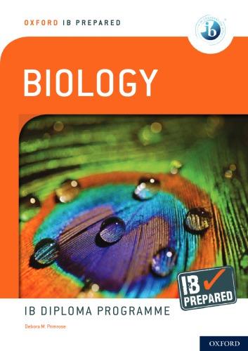
An example of anabolism is the formation of sucrose, common sugar. A six-carbon monosaccharide glucose molecule joins a six-carbon monosaccharide fructose molecule to form the disaccharide sucrose, with the loss of one molecule of water
You should be able to identify biochemicals such as sugars, lipids or amino acids from molecular diagrams You will only be asked to identify a molecule type from a series of molecular structures you already know
• In anablsm, two or more molecules join to form a larger molecule
• In caablsm, molecules are broken down into smaller molecules.
Fgue 2.1.1. The formation of sucrose (a disaccharide) from the condensation of glucose and fructose (two monosaccharides)
2 . 2 W A T E R
Yu shuld knw:
✔ water molecules are polar, they have a positive end and a negative end
✔ water’s cohesive, adhesive, thermal and solvent properties can be explained by hydrogen bonding and dipolarity
✔ hydrophilic substances are attracted to water (water-loving) and hydrophobic substances are repelled by water (water-fearing)
Yu shuld be able :
✔ compare the thermal properties of water with those of methane
✔ explain how water acts as a coolant in sweat
Carbon compounds and carbon cycling will be studied in Topic 4.3. Topic 4 4 describes how carbon dioxide released from metabolic reactions causes climate change
Fgue 2 2 1 Hydrogen bonding between two water molecules
Water is formed from one atom of oxygen and two atoms of hydrogen joined by covalent bonds The hydrogen atoms form a V shape with the oxygen in the vertex. The atom of oxygen is electronegative while the hydrogen atoms are electropositive. The negative part of one water molecule is attracted to the positive part of another water molecule, forming electromagnetic bonds called hydrogen bonds. These bonds are weak, but many bonds together form a strong bonding between water molecules
Water has particular thermal properties Each hydrogen bond has an average energy of 20 kJ mol 1 , therefore water needs a lot of heat to change state, causing the boiling and melting points to be high Water has a high specific heat capacity, a high heat of vaporization and a high heat of fusion.
Example 2.2.1.
a) Complete the table to distinguish the solubility of molecules. Place a tick (✓) in the cases where the molecules are soluble in water
Solution
a) Mlecule glucose amino acids fats cholesterol oxygen sodium chloride
• Hea capacy is the energy required to raise the temperature of 1 g of substance by 1°C
• Hea f vapan is the energy required to change 1 g of liquid to vapour.
• Hea f fusn is the energy required to change 1 g of solid to liquid.
b) Explain the relationship between the solubility in water and the possibility of transport in blood plasma of these molecules
Solution
b) Water-soluble substances can be transported in blood plasma which is composed mainly of water Substances that are not soluble in water, such as lipids, need to be transported through vesicles or attached to soluble substances such as proteins Cholesterol is an example of an insoluble molecule that has to be attached to proteins, forming high-density lipoprotein cholesterol, for transport.
2 . 3 C A R B O H Y D R A T E S A N
Yu shuld knw:
✔ monosaccharide monomers are joined by condensation reactions to form disaccharides and polysaccharides.
✔ fatty acids can be saturated, monounsaturated or polyunsaturated
✔ unsaturated fatty acids can be cis or trans isomers.
✔ triglycerides are formed by condensation from three fatty acids and one glycerol
D L I P I D S
Yu shuld be able :
✔ identify and analyse the structure and function of cellulose, starch and glycogen
✔ compare and contrast cellulose, starch and glycogen with respect to their structure and function, using molecular visualization software
✔ explain the health risks of trans fats and saturated fatty acids in the diet.
✔ compare lipids and carbohydrates in terms of energy storage
✔ evaluate evidence and the methods used to obtain the evidence for health claims made about lipids
✔ determine body mass index by calculation or use of a nomogram.
Monosaccharide monomers, such as glucose and fructose, can be linked together with glycosidic bonds to form disaccharides, such as sucrose Because this reaction liberates water it is called a condensation reaction Sucrose is found in plants. Lactose, the sugar found in milk, and maltose, the sugar found in seeds, are also disaccharides. Lactose is formed from glucose and galactose. Maltose is composed of two glucose molecules and is produced by the hydrolysis of starch.
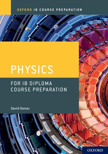
Polysaccharide polymers are composed of long chains of monosaccharides The most abundant polysaccharides in nature are starch and cellulose, which are found in plants, and glycogen, which is mainly found in animals. Starch is composed of two different polymers of α-glucose, amylose and amylopectin Amylose is a linear molecule, while amylopectin is branched. In amylose, the glucose molecules are joined through α-1,4-glycosidic bonds In amylopectin, these same bonds are formed in the main branch, but other α-glucose molecules are joined at carbon 6 forming α-1,6-glycosidic bonds
Fgue 2.3.1. Structure of starch
Cellulose is a polymer of β-glucose molecules joined by 1,4-glycosidic bonds. Because the hydroxyl group on carbon 1 is on the opposite side of the chain to the hydroxyl group on carbon 4, every second glucose is upside down in order to position these two hydroxyl groups together. This makes cellulose a strong molecule, difficult to digest in the human body Many cellulose molecules join together to form microfibres. The fibres found in the cell walls of plants are made from these cellulose microfibres
Example 2.3.2.
a) Compare and contrast the structure of glycogen and starch.
b) Explain how the bonding in starch and cellulose molecules affects their structure
Solution
a) Glycogen, found mostly in animals, and starch, found in plants, are polymers of glucose molecules Starch is formed by two molecules, amylose and amylopectin. Amylose is a linear molecule while amylopectin is branched In both starch and glycogen, the bond joining the main chain of glucose molecules is an α-1,4-glycosidic bond The branching occurs at carbon 6 with an α-1,6-glycosidic bond Glycogen is also formed from branched molecules, but in glycogen the branching is greater than in starch, therefore the molecule is more compact
b) Starch is formed by α-glucose while cellulose is formed by β-glucose. In α-glucose the hydroxyl group on carbon 1 is on the same side of the chain as the one on carbon 4, allowing the
• Cabhydaes are carbon compounds containing hydrogen and oxygen They serve as energy storage and structural components of organisms.
• Mnsacchades are simple sugars and are the basic building blocks of carbohydrates.
• Dsacchades are two monosaccharides joined together
• Plysacchades are polymers of monosaccharides.
Example 2.3.1.
A disaccharide is broken down into glucose and fructose.
a) State the name of the disaccharide
b) Identify the type of bond present in the disaccharide.
Solution
a) Sucrose
b) Glycosidic bond (formed by condensation)
Lipids in the diet have been said to be bad for human health Nevertheless, they are necessary for many metabolic processes Health problems linked to dietary lipids include obesity and coronary heart disease
• Lpds are carbon compounds containing mainly hydrogen and some oxygen. They form a diverse group containing oils, fats, membrane components and hormones
• Fay acds are formed by long chains of carbons (4 to 34) with hydrogens attached and an acid group at one end
• Unsauaed fatty acids have double or triple bonds between two or more carbons
• Sauaed fatty acids do not have any double or triple bonds.
• Cis fay acds have the two hydrogen atoms adjacent to the double bond located on the same side of the chain.
• Trans fay acds have the two hydrogen atoms adjacent to the double bond located on opposite sides of the chain.
fatty acid
Fgue 2 3 2 Saturated fatty acid with no carbon–carbon double bonds and unsaturated fatty acid showing one carbon–carbon double bond
formation of the 1,4-glycosidic bond In β-glucose, the hydroxyl groups are found on different sides of the chain In order for the β-1,4-glycosidic bonds to form, every second β-glucose needs to be upside-down in order to position the hydroxyl groups on the same side of the chain.
Lipids are carbon compounds such as glycerides and steroids that are insoluble in water. They contain double the energy of carbohydrates. Fatty acids are the main components of simple lipids They are formed by a long chain of carbons and hydrogens with an acid group at carbon 1. The carbons can be joined by single or double bonds. When the fatty acid has no double bonds it is said to be saturated; if it has one or more double bonds it is said to be unsaturated. Monounsaturated fatty acids have only one double bond while polyunsaturated fatty acids have two or more double bonds.
Unsaturated fatty acids can have the hydrogen atoms next to the double bond on the same side or on opposite sides of the chain If the hydrogen atoms are on the same side of the chain, the unsaturated fatty acid is said to be cis; if they are on opposite sides of the chain it is trans. The tail in cis fatty acids kinks, separating the molecules. This gives cis fatty acids a lower melting point than trans fatty acids
There are many types of lipids Some have a simple structure, such as the monoglycerides, formed from glycerol and one fatty acid, while others have very complex molecules, such as steroids You need to know only the structure of triglycerides Triglycerides are formed by condensation from three fatty acids and one glycerol molecule. These fatty acids do not need to be the same In some cases, one carbon joins to a phosphate group instead of a fatty acid, forming a phospholipid This phosphate group can at the same time join to another group, for example choline, forming phosphatidylcholine, a phospholipid that forms an important part of the cell membrane
Example 2.3.3.
Explain the health risks of trans fats and saturated fatty acids in the diet.
Solution
Triglycerides formed from saturated fatty acids or from trans unsaturated fatty acids (fats made with these fatty acids are called trans fats) form rods that pack tightly together, forming a high density of intermolecular contacts. Hydrocarbons with cis double bonds or branches are irregular in shape and cannot pack so tightly. High levels of saturated or trans fats may contribute to hardening of the arteries (arteriosclerosis) or the formation of plaques or atheroma on the artery walls (atherosclerosis) An atheroma consists mainly of macrophages, lipids and connective tissue It forms a swelling in the artery wall which slows down or even blocks the flow of blood. The loss of blood flow to the heart can result in coronary heart disease, a heart attack or myocardial infarction, or other cardiovascular diseases
SAMPLE StUDENt ANSWEr
The image shows a nomogram
a) Using the nomogram, state the lower weight limit for a woman with the height of 155 cm to be classified as overweight, giving the units. [1]
This answer could have achieved 1/1 marks:
Lower weight limit: 60 kg
b) Draw the structure of a saturated fatty acid [2]
This answer could have achieved 1/2 marks:
▲ On the nomogram the point where the 155 cm height line intersects the line between normal and overweight is at 60 kg weight
▼ This structure does not show the acid group
▲ One mark was given for the acid group and hydrogens shown
▼ The structure on the left only has three carbons and the shortest fatty acid known is butyric acid, with 4 carbons
Yu shuld knw:
✔ polypeptides are formed from amino acids linked together by condensation
✔ polypeptides are synthesized in ribosomes from 20 different amino acids.
✔ a huge range of polypeptides is possible because the amino acids can be linked together in any sequence.
✔ genes code for the amino acid sequence of polypeptides.
✔ a protein may consist of one polypeptide or of multiple polypeptides linked together
✔ the three-dimensional conformation of a protein is determined by the amino acid sequence.
The synthesis of proteins through translation will be studied in Topic 2.7. The industrial use of organisms to produce proteins will be discussed in Topic B.1.
Most organisms assemble proteins from the same 20 amino acids However, pyrrolysine and selenocysteine, which were discovered fairly recently, are two additional amino acids that are not present in all organisms, and they are genetically encoded in a dierent way from the other 20.
• the α-carbon in the middle with H and R group attached;
• the peptide bond correctly drawn between N and C = 0
Marks would also have been awarded for the acid group at one end of the dipeptide and the amine at the other end, plus another mark for showing the loss of water
✔ proteins have a very wide range of functions in living organisms.
✔ a proteome is the full set of proteins in an individual; it is unique to each individual
Yu shuld be able :
✔ describe the functions of rubisco, insulin, immunoglobulins, rhodopsin, collagen and spider silk
✔ explain how heat and changes to pH can denature a protein.
✔ draw molecular diagrams to show the formation of a peptide bond
Amino acids are the building blocks of proteins There are 20 different amino acids. These amino acids are joined by condensation in the ribosomes to form polypeptides The bond between amino acids is called a peptide bond A DNA molecule is used as a blueprint to produce RNA that will carry the information to produce a polypeptide in a process called translation The three-dimensional shape of a protein is determined by the amino acid sequence.
• rubsc is the short name for ribulose bisphosphate carboxylase/ oxygenase. It is the enzyme involved in photosynthesis that takes part in carbon dioxide xation
• insuln is a hormone that promotes the absorption of glucose by the liver.
• immunglbulns are globular proteins that function as antibodies They have a number of roles in the body’s immune defence.
• rhdpsn is a pigment involved in light detection
• Cllagen is a brous protein found in connective tissues
• Spde slk is a brous protein made by spiders to spin their web
• Peme is all of the proteins produced by a cell, a tissue or an organism.
• Gel elecphess is a method used to separate proteins according to their size.
a) Draw molecular diagrams to show the condensation reaction between two amino acids to form a dipeptide. [4]
This answer could have achieved 4/4 marks:
SAMPLE StUDENt ANSWEr
Explain how heat can denature a protein.
Solution
Heat can modify the three-dimensional structure of a protein, thereby affecting its function. In the case of an enzyme such as rubisco, it would cease to catalyse the reaction When temperature is increased, the increased vibrations within the molecule can cause the interactions between the R groups of different amino acids to be broken, changing the structure of the protein
To detect the proteome of a tissue, proteins are extracted then separated in a gel electrophoresis run.
2 . 5 E N Z Y M E S
Yu shuld knw:
✔ enzymes have an active site to which specic substrates bind
✔ molecular movement and collision of the enzyme ’ s active site with the substrate are required for catalysis
✔ the rate of activity of enzymes depends on temperature, pH and substrate concentration.
✔ enzymes can be denatured.
✔ enzymes can be immobilized to be used in industry
It is possible to provide more correct answers than there are marks available in the question If you write a good answer, it won’t matter if you missed something on the mark scheme because you can still get full marks for another correct point
Yu shuld be able :
✔ design experiments to test the effect of temperature, pH and substrate concentration on the activity of enzymes
✔ sketch graphs to show the expected effects of temperature, pH and substrate concentration on the activity of enzymes.
✔ describe methods to produce lactose-free milk and its advantages
Enzymes control cell metabolism They are globular proteins that have a catalytic zone called the active site. This site can consist of around three to twelve amino acids Changes in temperature and pH can alter the spatial disposition of the protein, affecting the rate of reaction of the enzyme. If the change is extreme, the protein can denature, leaving the enzyme unable to work
How enzymes are used in metabolism, cell respiration and photosynthesis will be discussed in Topic 8
• An enyme is a globular protein that acts as a biological catalyst.
• A subsae is the molecule changed by the enzyme
• The acve se is the part of the enzyme that binds to the substrate
• Denauan is the loss of the tertiary structure of the enzyme
• opmum describes the ideal conditions required for an enzyme to work
▲ The student identied the optimum temperature in the range 44 to 48°C and the pH between 7 8 and 8 5
▲ Marks could be scored for mentioning changes in colour or absorbance or any chemical changes indicating the presence of amino acids
▲ The answers are correct. The amount of buffer would have also scored a mark, but not temperature or pH, as these were the variables of the experiment
SAMPLE StUDENt ANSWEr
Keratin is a protein found in hair, nails, wool, horns and feathers The graphs show the relative keratinase activity obtained in experiments of keratin digestion at dierent pH values and at dierent temperatures.
Fgue 2 5 2 Effect of the substrate concentration on the rate of reaction of an enzyme
a) Determine the optimum pH and temperature of keratinase [1]
This answer could have achieved 1/1 marks:
Optimum pH = 8
Optimum temperature: 45°C.
b) Suggest w changes occurring in the reaction vessel that could be used to indicate keratinase activity [2]
This answer could have achieved 2/2 marks:
The change in mass of keratin
The change in mass of the newly produced amino acids
c) State two conditions that should be kept constant in both experiments [2]
This answer could have achieved 2/2 marks:
The mass of keratinase used
The time of keratinase–keratin reaction
Increasing substrate concentration will increase the rate of reaction of enzymes, but only up to a point, as all the active sites will become occupied and the enzyme will not be able to react any faster
Example 2.5.1.
Describe a method to obtain a named immobilized enzyme, pointing out the advantages and disadvantages of the method Solution
Lactase is an enzyme widely used in industry in an immobilized form to produce lactose-free milk. Lactase is immobilized in alginate beads in a column Milk is poured through this column Lactase digests the lactose found in milk into glucose and galactose, reducing the amount of lactose sugar in milk, making it suitable for lactose-intolerant people. The advantages of immobilization are that the enzymes can be used many times, as they can be separated from the product. In many cases, immobilization stabilizes the enzyme, therefore the reactions can occur at different temperatures or pH values. A disadvantage of the method is that it is more expensive and in some cases the enzyme has its active site sequestered in the matrix, reducing its reactivity