Buy ebook An introduction to orthodontics 5th edtion edition simon j. littlewood cheap price
An Introduction
to Orthodontics
5th Edtion Edition Simon J. Littlewood
Visit to download the full and correct content document: https://ebookmass.com/product/an-introduction-to-orthodontics-5th-edtion-edition-sim on-j-littlewood/
More products digital (pdf, epub, mobi) instant download maybe you interests ...
An Introduction to PHP: Learn PHP 8 to Create Dynamic Websites 1st Edition Mark Simon
Consultant Orthodontist, St Luke’s Hospital, Bradford, UK
Honorary Senior Clinical Lecturer, Leeds Dental Institute, Leeds, UK
3
Great Clarendon Street, Oxford, OX2 6DP, United Kingdom
Oxford University Press is a department of the University of Oxford. It furthers the University’s objective of excellence in research, scholarship, and education by publishing worldwide. Oxford is a registered trade mark of Oxford University Press in the UK and in certain other countries
The moral rights of the authors have been asserted
Second edition 2001
Third edition 2007
Fourth edition 2013
Impression: 1
All rights reserved. No part of this publication may be reproduced, stored in a retrieval system, or transmitted, in any form or by any means, without the prior permission in writing of Oxford University Press, or as expressly permitted by law, by licence or under terms agreed with the appropriate reprographics rights organization. Enquiries concerning reproduction outside the scope of the above should be sent to the Rights Department, Oxford University Press, at the address above
You must not circulate this work in any other form and you must impose this same condition on any acquirer
Published in the United States of America by Oxford University Press 198 Madison Avenue, New York, NY 10016, United States of America
British Library Cataloguing in Publication Data
Data available
Library of Congress Control Number: 2018954270
ISBN 978–0–19–253958–8
Printed in Great Britain by Bell & Bain Ltd., Glasgow
Oxford University Press makes no representation, express or implied, that the drug dosages in this book are correct. Readers must therefore always check the product information and clinical procedures with the most up-to-date published product information and data sheets provided by the manufacturers and the most recent codes of conduct and safety regulations. The authors and the publishers do not accept responsibility or legal liability for any errors in the text or for the misuse or misapplication of material in this work. Except where otherwise stated, drug dosages and recommendations are for the non-pregnant adult who is not breast-feeding
Links to third party websites are provided by Oxford in good faith and for information only. Oxford disclaims any responsibility for the materials contained in any third party website referenced in this work.
Preface for fifth edition
Orthodontics is both an art and a science, and, like most great works of art, at its best orthodontics can appear both deceptively simple and wonderfully aesthetic. The reality is of course that behind that apparent simplicity, there is real complexity that takes years to master. Gaining expertise in any subject requires sound foundations on which to build on, and we hope that this introduction to orthodontics provides these foundations.
In this new, significantly updated edition, we have tried to stay true to the ethos of the previous editions, providing key basic science and clinical information that is based on the best current evidence. We hope it will be useful to anyone involved in the treatment of orthodontic patients: undergraduate dental students, postgraduate students specializing in orthodontics, dentists with an interest in orthodontics, orthodontic therapists and orthodontic nurses, and perhaps even those more experienced orthodontists who would welcome a succinct evidence-based, sensible, and contemporary update on the subject of orthodontics.
We hope you enjoy it!
Simon J. Littlewood and Laura Mitchell
Acknowledgements
We would like to thank everyone who has assisted in completing this book, in particular our new contributing authors, Benjamin R. K. Lewis, Sophy K. Barber, and Fiona R. Jenkins. It has been a pleasure to work with these talented orthodontists on this project. We would also like to thank all those authors who have contributed to previous versions. Individual credits to clinicians who have provided figures for this edition are provided in the respective legends throughout the book. We would also like to sincerely thank all those patients who have provided consent to show their photos.
Working with busy authors is not always easy, so we would like to thank all those clinical and support staff who work with us on a daily basis.
For all those inspiring clinicians, teachers, and colleagues who have shared with us their knowledge, ideas, and experience throughout our careers, thank you.
We would also like to thank the staff of Oxford University Press for their help, patience, and expertise in guiding us through the publishing process.
And finally, to our respective families—Emma and Jack Littlewood, and David Mitchell—this book is dedicated to you.
Simon J. Littlewood and Laura Mitchell
Online Resources
Further reading and references (including Cochrane Reviews) can also be found at: www.oup.com/uk/orthodontics5e.
Where possible, these are presented as active links which direct you to the electronic version of the work, to help facilitate onward study. If you are a subscriber to that work (either individually or through an institution), and depending on your level of access, you may be able to peruse an abstract or the full article if available.
Detailed contents
9
18
18.1
appliances (Benjamin R. K. Lewis)
18.5
18.6
18.7
18.8
19 Functional appliances (S. J. Littlewood)
19.1
19.3
19.4
19.5
19.6
19.9
19.10
20.1
20.2
20.3
20.4
1 The rationale for orthodontic treatment
Learning objectives for this chapter
• Gain an understanding of the differences between need and demand for treatment.
• Gain an appreciation of the benefits and risks of orthodontic treatment.
• Gain an appreciation of the importance of discussing the risks and benefits of treatment with patients and their families.
1.1 Orthodontics
Orthodontics is the branch of dentistry concerned with facial growth, development of the dentition and occlusion, and the diagnosis, interception, and treatment of occlusal anomalies.
1.2 Malocclusion
‘Ideal occlusion’ is the term given to a dentition where the teeth are in the optimum anatomical position, both within the mandibular and maxillary arches (intramaxillary) and between the arches when the teeth are in occlusion (intermaxillary). Malocclusion is the term used to describe dental anomalies and occlusal traits that represent a deviation from the ideal occlusion. In reality, it is rare to have a truly perfect occlusion and malocclusion is a spectrum, reflecting variation around the norm.
The prevalence of malocclusion and particular occlusal anomalies depends on the population studied (e.g. age and racial characteristics), the criteria used for assessment, and the methods used by the examiners (e.g. whether radiographs were employed). In the UK, it is estimated 9% of 12-year-olds and 18% of 15-year-olds are undergoing orthodontic treatment, with a further 37% of 12-year-olds and 20% of 15-year-olds requiring treatment (Table 1.1). This suggests the overall prevalence of moderate–severe malocclusion is around 40–50% in adolescents (Table 1.1).
Table
1.1 England, Wales, and Northern Ireland Child Dental Health Survey 2013
Age band 12 years 15 years
Children undergoing orthodontic treatment at the time of the survey
Children not undergoing treatment but in need of treatment (IOTN dental health component)
Source data from Child Dental Health Survey 2013, England, Wales and Northern Ireland, 2015, Health and Social Care Information Centre.
1.3 Rationale for orthodontic treatment
Malocclusion may cause concerns related to dental health and/or oral-health-related quality of life issues arising from appearance, function, and the psychosocial impact of the teeth. The need for treatment depends on the impact of the malocclusion and whether treatment is likely to provide a demonstrable benefit to the patient. To judge treatment need, potential benefits of treatment are balanced against the risk of possible complications and side-effects in a risk–benefit analysis (Table 1.2).
1.3.1 Need for orthodontic treatment
Health and well-being benefits are the most appropriate determinant of treatment need. Orthodontic indices have been developed to help objective and systematic evaluation of the potential risk to dental health posed by the malocclusion and the possible benefits of orthodontic treatment (see Section 2.3). While indices were largely developed to measure treatment need, due to high treatment demand in many
Table
1.2 Risk–benefit analysis for orthodontics
Benefits of treatment versus Risks
Improved dental health
Improved oral healthrelated quality of life (OHRQoL)
Improved aesthetics
Improved function
Worsening of dental health
Failure to achieve aims of treatment Relapse
countries, indices are also used to manage demand and support prioritization through some form of rationing. For example, in the UK acceptance for NHS orthodontic treatment is predominantly based on need for treatment determined by the Index of Orthodontic Treatment Need (IOTN) (see Section 2.3.3). Similarly, in Sweden treatment priority is estimated using a Priority Index developed by the Swedish Orthodontic
Board and the Medical Board, which aims to identify and treat the malocclusions judged to be most severe.
Unmet treatment need varies within and across countries, depending on individuals’ desire for treatment and organizational factors, such as availability of treatment, access to services, and cost of treatment. In the UK, the unmet orthodontic treatment need for children from deprived households is higher than average; 40% for 12-year-olds and 32% for 15-year-olds. Similar patterns of inequality in access to treatment are seen in other countries.
1.3.2 Demand for orthodontic treatment
It can readily be appreciated that demand for treatment does not necessarily reflect objective treatment need. Some patients are very aware of minor deviations, such as mild rotations of the upper incisors, whilst others refuse treatment for malocclusions that are considered to be severe.
Research shows awareness of malocclusion and willingness to undergo orthodontic treatment is greater in females and those from higher socioeconomic backgrounds. Demand is also higher in areas with a smaller population to orthodontist ratio, presumably due to increased awareness and acceptance of orthodontic appliances.
The demand for treatment is increasing, particularly among adults who are attracted by the increasing availability of less visible appliances, such as ceramic brackets and lingual fixed appliances (see Section 20.6) and orthodontic aligners (see Chapter 21). Orthodontic treatment has a useful adjunctive role to restorative work and as people are keeping their teeth for longer, this is contributing to more requests for interdisciplinary care (see Section 20.5). Increasing dental awareness and the desire for straight teeth, combined with the acceptability of orthodontic appliances and awareness of different types of orthodontic treatment means many adults who did not have treatment during adolescence are now seeking treatment.
1.4 Potential benefits to dental health
To determine whether orthodontic treatment is likely to carry a dental health benefit, it is necessary to consider first whether the malocclusion is likely to cause problems to dental health and secondly, whether orthodontic treatment is likely to address the problem.
There are specific occlusal anomalies where evidence suggests orthodontic treatment may provide a dental health benefit (Box 1.1).
For other dental conditions, such as caries, plaque-induced periodontal disease, and temporomandibular joint dysfunction syndrome (TMD), there is currently insufficient evidence to suggest orthodontic treatment is beneficial. These conditions are complex and multifactorial in origin and as such, direct causal relationship with malocclusion is difficult to measure effectively.
1.4.1 Localized periodontal problems
Certain occlusal anomalies may predispose individuals to periodontal problems, particularly where the gingival biotype is thin, and in these cases orthodontic intervention may have a long-term health benefit. These include:
Box 1.1 Occlusal anomalies where evidence suggests orthodontic correction would provide long-term dental health benefit
Localized periodontal problems
• Crowding causing tooth/teeth to be pushed out of the bony trough, resulting in recession
• Periodontal damage related to tramatic overbites
• Anterior crossbites with evidence of compromised buccal periodontal support on affected lower incisors
• Increased overjet with increased risk of dental trauma
• Unerupted impacted teeth with risk of pathology
• Crossbites associated with mandibular displacement
• Crowding where one or more teeth are pushed buccally or lingually out of the alveolar bony trough, resulting in reduced periodontal support and localized gingival recession.
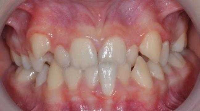
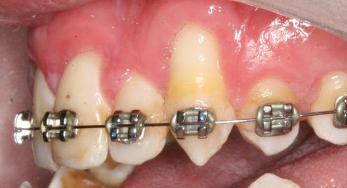
• Class III malocclusion where lower incisors in crossbite are pushed labially (Fig. 1.1).
• Traumatic overbites, which occur when teeth bite onto the gingiva, can lead to gingival inflammation and loss of periodontal support over time and this is accelerated by suboptimal plaque control.
1.4.2 Dental trauma
There is evidence that increased overjet is associated with trauma to the upper incisors. Two systematic reviews have found that the risk of injury is more than doubled in individuals with an overjet greater than 3 mm and the risk of injury appears to increase with overjet size and lip incompetence. Surprisingly, overjet is a greater contributory factor in girls than boys despite traumatic injuries being more common in boys. Orthodontic intervention may be indicated where assessment and history indicate the young person is at increased risk of dental trauma (see Section 9.2.2). Mouthguards are also important in reducing the risk of dental trauma, particularly for those participating in contact sports (see Section 8.9).
1.4.3 Tooth impaction
Tooth impaction occurs when normal tooth eruption is impeded by another tooth, bone, soft tissues, or other pathology. Supernumerary teeth can cause impaction and if judged to be impeding normal dental development, orthodontic input may be required (see Section 3.3.6).
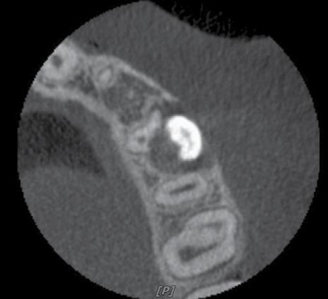
Ectopic teeth are teeth that have formed, or subsequently moved, into the wrong position; often ectopic teeth become impacted. Unerupted impacted teeth may cause localized pathology, most commonly resorption of adjacent roots or cystic change. This is most frequently seen in relation to ectopic maxillary canine teeth, which can resorb roots of the incisors and premolars (Fig. 1.2). Orthodontic management of impacted teeth may be indicated to reduce the risk of pathology (see Section 14.8).
1.4.4 Caries
Caries experience is directly influenced by oral hygiene, fluoride exposure, and diet; however, research has failed to demonstrate a significant association between malocclusion and caries. Caries reduction is therefore rarely an appropriate justification for orthodontic treatment and placement of orthodontic appliances in an individual with uncontrolled caries risk factors is likely to cause significant harm.
In caries-susceptible children, for example those with special needs, malalignment may reduce the capacity for natural tooth cleansing and potentially increase the risk of caries. In these cases, an orthodontic opinion may be sought regarding methods for reducing food stagnation, such as extraction or simple alignment to alleviate localized crowding.
1.4.5 Plaque-induced periodontal disease
The association between malocclusion and plaque-induced periodontal disease is weak, with research indicating that individual motivation has more impact than tooth alignment on effective tooth brushing. In people with consistently poor plaque control, inadequate oral hygiene is more critical than tooth malalignment in the propagation of periodontal disease. Although patients report increased dental awareness and positive habits around diet and oral hygiene patients following orthodontic treatment, poor plaque control is a contraindication for orthodontic treatment. It is essential that oral hygiene is satisfactory and any periodontal disease is controlled prior to considering orthodontic treatment to prevent worsening of dental health.
Fig. 1.1 (a) A 12-year-old male presented with gingival recession on the left mandibular central incisor resulting from an anterior crossbite pushing the tooth labially. (b) Orthodontic treatment was indicated to prevent further damage to the periodontal tissues. Initially upper arch alignment was provided to correct the anterior crossbite. A small improvement was noted in the gingival recession. (c) Comprehensive treatment was provided and following treatment, the gingival condition of the left mandibular central incisor is similar to the other mandibular incisors.
(a)
(b)
(c)
(a) (b)
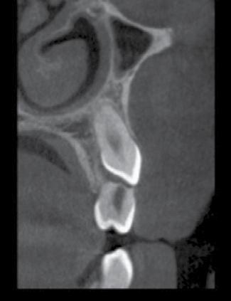
Fig. 1.2 (a) Periapical radiograph from a 14-year-old female patient who presented with resorption of the left maxillary first premolar caused by a transposed and ectopic canine. (b) Cone-beam computed tomography shows the extent of the root resorption of the first premolar more clearly.
For people with reduced dexterity or restricted access for cleaning, it is possible that irregular teeth may hinder effective brushing. In these cases, orthodontic alignment may aid plaque control but appliance treatment must be approached carefully to minimize the risk of periodontal damage during treatment.
The aetiology and management of TMD has caused considerable controversy in all branches of dentistry. TMD comprises a group of related disorders with multifactorial aetiology including psychological, hormonal, genetic, traumatic, and occlusal factors. Research suggests that depression, stress, and sleep disorders are major factors in the aetiology of TMD and that parafunctional activity, for example bruxism, can contribute to muscle pain and spasm. Some authors maintain that minor occlusal imperfections can lead to abnormal paths of closure and/or bruxism, which then result in the development of TMD; however if this
were the case, a much higher prevalence of TMD would be expected to reflect the level of malocclusion in the population.
The role of orthodontics in TMD has been extensively debated, with some authors claiming that orthodontic treatment can cause TMD, while others advocate appliance therapy to manage TMD. After considerable discussion in the literature, the consensus view is that orthodontic treatment, either alone or in combination with extractions, cannot be reliably shown to either ‘cause’ or ‘cure’ TMD.
The alleged success of a wide assortment of treatment modalities for TMD highlights both the multifactorial aetiology and the self-limiting nature of the condition. Given this, conservative and reversible approaches are advised to manage TMD in the first instance. It is advisable to carry out a TMD screen for all potential orthodontic patients, including questions about symptoms, examination of the temporomandibular joint and associated muscles, and a record of the range of opening and movement (see Section 5.4.6). Where signs or symptoms of TMD are found it is wise to refer the patient for a comprehensive assessment and specialist management before embarking on orthodontic treatment.
1.5 Potential benefits for oral health-related quality of life
The other key area where orthodontics may be beneficial is in improving oral health-related quality of life (OHRQoL). Research focussing on the effect of malocclusion suggests OHRQoL can be negatively affected by issues relating to dental appearance, masticatory function, speech, and psychosocial well-being.
1.5.1 Appearance
Dissatisfaction with dental appearance is often the principal reason people seek orthodontic treatment and, in most cases, treatment is able to deliver a positive change. Although improved dental appearance may be cited as the main goal of treatment by patients, it is likely that the perceived benefit is not a change in appearance per se, but the anticipated psychosocial benefit associated with improved appearance.
1.5.2
Masticatory function
Patients with significant inter-arch discrepancy including anterior open bites (AOB) and markedly increased or reverse overjet often report difficulty with eating, particularly when incising food (Fig. 1.3). This may manifest as avoidance of certain foods, such as sandwiches or apples, or embarrassment when eating in public. Patients with severe hypodontia may also experience problems with eating due to fewer teeth to bite on and concerns about dislodging mobile primary teeth and prosthetic teeth (see Chapter 21). Limited masticatory function rarely results in a complete inability to eat, but it can contribute to significant quality of life issues and this may be a driver for orthodontic treatment.
1.5.3 Speech
Speech is a complex neuromuscular process involving respiration, phonation, articulation, and resonance. Articulation is the formation of different sounds through variable contact of the tongue with surrounding structures, including the palate, lips, alveolar ridge, and dentition. It is unlikely that orthodontic treatment will significantly change speech in most cases, as speech patterns are formed early in life before the
permanent dentition is present and the teeth are only one component in the complex system. However, where patients cannot attain contact between the incisors anteriorly, this may contribute to the production of a lisp (interdental sigmatism). In these cases correcting the incisor relationship and reducing interdental spacing may reduce lisping and improve confidence to talk in public.
1.5.4 Psychosocial well-being
Extensive research has been undertaken to examine the effect of malocclusion on psychosocial well-being in terms of self-perception, quality of life, and social interactions. Malocclusion has been linked to reduced self-confidence and self-esteem, with more severe malocclusion and dentofacial deformities causing higher levels of oral impacts. However, other research suggests visible malocclusion has no discernible negative effect on long-term social and psychological well-being. A possible explanation for this is that self-esteem is a mediator in the response to malocclusion, rather than a consequence of malocclusion. Furthermore, self-reported impact of malocclusion may not always reflect objective measurement of the severity of occlusal deviations; this has been attributed to an individual’s resilience, ability to cope, as well as social and cultural factors.
Dental appearance can evoke social judgements that affect peer relations and childhood emotional and social development. People with an attractive dentofacial appearance have been judged to be friendlier, more interesting and intelligent, more successful, and more socially competent. On the other hand, deviation from the norm can cause stigmatization and a high correlation has been found between victimization, malocclusion, and quality of life. The incidence of peer victimization in adolescent orthodontic patients with untreated malocclusion has been estimated to be around 12% in the UK. The extent of malocclusion may not be proportionate to the psychosocial impact, for example, more severe forms of facial deformity can elicit stronger reactions such as pity or revulsion, while milder malocclusions can lead to ridicule and teasing.
Fig. 1.3 A significant skeletal discrepancy can impact on masticatory function. This 28-year-old female patient reported that her Class III incisor relationship and bilateral buccal crossbite made incising and chewing food difficult.
Table 1.3 Potential risks of orthodontic treatment
Problem Avoidance/Management of risk
Intra-oral damage
Root resorption
Loss of periodontal support
Demineralization
Enamel damage
Avoid treatment in patients with resorbed, blunted, or pipette-shaped roots
In teeth judged to be at risk, roots should be monitored radiographically and treatment terminated if root resorption is evident
Maintain high level of oral hygiene
Avoid moving teeth out of alveolar bone
Diet control, high level of oral hygiene, regular fluoride exposure
Abandon treatment
Avoid potentially abrasive components e.g. ceramic brackets where there is a risk of occlusal contact
Use of appropriate instruments and burs to remove appliances and adhesives
Soft tissue damage
Loss of vitality
Extra-oral damage
Worsening facial profile
Soft tissue damage
Avoid traumatic components
Orthodontic wax or silicone to protect against ulceration
Manage allergic reaction promptly
If history of previous trauma to incisors, counsel patient
Careful treatment planning and appropriate mechanics
Use of appropriate safety measures with headgear
Manage allergy promptly
Ineffective treatment
Relapse Avoidance of unstable tooth positions at end of treatment
Failure to achieve treatment objectives
1.6 Potential risks of orthodontic treatment
Like any other branch of medicine or dentistry, orthodontic treatment is not without potential risks. These risks need to be explained to patients during the decision-making process and where possible, steps taken to manage the risk (Table 1.3). Patients should be made aware of their role in treatment and any self-care or behaviour required to achieve success, such as modifications to diet, oral hygiene practice, or use of a sports guard for participation in contact sports.
1.6.1 Root resorption
It is now accepted that some root resorption is inevitable as a consequence of tooth movement, but there are factors that increase the risk of more severe root resorption (Box 1.2).
Long-term retention
Thorough assessment and accurate diagnosis
Effective treatment planning
Appropriate use of appliances and mechanics
Box 1.2 Recognized risk factors for root resorption during orthodontic treatment
• Shortened roots with evidence of previous root resorption
• Pipette-shaped or blunted roots
• Teeth which have suffered a previous episode of trauma
• Patient habits (e.g. nail biting)
• Iatrogenic—use of excessive forces, intrusion, and prolonged treatment time
(a) (b)
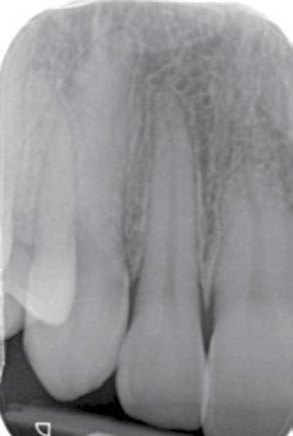
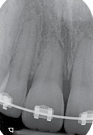
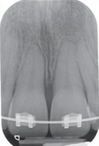
On average, during the course of a conventional 2-year fixed-appliance treatment, around 1 mm of root length will be lost and this amount is not usually clinically significant. However, this average finding masks a wide range of individual variation, as some patients appear to be more susceptible and undergo more marked root resorption. Evidence would suggest a genetic basis in these cases. In teeth with periodontal attachment loss or already shortened roots, the impact of root resorption will be higher (Fig. 1.4).
1.6.2 Loss of periodontal support
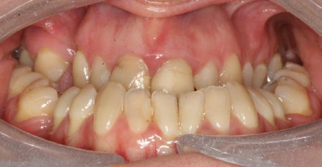
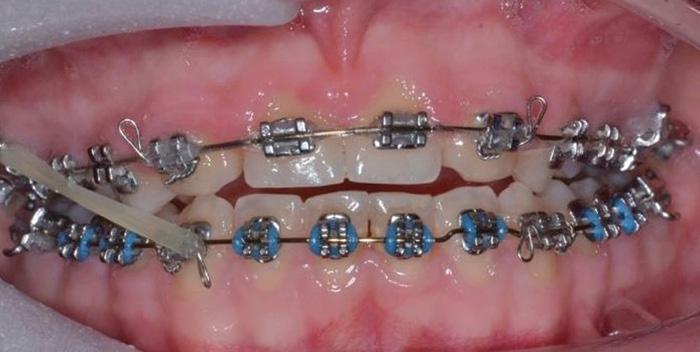
An increase in gingival inflammation is commonly seen following the placement of fixed appliances as a result of reduced access for cleaning and if oral hygiene is consistently poor, gingival hyperplasia may develop
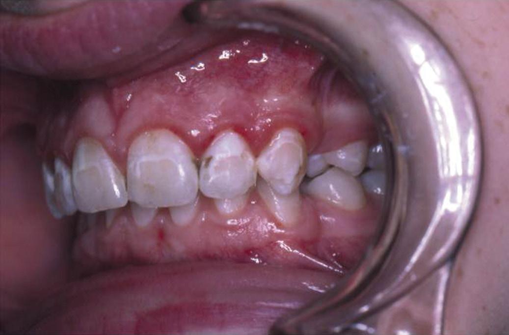
(Fig. 1.5). This normally reduces or resolves following removal of the appliance, but some apical migration of periodontal attachment and alveolar bony support is usual during a 2-year course of orthodontic treatment. In most patients this is minimal but in individuals who are susceptible to periodontal disease, more marked loss may occur. Removable appliances may also be associated with gingival inflammation, particularly of the palatal tissues, in the presence of poor oral hygiene.
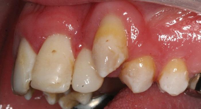
Orthodontic movement of teeth outside the envelope of alveolar bone can result in loss of buccal or less commonly lingual bone, increasing the risk of bony dehiscence and gingival recession. The risk is higher in patients with a narrow alveolus, thin gingival biotype, or existing crowding where teeth have been pushed outside the alveolar bone (Fig. 1.6).
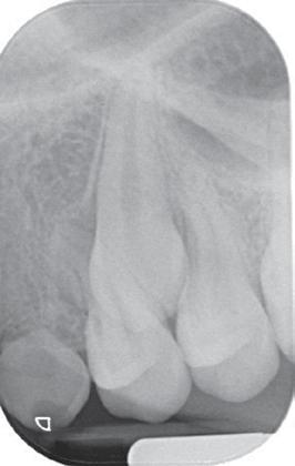
Fig. 1.4 (a) A patient with a shortened right maxillary central incisor root pre-treatment. A risk–benefit analysis is necessary to determine whether the risk of further resorption is justified by the potential benefit of treatment. (b) A monitoring periapical radiograph of the right central and lateral incisor 6 months into treatment shows little further resoprtion of the central incisor root; however, some resoprtion of the apical tip of the lateral incisor root was noted. (c) A further radiograph of the incisors 6 months later confirmed there was no significant progress in the root shortening.
Fig. 1.5 Gingival hyperplasia in the upper labial segment during fixed appliance treatment (a) and at the time of appliance removal (b). The gingival hyperplasia is expected to fully resolve following removal of the appliance.
Fig. 1.6 Teeth that are buccally positioned outside the alveolar bone due to crowding (a) are at increased risk of gingival recession during orthodontic alignment (b). At-risk patients must be informed of potential worsening of the gingival recession prior to commencing orthodontic treatment.
(a)
(b)
(c)
(a)
(b)
(a)
(b)
1.6.3 Demineralization
Demineralized white lesions are an early, reversible stage in the development of dental caries, which occur when a cariogenic plaque accumulates in association with a high-sugar diet. If white spot lesions are not managed early and effectively they can cause permanent damage and even progress to frank caries. The presence of a fixed appliance predisposes to plaque accumulation, as tooth cleaning around the components of the appliance is more difficult. Demineralization during treatment with fixed appliances is a real risk, with a reported prevalence of between 2% and 96% (see Section 18.7). Although there is evidence to show that the lesions regress following removal of the appliance, patients may still be left with permanent ‘scarring’ of the enamel (Fig. 1.7).
1.6.4 Enamel damage
Enamel damage can occur as a result of trauma or wear from the orthodontic appliances. Band seaters, band removers, and bracket removal can cause fracture of enamel, or even whole cusps in heavily restored teeth. During removal of adhesives, the debonding burs can cause enamel damage, particularly if used in a high-speed handpiece. Certain components of orthodontic appliances can cause wear to opposing tooth enamel if there is heavily occlusal contact during function. This is a particular concern if ceramic brackets are used in the lower arch in cases with a deep overbite or where buccal crossbites are present.
1.6.5 Intra-oral soft tissue damage
Ulceration can occur during treatment as a result of direct trauma from both fixed and removable appliances, although it is more commonly seen in association with fixed components as an uncomfortable removable appliance is usually removed. Lesions generally heal within a few days without lasting effect.
Intra-oral allergic reactions to orthodontic components are rare but have been reported in relation to nickel, latex, and acrylate.
on the buccal surfaces of the incisor and canine teeth during fixed appliance treatment. After repeated attempts to control risk factors, treatment was abandoned to prevent further enamel damage.
Management depends on the location and severity of the allergic reaction and the scope for modifying treatment.
1.6.6 Pulpal injury
Excessive apical root movement can lead to a reduction in blood supply to the pulp and even pulpal death. Teeth which have undergone a previous episode of trauma appear to be particularly susceptible, probably because the pulpal tissues are already compromised. Any teeth that have previously suffered trauma or that are judged to be at risk of pulpal injury require thorough examination prior to orthodontic treatment, and any orthodontic treatment should be delivered with light force and careful monitoring.
1.6.7 Extra-oral damage
Some authors have expressed concern over detrimental effects to the facial profile as a result of orthodontics, particularly retraction of anterior teeth in conjunction with extractions. While a number of studies have shown little difference in profile between extraction and non-extraction treatment, it is important that when treatment planning to correct malocclusion, the impact on overall facial appearance is considered.
Contact dermatitis is reported in approximately 1% of the population and allergic reactions may be seen on facial skin in response to components of appliances, usually nickel. This may be managed by covering metal components with tape to prevent contact, or alternative treatment methods may be sought depending in the severity of the reaction. Recoil injury from the elastic components of headgear poses a rare but potentially severe risk of damage to the eyes. This is discussed in more detail in Chapter 15 (see Section 15.5.3). Iatrogenic skin damage, such as burns from acid etch or hot instruments, are avoidable using the usual precautions employed in other fields of dentistry.
1.6.8 Relapse
Relapse is defined as the return of features of the original malocclusion following correction. Retention is a method to retain the teeth in their corrected position, and it is now accepted that without retention there is a significant risk the teeth will move. The extent of relapse is highly variable and difficult to predict but any undesirable tooth movement following orthodontic treatment will reduce the net benefit of orthodontic treatment. Relapse and retention are covered in detail in Chapter 16.
1.6.9 Failure to achieve treatment objectives
When deciding whether orthodontic treatment is likely to be beneficial it is important to consider the effectiveness of appliance therapy in correcting the malocclusion. There are a number of operator- and patient-related factors that may prevent treatment achieving a worthwhile improvement (Table 1.4).
Errors in diagnosis, treatment planning, and delivery can lead to poor selection of appliances and ineffective treatment. It is essential to determine whether planned tooth movements are attainable within the constraints of the skeletal and growth patterns of the individual patient, as excessive tooth movement or failure to anticipate adverse growth changes will reduce the chances of success (Chapter 7). There is evidence that orthodontic treatment is more likely to achieve a pleasing and successful result if the operator has had some postgraduate
Fig. 1.7 Demineralization
Table 1.4
Failure to achieve treatment objectives
Operator factors Patient factors
Errors of diagnosis Poor oral hygiene/diet
Errors of treatment planning Failure to wear appliances/elastics
Anchorage loss
Technique errors
Repeated appliance breakages
Failure to attend appointments
Poor communication Unexpected unfavourable growth
Inadequate experience/ training
training in orthodontics, as this supports appropriate appliance selection and use.
Patient co-operation is essential to achieve a successful outcome. Patients must attend appointments, look after their teeth and appliances, and comply with wear and care instructions. Patients are more likely to co-operate if they, and their family, fully understand the process and their role from the outset. This should be explicitly stated during the consent process. It is important to establish that the patient and family feel willing and able to adhere to the agreed treatment plan before commencing treatment. Long-term effectiveness of treatment depends on patients’ commitment to life-long retainer wear and this must be stressed at the beginning of discussions about orthodontic treatment (see Chapter 16).
1.7 Discussing orthodontic treatment need
It is important that patients and families are involved in the discussion about whether orthodontic treatment is needed and justified. Patients and their families have a key role in providing information about the impact of malocclusion, expectations from treatment, and their desired outcome. The clinician’s role is to provide unbiased information about the potential risks and benefits of treatment based on best available evidence and their own clinical experience. General information should
be tailored to the individual’s clinical presentation and personal circumstance. Patients and families should be supported to participate in the decision about whether treatment is likely to provide sufficient benefit to outweigh any risks. Patients also have a vital role in determining whether they are likely to be able to comply with treatment adequately to achieve a satisfactory outcome. Treatment planning and consent are covered in more detail in Chapter 7.
Key points
• The decision whether to embark on orthodontic treatment is essentially a risk–benefit analysis.
• The perceived benefits of orthodontic intervention should outweigh any potential risks associated with treatment.
• Patients and families have an important role in determining whether treatment is likely to address issues caused by the malocclusion.
Relevant Cochrane reviews
Benson, P. E., Parkin, N., Dyer, F., Millett, D.T., Furness, S., and Germain, P. (2013). Fluorides for the prevention of early tooth decay (demineralised white lesions) during fixed brace treatment. Cochrane Database of Systematic Reviews, Issue 12. Art. No.: CD003809. DOI: 10.1002/14651858.CD003809.pub3. https://www.cochranelibrary.com/cdsr/doi/10.1002/14651858.CD003809.pub3/full
The authors report that (1) fluoride varnish applied every 6 weeks provided moderate-quality evidence of around 70% reduction in demineralized white lesions, and (2) no difference was found between different formulations of fluoride toothpaste and mouth rinse on white spot index, visible plaque index, and gingival bleeding index.
Principal sources and further reading
American Journal of Orthodontics and Dentofacial Orthopedics, 1992, 101(1).
This is a special issue dedicated to the results of several studies set up by the American Association of Orthodontists to investigate the link between orthodontic treatment and the temporomandibular joint.
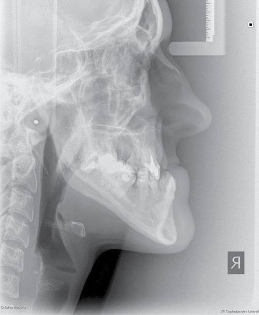
Davies, S. J., Gray, R. M. J., Sandler, P. J., and O’Brien, K. D. (2001). Orthodontics and occlusion. British Dental Journal, 191, 539–49. [DOI: 10.1038/ sj.bdj.4801229] [PubMed: 11767855] This concise article is part of a series of articles on occlusion. It contains an example of an articulatory examination.
The rationale for orthodontic treatment
DiBiase, A. T. and Sandler, P. J. (2001). Malocclusion, orthodontics and bullying. Dent Update, 28, 464–6. [DOI: 10.12968/denu.2001.28.9.464] [PubMed: 11806190]
An interesting discussion around bullying and the ‘victim type’.
Egermark, I., Magnusson, T., and Carlsson, G. E. (2003). A 20-year follow-up of signs and symptoms of temporomandibular disorders in subjects with and without orthodontic treatment in childhood. Angle Orthodontist, 73, 109–15. [DOI: 10.1043/0003-3219(2003)73<109:AYFOSA>2.0.CO] [PubMed: 12725365].
A long-term cohort study, which found no statistically significant difference in TMD signs and symptoms between subjects with or without previous experience of orthodontic treatment.
Guzman-Armstrong, S., Chalmers, J., Warren, J. J. (2011). Readers’ forum: White spot lesions: prevention and treatment. American Journal of Orthodontics and Dentofacial Orthopedics, 138, 690–6. [DOI: 10.1016/j. ajodo.2010.07.007] [PubMed: 21171493]
An interesting and informative read on decalcification during orthodontic treatment.
Helm, S. and Petersen, P. E. (1989). Causal relation between malocclusion and caries. Acta Odontologica Scandinavica, 47, 217–21. [DOI: 10.3109/00016358909007704] [PubMed: 2782059]
A historic paper that demonstrates no link between malocclusion and caries.
Joss-Vassalli, I., Grebenstein, C., Topouzelis, N., Sculean, A., and Katsaros, C. (2010). Orthodontic therapy and gingival recession: a systematic review. Orthodontics and Craniofacial Research, 13, 127–41. [DOI: 10.1111/j.1601-6343.2010.01491.x] [PubMed: 20618715]
Kenealy, P. M., Kingdon, A., Richmond, S., and Shaw, W. C. (2007). The Cardiff dental study: a 20-year critical evaluation of the psychological health gain from orthodontic treatment. British Journal of Health Psychology, 12, 17–49. [DOI: 10.1348/135910706X96896] [PubMed: 17288664]
An interesting paper highlighting the complexities of self-esteem.
Luther, F. (2007). TMD and occlusion part I. Damned if we do? Occlusion the interface of dentistry and orthodontics. British Dental Journal, 202, E2.
Luther, F. (2007). TMD and occlusion part II. Damned if we don’t? Functional occlusal problems: TMD epidemiology in a wider context. British Dental Journal, 202, E3. These two articles are well worth reading.
Maaitah, E. F., Adeyami, A. A., Higham, S. M., Pender, N., and Harrison, J. E. (2011). Factors affecting demineralization during orthodontic treatment: a post-hoc analysis of RCT recruits. American Journal of Orthodontics and Dentofacial Orthopedics, 139, 181–91. [DOI: 10.1016/j. ajodo.2009.08.028] [PubMed: 21300246]
A useful study that concludes that pre-treatment age, oral hygiene, and status of the first permanent molars can be used as a guide to the likelihood of decalcification occurring during treatment.
Mizrahi, E. (2010). Risk management in clinical practice. Part 7. Dento-legal aspects of orthodontic practice. British Dental Journal, 209, 381–90. [DOI: 10.1038/sj.bdj.2010.926] [PubMed: 20966997].
Murray, A. M. (1989). Discontinuation of orthodontic treatment: a study of the contributing factors. British Journal of Orthodontics, 16, 1–7. [DOI: 10.1179/bjo.16.1.1] [PubMed: 2647133].
Nguyen, Q. V., Bezemer, P. D., Habets, L., and Prahl-Andersen, B. (1999). A systematic review of the relationship between overjet size and traumatic dental injuries. European Journal of Orthodontics, 21, 503–15. [DOI: 10.1093/ejo/21.5.503] [PubMed: 10565091].
Petti, S. (2015). Over two hundred million injuries to anterior teeth attributable to large overjet: a meta-analysis. Dental Traumatology, 31, 1–8. [DOI: 10.1111/edt.12126] [PubMed: 25263806]
Two systematic reviews that demonstrate the relationship between increased overjet and dental trauma.
Roberts-Harry, D. and Sandy, J. (2003). Orthodontics. Part 1: who needs orthodontics? British Dental Journal, 195, 433. [DOI: 10.1038/ sj.bdj.4810592] [PubMed: 14576790]
A summary of the potential benefits of orthodontic treatment.
Seehra, J., Newton, J. T., and Dibiase A. T. (2011). Bullying in schoolchildren – its relationship to dental appearance and psychosocial implications: an update for GDPs. British Dental Journal, 210, 411–15. [DOI: 10.1038/ sj.bdj.2011.339] [PubMed: 21566605]
A useful summary of bullying and its relationship to malocclusion.
Steele, J., White, D., Rolland, S., and Fuller, E. (2015). Children’s Dental Health Survey 2013. Report 4: The burden of dental disease in children: England, Wales and Northern Ireland. Leeds: Health and Social Care Information Centre.
Tsakos, G., Hill, K., Chadwick B., and Anderson, T. (2015). Children’s Dental Health Survey 2013. Report 1: Attitudes, behaviours and Children’s Dental Health: England, Wales and Northern Ireland. Leeds: Health and Social Care Information Centre.
The reports from the 2013 Child Dental Health Survey, highlighting orthodontic treatment need.
Travess, H., Roberts-Harry, D., and Sandy, J. (2004). Orthodontics. Part 6: Risks in orthodontic treatment. British Dental Journal, 196, 71–7. [DOI: 10.1038/sj.bdj.4810891] [PubMed: 14739957]
A follow-up to the previous article by the same authors to outline the risks of orthodontic treatment, illustrated with cases.
Weltman, B., Vig, K. W., Fields, H. W., Shanker, S., and Kaizar, E. E. (2010). Root resorption associated with orthodontic tooth movement: a systematic review. American Journal of Orthodontics and Dentofacial Orthopedics, 137, 462–76. [DOI: 10.1016/j.ajodo.2009.06.021] [PubMed: 20362905]
Wheeler, T. T., McGorray, S. P., Yurkiewicz, L., Keeling, S. D., and King, G. J. (1994). Orthodontic treatment demand and need in third and fourth grade schoolchildren. American Journal of Orthodontics and Dentofacial Orthopedics, 106, 22–33. [DOI: 10.1016/S0889-5406(94)70017-6] [PubMed: 8017346]
Contains a good discussion on the need and demand for treatment.
Zhang, M., McGrath, C., and Hägg, U. (2006). The impact of malocclusion and its treatment on quality of life: a literature review. International Journal of Paediatric Dentistry, 16, 381–7. [DOI: 10.1111/j.1365-263X.2006.00768.x] [PubMed: 17014535]
References for this chapter can also be found at: www.oup.com/uk/orthodontics5e. Where possible, these are presented as active links that direct you to the electronic version of the work to help facilitate onward study. If you are a subscriber to that work (either individually or through an institution), and depending on your level of access, you may be able to peruse an abstract or the full article if available.
2 The aetiology and classification of malocclusion
L. Mitchell
Learning objectives for this chapter
• Be aware of current understanding of the aetiology of malocclusion.
• Achieve an insight into classifying malocclusion.
• Gain an understanding of the commonly used classifications and indices.
2.1 The aetiology of
malocclusion
An ideal occlusion is defined as an anatomically perfect arrangement of the teeth. While previously orthodontists may have concentrated on achieving a static, anatomically correct occlusion, it is now accepted that a functional occlusion is more important (see Box 2.1). It is important to realize that malocclusion is not in itself a disease; rather, it describes variation around the ideal.
The aetiology of malocclusion is a fascinating subject about which there is still much to elucidate and understand. Theoretically, malocclusion can occur as a result of genetically determined factors which are inherited, or environmental factors, or a combination of both inherited and environmental factors acting together. For example, failure of eruption of an upper central incisor may arise as a result of dilaceration following an episode of trauma during the deciduous dentition which led to intrusion of the primary predecessor—an example of environmental aetiology. Failure of eruption of an upper central incisor can also occur as a result of the presence of a supernumerary tooth—a scenario which questioning may reveal also affected the patient’s parent, suggesting an inherited problem. However, if in the latter example, caries (an environmental factor) has led to early loss of many of the deciduous teeth then forward drift of the first permanent molar teeth may also lead to superimposition of the additional problem of crowding.
While it is relatively straightforward to trace the inheritance of syndromes such as cleft lip and palate (see Chapter 24), it is more difficult to determine the aetiology of features which are in essence part of normal variation, and the picture is further complicated by the compensatory mechanisms that exist. Evidence for the role of inherited factors in the aetiology of malocclusion has come from studies of families and twins. The facial similarity of members of a family, for example, the prognathic mandible of the Hapsburg royal family, is easily appreciated. However, more direct testimony is provided in studies of twins and triplets, which indicate that skeletal pattern and tooth size and number are largely genetically determined.
Examples of environmental influences include digit-sucking habits and premature loss of teeth as a result of either caries or trauma. Soft tissue pressures acting upon the teeth for more than 6 hours per day can also influence tooth position. However, because the soft tissues including the lips are by necessity attached to the underlying skeletal framework, their effect is also mediated by the skeletal pattern.
Crowding is extremely common in Caucasians, affecting approximately two-thirds of the population. As was mentioned above, the size of the jaws and teeth are mainly genetically determined; however, environmental factors, for example, premature deciduous tooth loss, can precipitate or exacerbate crowding. In evolutionary terms both jaw size and tooth size appear to be reducing. However, crowding is much more
prevalent in modern populations than it was in prehistoric times. It has been postulated that this is due to the introduction of a less abrasive diet, so that less interproximal tooth wear occurs during the lifetime of an individual. However, this is not the whole story, as a change from a rural to an urban lifestyle can also apparently lead to an increase in crowding after about two generations.
Although this discussion may at first seem rather theoretical, the aetiology of malocclusion is a vigorously debated subject. This is because if one believes that the basis of malocclusion is genetically determined, then it follows that orthodontics is limited in what it can achieve. However, the opposite viewpoint is that every individual has the potential for ideal occlusion and that orthodontic intervention is required to eliminate those environmental factors that have led to a particular malocclusion. It is now acknowledged that the majority of malocclusions are caused by both inherited polygenic and environmental factors and the interplay between them. Malocclusion is not a single disease, but a collection of abnormal traits. These traits can be the result of complex interactions between different genes, interactions between genes and the environment (epigenetics), and distinct environmental factors.
When planning treatment for an individual patient, it is often helpful to consider the role of the following in the aetiology of their malocclusion. Further discussion of these factors will be considered in the forthcoming chapters covering the main types of malocclusion:
1. Skeletal pattern—in all three planes of space
2. Soft tissues
3. Dental factors.
Of necessity, the above is a brief summary, but it can be appreciated that the aetiology of malocclusion is a complex subject. The reader seeking more information is advised to consult the publications listed in the section on ‘Principal sources and further reading’ at the end of this chapter.
Box 2.1 Functional occlusion
• An occlusion which is free of interferences to smooth gliding movements of the mandible with no pathology.
• Orthodontic treatment should aim to achieve a functional occlusion.
• But there is a lack of evidence to indicate that if an ideal functional occlusion is not achieved that there are deleterious long-term effects on the temporomandibular joints.
2.2 Classifying malocclusion
The categorization of a malocclusion by its salient features is helpful for describing and documenting a patient’s occlusion. In addition, classifications and indices allow the prevalence of a malocclusion within a population to be recorded, and also aid in the assessment of need, difficulty, and success of orthodontic treatment.
Malocclusion can be recorded qualitatively and quantitatively. However, the large number of classifications and indices which have been devised are testimony to the problems inherent in both these approaches. All have their limitations, and these should be borne in mind when they are applied (Box 2.2).
2.2.1
Qualitative assessment of malocclusion
Essentially, a qualitative assessment is descriptive and therefore this category includes the diagnostic classifications of malocclusion. The main drawback to a qualitative approach is that malocclusion is a continuous variable so that clear cut-off points between different categories do not always exist. This can lead to problems when classifying borderline malocclusions. In addition, although a qualitative classification is a helpful shorthand method of describing the salient features of a malocclusion, it does not provide any indication of the difficulty of treatment.
Qualitative evaluation of malocclusion was attempted historically before quantitative analysis. One of the better-known classifications was devised by Angle in 1899, but other classifications are now more
Box 2.2 Important attributes of an index
• Validity—can the index measure what it was designed to measure?
• Reproducibility—does the index give the same result when recorded on two different occasions and by different examiners?
• Acceptability—is the index acceptable to both professionals and patients?
• Ease of use—is the index straightforward to use?
widely used, for example, the British Standards Institute (1983) classification of incisor relationship.
2.2.2 Quantitative assessment of malocclusion
In quantitative indices, two differing approaches can be used:
• Each feature of a malocclusion is given a score and the summed total is then recorded (e.g. the Peer Assessment Rating (PAR) Index).
• The worst feature of a malocclusion is recorded (e.g. the Index of Orthodontic Treatment Need (IOTN)).
2.3 Commonly used classifications and indices
2.3.1
Angle’s classification
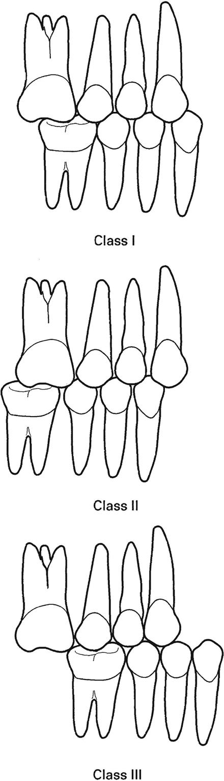
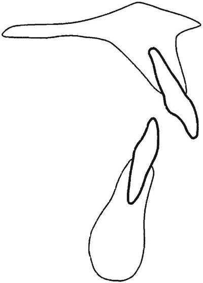
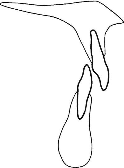
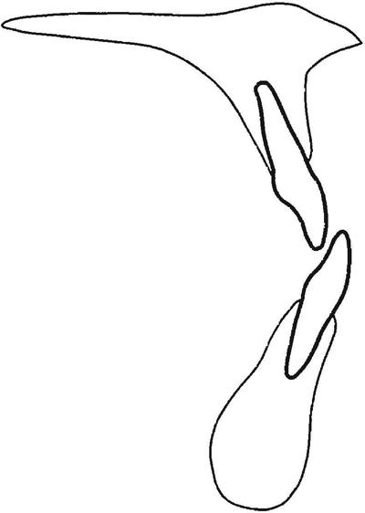
Angle’s classification was based upon the premise that the first permanent molars erupted into a constant position within the facial skeleton, which could be used to assess the anteroposterior relationship of the arches. In addition to the fact that Angle’s classification was based upon an incorrect assumption, the problems experienced in categorizing cases with forward drift or loss of the first permanent molars have resulted in this particular approach being superseded by other classifications. However, Angle’s classification is still used to describe molar relationship, and the terms used to describe incisor relationship have been adapted into incisor classification. Angle described three groups (Fig. 2.1):
• Class I or neutrocclusion—the mesiobuccal cusp of the upper first molar occludes with the mesiobuccal groove of the lower first molar. In practice, discrepancies of up to half a cusp width either way were also included in this category.
• Class II or distocclusion—the mesiobuccal cusp of the lower first molar occludes distal to the Class I position. This is also known as a postnormal relationship.
• Class III or mesiocclusion—the mesiobuccal cusp of the lower first molar occludes mesial to the Class I position. This is also known as a prenormal relationship.
2.3.2 British Standards Institute classification
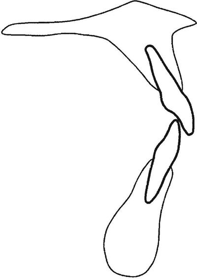
This is based upon incisor relationship and is the most widely used descriptive classification. The terms used are similar to those of Angle’s classification, which can be a little confusing as no regard is taken of molar relationship. The categories defined by British Standard 4492 are shown in Box 2.3 (see also Figs 2.2, 2.3, 2.4, and 2.5).
As with any descriptive analysis, it is difficult to classify borderline cases. Some workers have suggested introducing a Class II intermediate category for those cases where the upper incisors are upright and the overjet increased to between 4 and 6 mm. However, this approach has not gained widespread acceptance.
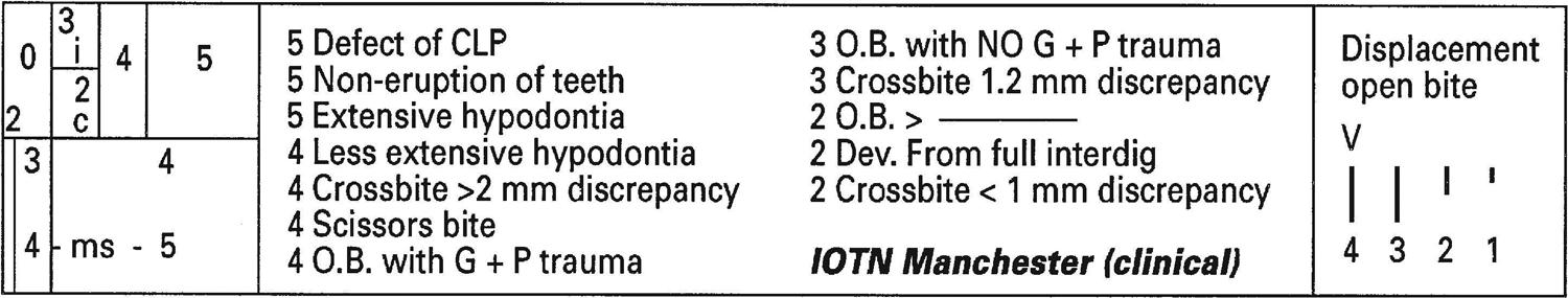
2.3.3 Index of Orthodontic Treatment Need (IOTN)
The IOTN was developed as a result of a government initiative. The purpose of the index was to help determine the likely impact of a malocclusion on an individual’s dental health and psychosocial well-being. It comprises two elements.
Dental health component
This was developed from an index used by the Dental Board in Sweden designed to reflect those occlusal traits which could affect the function
14 The aetiology and classification of malocclusion
Box 2.3 British Standards incisor classification
• Class I—the lower incisor edges occlude with or lie immediately below the cingulum plateau of the upper central incisors.
• Class II—the lower incisor edges lie posterior to the cingulum plateau of the upper incisors. There are two subdivisions of this category:
• Division 1—the upper central incisors are proclined or of average inclination and there is an increase in overjet.
• Division 2—the upper central incisors are retroclined. The overjet is usually minimal or may be increased.
• Class III—the lower incisor edges lie anterior to the cingulum plateau of the upper incisors. The overjet is reduced or reversed.
Permission to reproduce extracts from British Standards is granted by BSI. British Standards can be obtained in PDF or hard copy formats from the BSI online shop: www.bsigroup.com/Shop or by contacting BSI Customer Services for hardcopies only: Tel: +44 (0)20 8996 9001, Email: cservices@bsigroup.com
Fig. 2.1 Angle’s classification.
Fig. 2.2 Incisor classification—Class I.
Fig. 2.3 Incisor classification—Class II division 1.
Fig. 2.4 Incisor classification—Class II division 2.
Fig. 2.5 Incisor classification—Class III.
and longevity of the dentition. The single worst feature of a malocclusion is noted (the index is not cumulative) and categorized into one of five grades reflecting need for treatment (Box 2.4):
The SCAN scale was first published in 1987 by the European Orthodontic Society (Ruth Evans and William Shaw, Preliminary evaluation of an illustrated scale for rating dental attractiveness. European Journal of Orthodontics 9: 314 – 318.)
A ruler has been developed to help with assessment of the dental health component (Fig. 2.6), and these are available commercially. As only the single worst feature is recorded, an alternative approach is to look consecutively for the following features (known as MOCDO):
• Missing teeth
• Overjet
• Crossbite
• Displacement (contact point)
• Overbite.
Aesthetic component
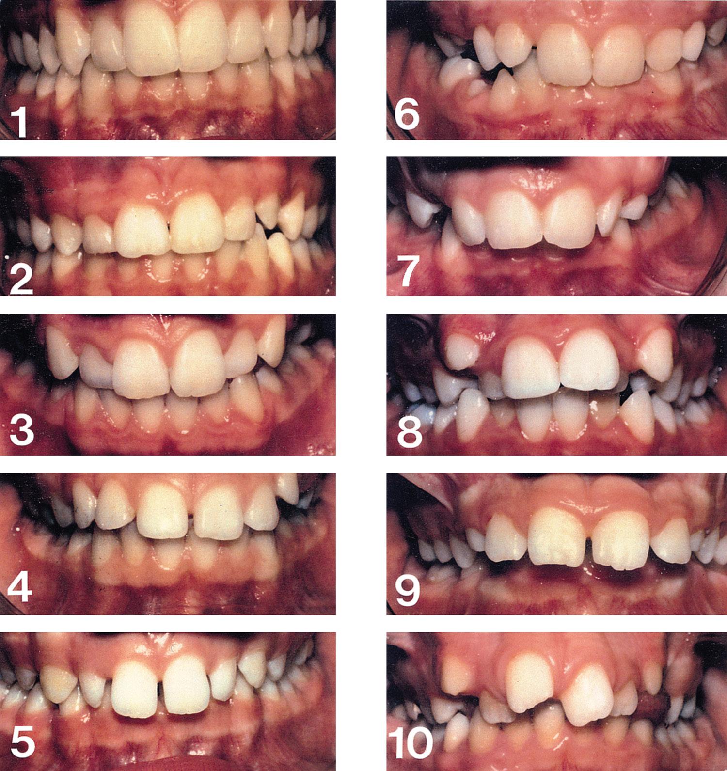
This aspect of the index was developed in an attempt to assess the aesthetic handicap posed by a malocclusion and thus the likely psychosocial impact upon the patient—a difficult task (see Chapter 1). The aesthetic component comprises a set of ten standard photographs (Fig. 2.7), which are also graded from score 1, the most aesthetically pleasing, to score 10, the least aesthetically pleasing. Colour photographs are available for assessing a patient in the clinical situation and black-andwhite photographs for scoring from study models alone. The patient’s teeth (or study models), in occlusion, are viewed from the anterior aspect and the appropriate score determined by choosing the photograph that is thought to pose an equivalent aesthetic handicap. The scores are categorized according to need for treatment as follows:
• Score 1 or 2—none
• Score 3 or 4—slight
• Score 5, 6, or 7—moderate/borderline
• Score 8, 9, or 10—definite.
(Reproduced from Evans, R. and Shaw, W. C., A preliminary evaluation of an illustrated scale for rating dental attractiveness. European Journal of Orthodontics , 9, pp. 314–318. Copyright (1987) with permission from Oxford University Press.)
An average score can be taken from the two components, but the dental health component alone is more widely used. The aesthetic component has been criticized for being subjective—particular difficulty is experienced in accurately assessing Class III malocclusions or anterior open bites, as the photographs are composed of Class I and Class II cases, but studies have indicated good reproducibility.
2.3.4 Peer Assessment Rating (PAR)
The PAR index was developed primarily to measure the success (or otherwise) of treatment. Scores are recorded for a number of parameters (listed below), before and at the end of treatment using study models. Unlike IOTN, the scores are cumulative; however, a weighting is accorded to each component to reflect current opinion in the UK as to their relative importance. The features recorded are listed as follows, with the current weightings in parentheses:
• Crowding—by contact point displacement (×1)
• Buccal segment relationship—in the anteroposterior, vertical, and transverse planes (×1)
• Overjet (×6)
• Overbite (×2)
• Centrelines (×4).
The difference between the PAR scores at the start and on completion of treatment can be calculated, and from this the percentage change in PAR score, which is a reflection of the success of treatment, is derived. A high standard of treatment is indicated by a mean percentage reduction of greater than 70%. A change of 30% or less indicates that no appreciable improvement has been achieved. The size of the PAR score at the beginning of treatment gives an indication of the severity of a malocclusion. Obviously it is difficult to achieve a significant reduction in PAR in cases with a low pre-treatment score.
2.3.5 Index of Complexity, Outcome and Need (ICON)
This index incorporates features of both the IOTN and the PAR. The following are scored and then each score is multiplied by its weighting:
• Aesthetic component of IOTN (×7)
• Upper arch crowding/spacing (×5)
• Crossbite (×5)
• Overbite/open bite (×4)
• Buccal segment relationship (×3).
The total sum gives a pre-treatment score, which is said to reflect the need for, and likely complexity of, the treatment required. A score of more than 43 is said to indicate a demonstrable need for treatment. Following treatment, the index is scored again to give an improvement grade and thus the outcome of treatment.
This ambitious index has been criticized for the large weighting given to the aesthetic component and has not gained widespread acceptability.
2.3.6 Index of Orthognathic Functional Treatment Need (IOFTN)
Although the IOTN has proved a reliable method of assessing malocclusion, like any index, it does have its limitations. Many of these relate to
Box
The aetiology and classification of malocclusion
2.4 The Index of Orthodontic Treatment Need
Grade 5 (Very Great)
5a Increased overjet greater than 9 mm.
5h Extensive hypodontia with restorative implications (more than one tooth missing in any quadrant) requiring pre-restorative orthodontics.
5i Impeded eruption of teeth (with the exception of third molars) due to crowding, displacement, the presence of supernumerary teeth, retained deciduous teeth, and any pathological cause.
5m Reverse overjet greater than 3.5 mm with reported masticatory and speech difficulties.
5p Defects of cleft lip and palate.
5s Submerged deciduous teeth.
Grade 4 (Great)
4a Increased overjet 6.1–9 mm.
4b Reversed overjet greater than 3.5 mm with no masticatory or speech difficulties.
4c Anterior or posterior crossbites with greater than 2 mm discrepancy between retruded contact position and intercuspal position.
4d Severe displacement of teeth, greater than 4 mm.
4e Extreme lateral or anterior open bites, greater than 4 mm.
4f Increased and complete overbite with gingival or palatal trauma.
4h Less extensive hypodontia requiring pre-restorative orthodontic space closure to obviate the need for a prosthesis.
4l Posterior lingual crossbite with no functional occlusal contact in one or both buccal segments.
4m Reverse overjet 1.1–3.5 mm with recorded masticatory and speech difficulties.
4t Partially erupted teeth, tipped and impacted against adjacent teeth.
4x Supplemental teeth.
Grade 3 (Moderate)
3a Increased overjet 3.6–6 mm with incompetent lips.
3b Reverse overjet 1.1–3.5 mm.
3c Anterior or posterior crossbites with 1.1–2 mm discrepancy.
3d Displacement of teeth 2.1–4 mm.
3e Lateral or anterior open bite 2.1–4 mm.
3f Increased and complete overbite without gingival trauma.
Grade 2 (Little)
2a Increased overjet 3.6–6 mm with competent lips.
2b Reverse overjet 0.1–1 mm.
2c Anterior or posterior crossbite with up to 1 mm discrepancy between retruded contact position and intercuspal position.
2d Displacement of teeth 1.1–2 mm.
2e Anterior or posterior open bite 1.1–2 mm.
2f Increased overbite 3.5 mm or more, without gingival contact.
2g Prenormal or postnormal occlusions with no other anomalies; includes up to half a unit discrepancy.
Grade 1 (None)
1 Extremely minor malocclusions including displacements less than 1 mm.
severe malocclusions which are not amenable to routine orthodontic appliances alone. For example, many severe Class III malocclusions have dento-alveolar compensation, that is, retroclination of the lower incisors and/or proclination of the upper incisors which masks the severity of the underlying skeletal discrepancy. As a result, when assessed with the IOTN these malocclusions may only score dental health component grade 3. Also, significant, unsightly excessive upper incisor show, which
Fig. 2.7 Aesthetic component of IOTN. Reproduced from Evans, R. and Shaw, W. C., A preliminary evaluation of an illustrated scale for rating dental attractiveness. European Journal of Orthodontics, 9: 314–318. Copyright (1987) with permission from Oxford University Press.
can lead to potential gingival and periodontal problems, is not reflected in the scoring parameters.
A new index was developed from the IOTN with the aim of trying to address these concerns and to reflect the functional issues that arise with these severe surgical cases. Like IOTN, it is based on a 5-point scale with Grade 5 reflecting a ‘Very Great Need to Treatment’ and Grade 1 ‘No Need for Treatment’ (see Box 2.5).