Regional Anesthesia and Acute Pain Medicine-A Problem-Based Learning Approach (ANESTHESIOLOGY A PROBLEM-BASED LEARNING) (Apr 21, 2023)_(0197518516)_(Oxford University Press) Magdalena Anitescu (Author)
Visit to download the full and correct content document: https://ebookmass.com/product/regional-anesthesia-and-acute-pain-medicine-a-probl em-based-learning-approach-anesthesiology-a-problem-based-learning-apr-21-2023_ 0197518516_oxford-university-press-magdalena-anitescu-author/
More products digital (pdf, epub, mobi) instant download maybe you interests ...
Neuroanesthesia: A Problem-Based Learning Approach
ANESTHESIOLOGY: A PROBLEM- BASED LEARNING APPROACH
Series Editor: Magdalena Anitescu, MD
Published and Forthcoming Titles
Pain Management, edited by Magdalena Anitescu
Anesthesiology, edited by Tracey Straker and Shobana Rajan
Pediatric Anesthesia, edited by Kirk Lalwani, Ira Todd Cohen, Ellen Y. Choi, and Vidya T. Raman
Neuroanesthesia, edited by David E. Traul and Irene P. Osborn
Cardiac Anesthesia, edited by Mohammed M. Minhaj
Critical Care, edited by Taylor Johnston and Steven Miller
Perioperative Care, edited by Deborah Richman and Debra Pulley
Regional Anesthesia and Acute Pain Medicine, edited by Nabil Elkassabany and Eman Nada
REGIONAL ANESTHESIA AND ACUTE PAIN MEDICINE
A PROBLEM- BASED LEARNING APPROACH
EDITED BY
Nabil Elkassabany and Eman Nada
Oxford University Press is a department of the University of Oxford. It furthers the University’s objective of excellence in research, scholarship, and education by publishing worldwide. Oxford is a registered trade mark of Oxford University Press in the UK and certain other countries.
Published in the United States of America by Oxford University Press 198 Madison Avenue, New York, NY 10016, United States of America.
All rights reserved. No part of this publication may be reproduced, stored in a retrieval system, or transmitted, in any form or by any means, without the prior permission in writing of Oxford University Press, or as expressly permitted by law, by license, or under terms agreed with the appropriate reproduction rights organization. Inquiries concerning reproduction outside the scope of the above should be sent to the Rights Department, Oxford University Press, at the address above.
You must not circulate this work in any other form and you must impose this same condition on any acquirer.
CIP data is on file at the Library of Congress
ISBN 978–0–19–751851–9
DOI: 10.1093/med/9780197518519.001.0001
This material is not intended to be, and should not be considered, a substitute for medical or other professional advice. Treatment for the conditions described in this material is highly dependent on the individual circumstances. And, while this material is designed to offer accurate information with respect to the subject matter covered and to be current as of the time it was written, research and knowledge about medical and health issues is constantly evolving and dose schedules for medications are being revised continually, with new side effects recognized and accounted for regularly. Readers must therefore always check the product information and clinical procedures with the most up-to-date published product information and data sheets provided by the manufacturers and the most recent codes of conduct and safety regulation. The publisher and the authors make no representations or warranties to readers, express or implied, as to the accuracy or completeness of this material. Without limiting the foregoing, the publisher and the authors make no representations or warranties as to the accuracy or efficacy of the drug dosages mentioned in the material. The authors and the publisher do not accept, and expressly disclaim, any responsibility for any liability, loss, or risk that may be claimed or incurred as a consequence of the use and/or application of any of the contents of this material.
Printed by Integrated Books International, United States of America
We dedicate this book to our late parents, family, teachers, and mentors for their continued support and care, and to the entire regional anesthesia and acute pain medicine community
CONTENTS
Contributors ix
SECTION 1: GENERAL CONSIDERATIONS
Jinlei Li
1. Topographic Anatomy and Physiologic Considerations 3
N. Robert Harvey and Sylvia H. Wilson
2. Perioperative Considerations for Success of Regional Anesthesia 17
Richa Wardhan and Janet Jira
3. Regional Anesthesia Equipment 25
Thomas Halaszynski
4. Setting Up a Modern Acute Pain Service (APS) 37
Jeffrey J. Mojica, Sean Washek, and Eric S. Schwenk
SECTION 2: PHARMACOLOGY
Kamen V. Vlassakov
5. Multimodal Analgesia 51
Archana O’Neill and Philipp Lirk
6. Local Anesthetic Systemic Toxicity 65 Osemeke Edobor, Veena Graff, and Taras Grosh
7. Prolonging Nerve Blocks 73
Brenton Alexander and Rodney Gabriel
SECTION 3: HEAD AND NECK
Kamen V. Vlassakov
8. Awake Intubation and Airway Blocks 85
Choopong Luansritisakul, Kamen V. Vlassakov, and Nantthasorn Zinboonyahgoon
9. Scalp Blocks: An Overview of Indications, Anatomy, and Technique 99
Arthur Formanek
10. Cervical Plexus Block for Carotid Endarterectomy 107
Shimon Gabriel Farkas and Kamen V. Vlassakov
11. Eye Blocks 117
Gustavo A. Lozada and Alvaro Andres Macias
SECTION 4: CARDIAC, PULMONARY AND CHEST WALL SURGERIES AND TRAUMA
Jinlei Li
12. Regional Techniques for Cardiothoracic and CardiacRelated Procedures 131
Ali N. Shariat and Himani V. Bhatt
13. Video-Assisted Thoracoscopy and Thoracotomy 141
Davies G. Agyekum and Taras Grosh
14. The Trauma Patient with Fractured Ribs 151
Dennis J. Warfield Jr. and Sanjib Das Adhikary
15. Breast Surgery 161
Franklin Chiao, Simon Chin, and Eman Nada
SECTION 5: ABDOMEN, PELVIS AND PERINEUM
Eman Nada
16. Emergency Exploratory Laparotomy 177
Francis V. Salinas
17. Postoperative Pain Management for Living Liver Donor 189
Victor Polshin and Engy T. Said
18. Postoperative Pain Management of Open Nephrectomy 199
Stanislav Sidash and Yatish Ranganath
19. Regional Anesthesia for Abdominoplasty 211
Sanchit Ahuja and Sree Kolli
20. Inguinal Hernia Repair 223
Derek Blankenship, Mitchell Cahan, and Gustavo Angaramo
21. Anesthesia for Anorectal Surgery 233
Luiz Eduardo Imbelloni, José Eduardo De Aguilar-Nascimento, Iris Chu, Engy T. Said, and Eman Nada
SECTION 6: UPPER EXTREMITY
Linda Thi Le-Wendling
22. Shoulder Arthroscopy 247
Alberto E. Ardon, Steven B. Porter, and Robert L. McClain
27. Regional Anesthesia and Pain Management for Total Hip Arthroplasty 307
Bradley H. Lee, Marko Mamic, and Jiabin Liu
28. Analgesia for Knee Arthroscopy and Anterior Cruciate Ligament Reconstruction 319
Michael Feduska and Colin Feduska
29. Enhanced Recovery After Total Knee Replacement 329
Jamie-Lee Metesky and Meg A. Rosenblatt
30. Regional Anesthesia for Ischemic Pain: Below Knee Amputation and Above Knee Amputation 341
Shanthi Reddy, Cameron Sumner, and James Kim
31. Regional Anesthesia for Open Reduction and Internal Fixation of an Ankle Fracture 351
Maryam Nilforoshan, Stephanie Huang, and Nabil Elkassabany
32. Regional Anesthesia for Bunionectomy 363
Daniel Abraham
SECTION 8: OBSTETRIC ANESTHESIA
Eman Nada
33. Epidural for Labor in a Morbidly Obese Patient and Neuraxial Ultrasound 375
Manuel C. Vallejo
34. Inadequate Labor Epidural Analgesia 387
Mohamed Ibrahim and Rakesh Vadhera
35. Regional Anesthesia and Analgesia for Cesarean Delivery 399 Caroline Martinello, Matthew Williams, and Jill M. Mhyre
36. Post-Dural Puncture Headache 411
Lauren Sayre and Robert Gaiser
SECTION 9: PEDIATRICS
Kamen V. Vlassakov
37. Regional Anesthesia Techniques for Circumcision and Congenital Inguinal Hernia Repair 423
Leah Margalit Winters Webb and Melissa Brooks Peterson
38. Epidural and Neuraxial Techniques in Newborns and Children 435
Balazs Horvath and Benjamin Kloesel
39. Regional Anesthesia for Major Abdominal Surgery in Pediatric Patients 447
Alina Lazar
SECTION 10: COMPLEX ACUTE PAIN PATIENTS
Nabil Elkassabany
40. Orthopedic Trauma in a Patient With Complex Regional Pain Syndrome 463
Reda Tolba and Mohamed Fayed
41. Surgery in the Presence of Active Addiction or Medication Assisted Addiction Treatment 475
Neil Batta, Nabil Elkassabany, and Ignacio Badiola
42. The Painful Vaso-Occlusive Sickle Cell Episode 483
Amberly Orr, Dalia Elmofty, and Lynn Kohan
SECTION 11: ANTICOAGULATION AND REGIONAL ANESTHESIA
Jinlei Li
43. Epidural Placement and Anticoagulation 497
Maryam Nilforoshan, Veena Graff, and Taras Grosh
44. Acute Coronary Syndrome in a Patient With a Thoracic Epidural Catheter 511
Yan Lai and Samiat Jinadu
45. Elective Abdominal Aortic Aneurysm With Epidural Analgesia 523
Michael Akerman, Ariel Anderson, and Ryan Norman
46. Lower Extremity Weakness in a Patient With Epidural Analgesia 531
Erik Helander and Yury Zasimovich
47. Big Toe Amputation With Diabetic Peripheral Neuropathy 539
Eman Nada and Sandra L. Kopp
48. Cauda Equina Syndrome After Spinal Anesthesia 553
Lauren Steffel and Joseph M. Neal
49. Nerve Blocks in a Patient With Multiple Sclerosis 561
Lisa L. Klesius and Shelly B. Borden
50. Nerve Injury After Plexus and Peripheral Nerve Blocks for Regional Anesthesia and Medicolegal Implications 573
H. David Hardman
Index 587
CONTRIBUTORS
Daniel Abraham, MD
Assistant Professor
Department of Anesthesiology and Critical Care
University of Pennsylvania Philadelphia, PA, USA
Sanjib Das Adhikary, MBBS, MD
Professor of Anesthesiology, Orthopedics, & Rehabilitation
Vice Chair for Research and Innovation
Division Director for Orthopedic Anesthesia, & Regional Anesthesia and Acute Pain Medicine
Department of Anesthesiology & Perioperative Medicine
Penn State College of Medicine Hershey, PA, USA
José Eduardo De Aguilar-Nascimento, TCBC-MT
Professor
Titular do Departamento de Cirurgia da Faculdade de Medicina da Universidade Federal De Mato Grosso Várzea Grande, MT, Brazil
Davies G. Agyekum, MD, PhD
Anesthesiologist
Department of Anesthesiology Newton-Wellesley Hospital Newton, MA, USA
Sanchit Ahuja, MD Fellow
Department of Cardiothoracic Anesthesia
Anesthesiology Institute Cleveland Clinic Cleveland, OH, USA
Michael Akerman, MD
Assistant Professor
Department of Anesthesiology
Cornell Medical Center New York, NY, USA
Brenton Alexander, MD
Attending Physician
Department of Regional Anesthesiology and Acute Pain Management
University of California, San Diego San Diego, CA, USA
Ariel Anderson, MD
Clinical Instructor of Anesthesiology Keck School of Medicine
University of Southern California Los Angeles, CA, USA
Gustavo Angaramo, MD
Associate Professor of Anesthesiology and Critical Care Director of Regional Anesthesia Division Director of Peri-operative Units
University of Massachusetts Medical School Worcester, MA, USA
Alberto E. Ardon, MD, MPH
Assistant Professor
Department of Anesthesiology and Perioperative Medicine Mayo Clinic Jacksonville, FL, USA
Ignacio Badiola, MD
Assistant Professor of Anesthesiology and Critical Care Department of Anesthesiology and Critical Care Perelman School of Medicine University of Pennsylvania Philadelphia, PA, USA
Neil Batta
Resident in Physical Medicine and Rehabilitation Interventional Pain Management NeoSpine Gig Harbor, WA, USA
Himani V. Bhatt, DO, MPA, FASE, FASA Associate Professor of Anesthesiology Perioperative and Pain Medicine Director, Division of Cardiac Anesthesiology
Mount Sinai Morningside Medical Center
Associate Professor of Cardiovascular Surgery
Icahn School of Medicine at Mount Sinai Director, Division of Cardiac Anesthesiology
Mount Sinai St. Luke’s Hospital New York, NY, USA
Derek Blankenship, MD Anesthesiologist
Anne Burnett Marion School of Medicine at Texas Christian University Baylor Scott &White All Saints Medical Center
NorthStar Anesthesia Irving, TX, USA
Shelly B. Borden, MD
Clinical Anesthesiologist
William S. Middleton Memorial Veterans Hospital Madison, WI, USA
Mitchell Cahan, MD Chairman, Department of Surgery Mount Auburn Hospital Vice Chairman
Roberta and Stephen R. Weiner Department of Surgery
Beth Israel Deaconess Medical Center Professor of Surgery (Adjunct) Department of Surgery University of Massachusetts Chan Medical School Worcester, MA, USA
Franklin Chiao, MD, MS, MBA, LAc, FASA Director of Acute Pain Management Medical Director
Westchester Medical Center New York, NY, USA
Simon Chin, MD
Clinical Assistant Professor Department of Surgery
Donald and Barbara Zucker School of Medicine at Hofstra/Northwell Hempstead, NY, USA
Iris Chu, MD
Assistant Professor
Department of Anesthesiology
Geisel School of Medicine at Dartmouth College Dartmouth-Hitchcock Lebanon, NH, USA
Peter Dienhart, MD
Assistant Professor Department of Anesthesiology Ohio State University Wexner Medical Center Columbus, OH, USA
Osemeke Edobor, MD Anesthesiologist Department of Anesthesiology Morristown Medical Center Atlantic Health System Morristown, New Jersey, USA
Nabil Elkassabany, MD, MSCE Professor and Vice Chair of Clinical Operations Department of Anesthesiology University of Virginia Charlottesville, VA, USA
Dalia Elmofty, MD
Associate Professor
Department of Anesthesia and Critical Care University of Chicago Chicago, IL, USA
Shimon Gabriel Farkas, MD
Staff Anesthesiologist
United Anesthesia Services PC Paoli, PA, USA
Mohamed Fayed, MD
Resident
Henry Ford Health System Detroit, Michigan, USA
Colin Feduska MD
Assistant Professor
Department of Anesthesiology and Critical Care University of Pennsylvania Philadelphia, PA, USA
Michael Feduska, MD
Assistant Professor Department of Anesthesiology and Critical Care University of Pennsylvania Philadelphia, PA, USA
Arthur Formanek, MD
Instructor
Brigham and Women’s Hospital Harvard Medical School Boston, MA, USA
Rodney Gabriel, MD
Chief of Department of Regional Anesthesiology and Acute Pain Management
University of California, San Diego
San Diego, CA, USA
Robert Gaiser, MD Professor
Department of Anesthesiology
Yale University
New Haven, CT, USA
Veena Graff, MD, MS
Assistant Professor
Department of Anesthesiology and Critical Care
Perelman School of Medicine, University of Pennsylvania Philadelphia, PA, USA
Taras Grosh, MD
Assistant Professor
Department of Anesthesiology and Critical Care
Perelman School of Medicine, University of Pennsylvania
Hospital of the University of Pennsylvania Philadelphia, PA, USA
Thomas Halaszynski, DMD, MD, MBA Professor
Department of of Anesthesiology
Yale University
New Haven, CT, USA
H. David Hardman, MD, MBA, FASA Professor
Department of Anesthesiology University of North Carolina Chapel Hill, NC, USA
N. Robert Harvey, MD
Assistant Professor
Department of Anesthesia and Perioperative Medicine
Medical University of South Carolina Charleston, SC, USA
Erik Helander, MD Fellow
Department of Anesthesiology University of Florida College of Medicine Gainesville, FL, USA
Balazs Horvath, MD, FASA Associate Professor Department of of Anesthesiology University of Minnesota
M Health Fairview University of Minnesota Masonic Children’s Hospital Minneapolis, MN, USA
Stephanie Huang, MD
Assistant Professor
Department of Clinical Anesthesiology and Critical Care Perelman School of Medicine, University of Pennsylvania Philadelphia, PA, USA
Mohamed Ibrahim, MD, PhD
Assistant Professor
Obstetric Anesthesiology Fellowship Program Director University of Texas Medical Branch Galveston, TX, USA
Luiz Eduardo Imbelloni Professor of Anesthesiology Director
Institute for Regional Anesthesia Hospital de Base-FAMERP
São José do Rio Preto, SP, Brazil
Samiat Jinadu, MD
Assistant Professor
Department of Anesthesiology and Perioperative Medicine, School of Medicine
Oregon Health and Science University Portland, OR, USA
Janet Jira, MD
Regional Anesthesiologist Long Island Jewish Medical Center New Hyde Park, NY, USA
James Kim, MD Assistant Professor
Department of Anesthesiology and Critical Care University of Pennsylvania Health System Philadelphia, PA, USA
Lisa L. Klesius, MD
Assistant Professor
Division Chief of Regional Anesthesia and Acute Pain Medicine Department of Anesthesiology
University of Wisconsin School of Medicine and Public Health Madison, WI, USA
Benjamin Kloesel, MD, MSBS
Assistant Professor
Department of of Anesthesiology University of Minnesota
M Health Fairview University of Minnesota Masonic Children’s Hospital Minneapolis, MN, USA
Lynn Kohan, MD, MS
Associate Professor
Department of of Anesthesiology University of Virginia Charlottesville, VA, USA
Sree Kolli, MD
Staff Anesthesiologist Department of Anesthesiology, Acute Pain and Regional Anesthesia Cleveland Clinic Cleveland, OH, USA
Sandra L. Kopp, MD Professor of Anesthesiology Department of Anesthesiology and Perioperative Medicine
Mayo Clinic Rochester, MN, USA
Michael Kushelev, MD Assistant Professor Department of Anesthesiology
Ohio State University Wexner Medical Center Columbus, OH, USA
Yan Lai, MD, MPH, FASA Assistant Professor Department of Anesthesiology Icahn School of Medicine
Mount Sinai Medical Center
New York, NY, USA
Alina Lazar, MD Assistant Professor
Department of Anesthesia and Critical Care University of Chicago Chicago, IL, USA
Bradley H. Lee, MD Assistant Attending Anesthesiologist Hospital for Special Surgery Clinical Instructor of Anesthesiology
Weill Cornell Medical College Department of Anesthesiology Critical Care and Pain Management Hospital for Special Surgery
New York, NY, USA
Linda Thi Le-Wendling
Associate Professor Department of Anesthesiology University of Florida Florida, FL, USA
Philipp Lirk, MD, MSc, PhD
Associate Professor of Anaesthesia Department of Anesthesiology, Perioperative and Pain Medicine Brigham and Women’s Hospital Harvard Medical School Boston, MA, USA
Jiabin Liu, MD PhD
Associate Attending Anesthesiologist Hospital for Special Surgery Clinical Associate Professor Department of Anesthesiology
Weill Cornell Medical College New York, NY, USA
Gustavo A. Lozada, MD, MSEd Clinical Director of Longwood Massachusetts Eye and Ear Instructor, Harvard Medical School Boston, MA, USA
Choopong Luansritisakul, MD Staff Anesthesiologist Department of Anesthesiology Siriraj Hospital Mahidol University Bangkok, Thailand
Alvaro Andres Macias, MD Chief of Anesthesia Massachusetts Eye and Ear Assistant Professor Harvard Medical School Boston, MA, USA
Marko Mamic, MD DuPage Valley Anesthesiologists Department of Anesthesiology Naperville, IL, USA
Caroline Martinello, MD Assistant Professor Department of Anesthesiology University of Arkansas for Medical Sciences Little Rock, AR, USA
Jennifer Matos, MD Assistant Professor Department of Anesthesia and Perioperative Medicine
Medical University of South Carolina Charleston, SC, USA
Robert L. McClain, MD Assistant Professor Department of Anesthesiology and Perioperative Medicine
Mayo Clinic Jacksonville, FL, USA
Jamie-Lee Metesky, MD
Assistant Professor
Department of Anesthesiology, Perioperative & Pain Medicine
Mount Sinai Morningside and Mount Sinai West New York, NY, USA
Jill M. Mhyre, MD
The Dola S. Thompson Professor and Chair Department of Anesthesiology University of Arkansas for Medical Sciences Little Rock, AR, USA
Jeffrey J. Mojica, DO
Clinical Assistant Professor
Department of Anesthesiology
Sidney Kimmel Medical College at Thomas Jefferson University Philadelphia, PA, USA
Eman Nada, MD, PhD
Associate Professor
Chief of Regional Anesthesia Department of Anesthesiology
Renaissance School of Medicine
Stony Brook University Stony Brook, NY, USA
Joseph M. Neal, MD Emeritus Anesthesiology Faculty
Affiliate Investigator (Benaroya Research Institute)
Virginia Mason Medical Center Seattle, WA, USA
Mark D. Neuman, MD, MSc
Associate Professor
Department of Anesthesiology and Critical Care
Perelman School of Medicine University of Pennsylvania Philadelphia, PA, USA
Maryam Nilforoshan, MD
Assistant Professor
Department of Anesthesiology and Critical Care Perelman School of Medicine University of Pennsylvania Philadelphia, PA, USA
Olga “Kiki” Nin, MD Clinical Associate Professor Medical Director
Department of Anesthesiology
Medical Director Florida Surgical center
Florida Surgical Center
University of Florida College of Medicine Gainesville, FL, USA
Ryan Norman Fellow
Regional Anesthesiology and Acute Pain Medicine
Massachusetts General Hospital
Harvard Medical School Boston, MA, USA
Jason B. Ochroch, MD
Assistant Professor
Department of Anesthesiology and Critical Care Perelman School of Medicine, University of Pennsylvania Philadelphia, PA, USA
Archana O’Neill, MD
Instructor in Anaesthesia
Department of Anesthesiology, Perioperative and Pain Medicine Brigham and Women’s Hospital Harvard Medical School Boston, MA, USA
Amberly Orr, MD Pain Fellow
Department of Anesthesia and Critical Care University of Chicago Chicago, IL, USA
Melissa Brooks Peterson, MD Associate Professor
Department of Pediatric Anesthesiology Children’s Hospital Colorado University of Colorado, Anschutz Medical Campus Aurora, CO, USA
Victor Polshin, MD
Anesthesiologist at Beth-Israel Deaconess Medical Center Instructor Harvard Medical School Boston, MA, USA
Steven B. Porter, MD
Assistant Professor
Department of Anesthesiology and Perioperative Medicine Mayo Clinic Jacksonville, FL, USA
Yatish Ranganath, MD
Associate Professor
Division Chief | Regional Anesthesia & Acute Pain Medicine Department of Anesthesia Indiana University School of Medicine Indianapolis, IN, USA
Shanthi Reddy, MD
Anesthesiologist
University of Virginia Health System Valley Anesthesiology Consultants Phoenix, AZ, USA
Meg A. Rosenblatt, MD, FASA
Professor of Anesthesiology and Orthopedics
Chair of the Department of Anesthesiology, Perioperative & Pain Medicine
Mount Sinai Morningside and West Hospitals New York, NY, USA
Engy T. Said, MD
Clinical Associate Professor of Anesthesiology
Division of Regional Anesthesia & Acute Pain
University of California, San Diego San Diego, CA, USA
Francis V. Salinas, MD
Staff Anesthesiologist
Medical Director
Interventional Platform Swedish Issaquah Hospital
Issaquah, WA, USA
Lauren Sayre, MD
Anesthesiologist
Department of Anesthesiology
Seven Hills Anesthesia, St. Elizabeth Division
Edgewood, KY, USA
Eric S. Schwenk, MD
Associate Professor
Department of Anesthesiology and Orthopedic Surgery
Sidney Kimmel Medical College at Thomas Jefferson University
Philadelphia, PA, USA
Ali N. Shariat, MD
Assistant Professor
Department of Anesthesiology
Perioperative and Pain Medicine
Mount Sinai Morningside Medical Center
Icahn School of Medicine at Mount Sinai
New York, NY, USA
Stanislav Sidash, MD
Assistant Professor
Department of Anesthesiology
Geisel School of Medicine
Dartmouth-Hitchcock Medical Center
Lebanon, NH, USA
Lauren Steffel, MD
Acting Assistant Professor
Department of Anesthesiology
University of Washington
Veterans Affairs Puget Sound Health Care System Seattle, WA, USA
Cameron Sumner, MD
Anesthesiologist at US Anesthesia Partners Denver, CO, USA
Reda Tolba, MD
Clinical Professor
Department of Anesthesiology
Cleveland Clinic Lerner College of Medicine, Cleveland
Chair of department of Pain Management, Cleveland Clinic
Abu Dhabi, UAE
Rakesh Vadhera, MD, FRCA, FFARCSI
Professor of Anesthesiology and Director of Obstetric Anesthesia
University of Texas, UTMB
Galveston, TX, USA
Manuel C. Vallejo, MD, DMD
Designated Institutional Official
Associate Dean
Professor of Medical Education, Anesthesiology, Obstetrics and Gynecology
Department of Anesthesiology
West Virginia University School of Medicine
Morgantown, WV, USA
Kamen V. Vlassakov, MD
Chief, Regional and Orthopedic Anesthesiology
Brigham and Women’s Hospital
Assistant Professor
Department of Anesthesiology, Perioperative and Pain Medicine
Harvard Medical School Boston, MA, USA
Richa Wardhan, MD
Associate Professor
Department of Anesthesiology College of Medicine-University of Florida Gainesville, FL, USA
Dennis J. Warfield Jr., MD
Assistant Professor of Anesthesiology
Department of Anesthesiology & Perioperative Medicine
Penn State College of Medicine Hershey, PA, USA
Sean Washek, DO
Clinical Instructor of Anesthesiology
Sidney Kimmel Medical College at Thomas Jefferson University Philadelphia, PA, USA
Leah Margalit Winters Webb, MD
Assistant Professor
Department of Pediatric Anesthesiology Children’s Hospital Colorado University of Colorado, Anschutz Medical Campus Aurora, CO, USA
Matthew Williams, MD
Assistant Professor
Department of Anesthesiology University of Arkansas for Medical Sciences Little Rock, AR, USA
Sylvia H. Wilson, MD
Associate Professor
Department of Anesthesia and Perioperative Medicine Medical University of South Carolina Charleston, SC, USA
Yury Zasimovich, MD
Assistant Professor
Department of Anesthesiology University of Florida College of Medicine Gainesville, FL, USA
Nantthasorn Zinboonyahgoon, MD
Associate Professor
Department of Anesthesiology
Siriraj Hospital
Mahidol University Bangkok, Thailand
SECTION 1 GENERAL CONSIDERATIONS
Jinlei Li
TOPOGRAPHIC ANATOMY AND PHYSIOLOGIC CONSIDERATIONS
N. Robert Harvey and Sylvia H. Wilson
STEM CASE AND KEY QUESTIONS
A 62-year-old 100-kg male is involved in a motor vehicle collision and sustains bilateral T5–8 rib fractures, a right radius fracture, a small right pneumothorax, and multiple lower extremity lacerations. The intensive care team contacts the regional acute pain service to assist with pain management for the patient’s numerous injuries. Specifically, he is hypoventilating and splinting from his four rib fractures. The intensive care team is concerned that the patient will require intubation soon if his rib fracture pain is not promptly controlled.
(1) WHAT SURFACE LANDMARKS MAY ASSIST WITH SELECTING THE OPTIMAL THORACIC LEVEL FOR EPIDURAL ANALGESIA?
As this patient has T5–8 rib fractures, the inferior angles of the scapula are marked to identify T7 spinous process. You then palpate one interspace above and mark the T6–T7 interspace. This anatomic level is further verified by palpating the spinous process at the vertebral prominence at C7. You then palpate down the spine to confirm the previously marked T6–T7 interspace. Last, you palpate the patient’s iliac crest as a marker for the L4 spinous process and L4–L5 interspace. The spine is then palpated cephalad to again confirm the T6–T7 interspace. With the T6–T7 interspace confirmed, the patient is prepped and draped for epidural placement.
(2) WHAT IS THE NEURAXIAL ANATOMY TRANSVERSED WITH EPIDURAL PLACEMENT?
Following placement of a subcutaneous local anesthetic (LA) skin wheal, a 17-gauge Tuohy is advanced through the skin, subcutaneous fat, supraspinous ligament, and interspinous ligament. As an increased resistance is noted, a glass syringe with saline is connected to the Tuohy needle and a loss of resistance technique is used to advance the needle through the ligamentum flavum. The saline suddenly is easy to inject at 6 cm and the epidural space is identified. An epidural catheter is easily threaded into the epidural space and the Tuohy needle is removed. As the loss of resistance was noted at 6 cm, the catheter is secured at 11 cm using a sterile, clear, occlusive dressing.
(3) WHAT FACTORS IMPACT THE NEURAXIAL BLOCK HEIGHT?
After negative catheter aspiration for blood and cerebrospinal fluid (CSF), a 3 ml test dose (lidocaine 1.5% with epinephrine 5µg/ml) is administered. The patient’s heart rate is stable (70–73 bpm) and he denies perioral numbness, a metallic taste, or any heaviness is his lower extremities or gluteal region. You note that the patient is over 60 years old and an average height at 5′8″. Since the patient has been hemodynamically stable (blood pressure 112/71), he is bolused with 5 ml of 0.15% bupivacaine in two divided aliquots. Vital signs are assessed every 5 minutes and the patient remains hemodynamically stable. A continuous LA infusion (bupivacaine 0.1%) is started at a rate of 6 ml/h.
(4) WHAT SURFACE ANATOMY WOULD INDICATE THE EPIDURAL IS COVERING THE APPROPRIATE DERMATOMES? WHAT METHODS ARE USED TO EVALUATE BLOCK COVERAGE?
After epidural catheter placement and dosing, the patient reports reduced pain at both rest (1/ 10 from 6/ 10) and with deep inspiration (3/ 10 from 10/ 10). On a physical exam, he has bilateral decreased temperature sensation to ice from the skin at his nipple line to the skin superior to his umbilicus. He maintains normal handgrip, hip flexor, and quadricep muscle strength bilaterally. Last, the patient is able to demonstrate 1500 ml inspiratory volumes consistently using a bedside incentive spirometer with reasonable comfort.
(5) WHAT ARE OTHER REGIONAL ANESTHETIC OPTIONS FOR POLY- TRAUMA PATIENTS?
Now that the patient’s pain is well controlled, he asks what regional anesthetic options are possible for his multiple lacerations and fractured radius. As he has multiple injuries in several anatomic areas, you are concerned that performing regional techniques to cover all of his injuries would lead to LA toxicity. You recommend starting systemic non- opioid analgesic medications. You prescribe a combination of scheduled oral acetaminophen, oral gabapentin,
and intravenous ketorolac with breakthrough tramadol for moderate pain and oxycodone for severe pain. You assure him that the regional and acute pain service will continue to follow him while his epidural is in place and can reassess his analgesic regimen daily. You also discuss brachial plexus blocks for his radius fracture if oral therapy fails, as this is the area causing the most discomfort now that his ribs are anesthetized.1
(6) DESCRIBE THE USE OF LOCAL ANESTHETICS FOR ANALGESIC VERSUS ANESTHETIC BLOCKS.
The patient requires open reduction and internal fixation of his radius fracture the next day. He and his surgeon both wish to avoid general anesthesia given his numerous rib fractures and small pneumothorax. However, he is doing well with his epidural and systemic regimen and does not think he needs an indwelling perineural catheter. You agree and place a single injection, brachial plexus block (axillary approach) for surgical anesthesia using 20 ml of ropivacaine 0.5%. A medical student asks why ropivacaine 0.5% was chosen rather than ropivacaine 0.2% or 1.0% or lidocaine 1.5%. You explain that ropivacaine 0.5% will provide surgical anesthesia coupled with postoperative analgesia for approximately 6–12 hours. Conversely, ropivacaine 0.2% would provide some postoperative analgesia but would not provide a block dense enough to deliver surgical anesthesia. Ropivacaine 1.0% would also provide surgical anesthesia with postoperative pain control, but this patient is also receiving an epidural infusion of LA. Total LA dose delivered to a patient must be considered, especially in patients receiving multiple regional anesthetic techniques. While lidocaine 1.5% could also provide surgical anesthesia, the anesthetic effects would only last 1–2 hours. Thus, lidocaine would provide dense surgical anesthesia, but without the benefit of postoperative pain control for the patient.
DISCUSSION
Comprehension of the relationship between surface anatomy and nervous system is one of the most essential building blocks for regional anesthesia procedures and assessment. Neuraxial techniques, in particular, are an excellent way to understand this relationship.
NEURAXIAL TECHNIQUES
Neuraxial anesthesia has been a common regional technique for over five decades and involves the injection of medicines into the epidural and/or subarachnoid space. Thus epidural, spinal, and combined spinal epidural (CSE) anesthesia represent the different types of regional neuraxial techniques. Neuraxial blocks may be used as the primary anesthetic (e.g., lower extremity arthroplasty or cesarean delivery), to minimize postoperative pain (e.g., esophagectomy or ventral hernia repair), or for analgesia separate from a surgical or interventional procedure (e.g., rib fractures or labor analgesia).
Neuraxial Anatomy
Detailed knowledge of anatomical layers, bones, and ligaments superficial to and surrounding the neuraxis is paramount to avoid complications, expedite procedures, and trouble-shoot techniques in complicated patients. The spine is made up of 24 true (unfused) vertebrae and 9 fused vertebrae (sacral and coccygeal). The first 7 vertebrae are termed cervical and are located in the neck. The next 12 are thoracic and attach to the ribs. The remaining 5 vertebrae are found in the lumbar region. Of note, the spine curves in a convex manner (lordosis) at the cervical and lumbar levels, while it is concave (kyphosis) in the thoracic and sacral portions. Notably, the spinal cord, originating from the foramen magnum, terminates (conus medullaris) at approximately L1 in adults but at L3 in newborns and infants. As neuraxial procedures at the lumbar level have the advantage of being below the conus medullaris, this is an important consideration.
Below the skin and subcutaneous fat, several ligaments provide stability to the vertebral column superficial to the meninges and spinal cord. These are commonly presented in a superficial to deep manner. First, the supraspinous ligament spans the tips of the spinous processes. Between the spinous processes runs the interspinous ligament, which fuses with the supraspinous ligament posteriorly (superficial) and the ligamentum flavum anteriorly (deep). Deep to the ligamentum flavum lie the meninges: dura mater, arachnoid mater, and pia mater (superficial to deep). The epidural space is a potential space deep to the ligamentum flavum and superficial to the dura mater. The subarachnoid space is deep to the arachnoid mater and contains CSF. Notably, further stability to the spine is provided by the anterior and posterior longitudinal ligaments that surround the anterior and posterior vertebral bodies, respectively. These ligaments are not encountered with neuraxial block placement.2
Surface Anatomy
As delivery of neuraxial LA to a targeted group of dermatomes can lower drug requirements and medication related side effects, knowledge of surface and dermatomal anatomy is essential for neuraxial procedures. Dermatomes are skin areas that are mainly supplied by sensory (afferent) nerve fibers from a single dorsal spinal nerve root. While dermatomes will be discussed in more detail with neuraxial block assessment, the dermatomes impacted by pain must be considered when selecting a needle insertion site for a neuraxial procedure to deliver analgesia to the appropriate area. While ultrasonography of the neuraxis has experienced growing popularity, neuraxial procedures are classically performed with palpation of surface landmarks and are considered “blind techniques.” Consequently, surface landmarks along the posterior spine remain essential to recognize the optimal site for needle insertion.
Four key surface landmarks are utilized to identify the appropriate level to perform neuraxial procedures (Table 1.1). The bony knob palpated in the distal neck is known as the vertebral prominence and represents the C7 spinous process.
Table
LEVEL SURFACE ANATOMY
C7
T7
Vertebral prominence
Inferior angle of scapula
L4 Iliac crest
S2
Posterior, superior iliac spine
While, this landmark is consistent, it can be challenging to palpate in the obese and morbidly obese patient populations. The inferior angle of the scapula represents the T7 spinous process and most commonly establishes the correct interspace for thoracic epidural placement. However, studies have shown that there is patient variability, and the inferior angle of the scapula may also correspond to T6 spinous process (25%), T8 spinous process (18%), and less commonly to T4 spinous process (1%), T5 spinous process (1%), or T9 spinous process (1%).3 In the lumbar region, the line between iliac crests is termed the intercristal line or Tuffier’s line. This landmark corresponds to the level of the L4 spinous process or L4–L5 interspace and is employed for lumbar epidural placement. However, similar to the thoracic spine, anatomic variability is possible and should be anticipated in parturients. In a 2011 study of 45 pregnant women, the intercristal line was noted by ultrasonography to be above the L4–L5 interspace in 100% of subjects. While the intercristal line most often corresponded to immediately below the L2–L3 interspace, it ranged from immediately above the L1–L2 to immediately cephalad to the L4–L5 interspace.4 Last, a line drawn between the posterior superior iliac spines represents the level of the S2 and the caudal limit of the dural sac for adult patients. When a targeted area of analgesia is required at an interspace other than these four sites, these surface landmarks are first marked and then the spinous processes palpated either cephalad or caudad to identify the desired interspace for needle insertion. Given the anatomic variability between patients, it is advisable to utilize more than one surface landmark when identifying a neuraxial level for needle placement (Figure 1.1).
Neuraxial Block Placement
The midline approach for neuraxial blocks is a popular technique, and the needle transverses several layers before reaching the epidural and subarachnoid spaces. All neuraxial techniques begin with identification of the correct interspace, aseptic preparation of the skin, sterile drape placement, and placement of an LA skin wheal over the intended needle insertion site. For epidural placement, a 17–18 gauge Tuohy needle is inserted at the skin and passed through the subcutaneous fat, supraspinous ligament, interspinous ligament, and ligamentum flavum. Notably, once the needle reaches the interspinous ligament, it often becomes anchored and remains in place without external support. Next, the ligamentum flavum generally conveys an increased resistance, like pushing through
a rubber eraser. Since this indicates the ligamentous layer just superficial to the epidural space, the Tuohy is advanced slowly, using a loss of resistance technique to saline or air. Loss of resistance signals entering the epidural space, a potential space between the ligamentum flavum and the dura.5 After the epidural space is identified, a catheter (19–20 gauge based on the Tuohy size) is threaded. The catheter is often secured 4–5 cm deeper than the depth where the loss of resistance was encountered. This allows for some movement of the epidural catheter as the patient moves, while preserving the catheter in the epidural space. As epidural placement is a “blind” technique, it is important to ensure that the epidural catheter has not been inadvertently threaded into a blood vessel or into the subarachnoid space. This is accomplished with a “test dose” that is most often composed of lidocaine and epinephrine.6 Intravascular injection of lidocaine is associated with perioral numbness or a metallic taste, while intravascular injection of 10–15 µg epinephrine raises the heart rate 10–15 beats per minute. Conversely, a subarachnoid injection results in heaviness or numbness in the proximal lower extremities or gluteal regions. Patients should be queried for these signs or symptoms and the heart rate should be monitored for changes. Any change in patient sensation or hemodynamics should prompt procedure cessation and further investigation. After a negative test dose, the epidural catheter may be bolused to achieve the desired anesthesia or analgesia. Since dosing of the epidural is likely to cause hemodynamic changes (e.g., hypotension), it is essential that heart rate and blood pressure monitoring occur during this time period and immediately after. In obstetric anesthesia, fetal heart tone monitoring is also advisable.7
In a midline-approach subarachnoid block or spinal, the needle is advanced through the same layers as described with epidural placement, but then further advanced through the dura until CSF returns freely.5 Medication is then injected into the CSF. Common spinal needles are divided into blunt needles (e.g., Whitacre, Sprotte, and Gertie Marx) and cutting needles (e.g., Quincke). Because the dura is punctured in a subarachnoid block, this places the patient at risk for postdural puncture headache (PDPH). While the risk of PDPH is lower with blunt needles, it is also significantly lowered by a smaller needle gauge. As a result, spinal needles are typically 24–27 gauge. To a greater extreme than in epidural anesthesia, hypotension should be anticipated and expected with subarachnoid blocks. Common strategies to mitigate the hypotensive response include co-loading of intravenous fluids at the start of block placement and the initiation of vasopressors (e.g., phenylephrine or norepinephrine) upon successful intrathecal medication injection.8
Neuraxial techniques can also be combined. A CSE block allows providers to place a spinal prior to epidural placement. If a CSE is planned, the Tuohy is first placed in the epidural space as described above. The Tuohy then remains in place and an appropriately sized spinal needle (26–27 gauge based on the size and design of the Tuohy needle) is inserted into the Tuohy needle, through the dura, and into the subarachnoid space. Free CSF confirms position, and medications are injected. Following medication injection, the spinal needle is removed but the Tuohy needle left in place. The epidural
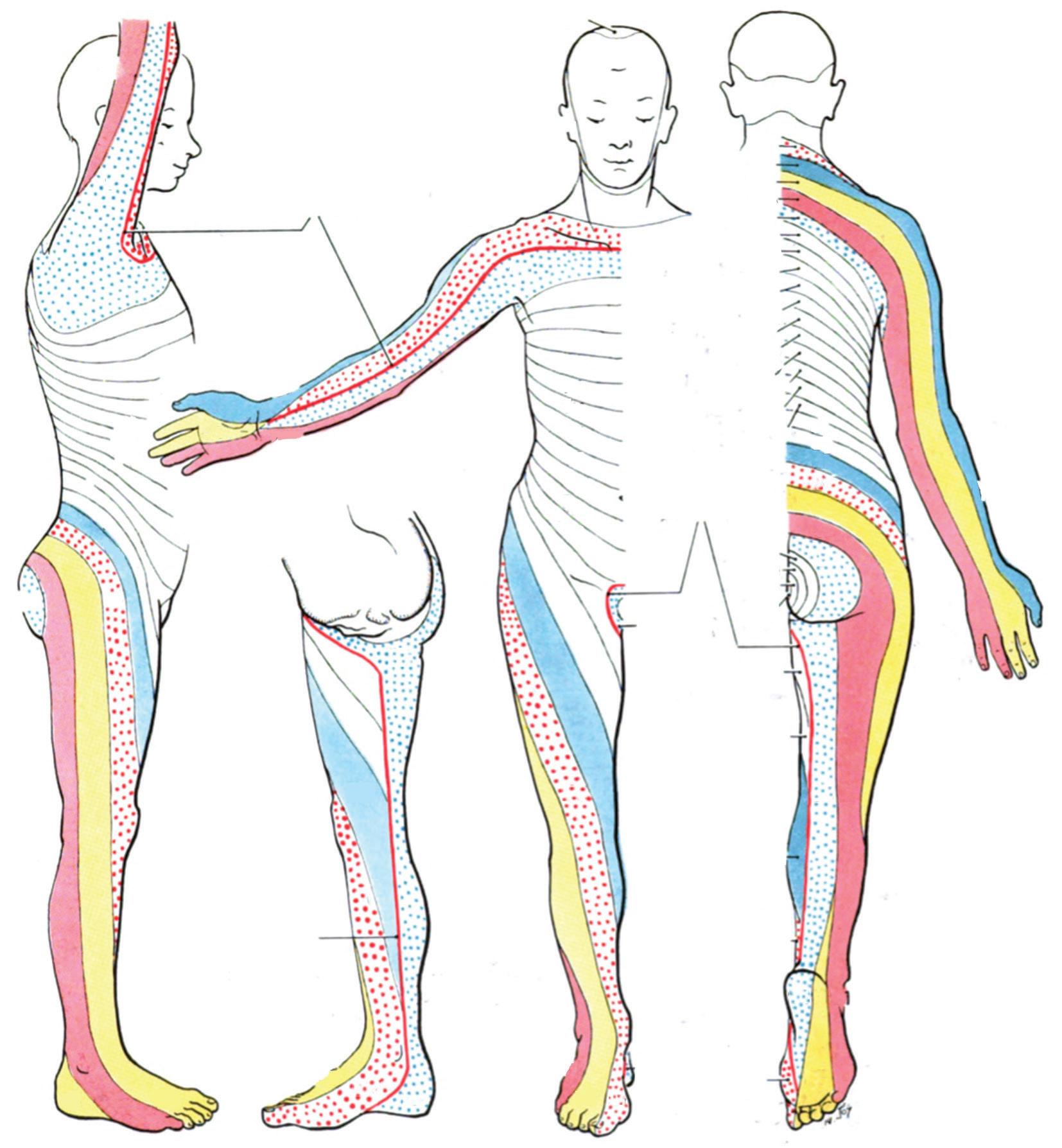
Grant’s Atlas, 1962. Wikipedia, public common domain. Accessed May 8, 2021. https://en.wikipedia.org/wiki/Dermatome_(anatomy)#/media/File:Grant_1 962_663.png.
catheter is then threaded and secured as with epidural placement. CSE permits multiple options, as a small LA dose or only opiates may be injected into the CSF to expedite analgesia. Additionally, spinal anesthetic may be placed and the epidural catheter threaded to provide continued anesthesia if the subarachnoid block resolves. A more recent variation of the CSE is a dural puncture epidural (DPE).8 In this technique, the Tuohy is placed in the epidural space and the dura punctured with the spinal needle, similar to the CSE; however, no medications are injected into the CSF. The spinal needle is removed and the epidural catheter placed, secured, and dosed. Despite the absence of injection of intrathecal medications, DPE has been associated with more rapid onset of labor analgesia and improved block quality (e.g., bilateral coverage and density) compared to a labor epidural without DPE. While DPE does not provide as rapid analgesia as CSE,
it is associated with fewer side effects, including less maternal hypotension and fetal bradycardia.9
For patients with narrow intervertebral spaces or who have difficulty with flexed positioning, alternatives to the midline approach may be useful. In the paramedian approach, the needle is inserted 1 cm lateral and 1 cm caudal to the traditional midline insertion site. The needle is then advanced in a cephalo-medial direction until loss of resistance is obtained and epidural space identified. With the paramedian technique, the ligamentum flavum is the first ligament encountered, as the needle does not pass through the supraspinous or interspinous ligaments. This technique may also be utilized for spinal anesthesia with the needle advanced until CSF flow is encountered, identifying the subarachnoid space. In the Taylor approach, the paramedian technique is used to enter the L5–S1 interspace. The skin is anesthetized 1 cm medial
Figure 1.1. Dermatomes.
and 1 cm caudal to the posterior superior iliac spine. The needle is advanced in a cephalo-medial direction at approximately a 55-degree angle. This approach is advantageous since it requires minimal patient flexion; however, it is technically complicated.10
Neuraxial Local Anesthetic Spread
Selection of LA type, concentration, and dose depends on the need for analgesia versus surgical anesthesia and block duration required. LA are classified into two main groups: esters and amides (Table 1.2).10 Amides are metabolized by the liver and generally have longer durations than esters, which are metabolized by pseudocholinesterase. Ester LAs include benzocaine, chloroprocaine, procaine, and tetracaine. Amide LAs include bupivacaine, levobupivacaine, lidocaine, mepivacaine, prilocaine, and ropivacaine.
LAs inhibit nerve conduction by binding with intracellular voltage-gated sodium channels in a reversible fashion. The greatest binding affinity of LAs occurs when the sodium channel is in the open state. When the sodium channel closes, the blinding affinity is the lowest.
For onset, LAs must become unionized and permeate the nerve lipid bilayer to reach the intracellular sodium channels. Thus, speed of onset is related to lipid solubility and pKa. Local anesthetics with a pKa closer to physiologic pH more rapidly reach a unionized state. However, an increased amount of the unionized form can also be achieved with a larger drug dose and to expedite anesthetic onset. A classic example of this is the administration of 20 ml of 3% chloroprocaine (pKa 8.7) by epidural catheter for emergent cesarean delivery rather than 20 ml of 2% lidocaine (pKa 7.9) with epinephrine. Despite having a pKa farther from physiologic pH, chloroprocaine provides a more rapid onset for surgical anesthesia since a 600 mg dose is delivered, compared to a 400 mg dose of lidocaine. Additionally, as lipid-soluble drugs more rapidly cross the nerve cell membrane, the most significant factor in determining potency is lipid solubility or hydrophobicity. More lipophilic molecules are also highly protein bound in the bloodstream, increasing duration of block. Notably, duration is also related to metabolism as esters are rapidly metabolized by pseudocholinesterase and amides more slowly metabolized by the liver.
Understanding neuraxial medication spread is important when administering medications as either a bolus or infusion. Several factors impact the spread of LAs in the epidural and subarachnoid spaces. Consequently, the area anesthetized may vary between patients.
Factors that impact neuraxial spread or block height may be categorized as drug factors, patient factors, and positioning. Above all other factors, the total mass of LA seems to be the most significant factor in determining the extent of sensory, sympathetic, and motor neural blockade. Patient factors resulting in increased neuraxial spread include age and gravity. In lumbar epidurals, several studies have reported spread 3–8 segments higher in patients over 60 years old compared with patients under 40 years old.11,12 Conversely, in thoracic epidurals, patients over 60 years old consistently have decreased epidural dose requirements (40% less) coupled with increased hemodynamic instability, compared with patients under 40 years old.13,14 Similarly, parturients require decreased LA doses compared to non-pregnant patients. As increased abdominal pressure results in engorged epidural veins, it is theorized that this explains the decreased neuraxial dose requirements for pregnant patients. However, weight (obesity) does not impact LA spread, and height only impacts spread for extremely short (increased spread) or extremely tall (decreased spread) subjects. Notably, the number of segments blocked does not differ significantly by insertion site (e.g., high thoracic, mid-thoracic, low thoracic, or lumbar) when age and gravity are considered. While the supine and sitting positions do not impact block height when dosing lumbar epidurals, the head-down position (15 degrees) results in an increased block height when dosing lumbar epidurals. In the same way, a sensory block of 0–3 segments produces greater results on the dependent side when epidurals are dosed in the lateral position. No differences are noted in block height for thoracic epidurals, regardless of patient position.15
Similar to epidural anesthesia, LA spread, or block height, is also impacted by drug factors, positioning, and patient factors for spinal anesthesia. The primary drug factors impacting block height are dose, baricity, and positioning. Increased LA dose correlates with greater block height. Patient position impacts block spread depending on the baricity utilized. Hyperbaric solutions spread with gravity to more dependent regions, while isobaric injectates spread evenly, and hypobaric
Table 1.2 LOCAL ANEST h ETICS FREQUENTLY USED FOR REGIONAL ANEST h ESIA 11
injectates spread against gravity. Isobaric solutions are generally not affected by positioning. Conversely, hyperbaric solutions lead to cephalad spread in the Trendelenburg position, but result in a saddle block if the patient is left sitting upright. The opposite occurs with hypobaric solutions, and a lateral position results in denser blockade of the non-dependent side. Neuraxial spread may be increased by patient factors including advanced age and pregnancy. As with epidurals, height is generally a factor only for patients at extreme ends of the spectrum. Scoliosis does not affect blockade spread; however, kyphosis may alter the spread of hyperbaric injectates in supine patients. Injection into the epidural space following a spinal anesthetic (as with a CSE) also appears to increase block height.2
Surface Anatomy and Dermatomes for Neuraxial Analgesic Assessment
Dermatomes are skin areas innervated by sensory (afferent) nerve fibers from a single spinal nerve. After epidural or subarachnoid block placement and dosing, loss of sensation must be assessed by dermatomal markers. The relationship between dermatomes and surface landmarks is useful to assess adequate analgesia or anesthesia. For neuraxial block assessment, sensory changes are assessed along dermatomal distributions in a systematic fashion. At the cephalad end and extremes of a block (e.g., cheek, or shoulder of a patient with a mid-thoracic epidural), sensation should be intact and patients should not
be able to discern a differential block. Studies have shown that blocks do not terminate completely over the course of one dermatome. Instead, patients report a change in sensation or fading of the block over the course of two dermatomes with the complete loss of cold sensation often extending slightly cephalad by 1–2 dermatomes compared to the loss of light touch and pinprick.16
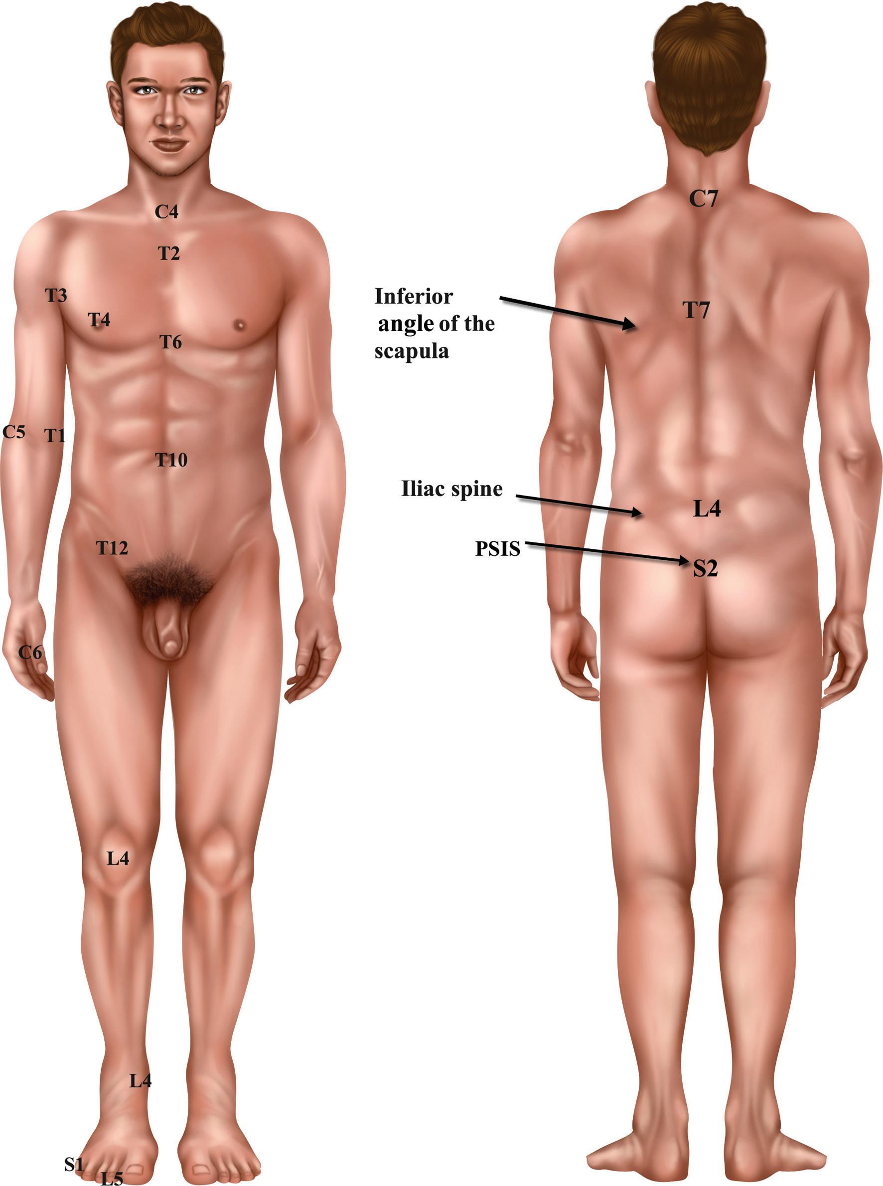
Several dermatomal markers are important for block assessment and for prevention or evaluation of potentially adverse events (Table 1.3). In terms of neuraxial block assessment, the nipple line (T4), xiphoid process (T6), and umbilicus (T10) are commonly used to assess thoracic epidural analgesia. In obstetric anesthesia, the xiphoid process (T6) and umbilicus (T10) are also frequently used to assess adequate anesthesia for cesarean delivery (T6) and labor analgesia (T10), respectively. Remarkably, obstetric analgesia almost exclusively involves lumbar neuraxial techniques, but uses both increased LA volumes combined with the decreased LA requirements needed for parturients to obtain thoracic level anesthesia and analgesia. In regard to adverse event diagnosis, numbness in the hands (C6– C8) or at the level of the sternal notch (C4) likely indicates high neuraxial blockade at or near the level of the phrenic nerve (C3– C5) or cardioaccelerator fibers (T1–T4). These signs and symptoms necessitate prompt diagnosis and changes in management or even interventions to prevent adverse events. Similarly, numbness over the upper thigh (L2–L3) or patella (L4) in patients ambulating with epidural
Table 1.3 DERMATOMES, CORRESPONDING SURFACE LANDMARKS, AND OT h ER
SIGNIFICANT INDICATIONS 2
DERMATOME SURFACE ANATOMY
C4 Sternal notch
C5 Lateral antecubital fossa
C6 Thumb
C7 Middle finger
C8 Little finger
T1
Medial antecubital fossa
T2 Manubrium
T3 Axillary apex
T4 Nipple line
T6 Xiphoid process
T10 Umbilicus
T12
Inguinal ligament
L1–2 Anterior, superior thigh
L4 Patella and medial malleolus
L5 Third metatarsal
S1 Little toe
BLOCK SIGNIFICANCE
Phrenic nerve likely compromised
Evaluate for phrenic nerve compromise
Evaluate for phrenic nerve compromise
Cardioaccelerator fiber blocked
Cardioaccelerator fiber blocked
Likely some cardioaccelerator fibers blocked
Likely some cardioaccelerator fibers blocked
Splanchnic fibers may be blocked (T5–L1)
Sympathetic nervous system block restricted to lower extremities
No sympathetic nervous system block
analgesia may indicate a need to decrease the epidural infusion rate to prevent a patient fall. For these reasons and others, dermatomal block distributions should be assessed after neuraxial block placement until block resolution and for the duration of epidural catheter maintenance. Dermatome levels may also be used to assess the density of brachial plexus (C5–T1) or individual peripheral nerve blocks (Figures 1.1 and 1.2).
PERIP h ERAL NERVE ANATOMY AND FIBER TYPES
As all regional techniques are commonly evaluated by asking the patient to discern peripheral sensory changes to light touch, cold, or pinprick, it is important to review basic peripheral nerve anatomy and nerve fiber types.
Peripheral nerves refer to the parts of spinal nerves distal to the nerve roots. Each spinal nerve is covered with an outer layer of protective epineurium and contains several fascicles covered by perineurium. Each fascicle is composed of afferent (sensory) and efferent (motor) nerve fibers (axons) with variable sympathetic input. Axons are further encircled by
connective tissue containing glial cells termed endoneurium Each fascicle also contains capillaries and fibroblasts. Nerves have a high metabolic activity and may require reliable blood flow. Peripheral nerves’ blood supply arises from collateral arterial branches of adjacent arteries that contribute to the vasa nervorum and anastomose to provide an uninterrupted circulation along the course of the nerve. Myelin, derived from Schwann cells, further insulates nerve axons and increases conduction velocity by forcing current to flow through periodic breaks in the myelin sheath termed nodes of Ranvier, creating saltatory conduction. LAs block nerve conduction reliably if at least three successive nodes of Ranvier are exposed to adequate concentrations of an LA. Thus, increased concentrations of LAs result in progressive interruption of autonomic, sensory, and motor impulses. Furthermore, LAs placed around a given peripheral nerve diffuse to block the outer fascicles first, before diffusing to the core bundles toward the center of the nerve. As outer fascicles correlate with more proximal sites, proximal structures become anesthetized prior to the more distal areas that correlate with the central nerve fibers.
Peripheral nerves are classified based on function, size, and conduction velocity (Table 1.4). Increased nerve diameter
Figure 1.2. Surface anatomy landmarks in relation to dermatomal coverage in neuraxial blocks.
FIBER TYPE SIZE CONDUCTION
A-α and A-β 2–33 microns 30–120 m/sec Motor and proprioception
A-γ fibers 3– 6 micron 15–35 m/sec Muscle tone
A-δ fibers 1– 4 micron 5–25 m/sec Pain, touch, and temperature
B-fibers <3micron 3–15 m/sec
C-fibers 0.3–1.3 micron 2 m/sec
and the presence of myelin, which acts as an insulator and promotes saltatory conduction, both increase conduction velocity. The largest myelinated fibers, A-fibers, are able to conduct motor and sensory signals at high speeds. The fastest and largest of these are A-α and A-β fibers, which conduct proprioception and motor signals. A-γ fibers are slightly smaller and supply muscle tone by innervating the muscle spindle. Last, A-δ fibers convey afferent information concerning pain, touch, and temperature. Notably, light touch is mediated by A-β fibers, whereas pinprick and cold are both mediated by A-δ fibers.17 Nerve fibers carrying autonomic information, Band C-fibers, are smaller than A-fibers. B-fibers are preganglionic sympathetic fibers and the smallest myelinated nerves. The smallest of all fibers are C-fibers, which are unmyelinated and carry postganglionic sympathetic and afferent sensory information. Note that while A-δ fibers and C-fibers respond to pain, A-δ fibers respond to early, superficial pain and require a rapid response. Conversely, C-fibers react to late, intense noxious stimuli that may be chemical, mechanical, or thermal.17
PERIP h ERAL NERVE BLOCKS
Regional anesthesia can provide significant analgesia to patients both in and out of the operating room. In addition to neuraxial blocks, common regional techniques include plexus blocks, selective peripheral nerves blocks, and plane blocks.
BRACHIAL PLEXUS
The brachial plexus provides motor and sensory information to and from the upper extremity, back, and chest. Formed from the spinal nerve roots of C5–T1, the plexus is organized into roots, trunks, divisions, cords, and branches. The spinal nerve roots pass through the intervertebral foramina to form the superior (C5– C6), middle (C7), and inferior (C8–T1) trunks. The long thoracic nerve is also formed from contributions from C5– C7 nerve roots, while the subclavian and suprascapular nerves arise from the superior trunk. The cords then form from anterior and posterior divisions from the three trunks. The lateral cord arises from the anterior divisions of the superior and middle trunks, while the medial
Preganglionic sympathetic fibers
Postganglionic sympathetic; afferent sensory (autonomics, pain, and temperature)
cord is formed by the anterior division of the inferior trunk. The posterior cord is formed by the contributions from the posterior divisions of all three trunks. The lateral cord further divides into the musculocutaneous, lateral root of the median and lateral pectoral nerves. The medial cord gives rise to the ulnar, medial root of the median, medial pectoral, medial antebrachial cutaneous, and medial brachial cutaneous nerves. Finally, the posterior cord gives off the radial, axillary, thoracodorsal, inferior subscapular, and superior subscapular nerves2 (Figure 1.3).
Approaches to brachial plexus blocks are generally described by their anatomical block location. The interscalene approach blocks roots C5– C7 between the anterior and middle scalene muscles. In the supraclavicular approach, the block is placed above the clavicle as the trunks-divisions lie lateral to the subclavian artery just above the first rib. The infraclavicular approach blocks the cords below the clavicle as they surround the infraclavicular artery. Finally, the axillary approach involves LA injection at the level of the axilla as the median, ulnar, and radial nerves surround the axillary artery, while the musculocutaneous nerve is superior-lateral in the muscular plane between the biceps and the coracobrachialis muscles.18
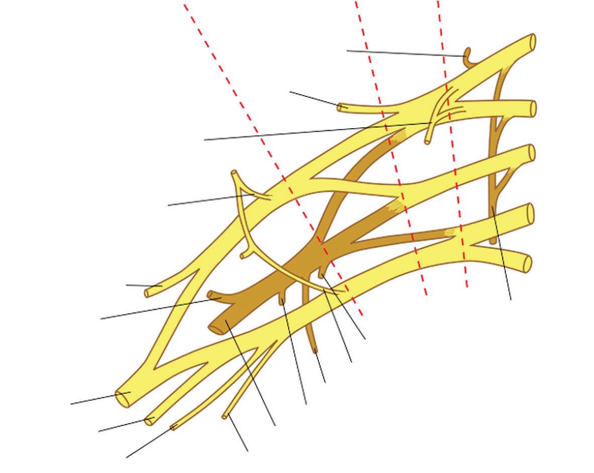
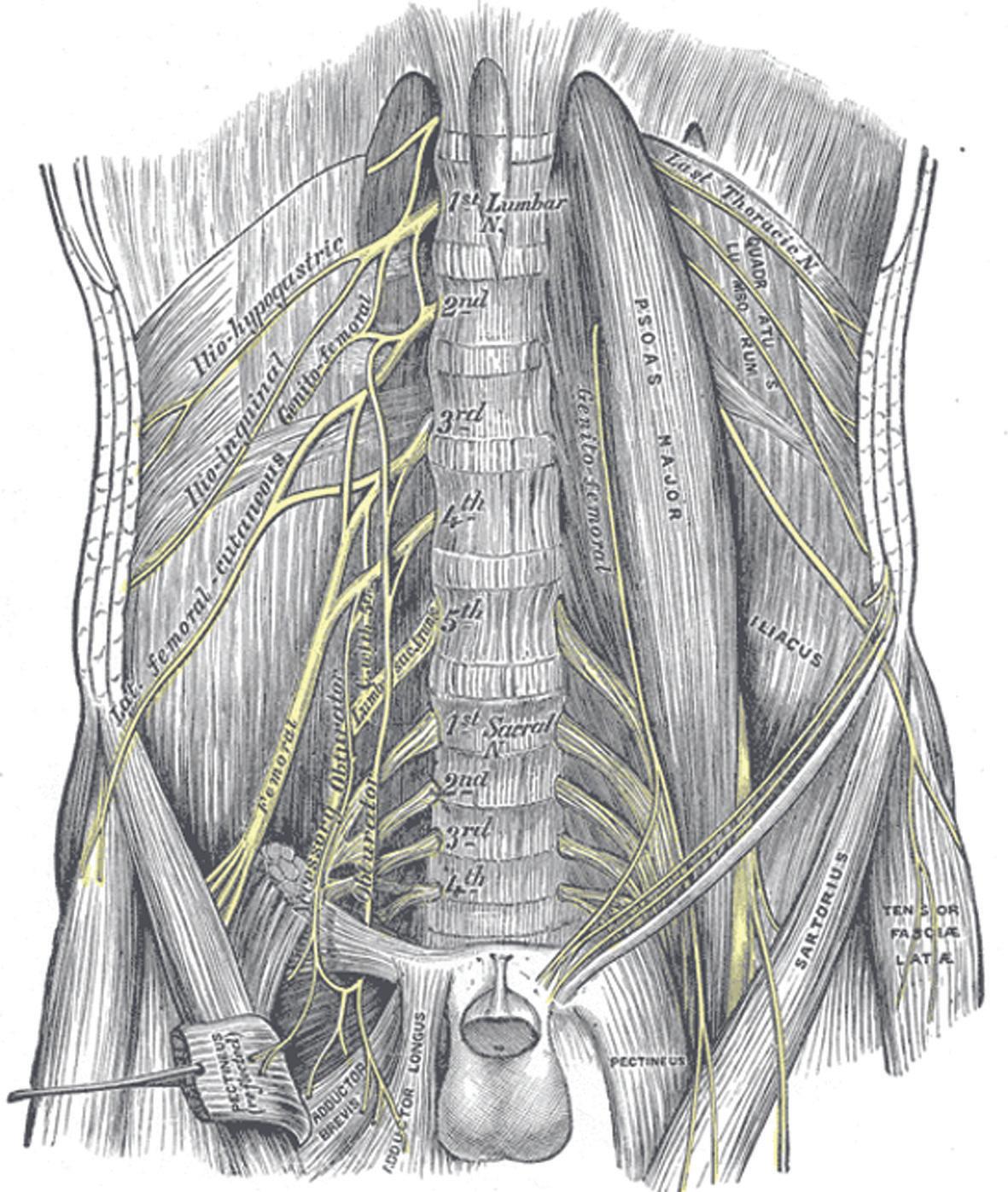
LUMBAR PLEXUS AND SACRAL PLEXUS
The lumbar and sacral plexuses are closely related, with each giving contribution to the other with high variability.
The lumbar plexus is formed from spinal nerve roots of L1–L5. Iliohypogastric and ilioinguinal nerves arise from T12–L1, while the genitofemoral nerve is formed by L1–L2 and the lateral femoral cutaneous nerve of the thigh from L2–L3. Both the obturator and femoral nerves receive contributions from L2–L4. The lumbar plexus is commonly blocked for hip and proximal leg procedures, although the gluteal and posterior thigh regions will be spared as these arise from the sacral plexus. The femoral nerve and its distal branch, the saphenous nerve, are often blocked for knee analgesia. Notably, the saphenous nerve is the only branch of the lumbar plexus that innervates areas below the knee. This sensory branch covers the medial leg and ankle and must be blocked for medial ankle coverage2 (Figure 1.4).
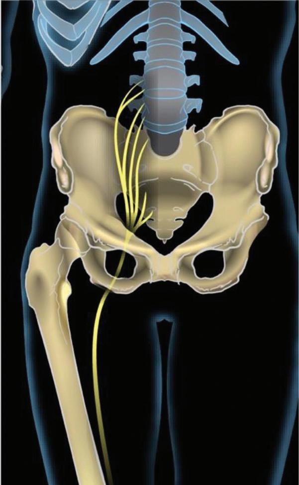
The sacral plexus is formed by the spinal nerve roots of S1–S5, with contribution from L4–L5 in the form of the Table 1.4
Cords
Dorsal scapular ner ve
Subrascapular nerve
Nerve to subclavius
Lateral pectoral ner ve
Musculocutaneous ner ve
Axillar y ner ve
Median ner ve
Ulnar nerve
Lateral Cord
Posterior Cord Medial Cord
Upper subscapular ner ve
Medial pectoral nerve
oracodorsal nerve
Lower subscapular ner ve
Radial ner ve
Medial cutaneous ner ve of the forearm Medial cutaneous ner ve of the arm
Long thoracic nerve
1.3. Brachial plexus anatomy illustration. Modified by Mattopaedia from the 1918 edition of Gray’s Anatomy. Wikipedia Public Domian. Accessed May 8, 2021. https://en.wikipedia.org/wiki/File
Figure 1.4. Lumbar plexus anatomy. Henry Gray, Anatomy of the Human Body (1918). Wikipedia Public Domain. Accessed May 8, 2021. https://en.wikipedia.org/wiki/Radiation-induced_lumbar plexopa thy#/media/File:Gray823.png
Figure
Figure 1.5. Sacral plexus and sciatic nerve. Wikimedia Commons. Accessed May 8, 2021. https://en.wikipe dia.org/wiki/ Sciatic_nerve#/media/File:Sciatic ner ve2.jpg
lumbosacral trunk (Figure 1.5). The largest and longest nerve in the body, the sciatic nerve (L4–S3), arises from the sacral plexus and later divides into the common fibular (peroneal) and tibial nerves proximal to the popliteal fossa. Notably, the sciatic nerve innervates the entire lower leg and foot below the knee, with the exception of the medial leg and ankle which is innervated by the saphenous nerve, a branch of the femoral nerve. Other major nerves arising from the sacral plexus include superior gluteal (L4–S1), inferior gluteal (L5–S2), and posterior femoral cutaneous (S1–S3) nerves. Nerves to quadratus femoris (L4–S1), inferior gemellus (L4–S1), obturator internus (L5–S2), superior gemellus (L5–S2), and piriformis (S1–S2) also arise from the sacral plexus. The sciatic nerve is commonly blocked for anesthesia and analgesia. While the Labat and parasacral approaches cover the sciatic nerve and additional portions of the sacral plexus, the popliteal approach is quite popular for superficial location and ease of block placement.2
SELECTIVE PERIPHERAL NERVE BLOCKS
Selective peripheral nerve block may be used to target analgesia for a specific nerve. This may be preferred in order to preserve proximal motor function. Adductor canal blocks are commonly used to target the saphenous nerve and achieve knee analgesia while minimizing the quadriceps weakness observed with a femoral nerve block. Alternatively, selective peripheral nerve blocks may be chosen to augment a plexus block that is not sufficient. For example, a distal median nerve block at the elbow may be used to supplement a brachial plexus block if analgesia is inadequate in the median distribution.
PLANE BLOCKS
Plane blocks are a more recent regional phenomenon. They involve injection of LA into an intramuscular plane rather than directing the injection in the area of a specific nerve or
group of nerves. Types include serratus anterior plane (SAP), pectoralis (PEC I and II), transverse abdominis plane (TAP), quadratus lumborum (QL), and erector spinae (ESP) blocks, to name a few. Generally, these blocks are easily taught and performed with less risk of nerve injury as they deliver LA in an intramuscular plane. Conversely, since LAs are not delivered in a targeted fashion near the neuraxis, a plexus, or a target peripheral nerve, the analgesic impact may not be as profound. However, plane blocks may offer a reasonable alternative for analgesia to patients who are not candidates for deeper plexus or neuraxial techniques.
BLOCK SELECTION
Regional anesthesia techniques are chosen based on patient and surgical factors. Patient factors include comorbidities or anticoagulation status. In cases of anticoagulation, certain regional techniques may still be appropriate while others are contraindicated. Surgical factors must also be evaluated. These factors include the location of surgery and the need for neurologic assessment in the postoperative period.
LOCAL ANESTHESIA INFILTRATION
LA infiltration may also be performed by a surgeon during a surgical procedure for postoperative analgesia, intraoperative analgesia, or to supplement a regional technique. Advantages of infiltration include low cost, technical ease compared to other regional techniques, less motor blockade than other regional techniques, and relative lack of contraindications. Disadvantages include a short duration of analgesia, need for a high volume of LA, inadequate analgesia in areas without complete infiltration, and possible toxicity to surrounding structures. Notably, the high LA volume required may inhibit the use of other regional anesthesia options postoperatively and possibly increase the risk of local anesthetic systemic toxicity (LAST). Finally, infiltration may be ineffective at surgical sites with ongoing infections due to the decreased pH of the surrounding tissues rendering the LAs ineffective.19
RISKS OF REGIONAL ANESTHESIA PROCEDURES
Adverse outcomes for regional anesthesia are rare but can be catastrophic for patients; therefore, informed consent is essential. The most common risks include infection, bleeding, reactions to medications, and injury to structures below the skin. Infectious risks can range from a superficial cellulitis at the skin to an epidural abscess or meningitis.20 Bleeding risks must be assessed by the type of block and the patient’s coagulation status. The American Society of Regional Anesthesia and other societies have published guidelines to assist with decision-making for patients with altered coagulation.21,22 These guidelines should be reviewed and understood prior to offering patients any regional technique. Reactions to medications for regional anesthesia can vary from the expected hypotension with spinal anesthesia to an unintentional high spinal or LAST. The knowledge to diagnose and treat these must be
in the repertoire of every regional anesthesiologist. Specific to LAST, intralipid should be immediately available at any site where regional anesthesia is administered, and treatment guidelines with a checklist should be on every regional supply cart.23 Finally, needle insertion can result in trauma to structures below the skin including muscles, blood vessels, and nerves. Dural puncture with epidural insertion can lead to post-dural puncture headache. Although neural injury is rare, any nerve injury, even transiently, can cause emotional distress to the patient, and the risk must be discussed prior to block placement.
REGIONAL ANEST h ESIA VERSUS ANALGESIA
The decision whether to place an analgesic versus an anesthetic block depends on surgical procedure, contraindications, and patient-specific factors.24 Some procedures are well tolerated under a regional anesthesia technique, while others are not. Similarly, a need for early neurological assessment in the immediate postoperative period may make some regional techniques less desirable. Additionally, patient factors may impede or complicate the ability to perform a regional block. While individual patient anatomy may make some regional blocks more challenging, ultrasonography may assist with patient-specific anatomical evaluation. However, certain comorbidities may be contraindicated with specific regional blocks (e.g., spinal anesthesia in a patient with severe aortic stenosis or patients taking potent anticoagulants). Occasionally, low patient weights may limit total LA dose, making surgical blocks unattainable. This may occur more often in the extreme geriatric and pediatric populations. In these cases, regional analgesia for postoperative pain control should still be considered and discussed in cases where surgical anesthesia is not possible or contraindicated.
Analgesic blocks often utilize decreased concentrations of LAs (e.g., ropivacaine 0.2% for analgesia rather than 0.5% for surgical anesthesia). They provide pain relief and decreased sensation, but do not provide sufficient block density for a surgical procedure.17 In situations in which peripheral or neuraxial techniques are contraindicated, systemic analgesic medications and infiltration of LA at the surgical site may attenuate patient discomfort.
CONCLUSIONS
• A detailed understanding of anatomy is essential prior to performing any regional technique.
• Assessment of both neuraxial and peripheral nerve blocks require comprehension of dermatomal distributions and peripheral nerve innervation.
• Patient factors and procedure factors must both be accounted for when selecting a regional technique to ensure adequate anesthesia and analgesia and minimize patient risk.
REFERENCES
1. Capdevila M, Ramin S, Capdevila X. Regional anesthesia and analgesia after surgery in ICU. Curr Opin Crit Care. 2017;23(5):430–439.
3. Ambulkar R, Patil V, Doctor JR, Desai M, Shetty N, Agarwal V. Accuracy of ultrasound imaging versus manual palpation for locating the intervertebral level. J Anaesthesiol Clin Pharmacol. 2017;33(3):348–352.
4. Margarido CB, Mikhael R, Arzola C, Balki M, Carvalho JC. The intercristal line determined by palpation is not a reliable anatomical landmark for neuraxial anesthesia. Can J Anaesth. 2011;58(3):262–266.
5. Chin KJ, Karmakar MK, Peng P. Ultrasonography of the adult thoracic and lumbar spine for central neuraxial blockade. Anesthesiology. 2011;114(6):1459–1485.
6. Guay J. The epidural test dose: a review. Anesth Analg. 2006;102(3): 921–929.
7. Lappen JR, Myers SA, Bolden N, Mercer BM, Chien EKS. Maternal pulse pressure and the risk of postepidural complications: a randomized controlled trial. Obstet Gynecol. 2017;130(6): 1366– 1376.
8. Fitzgerald JP, Fedoruk KA, Jadin SM, Carvalho B, Halpern SH. Prevention of hypotension after spinal anaesthesia for caesarean section: a systematic review and network meta-analysis of randomised controlled trials. Anaesthesia. 2020 Jan;75(1):109–121.
9. Wilson SH, Wolf BJ, Bingham K, et al. Labor analgesia onset with dural puncture epidural versus traditional epidural using a 26-gauge Whitacre needle and 0.125% bupivacaine bolus: a randomized clinical trial. Anesth Analg. 2018;126(2):545–551.
10. Taylor JA. Lumbosacral subarachnoid tap. J Urol. 1940;43(4):561.
11. Wilson SH, Anderson MA. Local anesthetics and regional anesthesia. In: Reves JG, SR Barnett, JR McSwain, GA Rooke, eds. Geriatric Anesthesiology. 3rd ed. New York: Springer Science+Business Media; 2018:303–319.
12. Simon MJ, Veering BT, Stienstra R, van Kleef JW, Burm AG. The effects of age on neural blockade and hemodynamic changes after epidural anesthesia with ropivacaine. Anesth Analg. 2002;94(5): 1325–1330.
13. Hoyle J, Yentis SM. Assessing the height of block for caesarean section over the past three decades: trends from the literature. Anaesthesia. 2015;70(4):421–428.
14. Wink J, Wolterbeek R, Aarts LP, Koster SC, Versteegh MI, Veering BT. Upper thoracic epidural anaesthesia: effects of age on neural blockade and cardiovascular parameters. Acta Anaesthesiol Scand. 2013;57(6):767–775.
15. Yokoyama M, Hanazaki M, Fujii H, et al. Correlation between the distribution of contrast medium and the extent of blockade during epidural anesthesia. Anesthesiology. 2004;100(6):1504–1510.
16. Camorcia M, Capogna G. Sensory assessment of epidural block for Caesarean section: a systematic comparison of pinprick, cold and touch sensation. Eur J Anaesthesiol. 2006;23(7):611–617.
17. Curatolo M, Petersen-Felix S, Arendt-Nielsen L. Sensory assessment of regional analgesia in humans: a review of methods and applications. Anesthesiology. 2000;93(6):1517–1530.
18. Mian A, Chaudry I, Huang R, Rizk E, Tubbs RS, Loukas M. Brachial plexus anesthesia: a review of the relevant anatomy, complications, and anatomical variations. Clin Anat . 2014;27(2) :210– 221.
19. Moiniche S, Mikkelsen S, Wetterslev J, Dahl JB. A qualitative systematic review of incisional local anaesthesia for postoperative pain relief after abdominal operations. Br J Anaesth. 1998;81(3):377–383.
20. Practice advisory for the prevention, diagnosis, and management of infectious complications associated with neuraxial techniques: an updated report by the American Society of Anesthesiologists Task Force on Infectious Complications Associated with Neuraxial Techniques and the American Society of Regional Anesthesia and Pain Medicine. Anesthesiology. 2017;126(4):585–601.
21. Horlocker TT, Vandermeuelen E, Kopp SL, Gogarten W, Leffert LR, Benzon HT. Regional anesthesia in the patient receiving antithrombotic or thrombolytic therapy: American Society of Regional Anesthesia and Pain Medicine evidence-based guidelines (fourth edition). Reg Anesth Pain Med. 2018;43(3):263–309.
22. Leffert L, Butwick A, Carvalho B, et al. The Society for Obstetric Anesthesia and Perinatology consensus statement on the anesthetic management of pregnant and postpartum women receiving thromboprophylaxis or higher dose anticoagulants. Anesth Analg. 2018;126(3):928–944.
23. Neal JM, Woodward CM, Harrison TK. The American Society of Regional Anesthesia and Pain Medicine checklist for managing local anesthetic systemic toxicity: 2017 version. Reg Anesth Pain Med. 2018;43(2):150–153.
24. Bonnet F, Marret E. Influence of anaesthetic and analgesic techniques on outcome after surgery. Br J Anaesth. 2005;95(1):52–58.
REVIEW QUESTIONS
(1) What surface landmark is most often used to guide the needle insertion site for thoracic epidural placement?
A. Vertebral prominence
B. Nipple line
C. Scapular ridge
D. Inferior tip of scapula
(2) Which dermatome typically corresponds to the umbilicus?
A. T4
B. T6
C. T10
D. L2
(3) Decreased sensation to cold over the patella would indicate blockade of which nerve root?
A. T1
B. T12
C. L4
D. S3
(4) Through which ligament does the Tuohy needle pass just prior to entering the epidural space?
A. Supraspinous
B. Interspinous
C. Ligamentum flavum
D. Anterior cruciate
(5) Which nerve fibers exhibit the fastest conduction velocities?
A. Large myelinated
B. Large unmyelinated
C. Small myelinated
D. Small unmyelinated
(6) Which nerve root does NOT contribute to the brachial plexus?
A. C5
B. C6
C. T1
D. T2
(7) Which nerve arises from the lumbar plexus and provides innervation in the lower leg, below the knee?
A. Iliohypogastric
B. Ilioinguinal
C. Lateral femoral cutaneous
D. Saphenous
(8) At what pKa will local anesthetics most rapidly cross the lipid bilayer?
A. 6.5
B. 7.0
C. 7.5
D. 8.0
(9) Hypobaric bupivacaine is injected in the subarachnoid space at the L3– 4 interspace. If the patient lays in left lateral position (left side down), which of the following directions describes the predominant direction of local anesthetic spread?
A. Caudad
B. Cephalad
C. Left
D. Right
(10) Regional anesthesia by brachial plexus block is planned for open repair of a metacarpal fracture. Which of the following best describes is the lowest concentration of ropivacaine to provide surgical anesthesia?
A. 1.0%
B. 0.5%
C. 0.375%
D. 0.25%
ANSWERS
(1) D. The tips of the scapulae are often used as the landmark to place thoracic epidurals, as they most commonly align with the T7 interspace. Interspaces may be palpated and counted up or down, depending on the desired level for epidural placement. The vertebral prominence is at C7, and this landmark may be helpful to confirm a level counted from the tips of the scapulae. The nipple line is associated with the T4 dermatome, but is of limited utility in neuraxial placement due to its anterior location. The scapular ridge or spine of the scapula is aligned with T3 at the root of the spine of the scapula; however, this landmark may be more difficult to palpate and may vary more with patient positioning or movement.
Reference
Ambulkar R, Patil V, Doctor JR, Desai M, Shetty N, Agarwal V. Accuracy of ultrasound imaging versus manual palpation for locating the intervertebral level. J Anaesthesiol Clin Pharmacol. 2017;33(3):348–352.
(2) C. The umbilicus corresponds to the T10 dermatome. T4 corresponds to the nipple, T6 corresponds to the xiphoid process, and L2 corresponds to the anterior thigh.
(3) C. Decreased sensation to cold over the patella would correspond to blockade of the L4 nerve fibers. T1 corresponds to the antecubital fossa. T12 corresponds between the umbilicus at T10 and the groin (L1). S3 corresponds to the peri- anal area.
(4) C. The ligamentum flavum is the tough ligament passed through just before entering the epidural space, often known for a distinct increase in resistance just before encountering the loss of resistance of the epidural space. The supraspinous ligament runs along the tips of the spinous processes. The interspinous ligament runs between the spinous processes and is firm enough to anchor the Tuohy needle without requiring external support. The anterior cruciate ligament is in the knee.
Reference Chin KJ, Karmakar MK, Peng P. Ultrasonography of the adult thoracic and lumbar spine for central neuraxial blockade. Anesthesiology. 2011;114(6):1459–1485.
(5) A. Large myelinated fibers exhibit the highest conduction velocities, 30–120 m/sec. The largest are the A-〈 fibers and the A-β fibers which carry motor and proprioception information. Factors increasing conduction velocity include large fiber size and the presence of myelin. Myelin acts as an insulator, allowing the conduction to skip from myelin cell to myelin cell, resulting in saltatory conduction.
Reference
Curatolo M, Petersen-Felix S, Arendt-Nielsen L. Sensory assessment of regional analgesia in humans: a review of methods and applications. Anesthesiology. 2000;93(6):1517–1530.
(6) D. T2 does not contribute to the brachial plexus. T2 gives sensation to the chest wall, axilla, and the medial upper arm. The brachial plexus is formed by contributions from C5, C6, C7, C8, and T1.
(7) D. The lower leg is innervated (both motor and sensory) exclusively by branches of the sciatic nerve, except for the medial portion. The saphenous nerve, an extension of the femoral nerve below the knee, provides sensory input to the medial lower leg and ankle. It does not have motor input.
(8) C. Local anesthetics with a pKa closest to physiologic pH, 7.4, will demonstrate the most rapid onset. Local anesthetics must cross the lipid bilayer of the cell to reach their site of action, the Na+ channels. A pKa close to the physiologic pH allows the LA to convert to its un-ionized state more rapidly.
Reference
Wilson SH, Anderson MA. Local anesthetics and regional anesthesia. In. Reves JG, SR Barnett, JR McSwain, GA Rooke, ed. Geriatric anesthesiology. 3rd ed. New York: Springer Science+Business Media; 2018:303–319.
(9) D. Local anesthetic movement within the CSF is determined by baricity. Hypobaric solutions are less dense than the surrounding CSF, and thus will move against gravity. In this patient, the left side is down so the hypobaric bupivacaine will move against gravity to the right side (up side).
(10) B. Analgesic blocks utilize a lower concentration of local anesthetic to provide pain relief, but are not dense enough to tolerate surgical procedures. Increasing concentration of local anesthetic increases the density of the block, allowing for surgical manipulation. A concentration of 0.5% ropivacaine would provide adequate density for an anesthetic block. A 1% ropivacaine solution would increase likelihood of neurotoxicity and also for a potential LAST reaction.
Reference
Curatolo M, Petersen-Felix S, Arendt-Nielsen L. Sensory assessment of regional analgesia in humans: a review of methods and applications. Anesthesiology. 2000;93(6):1517–1530.
PERIOPERATIVE CONSIDERATIONS FOR SUCCESS OF REGIONAL ANESTHESIA
Richa Wardhan and Janet Jira
STEM CASE AND KEY QUESTIONS
A 65-year-old patient with a history of hypertension, hypercholesterolemia, diabetes, and osteoarthritis presents to the hospital for a right below-knee amputation. Patient has been seen by the preoperative anesthesia clinic and in agreement with the surgeon is expecting to receive a general anesthetic for surgery with sciatic nerve catheter and a femoral nerve catheter for postoperative pain management.
(1) WHAT ARE THE KEY ELEMENTS IN PREPARING A PATIENT FOR A PERIPHERAL NERVE BLOCK FOR POSTOPERATIVE PAIN MANAGEMENT?
When patients receive timely information regarding nerve blocks at the surgeon’s office, they are less likely to be apprehensive on the day of surgery. Discussion regarding the nerve block should start at the surgeon’s office.
The patient should be scheduled to arrive an hour to two hours in advance if the perineural catheter is to be placed. Arrival of the patient earlier than the scheduled time for their surgery provides the extra time needed to place the nerve block.
An organized acute pain team is key in accomplishing the goals of pain management without placing excessive strain on the infrastructure. A well- established acute pain team includes anesthesiologists, surgeons, nurses, and physical therapists.1
A succinct time-out checklist prior to nerve block placement is helpful in preventing mistakes and complications. The critical items on a nerve block time-out sheet should include the site of surgery, updated and valid surgical and anesthesia consents, and allergies. The “Safe Surgery Saves Lives” project developed by the World Health Organization (WHO) designed a 19-item perioperative surgical checklist to minimize the risk of adverse events during non-cardiac surgery. It advises practitioners to address surgical issues orally at three time points: “sign-in,” “time-out,” and “sign out.” Studies using this checklist have shown a significant decrease in both surgery-related complications and patient deaths.2
A dedicated, supportive, and trained nursing staff is the mainstay of a block procedure. Sedation administered safety
increases patient satisfaction and block success. Deep sedation or general anesthesia in adults adds risk of unintended intraneural injection without the patient having the ability to communicate pain/paresthesia when the needle is placed in the wrong location.3
(2) WHAT DOCUMENTS ARE WARRANTED IN ORDER TO SAFELY CONDUCT A REGIONAL ANESTHETIC PREOPERATIVELY?
History and physical examination (H&P) from the surgeon, a preoperative note from the anesthesiologist, a surgical consent, and an anesthesiology consent should all be available and accurate prior to proceeding with a nerve block procedure preoperatively. The procedure should absolutely not commence without surgical and anesthesia (verbal or written) consent. Informed consent is a legal requirement before performance of any medical/ surgical procedure, except for emergency situations. Proceeding with an elective procedure without consent constitutes assault. 4 Nerve blocks are almost never an emergency procedure; therefore the proceduralist is ethically and legally obligated to obtain consent. In obtaining consent it is important to ensure that the patient is competent and has adequate decision-making capacity.
Consent can be written or verbal. A written consent serves as evidence that consent was obtained, assuming that the patient had adequate capacity and information and there was no coercion.4
Elements of consent include capacity, disclosure, and voluntariness:5
• Capacity refers to the patient’s ability to understand information offered to them and to make decisions regarding their care.
• Disclosure is the process by which physicians describe the procedure in terms that are comprehensible to the patient, including alternatives and their associated benefits and risks.
• Voluntariness demands that a patient is allowed to consent or dissent without any coercion, allowing for patient autonomy.