[Ebooks PDF] download Kaufman’s clinical neurology for psychiatrists (major problems in neurology) f
Visit to download the full and correct content document: https://ebookmass.com/product/kaufmans-clinical-neurology-for-psychiatrists-major-pr oblems-in-neurology/
More products digital (pdf, epub, mobi) instant download maybe you interests ...
Kaufman’s Clinical Neurology for Psychiatrists David Myland Kaufman
Kaufman’s Clinical Neurology for Psychiatrists discusses medications, testing, procedures, and other aspects of medical care. Despite their purported effectiveness, many are fraught with side effects and other adverse outcomes. Discussions in this book neither recommend nor offer medical advice, and they do not apply to individual patients. The physician, who should consult the package insert and the medical literature, remains responsible for medications’ indications, dosage, contraindications, precautions, side effects, adverse reactions, and alternatives,
including doing nothing. Some aspects of medical care that this book discusses are widely and successfully used for particular purposes not approved by the Food and Drug Administration (FDA). Regarding these “off-label” treatments, as well as conventional ones, this book is reporting – not endorsing – their use by neurologists or other physicians. Finally, because medical practices rapidly evolve, readers should expect that sooner or later new diagnostic criteria and treatments will replace those discussed in this edition.
PREFACE
PURPOSE
We have written Kaufman’s Clinical Neurology for Psychiatrists – a collegial straightforward guide – from our perspective as neurologists at a major, urban academic medical center. In a format combining traditional neuroanatomic correlations with symptom-oriented discussions, the book will assist psychiatrists in learning modern neurology. It emphasizes neurologic conditions that are frequently occurring, common to psychiatry and neurology, illustrative of a scientific principle, or have prominent psychiatric manifestations. It also includes descriptions of numerous neurologic conditions that may underlie aberrant behavior, disturbances in mood, or cognitive impairment – symptoms that prompt patients or medical colleagues to solicit psychiatry consultations. Kaufman’s Clinical Neurology for Psychiatrists does not intend to replace comprehensive neurology textbooks or convert psychiatrists into semiprofessional neurologists; however, this book contains essential information required of psychiatrists.
ORGANIZATION AND CONTENT
The organization and content of Kaufman’s Clinical Neurology for Psychiatrists arose from our experience as faculty at the Albert Einstein College of Medicine, attending physicians at Montefiore Medical Center, and supervisors of numerous neurology and psychiatry residents; consultation with our colleagues, many of whom are worldrenowned physicians; and feedback from many of the 20,000 psychiatrists who have attended the course, “Clinical Neurology for Psychiatrists,” and the more than 50,000 individuals who have purchased previous editions of this book. Learning the material in this book should help readers prepare for examinations, perform effective consultations, and improve their practice and teaching. Section 1 reviews classic anatomic neurology and describes how to approach patients with a suspected neurologic disorder, identify central or peripheral nervous system disease, and correlate physical signs. Section 2 discusses common and otherwise important clinical areas, emphasizing aspects a psychiatrist may encounter. Topics include neurologic illnesses, such as multiple sclerosis, brain tumors, strokes, and traumatic brain injury; and common symptoms, such as headaches, chronic pain, epilepsy, and involuntary movement disorders. For each topic, chapters describe the relevant symptoms including psychiatric comorbidity, easily performed office and bedside examinations, appropriate laboratory tests, differential diagnoses, and some management options.
Many chapters contain outlines for a bedside examination; reproductions of standard bedside tests, such as the Montreal Cognitive Assessment (MoCA) and Abnormal Involuntary Movement Scale (AIMS), references to recent medical literature, and pertinent web sites. One chapter provides a compilation of computed tomography (CT), magnetic resonance imaging (MRI), and positron emission tomography (PET) images that other chapters reference. Appendices contain information pertaining to most chapters: Patient and Family Support Groups (Appendix 1); Costs of Various Tests and Treatments (Appendix 2); Diseases Transmitted by Chromosome or Mitochondria Abnormalities (Appendix 3); and Chemical and Biological Neurotoxins (Appendix 4).
In addition, the book reviews neurologic conditions that have entered the public arena because, willingly or unwillingly, psychiatrists are liable to be drawn into debates involving their own patients or the medical community. Psychiatrists should be well versed in the intricacies of the following conditions that this book describes:
• Amyotrophic lateral sclerosis and multiple sclerosis as battlegrounds of assisted suicide
• Meningomyelocele with Arnold–Chiari malformation as an indication for abortion and the value of spending limited resources on this fatal or severely debilitating condition
• Chronic pain as the fulcrum for legalizing marijuana and heroin
• Parkinson disease, spinal cord injury, and other disorders amenable to research and treatment with stem cells
• Persistent vegetative state and continuing life-support technology
• Cost of medical testing and treatment.
ADDITIONS AND OTHER CHANGES FOR THE EIGHTH EDITION
The first seven editions of Kaufman’s Clinical Neurology for Psychiatrists have enjoyed considerable success in the United States, Canada, and abroad. The book has been translated into Japanese, Italian, Korean, and Spanish. In the eighth edition, written 3 years after the seventh, we have clarified the presentations, discussed recent developments in many areas, and added many clinical, anatomic, and radiologic illustrations. To give the question-andanswer sections greater power, we have increased the number of questions, refined them, expanded the discussions, and provided more illustrations. We have increased the usage of questions based on clinical vignettes because
they mimic the clinical experience and the trend of national specialty examinations.
In a major new feature of this edition, Kaufman’s Clinical Neurology for Psychiatrists refers to the diagnostic criteria for various neurologic disorders in the Diagnostic and Statistical Manual of Mental Disorder, 5th Edition (DSM-5). It compares and contrasts DSM-5 diagnostic criteria to neurologists’ diagnostic criteria, which admittedly remain for the most part uncodified and variable. With a few exceptions, DSM-5 criteria rely entirely on the nature and duration of symptoms, but neurologists, depending on the illness, rely on genetic testing, biopsy results, blood tests, various laboratory testing, or physical findings, but only sometimes exclusively on the patient’s symptoms, to make a diagnosis in their field.
This edition updates and expands most topics and adds new ones:
• New nomenclature for seizures and epilepsy
• Revised diagnostic criteria for multiple sclerosis
• New treatments for epilepsy, Alzheimer disease, Parkinson disease, multiple sclerosis, and headaches
• New diagnostic modalities and treatments for several movement disorders
• New imaging techniques
• New organization of sleep–wake disorders
• Current guidelines for the diagnosis of concussions and their management
• Psychiatric comorbidity of neurologic illnesses
• New paraneoplastic disorders.
DIDACTIC DEVICES: THE VISUAL APPROACH AND QUESTION-AND-ANSWER SECTIONS
Kaufman’s Clinical Neurology for Psychiatrists – like much of the practice of neurology – relies on a visual approach. It provides abundant illustrations, including numerous sketches of “patients” that personify or reinforce clinical descriptions, correlate the basic science with clinical findings, and serve as the basis for question-and-answer learning. The visual approach conforms to neurologists’ predilection to “diagnose by inspection.” For example, they rely on their observations for the diagnoses of gait abnormalities, psychogenic neurologic deficits, neurocutaneous disorders, strokes, and involuntary movements.
In addition, the book reproduces neurologic test results, which are also visual records, such as CT, MRI, and electroencephalography (EEG).
Kaufman’s Clinical Neurology for Psychiatrists complements the text with question-and-answer sections at the end of most chapters and at the conclusion of the book. Sections at the end of chapters generally refer to material discussed within that chapter, whereas those questions at the book’s conclusion tend to require comparison of neurologic disorders that have appeared under different headings. In Chapter 4, before the question-and-answer review of the preceding chapters’ material, the book offers a guide to preparing for standardized tests.
The Albert Einstein College of Medicine and many other medical schools rely on similar “problem-based interactive studying” – case-based question-and-answer problems – as the optimum meaningful and efficient learning strategy. Not merely quizzing the reader, the book’s questions-and-answers form an integral part of the learning experience. In fact, many readers find that these sections are the single most informative portion of the book and term them “high yield.” In keeping with the visual emphasis of the book, many of the questions are based on visual material, including sketches of patients and reproductions of MRIs, CTs, and EEGs.
ONE CAVEAT
Kaufman’s Clinical Neurology For Psychiatrists expects welleducated and thoughtful readers. It demands attention and work, and asks them to follow a rigorous course. Readers should find the book, like the practice of medicine, complex and challenging, but at the same time rich and fulfilling.
Even with the additions of text, illustrations, and questions, the eighth edition of Kaufman’s Clinical Neurology for Psychiatrists remains manageable in size, depth, and scope, but still succinct enough for psychiatrists to read and enjoy from cover to cover.
David Myland Kaufman, MD
Howard L. Geyer, MD, PhD
Mark J. Milstein, MD
FIRST ENCOUNTER WITH A PATIENT: EXAMINATION AND FORMULATION
Despite the ready availability of sophisticated tests, the “hands on” examination remains the fundamental aspect of neurology. Beloved by neurologists, the neurologic examination provides a vivid portrayal of both function and illness. When neurologists say they have seen a case of a particular illness, they mean that they have really seen a patient with it.
When a patient’s history suggests a neurologic illness, the neurologic examination may unequivocally demonstrate it. Even if psychiatrists themselves do not perform the examination, they should be able to appreciate neurologic signs and assess a neurologist’s conclusion. Neurologists systematically examine the nervous system’s major components, paying particular attention to areas of interest in an individual patient in light of his or her symptoms. Neurologists try to adhere to the routine while avoiding omissions and duplications. Despite obvious dysfunction of one part of the nervous system, they evaluate all major areas. A neurologist can usually complete an initial or screening examination in 20 minutes or less and return to perform detailed or otherwise special testing of particular areas, such as the mental status.
EXAMINATION
Neurologists usually begin by noting a patient’s age, sex, and handedness, and then review the primary symptom, present illness, medical history, family history, and social history. They explore the primary symptom, associated symptoms, and possible etiologic factors. If a patient cannot relate the history, the neurologist might interrupt the process to look for language, memory, or other cognitive deficits. Many chapters in Section 2 contain outlines of the standard questions that relate to common symptoms.
After obtaining the history, the neurologist should be able to anticipate the patient’s deficits and prepare to look for disease primarily of the central nervous system (CNS) or the peripheral nervous system (PNS). At this point, without yielding to rigid preconceptions, the physician should have developed some sense of the problem at hand.
Then neurologists should look for the site of involvement (i.e., “localize the lesion”). “Localization,” one of the initial goals of most neurologic examinations, is valuable in the majority of cases. However, it is often
somewhat of an art and inapplicable in several important neurologic illnesses, such as Alzheimer disease.
The examination is not only of historical interest, but also remains irreplaceable in diagnosis. It consists of a functional neuroanatomy demonstration: mental status, cranial nerves, motor system, reflexes, sensation, cerebellar system, and gait (Box 1.1). This format should be followed during most examinations. Trainees still mastering this structure may bring a printed copy to the patient’s bedside to serve both as a reminder and as a place to record neurologic findings.
The examination usually starts with an assessment of the mental status, because cognition is the most fundamental neurologic function and cognitive impairments may preclude an accurate assessment of other neurologic functions. The examiner should consider specific intellectual deficits, such as language impairment (see Aphasia, Chapter 8), as well as general cognitive impairment (see Dementia, Chapter 7). Tests of cranial nerves may reveal malfunction of nerves either individually or in groups, such as the ocular motility nerves (III, IV, and VI) or the cerebellopontine angle nerves (V, VII, and VIII) (see Chapter 4).
The examination of the motor system is usually performed more to detect the pattern than the severity of weakness. Whether weakness is mild to moderate (paresis) or complete (plegia), the pattern rather than severity offers more clues to localization. On a practical level, of course, the severity of the paresis determines the patient’s functional capacity, e.g., whether a patient walks, requires a wheelchair, or stays bedridden.
When neurologists detect paresis they attempt to classify its pattern. They frequently speak of three patterns. If the lower face, arm, and leg on one side of the body are paretic, they call the pattern hemiparesis. They usually attribute hemiparesis to damage in the contralateral cerebral hemisphere or brainstem. They call weakness of both legs paraparesis and usually attribute it to spinal cord damage. If the paresis predominantly involves the distal portion of all four limbs, distal quadriparesis, they usually ascribe it to PNS rather than CNS damage.
Eliciting two categories of reflexes assists in determining whether paresis – or other neurologic abnormality – originates in CNS or PNS injury. Deep tendon reflexes (DTRs) are normally present with uniform reactivity (speed and forcefulness) in all limbs, but neurologic injury often alters their activity or symmetry. In general,
BOX 1.1 Neurologic Examination
Mental status
Attention
Cooperation
Orientation (to month, year, place, and any physical or mental deficits)
with CNS injury that includes corticospinal tract damage DTRs are hyperactive, whereas with PNS injury DTRs are hypoactive.
In contrast to DTRs, pathologic reflexes are not normally elicitable beyond infancy. If these are found, they are a sign of CNS damage. The most widely recognized pathologic reflex is the famous Babinski sign. After plantar stimulation, the great toe normally moves downward (i.e., it has a flexor response). With brain or spinal cord damage, plantar stimulation typically causes the great toe to move upward (i.e., to have an extensor response). This reflex extensor movement, which is a manifestation of CNS damage, is the Babinski sign (see Fig. 19.3). Neurologists say that the Babinski sign and other pathologic reflexes are “present,” “found,” or “elicited,” but not “positive” or “negative.” The terminology is similar to a traffic stop sign: It may be present or absent, but not positive or negative.
Frontal release signs, which are also pathologic reflexes, reflect frontal lobe injury. When present they point to an “organic” basis for a change in personality and, to some degree, correlate with cognitive impairment (see Chapter 7).
Unlike abnormal DTRs and Babinski signs, which are reproducible, objective, and difficult to mimic, the sensory examination relies almost entirely on the patient’s report. Its subjective nature has led to the practice of disregarding reports of disturbances inconsistent with the rest of the examination. Under most circumstances, the best approach is to test the major sensory modalities in a clear anatomic order and tentatively accept the patient’s report.
Depending on the nature of the suspected disorder, physicians may first test light touch sensation with their finger-tips or a wood stick cotton swab, and then three sensations carried by the posterior columns of the spinal cord: position, vibration, and stereognosis (appreciation of an object’s form by touching it). Neurologists might test pain (pinprick) sensation, which is carried in the lateral columns, but only in a careful manner with a nonpenetrating, disposable instrument, such as with a broken wood shaft of the cotton swab.
Neurologists evaluate cerebellar function by observing several standard maneuvers that include the finger-to-nose test and rapid alternating movement test (see Chapter 2). These tests may demonstrate intention tremor or incoordination.
If at all possible, neurologists watch the patient walk, because a normal gait requires intact CNS and PNS motor pathways, coordination, proprioception, and balance. Moreover, all these systems must be wellintegrated. Examining the gait is probably the single most valuable assessment of noncognitive functions of the nervous system. Neurologists watch for gait abnormalities that characterize many neurologic illnesses (see Table 2.1). In addition, they should expect certain gait abnormalities to be comorbid with cognitive impairment. Whatever the abnormality, gait impairment is not merely a neurologic sign, but a condition that routinely leads to fatal falls and permanent incapacity for numerous people each year.
FORMULATION
Although somewhat ritualistic, a succinct and cogent formulation remains the basis of neurologic problem solving. A neurologist’s classic formulation consists of an appraisal of the four aspects of the examination: symptoms, signs, localization, and differential diagnosis. A neurologist might also have to support a conclusion that neurologic disease explains the patient’s symptoms and signs or, equally important, does not. For this step, neurologists at least tentatively separate psychogenic signs from neurologic (“organic”) ones.
Localization of neurologic disease requires the clinician not only to determine whether the illness affects the CNS, PNS, or muscles (see Chapters 2–6), but precise localization of lesions within these regions of the nervous system is also generally expected. The physician must also establish whether the illness affects the nervous system diffusely or in a focal, discrete area. The site and extent of neurologic damage generally indicates certain diseases. A readily apparent example is that strokes and tumors usually involve a discrete area of the brain, but
Alzheimer disease usually causes widespread, symmetrical changes.
Finally, neurologists create a differential diagnosis that lists the disease or diseases most consistent with the patient’s symptoms and signs. They should include unlikely but potentially life-threatening conditions. In addition, many neurologists, in a flourish of intellectualism, conclude with unlikely but fascinating explanations. However, even at tertiary care institutions, common conditions arise commonly. Just as “hoof beats are usually from horses, not zebras,” patients are more likely to have hemiparesis from a stroke than a mitochondrial disorder.
A typical formulation might be as follows: “Mr. Jones, a 56-year-old right-handed bartender, has had left-sided headaches for 2 months and, on the day before admission, had a generalized seizure. He is lethargic. He has papilledema, a right hemiparesis with hyperactive DTRs, and a Babinski sign. The lesion is probably situated in the left cerebral hemisphere. It is most likely a tumor or stroke, but possibly a bacterial abscess.” This formulation briefly recapitulates the salient elements of the history and physical findings. In this case, neurologists would tacitly assume that neurologic disease is present because of the obvious, objective physical findings. The history of seizures, the right-sided hemiparesis, and abnormal reflexes localize the lesion. Neurologists would base their differential diagnosis on the high probability that a discrete cerebral lesion is causing these abnormalities.
A house officer presenting a case to a superior is well advised to separate the wheat from the chaff and complete the presentation within 2 minutes, which is the limit of most listeners’ attention spans. The clinician should also practice the presentation before rounds, bearing in mind Benjamin Franklin’s proverb, “By failing to prepare, you are preparing to fail.”
In summary, the neurologist should present a succinct, well-rehearsed formulation that answers The Four Questions of Neurology:
• What are the symptoms of neurologic disease?
• What are the signs of neurologic disease?
• Where is the lesion?
• What is the lesion?
RESPONDING AS A NEUROLOGIST TO CONSULTATIONS
During their training, psychiatry residents often rotate through a neurology service where they are required to answer requests for neurology consultations. Consultants at all levels must work with a variation of the traditional summary-and-formulation format. While the patient’s interests remain paramount, the consultant’s “client” is the referring physician. Both the referring physician and consultant should be clear about the reason for the consultation. Reasons for consultations typically concern a neurologic symptom, the significance of a neuroimaging report, or a treatment recommendation. Sometimes physicians request a broad review, such as when they ask the consultant to provide a second opinion or offer a prognosis. On the other hand, the referring physicians may not want to know the diagnosis or treatment options, but
simply want the neurology service to assume the primary care of the patient.
Without belaboring the obvious, the consultation note must be organized, succinct, and practical. The primary physician in an acute care hospital should be able to digest it in 2 minutes. Long notes are usually boring and inadvertently hide useful information. Cutting and pasting information and conclusions in computerized medical records by a consultant is redundant, liable to repeat errors, and, if a previous physician made an astute diagnosis, appears to take credit for someone else’s idea. Notes that are bad, for whatever reason, reflect poorly on the consultant and the consultant’s service, and they hamper the patient’s care. At least in an academic setting, the consultant should offer at least one teaching point about the case and provide general guidelines for handling similar inquires.
Finally, consultants should show an awareness of the entire situation, which often contains incomplete and conflicting elements. They should also be mindful of the situation of the referring physician and patient. Consultants in emergency situations might help by ordering –not merely suggesting – routine tests, such as blood studies, and important but innocuous treatments, such as thiamine injections. Except in unusual circumstances, consulting residents should not suggest hazardous tests or treatments without first presenting the case to their supervisor. Consultants should not divert the primary physicians’ efforts from the patient’s most important medical problems. They should not suggest embarking on elaborate, time-consuming testing for obscure, unlikely diagnoses when the patient’s illness is obvious and requires the primary medical team’s full attention. The consultant should ask, “How can I help?”
NEUROLOGIC DIAGNOSIS
Neurologists confirm a clinical diagnosis using different frames of reference. For some diseases, such as migraine and chronic pain, neurologists rely almost entirely on a patient’s symptoms. For others, such as Parkinson disease, they base their diagnosis on physical abnormalities or constellations of findings. For many other diseases, regardless of the patient’s symptoms and signs, their diagnosis rests on an abnormal test result. For example, the diagnosis of stroke or a brain tumor requires imaging studies, and confirmation of seizures often necessitates an EEG. Neurologists diagnose many asymptomatic individuals as having a neurologic disease on the basis of a single test, such as genetic analysis or MRI (see Chapter 20).
The clinical formulation remains the mainstay of neurologic diagnosis, but abnormal findings on MRI or other studies routinely trump clinical impressions. For example, the clinical examination may indicate the presence, location, and etiology of a cerebral lesion, but if an MRI indicates a different process, neurologists generally forsake their clinical formulation and accept the MRI findings as the diagnosis.
Overall, neurologists’ and psychiatrists’ diagnoses routinely differ in several respects. Neurologists shift the basis of their diagnosis from clinical constellation, to
image, to pathologic specimen, or to another test –whichever is the most specific. In contrast, psychiatrists base diagnoses, with the exception of sleep disorders and perhaps a few others, entirely on their patient’s history and observable clinical presentation without performing a physical examination. Neurologists routinely diagnose illnesses in asymptomatic individuals, such as those with genetic mutations for Huntington disease or
a spinocerebellar ataxia, but psychiatrists almost always require symptoms. Finally, neurologists do not have a Diagnostic and Statistical Manual (DSM) of Neurologic Disorders. While the lack of a DSM prevents uniformity, neurologists remain flexible in their diagnostic criteria and freed from pigeonholing patients’ symptoms and signs, which may considerably vary with the same illness from patient to patient, into diagnostic boxes.
SIGNS OF CENTRAL NERVOUS SYSTEM DISORDERS
Disorders of the brain and the spinal cord – the two major components of the central nervous system (CNS) – typically cause readily recognizable combinations of paresis, sensory loss, visual deficits, and neuropsychologic disorders (Box 2.1). Such signs of CNS disorders differ from those of the peripheral nervous system (PNS) and both differ from the signs of psychogenic disorders. Neurologists formulate their preliminary diagnosis and often initiate treatment on the basis of the patient’s history and the examination, but if results of investigations – such as laboratory testing or magnetic resonance imaging (MRI) – contradict the initial clinical impression, they will usually revise or at least reconsider it.
SIGNS OF CEREBRAL HEMISPHERE LESIONS
Hemiparesis, usually accompanied by changes in reflexes and muscle tone, is one of neurology’s most prominent and reliable signs. Damage to the corticospinal tract, also called the pyramidal tract (Fig. 2.1), in the cerebrum or brainstem above (rostral to) the decussation of the pyramids, causes contralateral hemiparesis (Box 2.2) with weakness of the arm and leg – and, if the lesion is high enough, the lower face – opposite the side of the lesion. Damage to this tract within the spinal cord causes ipsilateral arm and leg or only leg paresis, but no face paresis. The division of the motor system into upper and lower motor neurons is a basic construct of clinical neurology. During the corticospinal tract’s entire path from the cerebral cortex to the motor cranial nerve nuclei and the anterior horn cells of the spinal cord, this tract consists of upper motor neurons (UMNs) (Fig. 2.2). The anterior horn cells, which are part of the PNS, begin the lower motor neuron (LMN). Cerebral lesions that damage the corticospinal tract cause signs of UMN injury (Figs. 2.2–2.5):
• Paresis with muscle spasticity
• Hyperactive deep tendon reflexes (DTRs)
• Babinski signs.
In contrast, PNS lesions, including motor neuron diseases (diseases of the anterior horn cells) and disorders of nerves (neuropathy), cause signs of LMN injury:
• Paresis with muscle flaccidity and atrophy
• Hypoactive DTRs
• No Babinski signs.
Another indication of a cerebral lesion is loss of certain sensory modalities over one half of the body, i.e., hemisensory loss (Fig. 2.6). A patient with a cerebral lesion characteristically loses contralateral position sensation, two-point discrimination, and the ability to identify
objects by touch (stereognosis). Neurologists often describe loss of those modalities as “cortical” sensory loss. Pain sensation, a “primary” sense, is initially received by the thalamus, from which it is relayed to the cortex, limbic system, and elsewhere. Because the thalamus is situated above the brainstem but below the cerebral cortex, most patients with cerebral lesions still perceive painful stimuli. For example, patients with cerebral infarctions may be unable to specify a painful area of their body, but they will still feel the pain’s intensity and discomfort (see Chapter 14).
Visual loss of the same half-field in each eye, homonymous hemianopia (Fig. 2.7), is a characteristic sign of a contralateral cerebral lesion. Other equally characteristic visual losses are associated with lesions involving the eye, optic nerve, or optic tract (see Chapters 4 and 12). Because they are situated far from the visual pathway, lesions in the brainstem, cerebellum, or spinal cord do not cause visual field loss.
Another conspicuous sign of a cerebral hemisphere lesion is focal (partial) or focal-onset seizures (see Chapter 10). In fact, the majority of focal seizures that alter awareness or induce psychomotor phenomena originate in the temporal lobe.
Signs of Damage of the Dominant, Nondominant, or Both Cerebral Hemispheres
Although hemiparesis, hemisensory loss, homonymous hemianopia, and focal seizures may result from lesions of either cerebral hemisphere, several neuropsychologic deficits are referable to either the dominant or nondominant hemisphere. Neurologists usually ask a patient’s handedness when taking a history, but if this information is unavailable, because approximately 85% of people are right-handed, they assume with reasonable confidence that the left hemisphere serves as the dominant hemisphere.
Lesions of the dominant hemisphere may cause language impairment, aphasia, a prominent and frequently occurring neuropsychologic deficit (see Chapter 8). Because the corticospinal tract sits adjacent to the language centers, right hemiparesis often accompanies aphasia (see Fig. 8.1).
Lesions of the nondominant parietal lobe tend to produce one or more striking neuropsychologic disturbances (see Chapter 8). For example, patients may neglect or ignore left-sided visual and tactile stimuli (hemiinattention). They may fail to use their left arm and leg because they neglect their limbs rather than because of
BOX 2.1 Signs of Common CNS Lesions
Cerebral hemisphere*
Hemiparesis with hyperactive deep tendon reflexes, spasticity, and Babinski sign
Hemisensory loss
Homonymous hemianopia
Focal (partial) seizures
Aphasia, hemi-inattention, and dementia
Pseudobulbar palsy
Basal ganglia*
Movement disorders: parkinsonism, athetosis, chorea, and hemiballismus
Postural instability
Rigidity
Brainstem
Cranial nerve palsy with contralateral hemiparesis
Hemiparesis with hyperactive deep tendon reflexes and a Babinski sign
Hemisensory loss
Homonymous hemianopia
Focal seizure
Dominant hemisphere
Aphasia: fluent, nonfluent, conduction, or isolation
Gerstmann syndrome: acalculia, agraphia, finger agnosia, and left–right confusion
Alexia without agraphia
Nondominant hemisphere
Hemi-inattention
Anosognosia
Constructional apraxia
Both hemispheres
Dementia
Pseudobulbar palsy
*Signs contralateral to lesions
paresis. When they have left hemiparesis, patients may not appreciate it (anosognosia). Many patients lose their ability to arrange matchsticks into certain patterns or copy simple forms (constructional apraxia, Fig. 2.8).
As opposed to signs resulting from unilateral cerebral hemisphere damage, bilateral cerebral hemisphere
Midbrain Pons
Internal capsule
Medulla
Spinal cord
FIGURE 2.1 ■ Each corticospinal tract originates in the cerebral cortex, passes through the internal capsule, and descends into the brainstem. The tracts cross in the pyramids, which are protuberances on the inferior portion of the medulla, to descend in the spinal cord mostly as the lateral corticospinal tract. The corticospinal tracts synapse with the anterior horn cells of the spinal cord, which give rise to peripheral nerves. Neurologists often call the corticospinal tract the pyramidal tract because it crosses in the pyramids. The extrapyramidal system, which modulates the corticospinal tract, originates in the basal ganglia and cerebellum, and remains within the brain.
damage produces several important disturbances that psychiatrists are likely to encounter in their patients. One of them, pseudobulbar palsy, best known for producing emotional lability, results from bilateral corticobulbar tract damage (see Chapter 4). The corticobulbar tract, like its counterpart the corticospinal tract, originates in the motor cortex of the posterior portion of the frontal lobe. It innervates the brainstem motor nuclei, which in turn innervate the head and neck muscles. Traumatic brain injury (TBI), multiple cerebral infarctions (strokes), and frontotemporal dementia (see Chapter 7), are apt to strike the corticobulbar tract, as well as the surrounding frontal lobes, and thereby cause pseudobulbar palsy. Damage to both cerebral hemispheres – from large or multiple discrete lesions, degenerative diseases, or metabolic abnormalities – also causes dementia (see Chapter 7). In addition, because CNS damage that causes dementia must be extensive and severe, it usually also produces at least subtle physical neurologic findings, such as hyperactive DTRs, Babinski signs, mild gait impairment, and frontal lobe release reflexes. However, many neurodegenerative illnesses that cause dementia, particularly
Afferent limb
Normal Quadriceps muscle tendon
efferent limb
UMN Brain Spinal cord LMN Peripheral nerve
FIGURE 2.2 ■ A, Normally, when neurologists strike a patient’s quadriceps tendon with a percussion hammer, the maneuver elicits a DTR. In addition, when they stroke the sole of the foot to elicit a plantar reflex, the big toe bends downward (flexes). B, When brain or spinal cord lesions injure the corticospinal tract, producing upper motor neuron (UMN) damage, DTRs react briskly and forcefully, i.e., DTRs are hyperactive. As another sign of UMN damage, the plantar reflex is extensor (a Babinski sign). C, In contrast, peripheral nerve injury causes lower motor neuron (LMN) damage, the DTR is hypoactive and the plantar reflex is absent.
FIGURE 2.3 ■ This patient shows right hemiparesis with weakness of the right arm, leg, and lower face. The right-sided facial weakness causes the flat nasolabial fold; however, the forehead muscles remain normal (see Chapter 4 regarding this discrepancy). The right arm moves little, and the elbow, wrist, and fingers take on a flexed position; the right leg is externally rotated; and the hip and knee are flexed.
FIGURE 2.4 ■ When the patient stands up, his weakened arm retains its flexed posture. His right leg remains externally rotated, but he can walk by swinging it in a circular path. This maneuver is effective but results in circumduction or a hemiparetic gait
FIGURE 2.5 ■ Mild hemiparesis may not be obvious. To exaggerate it, the physician has asked this patient to extend both arms with his palms held upright, as though his outstretched hand were supporting a pizza box. His weakened arm sinks (drifts) and his palm turns inward (pronates). The imaginary pizza box would slide to his right. His arm drift and pronation represent a forme fruste of the posture seen with severe paresis (Fig. 2.3).
FIGURE 2.7 ■ In homonymous hemianopia, the same half of the visual field is lost in each eye. In this case, damage to the left cerebral hemisphere has caused a right homonymous hemianopia. This sketch portrays visual field loss, as is customary, from the patient’s perspective; the colored area represents the defect (see Figs. 4.1 and 12.7).
Lateral spinothalamic tract
Temperature
Pain
Medulla
Cervical spinal cord
Fasciculus cuneatus
Position
Stereognosis
FIGURE 2.6 ■ Peripheral nerves carry pain and temperature sensations to the spinal cord. After a synapse, these sensations cross and ascend in the contralateral lateral spinothalamic tract (pink) to terminate in the thalamus. From there, tracts relay the sensations to the limbic system, reticular activating system, and other brainstem regions as well as the cerebral cortex. In parallel, the peripheral nerves also carry position and vibration sense and stereognosis to the ipsilateral fasciculus cuneatus and fasciculus gracilis, which together constitute the spinal cord’s posterior columns (cross-hatched) (Fig. 2.15). Unlike pain and temperature sensation, these sensations ascend in the spinal cord via ipsilateral tracts (black). They cross in the decussation of the medial lemniscus, which is in the medulla, synapse in the thalamus, and terminate in the parietal cortex. (To avoid spreading blood-borne illnesses, examiners should use a disposable instrument when testing pain.)
FIGURE 2.8 ■ A patient showing constructional apraxia from a right parietal lobe infarction was unable to complete a circle (top figure), draw a square on request (second figure), or even copy one (third figure). She spontaneously tried to draw a circle and began to retrace it (bottom figure). Her constructional apraxia consists of rotation of the forms, perseveration of certain lines, and the incompleteness of the second and lowest figures. In addition, the figures tend toward the right-hand side of the page, which indicates that she has neglect of the left-hand side of the page, i.e., left hemi-inattention (see Chapter 8).
Alzheimer disease, do not cause overt findings such as hemiparesis.
While certainly not peculiar to cerebral lesions and even typically absent in early Alzheimer disease, gait impairment is a crucial neurologic finding. Because walking requires intact and well-integrated strength, sensation, and coordination, testing the patient’s gait is the single most reliable assessment of a patient’s noncognitive neurologic function. Gait impairment constitutes the primary physical component of the subcortical dementias, such as vascular dementia, dementia with Lewy
Parietal cortex
TABLE 2.1 Gait Abnormalities Associated With Neurologic Disorders
bodies disease, and Parkinson disease dementia (see Chapter 7). Several distinct gait abnormalities are clues to specific neurologic disorders, such as normal pressure hydrocephalus (Table 2.1). As a general rule, slow gait speed, e.g., 0.7 m/sec or less, is associated with an increased risk of dementia, stroke, falls, disability, hospitalization, and death.
SIGNS OF BASAL GANGLIA LESIONS
The basal ganglia, located subcortically in the cerebrum, consist of the caudate and putamen (together constituting the striatum), globus pallidus, substantia nigra; and subthalamic nucleus (corpus of Luysii) (see Fig. 18.1). They give rise to the extrapyramidal motor system, which modulates the corticospinal (pyramidal) tract. It controls muscle tone, regulates motor activity, and generates postural reflexes. Its efferent fibers play on the cerebral cortex, thalamus, and other CNS structures. Because its efferent fibers are confined to the brain, the extrapyramidal tract does not act directly on the spinal cord or LMNs. Signs of basal ganglia disorders include a group of fascinating, often dramatic, involuntary movement disorders (see Chapter 18):
• Parkinsonism is the combination of resting tremor, rigidity, bradykinesia (slowness of movement) or akinesia (absence of movement), and postural abnormalities. It usually results from Parkinson disease and related neurodegenerative illnesses, exposure to dopamine receptor-blocking antipsychotic medications, or toxins.
• Athetosis is the slow, continuous, writhing movement of the fingers, hands, face, and throat. Kernicterus or other perinatal basal ganglia injury usually causes it.
• Chorea is intermittent, randomly located, jerking of limbs and the trunk. The best-known example occurs in Huntington disease (previously called “Huntington chorea”), in which the caudate nuclei characteristically atrophy.
• Hemiballismus is the intermittent flinging of the arm and leg of one side of the body. It is classically associated with small infarctions of the contralateral subthalamic nucleus, but similar lesions in other basal ganglia may be responsible.
In general, when damage is restricted to the extrapyramidal system, patients have no paresis, DTR abnormalities, or Babinski signs – hallmarks of corticospinal (pyramidal) tract damage. More important, in many of these conditions, such as hemiballismus and athetosis, patients have no cognitive impairment or other neuropsychologic disorder. On the other hand, several conditions – such as Huntington disease, Wilson disease, and advanced Parkinson disease – affect the cerebrum as well as the basal ganglia. In them, dementia, depression, and psychosis frequently accompany involuntary movements (see Box 18.4).
With unilateral basal ganglia damage, signs develop in the contralateral limbs. For example, an infarction of the subthalamic nucleus causes contralateral hemiballismus, and degeneration of the substantia nigra causes contralateral parkinsonism (“hemiparkinsonism”).
SIGNS OF BRAINSTEM LESIONS
The brainstem contains, among a multitude of structures, the cranial nerve nuclei, the corticospinal tracts, other “long tracts” that travel between the cerebral hemispheres and the limbs, and cerebellar afferent (inflow) and efferent (outflow) tracts. Combinations of cranial nerve and long tract signs, and the absence of signs of cerebral injury, such as visual field cuts and neuropsychologic deficits, indicate the presence and location of a brainstem lesion. For example, brainstem injuries cause diplopia (double vision) because of cranial nerve impairment, but visual acuity and visual fields remain normal because the visual pathway, which passes from the optic chiasm to the cerebral hemispheres, does not travel within the brainstem (see Fig. 4.1). Similarly, a right hemiparesis associated with a left third cranial nerve palsy localizes the lesion to the brainstem (particularly the left midbrain). Moreover, that pair of findings indicates that further examination will reveal neither aphasia nor dementia.
Several brainstem syndromes illustrate critical anatomic relationships, such as the location of the cranial nerve nuclei or the course of the corticospinal tract; however, none of them involves neuropsychologic abnormalities. Although each syndrome has an eponym, for practical purposes it is only necessary to identify the clinical findings and, if appropriate, attribute them to a lesion in one of the three major divisions of the brainstem: midbrain, pons, or medulla (Fig. 2.9). Whatever the localization, most brainstem lesions result from occlusion of a small branch of the basilar or vertebral arteries.
In the midbrain, where the oculomotor (third cranial) nerve fibers pass through the descending corticospinal
CSF Pathway
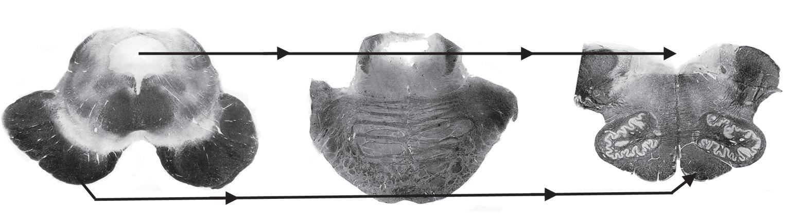
FIGURE 2.9 ■ Myelin stains of the three main divisions of the brainstem – midbrain, pons, and medulla – show several clinically important tracts, the cerebrospinal fluid (CSF) pathway, and motor nuclei of the cranial nerves. (Midbrain) The midbrain is identifiable by its distinctive silhouette and gently curved (pale, unstained in this preparation) substantia nigra (S). The aqueduct of Sylvius (A) is surrounded by the periaqueductal gray matter. Ventral to the aqueduct, near the midline, lie the oculomotor (3) and trochlear (not pictured) cranial nerve nuclei. The nearby MLF, which ascends from the pons, terminates in the oculomotor nuclei. The large, deeply stained cerebral peduncles, ventral to the substantia nigra, contain the corticospinal (pyramidal [Δ]) tract. Originating in the cerebral cortex, the corticospinal tract (Δ) descends ipsilaterally through the midbrain, pons, and medulla until it crosses in the medulla’s pyramids to continue within the contralateral spinal cord. CSF flows downward from the lateral ventricles through the aqueduct of Sylvius into the fourth ventricle (IV), which overlies the lower pons and medulla. CSF exits from the fourth ventricle into the subarachnoid space. (Also see a functional drawing [Fig. 4.5], computer-generated rendition [Fig. 18.2], and sketch [Fig. 21.1].) (Pons) The pons (Latin, bridge) houses the trigeminal motor division (5), abducens (6), facial (7), and acoustic/vestibular (not shown) cranial nerve nuclei and, inferior and lateral to the fourth ventricle, the locus ceruleus (*). In addition to containing the descending corticospinal tract (Δ), the basilar portion of the pons (basis pontis) contains large crisscrossing cerebellar tracts. (Also see a functional drawing [Fig. 4.7] and an idealized sketch [Fig. 21.2].) (Medulla) The medulla (Latin, marrow), readily identifiable by the pair of unstained scallop-shaped inferior olivary nuclei, includes the cerebellar peduncles (C), which contain afferent and efferent cerebellar tracts; the corticospinal tract (Δ); and the floor of the fourth ventricle (IV). It also contains the decussation of the medial lemniscus (M), the nuclei for cranial nerves 9–11 grouped laterally and 12 situated medially, and the trigeminal sensory nucleus (not pictured) that descends from the pons to the cervical–medullary junction. (Also see a functional drawing [Fig. 2.10].)
tract, a single small infarction can damage both pathways. Patients with oculomotor nerve paralysis and contralateral hemiparesis typically have a lesion in their midbrain ipsilateral to the paretic eye (see Fig. 4.9). In an analogous situation, patients with an abducens (sixth cranial) nerve paralysis and contralateral hemiparesis have a lesion in the pons ipsilateral to the paretic eye (see Fig. 4.11).
Lateral medullary infarctions create a classic but complex picture, the lateral medullary syndrome. Patients have dysarthria because of paralysis of the ipsilateral palate from damage to cranial nerves IX through XI; ipsilateral facial numbness (hypalgesia) (Greek, decreased sensitivity to pain) because of damage to cranial nerve V, with contralateral anesthesia of the body (alternating or crossed hypalgesia) because of ascending spinothalamic tract damage; and ipsilateral ataxia because of inferior cerebellar peduncle dysfunction. They also have nystagmus and vertigo from damage to the vestibulocochlear nerve and ipsilateral Horner syndrome (ptosis, miosis, anhydrosis) due to interruption of sympathetic fibers. In other words, the most important elements of this syndrome consist of damage to three groups of nuclei (V, VIII, and IX–XI) and three white matter tracts (spinothalamic, sympathetic, and inferior cerebellar peduncle). Although the lateral medullary syndrome commonly occurs and provides an excellent example of clinicalpathologic correlation, physicians need not recall all of its pathology or clinical features; however, they should know that lower cranial nerve palsies accompanied by alternating hypalgesia, without cognitive impairment or limb paresis, result from a lesion in the lower brainstem (Fig. 2.10). They should also know that the lateral medullary syndrome causes bulbar palsy (see Chapter 4).
Nystagmus, repetitive jerk-like eye movements that are usually conjugate (i.e., affecting both eyes equally and simultaneously), is not peculiar to the lateral medullary syndrome, but rather may result from any type of injury to the brainstem’s large vestibular nuclei. Nystagmus can be a manifestation of various disorders, including intoxication with alcohol, phenytoin (Dilantin), phencyclidine (PCP), or barbiturates; ischemia of the vertebrobasilar artery system; multiple sclerosis (MS); Wernicke–Korsakoff syndrome; or viral labyrinthitis. Among individuals who have ingested PCP, coarse vertical and horizontal (three directional or multidirectional) nystagmus characteristically accompanies an agitated delirium and markedly reduced sensitivity to pain and cold temperature. Unilateral nystagmus may be a component of internuclear ophthalmoplegia, which is usually a manifestation of MS or a small brainstem infarction (see Chapters 4 and 15).
SIGNS OF CEREBELLAR LESIONS
The cerebellum (Latin, diminutive of cerebrum) consists of two hemispheres and a central portion, the vermis Each hemisphere controls coordination of the ipsilateral limbs, and the vermis controls coordination of “axial” or “midline structures”: the head, neck, and trunk. Because the cerebellum controls coordination of the limbs on the same side of the body, it differs from the cerebrum wherein each hemisphere governs the contralateral body. Another unique feature of the cerebellum is that when one hemisphere is damaged, the other will eventually assume the functions for both. In other words, although loss of one cerebellar hemisphere will temporarily cause
Cerebellar peduncle
Sympathetic tract
Trigeminal nucleus
Spinothalamic tract
Nucleus ambiguus
FIGURE 2.10 ■ A, An occlusion of the right posterior inferior cerebellar artery (PICA) or its parent artery, the right vertebral artery, has caused an infarction of the lateral portion of the right medulla (stippled). This infarction damages important structures: the inferior cerebellar peduncle, the spinal trigeminal nerve (V) sensory nucleus, the spinothalamic tract (which arose from the contralateral side of the body), the nucleus ambiguus (cranial nerves IX and X motor nuclei), and poorly delineated sympathetic fibers. However, this infarction spares medial structures: the corticospinal tract, medial longitudinal fasciculus (MLF), and hypoglossal nerve (XII) nucleus. B, Because he has sustained an infarction of his right lateral medulla, this patient has a right-sided Wallenberg syndrome. He has a right-sided Horner syndrome (ptosis and miosis) because of damage to the sympathetic fibers (also see Chapter 12). He has rightsided ataxia because of damage to the ipsilateral cerebellar tracts. He has an alternating or crossed hypalgesia: diminished pain sensation on the right side of his face, accompanied by loss of pain sensation on the left trunk and extremities (shaded). Finally, he has hoarseness and paresis of the right soft palate because of damage to the right nucleus ambiguus. Because of the right-sided palate weakness, the palate deviates upward toward his left on voluntary phonation (saying “ah”) or in response to the gag reflex.
incapacitating ipsilateral incoordination, the patient’s deficit lessens as the remaining hemisphere compensates almost entirely. For example, patients who lose one cerebellar hemisphere to a stroke or TBI typically regain their ability to walk, although they may never dance.
Children who sustain such an injury are more resilient and often can learn to ride a bicycle and participate in athletic activities.
In addition to causing incoordination, cerebellar lesions cause subtle motor changes, such as muscle
hypotonia and pendular DTRs. However, cerebellar lesions do not cause paresis, hyperactive DTRs, or Babinski signs.
Although several technically sophisticated studies have shown that the cerebellum contributes to cognition and emotion, it does not play a discernible role in these functions in everyday endeavors. For example, lesions restricted to the cerebellum do not lead to dementia, language impairment, or other cognitive impairment. A good example is the normal intellect of children and young adults despite having undergone resection of a cerebellar hemisphere for removal of an astrocytoma (see Chapter 19).
On the other hand, several conditions damage the cerebrum as well as the cerebellum. For example, alcohol, phenytoin (Dilantin), lithium, and toluene may cause prominent ataxia and cognitive impairment.
For practical purposes, neurologists assess cerebellar function in tests of coordinated motor function. Thus, intention tremor, demonstrable in the finger-to-nose (Fig. 2.11) and heel-to-shin tests (Fig. 2.12), characterizes cerebellar dysfunction. This tremor is evident when the patient moves to a target but is absent when the patient rests. In a classic contrast, Parkinson disease causes a
resting tremor that is present when the patient sits quietly and reduced or even abolished when the patient moves (see Chapter 18). Physicians should not confuse the neurologic term “intention tremor” with “intentional tremor,” which would be a self-induced or psychogenic tremor.
Another sign of incoordination due to a cerebellar lesion is dysdiadochokinesia, impaired rapid alternating movements of the limbs. When asked to slap the palm and then the back of the hand rapidly and alternately on his or her own knee, for example, a patient with dysdiadochokinesia will do so with uneven force and irregular rhythm, and lose the alternating pattern.
Damage to either the entire cerebellum or the vermis alone causes incoordination of the trunk (truncal ataxia). This manifestation of cerebellar damage forces patients to place their feet widely apart when standing and leads to a lurching, unsteady, and wide-based pattern of walking (gait ataxia) (Table 2.1 and Fig. 2.13). A common example is the staggering and reeling of people intoxicated by alcohol. In addition, such cerebellar damage prevents people from walking heel-to-toe, i.e., performing “tandem gait.” Another common example of ataxia occurs in individuals who have inherited genetic mutations that cause combinations of cerebellar and spinal cord degeneration. In several disorders, patients have abnormalities beyond the nervous system (Fig. 2.14). Extensive damage of the cerebellum causes scanning speech, a variety of dysarthria. Scanning speech, which reflects incoordination of speech production, is characterized by poor modulation, irregular cadence, and
2.11 ■ This young man has a multiple sclerosis plaque in the right cerebellar hemisphere. During the finger-to-nose test, his right index finger touches his nose and then the examiner’s finger by following a coarse, irregular path. The oscillation in his arm’s movement is an intention tremor, and the irregularity in the rhythm is dysmetria
2.13 ■ Because this man has developed cerebellar degeneration from alcoholism, he has a typical ataxic gait His stance is broad-based. His gait is unsteady, and he is uncoordinated.
FIGURE
FIGURE 2.12 ■ In the heel-to-shin test, the patient with the rightsided cerebellar lesion in the previous sketch displays limb ataxia as his right heel wobbles when he pushes it along the crest of his left shin.
FIGURE
FIGURE 2.14 ■ The pes cavus foot deformity consists of a high arch, elevation of the dorsum, and retraction of the first metatarsal. When pes cavus occurs in families with childhood-onset ataxia and posterior column sensory deficits, it is a reliable sign of Friedreich ataxia, which is the most common hereditary ataxia in the United States and Europe.
matter on the inside with white outside – is opposite that of the cerebrum. Interruption of the myelinated tracts causes most of the signs of spinal cord injury, which neurologists call “myelopathy.”
The major descending pathway, entirely motor, is the lateral corticospinal tract.
The major ascending pathways, entirely sensory, include the following:
• Posterior columns (or dorsal columns), comprised of the fasciculi cuneatus and gracilis, carry position and vibration sensations to the thalamus.
• Lateral spinothalamic tracts carry temperature and pain sensations to the thalamus.
• Anterior spinothalamic tracts carry light touch sensation to the thalamus.
• Spinocerebellar tracts carry joint position and movement sensations to the cerebellum.
Spinal Cord Transection
FIGURE 2.15 ■ In this sketch of the spinal cord, the centrally located gray matter is stippled. The surrounding white matter contains myelin-coated ascending and descending tracts. Clinically important ascending tracts are the spinocerebellar tracts (SC), the lateral spinothalamic tract (ST), and the posterior columns [fasciculus cuneatus (FC), from the upper limbs, and fasciculus gracilis (FG), from the lower limbs]. The most important descending tract is the lateral corticospinal (CS) tract.
inability to separate adjacent sounds. Physicians should be able to distinguish dysarthria – whether caused by cerebellar injury, bulbar or pseudobulbar palsy, or other neurologic disorder – from aphasia (see Chapter 8).
Before considering the illnesses that damage the cerebellum (see Section 2), physicians must appreciate that the cerebellum normally undergoes age-related changes that appear between ages 50 and 65 years in the form of mildly impaired functional ability and abnormal neurologic test results. For example, as people age beyond 50 years, they walk less rapidly and less sure-footedly. They begin to lose their ability to ride a bicycle and to stand on one foot while putting on socks. During a neurologic examination they routinely topple during tandem walking.
SIGNS OF SPINAL CORD LESIONS
The spinal cord’s gray matter, which when viewed in the axial plane appears as a broad H-shaped structure in the center of the spinal cord, consists largely of neurons that transmit nerve impulses at one horizontal level. The spinal cord’s white matter, composed of myelinated tracts that convey information in a vertical direction, surrounds the central gray matter (Fig. 2.15). This pattern – gray
If an injury severs the spinal cord, the transection’s location – cervical, thoracic, or lumbosacral – determines the pattern of the ensuing motor and sensory deficits. Cervical spinal cord transection, for example, blocks all motor impulses from descending and sensory information from arising through the neck. This lesion causes paralysis of the arms and legs (quadriparesis) and, after 1–2 weeks, hyperactive DTRs, and Babinski signs. In addition, it prevents the perception of all limb, trunk, and bladder and bowel sensation. Similarly, a midthoracic spinal cord transection causes paralysis of the legs (paraparesis) with similar reflex changes, and sensory loss in the trunk and below (Fig. 2.16). In general, all spinal cord injuries disrupt bladder control and sexual function, which rely on delicate, intricate systems (see Chapter 16).
Another motor impairment attributable to spinal cord damage, whether from a specific lesion or a neurodegenerative illness, is pathologically increased muscle tone, which neurologists label hypertonicity or spasticity. It often creates more disability than the accompanying paresis. For example, because spasticity causes the legs to be straight, extended, and unyielding, patients tend to walk on their toes (see Fig. 13.3). Similarly, spasticity greatly limits the usefulness of patients’ hands and fingers.
In a variation of the complete spinal cord lesion, when a penetrating injury severs only the lateral half of the spinal cord, neurologists refer to the injury as a spinal cord hemitransection. The lesion causes the classic Brown–Séquard syndrome, which consists of ipsilateral paralysis of limb(s) from corticospinal tract damage and loss of vibration and proprioception from dorsal column damage combined with loss of temperature and pain (hypalgesia) sensation in the opposite limb(s) from lateral spinothalamic tract damage (Fig. 2.17). In the vernacular of neurology, one leg is weak and the other is numb.
Even with devastating spinal cord injury, cerebral function is preserved. In a frequently occurring and tragic example, soldiers surviving a penetrating gunshot wound of the cervical spinal cord, although quadriplegic, retain intellectual, visual, and verbal facilities. Nevertheless, veterans and other individuals with spinal cord injuries
FIGURE 2.16 ■ In a patient with a spinal cord injury, the “level” of hypalgesia indicates the site of the damage. The clinical landmarks are C4, T4, and T10. C4 injuries cause hypalgesia below the neck; T4 injuries, hypalgesia below the nipples; T10 injuries, hypalgesia below the umbilicus.
FIGURE 2.17 ■ In this case of hemitransection of the thoracic spinal cord from a knife wound, the patient shows the Brown–Séquard syndrome. Injury to the left lateral corticospinal tract results in the combination of left-sided leg paresis, hyperactive DTRs, and a Babinski sign; injury to the left posterior column results in impairment of left leg vibration and position sense. Most striking, injury to the left spinothalamic tract causes loss of temperature and pain sensation in the right leg. Loss of pain sensation contralateral to paresis is the signature of the Brown–Séquard syndrome.
often despair from isolation, lack of social support, and loss of their physical abilities.
Syringomyelia
A lesion that often affects only the cervical spinal cord consists of an elongated cavity, syringomyelia or simply a syrinx (Greek, syrinx, pipe or tube + myelos marrow). The syrinx occurs in the substance of the spinal cord, adjacent to its central canal, which is the thin tube running vertically within the gray matter. It usually develops, for unclear reasons, during adolescence. Traumatic intraspinal bleeding may cause a variety of syrinx, a hematomyelia These conditions produce clinical findings that reflect their neuroanatomy (Fig. 2.18). In both cases, as the cavity expands, its pressure rips apart the lateral spinothalamic tract fibers as they cross from one to the other side of the spinal cord. It also presses on the anterior horn cells of the anterior gray matter. The expansion not only causes neck pain, but a striking loss in the arms and hands of pain and temperature sensation, muscle bulk, and DTRs. Because the sensory loss is restricted to patients’ shoulders and arms, neurologists frequently describe it as cape- or shawl-like. Moreover, the sensory loss is characteristically restricted to loss of pain and temperature sensation because the posterior columns, merely displaced, remain functional.
NEUROLOGIC ILLNESSES
Several illnesses damage only specific ascending and descending spinal cord tracts (Fig. 2.19). The posterior columns – fasciculus gracilis and fasciculus cuneatus – seem particularly vulnerable. For example, tabes dorsalis (syphilis), combined system degeneration (vitamin B12 deficiency, see Chapter 5), and the spinocerebellar ataxias (SCAs) each damages the posterior columns alone or in combination with other tracts (also see Box 15.1). In these conditions, impairment of the posterior columns leads to a loss of position sense that prevents patients from being able to stand with their eyes closed (Romberg’s sign). When they walk, this position sense loss produces an ataxic gait or possibly a steppage gait (Fig. 2.20).
In another example, the human T-lymphotropic virus type 1 (HTLV-1) infects the spinal cord’s lateral columns. The infection, which is endemic in Caribbean islands, causes HTLV-1 associated myelopathy (or simply HTLV-1 myelopathy) in which patients develop spastic paraparesis that resembles MS. Perhaps more than in any other common myelopathy, the spasticity is disproportionately greater than the paresis.
Several toxic-metabolic disorders – some associated with substance abuse – damage the spinal cord. For example, nitrous oxide (N2O), a gaseous anesthetic that may be inhaled as a drug of abuse by thrill-seeking
FIGURE 2.18 ■ Left, A syringomyelia (syrinx) is an elongated cavity in the spinal cord. Its expansion disrupts the lateral spinothalamic tract as it crosses, and compresses the anterior horn cells of the gray matter. It does not impair the function of the posterior columns and corticospinal tracts. Right, The classic finding is a shawl-like pattern of loss of pain and temperature sensation in the arms and upper chest (in this case, C4–T4) that is accompanied by weakness, atrophy, and areflexia in the arms.
FIGURE 2.19 standard spinal cord preparation stains (white matter) black central H-shaped column gray. B, In combined system degeneration (vitamin B12 deficiency), posterior column and corticospinal tract demyelination causes their lack of stain. C, In tabes dorsalis (tertiary syphilis), damage to the posterior column leaves them unstained. D, MS leads to asymmetric, irregular, demyelinated unstained plaques.
dentists, causes a pronounced myelopathy by inactivating vitamin B12 (see Chapter 5). Copper deficiency, often from excess consumption of zinc by food faddists or inadvertently ingested from excess denture cream, leads to myelopathy. Also, unless physicians closely monitor and replace vitamins and nutrients following gastric bypass surgery, patients remain at risk of developing myelopathy for up to several years after the surgery.
FIGURE 2.20 ■ The steppage gait consists of the patient’s raising each knee excessively, as if perpetually climbing a staircase. This maneuver compensates for a loss of position sense by elevating the feet to ensure that they will clear the ground. Although the steppage gait is a classic sign of posterior column spinal cord damage from tabes dorsalis, peripheral neuropathies that impair position sense are a more frequent cause of this gait abnormality.
Most important, dementia accompanies myelopathy in several illnesses because of concomitant cerebral damage. Examples of this association include tabes dorsalis, vitamin B12 deficiency, AIDS, and, when disseminated throughout the cerebrum, MS.
Fasciculus cuneatus
Spinothalamic tract
PSYCHOGENIC NEUROLOGIC DEFICITS
Classic studies of hysteria, conversion disorders, and related conditions included patients who underwent only rudimentary physical examinations and minimal, if any, laboratory testing. Studies that re-evaluated the same patients after many years reported that in as many as 15% of them, specific neurologic conditions, such as movement disorders, multiple sclerosis (MS), or seizures, emerged. Another interesting aspect of these studies is that physicians in the first two-thirds of the 20th century assumed that many illnesses were entirely “psychogenic,” but today’s physicians consider these same illnesses to be neurologic disorders, such as Tourette disorder, writer’s cramp and other focal dystonias, erectile dysfunction, migraines, and trigeminal neuralgia.
Neurologists currently have an arsenal of high-tech tests, including computed tomography (CT), magnetic resonance imaging (MRI), functional MRI (fMRI), positron emission tomography (PET), electroencephalography (EEG), EEG-video monitoring, genetic analyses, and a full array of subspecialty consultants. Nevertheless, they still fail to reach 100% accuracy in their diagnoses. They also may hesitate before diagnosing a deficit as psychogenic, which is unfortunate, because delay in diagnosis adversely affects outcome. In some cases they may be forced to “undiagnose” a neurologic illness, such as MS, when further testing, observation, or consultation shows that the deficits actually constitute manifestations of a conversion disorder.
NEUROLOGIST’S ROLE
Even in the face of flagrant psychogenic signs, neurologists generally test for neurologic illness that could explain the patient’s symptoms, particularly those illnesses that would be serious or life-threatening. Although frequently observing, the course of the illness proves most informative, at the initial consultation neurologists tend to request extensive evaluations to obtain objective evidence of disease or its absence. They typically do not specify whether a patient’s symptoms and signs are of conscious or unconscious origin, subsuming both under the category of psychogenic disorders. Moreover, they consider malingering and exaggerations of a known neurologic deficit, embellishment, as psychogenic disorders. For example, they do not differentiate one person with “blindness” as a manifestation of an unconscious conflict from another one deliberately pretending to be blind to gain insurance money.
Within the framework of this potential oversimplification, neurologists reliably separate psychogenic disorders from ones that have a physiologic, organic basis. They also seek to recognize when patients have mixtures of neurologic and psychogenic deficits, disproportionately severe posttraumatic disabilities, and minor neurologic problems that preoccupy them.
When concluding that patients actually have a psychogenic disturbance, neurologists usually offer reassurances, suggest that the deficits will resolve by a certain date, and refer them for psychiatric consultation. Sometimes neurologists provide patients acceptable, facesaving exits by prescribing placebos or nonspecific treatment, such as physical therapy. They avoid ordering invasive diagnostic procedures, surgery, and medications, especially habit-forming or otherwise potentially dangerous ones. Occasionally, they hospitalize patients with conversion disorders to repeat an evaluation, provide treatment, and offer a refuge.
Neurologists working with psychiatrists will probably attribute most psychogenic symptoms or deficits to a conversion disorder (functional neurological symptom disorder), which constitutes a category under the new class of Somatic Symptom and Related Disorders in the Diagnostic and Statistical Manual of Mental Disorders, 5th edition (DSM-5). The diagnostic criteria for conversion disorder include one or more symptoms of altered voluntary motor or sensory function, incompatibility of the symptoms with neurologic and medical conditions, and distress or impairment in an important area of functioning. A defining characteristic of the class is that the symptoms provoke abnormal thoughts, feelings, and behaviors; however, the diagnosis rests on the neurologic examination. This chapter discusses several well-known, general psychogenic voluntary motor or sensory deficits. Other chapters describing neurologic illnesses discuss specific psychogenic deficits, such as psychogenic nonepileptic seizures (PNES) (see Chapter 10), psychogenic diplopia and blindness (see Chapter 12), and psychogenic tremors (see Chapter 18).
PSYCHOGENIC DEFICITS
What general clues prompt a neurologist to suspect a psychogenic deficit? When a deficit violates the laws of neuroanatomy, neurologists almost always deduce that it has a psychogenic origin. For example, if temperature sensation is preserved but pain perception is “lost,”
the deficit is nonanatomic and therefore likely to be psychogenic. Likewise, tunnel vision, which clearly violates these laws, is a classic psychogenic disturbance (see Fig. 12.7). One caveat is that migraine sufferers sometimes experience tunnel vision as an aura (see Chapter 9).
Other clues include a sudden onset and variable presence. For example, if someone who appears to have suddenly developed hemiparesis either walks when unaware of being observed or walks despite seeming to have paraparesis while in bed, neurologists conclude that the paresis has a psychogenic basis. A clear example occurs when someone with a “seizure” momentarily “awakens” and stops convulsive activity, but resumes it when assured of being observed. The psychogenic nature of a deficit can be confirmed if it is reversed during an interview while under the influence of hypnosis or hypnotic infusion.
Motor Signs
In general, signs of psychogenic weakness and gait impairment compared to signs of sensory loss are more sensitive and specific and carry greater predictive value. One indication of psychogenic weakness is a nonanatomic distribution of deficits, such as loss of strength in the arm and leg accompanied by blindness in one eye and deafness in one ear – all on the same side of the body. Another indication is the absence of functional impairment despite the appearance of profound weakness, such as ability to walk on heels and then toes even though manual testing seems to show marked ankle weakness.
Deficits that are intermittent also suggest a psychogenic origin. For example, a “give-way” effort, in which the patient offers a brief (several seconds) exertion before returning to an apparent paretic position, indicates a fluctuating condition that is probably psychogenic. Similarly, the face–hand test, in which the patient momentarily exerts
sufficient strength to deflect their falling hand from hitting their own face (Fig. 3.1), also indicates a psychogenic paresis.
An indication of psychogenic unilateral leg weakness is Hoover sign (Fig. 3.2). Normally, when someone attempts to raise a genuinely paretic leg, the other leg presses down. The examiner can feel the downward force at the patient’s normal heel and can use the straightened leg, as a lever, to raise the entire leg and lower body. In contrast, Hoover sign consists of the patient unconsciously pressing down with a “paretic” leg when attempting to raise the unaffected leg and failing to press down with the unaffected leg when attempting to raise the “paretic” leg.
A similar test involves abduction (separating) the legs. Normally, when asked to abduct one leg, a person reflexively and forcefully abducts both of them. Someone with genuine hemiparesis will abduct the normal leg, but will be unable to abduct the paretic one. In contrast, someone with psychogenic weakness will reflexively abduct the “paretic” leg when abducting the normal leg (abductor sign) (Fig. 3.3).
Gait Impairment
Many psychogenic gait impairments closely mimic neurologic disturbances, such as tremors in the legs, ataxia, or weakness of one or both legs. The most readily identifiable psychogenic gait impairment is astasia-abasia (Greek, inability to stand, inability to walk). In this disturbance, patients stagger, balance momentarily, and appear to be in great danger of falling; however, catching themselves at “the last moment” by grabbing hold of railings, furniture, and even the examiner, they never actually injure themselves (Fig. 3.4).
Another blatant psychogenic gait impairment occurs when patients drag a “weak” leg as though it were a
FIGURE 3.1 ■ In the face–hand test, a young woman with psychogenic right hemiparesis inadvertently demonstrates her preserved strength by deflecting her falling “paretic” arm from striking her face as the examiner drops it.
FIGURE 3.2 ■ A neurologist demonstrates Hoover sign in a 23-year-old man who has a psychogenic left hemiparesis. A, She asks him to raise his left leg as she holds her hand under his right heel. B, Revealing his lack of effort, the patient exerts so little downward force with his right leg that the neurologist easily raises it. C, When the neurologist asks him to raise his right leg while cupping his left heel, the patient reveals his intact strength as he unconsciously forces his left, “paretic” leg downward. D, As if to carry the example to the extreme, the patient forces his left leg downward with enough force to allow the neurologist to use his left leg as a lever to raise his lower torso.
completely lifeless object. In contrast, patients with a true hemiparetic gait swing their paretic leg outward with a circular motion, i.e., “circumduct” their leg (see Fig. 2.4). Still other characteristics that signify a psychogenic gait include a robotic pattern to walking and intermittent buckling at the knees.
Sensory Deficits
Although the sensory examination is the least reliable portion of the neurologic examination, several sensory abnormalities indicate a psychogenic deficit. For example, loss of sensation to pinprick* with a sharply demarcated boundary in the middle of the face and body constitutes the classic splitting the midline. This finding suggests a psychogenic loss because the sensory nerve fibers of the skin normally extend across the midline (Fig. 3.5). In a variation of this test, because vibrations naturally spread across bony structures, loss of vibration sensation over only half of the forehead, jaw, or sternum strongly suggests a psychogenic disturbance.
A similar abnormality is loss of sensation throughout the entire face but not past the hairline. This pattern is inconsistent with the anatomic distribution of the trigeminal nerve, which innervates the face and scalp anterior to the vertex but not the angle of the jaw (see Fig. 4.12).
As already mentioned, a discrepancy between pain and temperature sensations, which are normally carried together
*Neurologists avoid using actual pins to avoid blood-borne infections. Rather, they usually test for pain with nonpenetrating, disposable single-use instruments, such as broken cotton-tipped applicators.
by the peripheral nerves and then the lateral spinothalamic tracts, suggests psychogenic sensory loss. By contrast, discrepancy between pain and position sensations in the fingers is indicative of syringomyelia (Fig. 2.18). In a syringomyelia, the expanding central canal disrupts the central fibers of the spinal cord, which carry pain sensation.
Testing for sensory loss when the arms are twisted, placed out of sight behind the patient’s back, or viewed in a mirror may also expose psychogenic sensory deficits.
Finally, because sensory loss impairs function, patients with genuine sensory loss in their feet or hands cannot perform many tasks if their eyes are closed. Also, those with true sensory loss in both feet – from severe peripheral neuropathy or injury of the spinal cord posterior columns, usually from vitamin B12 deficiency, tabes dorsalis, or MS – tend to fall when standing erect with their eyes shut: the Romberg sign (see Chapter 2). By contrast, patients with psychogenic sensory loss can still generally button their shirts, walk short distances, and stand with their feet together and their eyes closed.
Special Senses
When patients’ blindness, tunnel vision, diplopia, or other disorders of vision violate the laws of neuroanatomy, which are firmly based on the laws of optics, neurologists diagnose those symptoms as psychogenic. Neurologists and ophthalmologists can readily separate psychogenic and neurologic visual disorders (see Chapter 12).
A patient with psychogenic deafness usually responds to unexpected noises or words. Unilateral hearing loss in the ear ipsilateral to a hemiparesis is highly suggestive of a psychogenic etiology because extensive auditory tract
FIGURE 3.3 ■ Upper series: Left hemiparesis from a stroke: The examiner’s hands are pushing the legs together, i.e., adducting them. A, When asked to abduct the left leg, that leg’s weakness cannot resist a physician’s pressure, which moves the leg toward the midline (adducts it). At the same time, the normal right leg reflexively abducts. B, When asked to abduct the normal right leg, the patient forcefully abducts it. The paretic left leg cannot resist the examiner’s pressure, and the examiner pushes the leg inward. Lower series: Psychogenic left hemiparesis: C, When asked to abduct both legs, the patient fails to resist the examiner’s hand pushing the left leg inward (adducting it). In addition, because of the patient’s failure to abduct the right leg, the examiner’s hand also presses it inward. D, When asked to abduct the normal right leg, the patient complies and forcefully abducts it; however, the left leg, which has psychogenic weakness, reflexively resists the physician’s inward pressure and abducts – the abductor sign
synapses in the pons ensure that some tracts reach the upper brainstem and cerebrum despite central nervous system (CNS) lesions (see Fig. 4.16). If doubts about hearing loss remain, neurologists often request audiometry, brainstem auditory evoked responses, and other objective tests.
Patients can genuinely lose the sense of smell (anosmia) from head injury (see Chapter 22), neurodegenerative disorders, or advanced age; however, these patients can usually still perceive noxious volatile chemicals, such as ammonia or alcohol, which irritate the nasal mucosa endings of the trigeminal nerve rather than the olfactory
nerve. This distinction is usually unknown to individuals with psychogenic anosmia, who typically claim inability to smell any substance.
Other Conditions
A distinct but common psychogenic disturbance, the hyperventilation syndrome, occurs in people with an underlying anxiety disorder, including panic disorder. It leads to lightheadedness and paresthesias around the mouth, fingers, and toes, and, in severe cases, to carpopedal spasm (Fig. 3.6). Although the disorder seems distinctive,
3.4 ■ A young man demonstrates astasia-abasia by seeming to begin to fall when walking, but then catching himself by balancing carefully. He even staggers the width of the room to grasp the rail. He sometimes clutches physicians and pulls them toward himself and then drags them toward the ground. While dramatizing his purported impairment, he actually displays good strength, balance, and coordination.
for
physicians should be cautious before diagnosing it because focal seizures and transient ischemic attacks (TIAs) may produce similar symptoms.
In this condition, hyperventilation first causes a fall in carbon dioxide tension that leads to respiratory alkalosis. The alkalosis produces hypocalcemia, which induces the
not to
the point of a
tetany of muscles and paresthesias. To demonstrate the cause of the spasms, physicians may recreate them by having a patient hyperventilate. This procedure may also induce giddiness, anxiety, or confusion. The conventional wisdom dictates that if hyperventilation produces those symptoms, the physician should abort the demonstration
FIGURE
FIGURE 3.5 ■ A young woman with psychogenic right hemisensory loss appears
feel
pin, which is used only
illustrative purposes, on her right forehead, but when it crosses to the left of center, she immediately appears to feel it, i.e., she splits the midline.
FIGURE 3.6 ■ Carpopedal spasm, which is the characteristic neurologic manifestation of hyperventilation, consists of flexion of the wrist and proximal thumb and finger joints. Also, although the thumb and fingers remain extended, they are drawn together and tend to overlap.
by having the patient continually rebreathe expired air from a paper bag cupped around the mouth. However, that remedy does not stop such attacks and physicians might mistakenly put a bag to the face of people breathing rapidly because of an asthma attack or congestive heart failure.
POTENTIAL PITFALLS
The examination of a patient suspected of having a psychogenic deficit requires particular sensitivity. It need not follow the conventional format and can be completed in two or more sessions. A threatening, embarrassing, or otherwise inept evaluation may obscure the diagnosis, harden the patient’s resolve, or precipitate a catastrophic reaction. An unreliable indication of a psychogenic deficit is a patient’s absence of concern or affect, la belle indifférence, concerning it. In fact, the expectation that patients with somatic symptoms and related disorders display abnormal thoughts, feelings, and behaviors would preclude the diagnosis in those with la belle indifférence. In any case, neurologists see this emotional posture in several neurologic disorders, particularly hemi-inattention, Anton syndrome, and frontal lobe injury. Psychiatrists should give la belle indifférence no diagnostic credibility in conversion disorders.
Although the neurologic examination itself seems rational and reliable, some findings are potentially misleading. For example, many anxious or “ticklish” individuals, with or without psychogenic hemiparesis, have brisk deep tendon reflexes and extensor plantar reflexes.
Neurologists may misdiagnose disorders as psychogenic when they are unique or bizarre, or when their severity is greater than expected. This error may simply reflect an individual neurologist’s lack of experience.
They may also misdiagnose disorders as psychogenic when a patient has no accompanying objective physical abnormalities. This determination might be faulty in illnesses where objective signs are often transient or subtle, such as MS, myasthenia gravis, focal seizures, and small strokes. Neurologists given an incomplete history may not appreciate certain disorders, such as transient hemiparesis induced by migraines, postictal paresis, or TIAs, or transient mental status aberrations induced by alcohol, medications, or seizures (see Box 9.3).
Another potential pitfall is dismissing an entire case because a patient is grossly exaggerating a deficit. Patients
FIGURE 3.7 ■ This young woman with a psychogenic nonepileptic seizure (PNES) is screaming during an entire 30-second episode. Several features reveal its nonneurologic nature. In addition to verbalizing throughout the episode rather than only at its onset (as in an epileptic cry), she maintains her body tone, which is required to keep her sitting upright. She has alternating flailing limb movements rather than organized bilateral clonic jerks. She has subtle but suggestive pelvic thrusting.
may feel that they must overstate a genuine medical problem to gain the necessary attention. In addition, the prospect of having developed a neurologic disorder may trigger overwhelming anxiety. For example, patients with a persistent headache may so fear a brain tumor that they embellish their history with additional symptoms to obtain an MRI.
Another common error is failure to recognize a protean disorder – MS is the classic example – because its early signs tend to be evanescent, exclusively sensory, or so disparate as to appear to violate several laws of neuroanatomy. Neurologists usually arrive at the correct diagnosis early and reliably – even in ambiguous cases –with MRI and sometimes visual evoked response testing and cerebrospinal fluid analysis (see Chapter 15). On the other hand, trivial sensory or motor symptoms accompanied by normal variations in these highly sensitive tests may lead to false-positive diagnoses of MS.
Neurologists are also liable to err in diagnosing involuntary movement disorders as psychogenic. These disorders, in fact, often carry some stigmata of psychogenic illness (see Chapter 2). For example, the movements may appear bizarre, precipitated or exacerbated by anxiety, or apparently relieved by tricks, such as when walking backwards alleviates a dystonic gait. Because laboratory tests are not available for many disorders – chorea, tics, tremors, and focal dystonia – the diagnosis rests on the neurologist’s clinical evaluation. As a general rule, physicians should assume, at least initially, that movement disorders are not psychogenic (see Chapter 18).
Physicians may misdiagnose epilepsy as psychogenic nonepileptic seizures (PNES) and vice versa (see Chapter 10). In general, but certainly not always, episodes of PNES are clonic and unaccompanied by incontinence, tongue biting, or loss of body tone (Fig. 3.7). Furthermore, while exceptions occur, patients tend to regain
awareness and have no retrograde or anterograde amnesia immediately after the movements cease. On the other hand, both seizures originating in the frontal lobe and mixtures of epileptic and psychogenic seizures notoriously mimic purely psychogenic ones. EEG-video monitoring almost always enables neurologists to categorize seizure-like events. It is imperative to keep in mind that many patients with PNES have epileptic seizures as well, so demonstrating that a single episode is psychogenic does not refute the diagnosis of epilepsy; conversely, a patient with epileptic seizures may also have episodes that are psychogenic.
REFERENCES
Baker, G. A., Hanley, J. R., Jackson, H. F., et al. (1993). Detecting the faking of amnesia: Performance differences between simulators and patients with memory impairments. Journal of Clinical and Experimental Neuropsychology, 15, 668–684.
Daum, C., Hubschmid, M., & Aybek, S. (2014). The value of “positive” clinical signs for weakness, sensory and gait disorders in conversion disorder: A systematic and narrative review. Journal of Neurology, Neurosurgery, and Psychiatry, 85, 180–190.
Eisendrath, S. J., & McNiel, D. E. (2002). Factitious disorders in civil litigation: Twenty cases illustrating the spectrum of abnormal
illness-affirming behavior. The Journal of the American Academy of Psychiatry and the Law, 30, 391–399.
Hayes, M. W., Graham, S., Heldorf, P., et al. (1999). A video review of the diagnosis of psychogenic gait. Movement Disorders: Official Journal of the Movement Disorder Society, 14, 914–921.
Jordbru, A. A., Smedstad, L. V., Klungsoyr, O., et al. (2014). Psychogenic gait disorder: A randomized controlled trial of physical rehabilitation with one-year follow-up. Journal of Rehabilitation Medicine, 46, 181–187.
Kanaan, R. A., & Wessely, S. C. (2010). Factitious disorders in neurology: An analysis of reported cases. Psychosomatics, 51, 47–51.
Letonoff, E. J., Williams, T. R., & Sidhu, K. S. (2002). Hysterical paralysis: a report of three cases and a review of the literature. Spine, 27, E441–E445.
Sonoo, M. (2004). Abductor sign: a reliable new sign to detect unilateral non-organic paresis of the lower limb. Journal of Neurology, Neurosurgery, and Psychiatry, 75, 121–125.
Stone, J., Carson, A., Duncan, R., et al. (2012). Which neurological diseases are most likely to be associated with “symptoms unexplained by organic disease.” Journal of Neurology, 259, 33–38.
Stone, J., Smyth, R., Carson, A., et al. (2006). La belle indifférence in conversion symptoms and hysteria: Systematic review. The British Journal of Psychiatry: The Journal of Mental Science, 188, 204–209.
Stone, J., Warlow, C., & Sharpe, M. (2010). The symptom of functional weakness: A controlled study of 107 patients. Brain: A Journal of Neurology, 133, 1537–1551.
Voon, V., Gallea, C., Hattori, N., et al. (2010). The involuntary nature of conversion disorder. Neurology, 74, 223–228.
CRANIAL NERVE IMPAIRMENTS
Various conditions may strike the cranial nerves individually, in pairs, or in groups. Moreover, when patients have symptoms of cranial nerve impairment, the underlying problem might not be damage to the cranial nerve itself, but rather a cerebral or brainstem lesion, neuromuscular junction problem, or psychogenic disturbance. Following custom, this chapter reviews the 12 cranial nerves and their disorders according to their Roman numeral designations, which readers may recall with the classic mnemonic device, “On old Olympus’s towering top, a Finn and German viewed some hops” (Box 4.1).
OLFACTORY (FIRST)
Olfactory nerves transmit the sensation of smell to the brain. As the work that led to the 2004 Nobel Prize in Physiology or Medicine has shown, olfaction begins with highly complex, genetically determined specific G protein-coupled olfactory receptors. Odiferous molecules bind to one or more receptors that lead to their identification. Rats, which live by their sense of smell, have about 1400 olfactory receptor genes. Humans have about 350 olfactory receptor genes, but they comprise almost 1.5% of our total genome.
From the olfactory receptors located deep in the nasal cavity, branches of the pair of olfactory nerves pass upward through the multiple holes in the cribriform plate of the skull to several areas of the brain. Some branches terminate in entorhinal cortex and other regions on the undersurface of the frontal lobe. Others terminate deep in the hypothalamus and amygdala – cornerstones of the limbic system (see Fig. 16.5). The olfactory nerves’ input into the limbic system accounts, at least in part, for the influence of smell on sexual behavior and memory. Also, unlike other sensations – touch, vision, and hearing –whose pathways synapse in the thalamus or geniculate bodies en route to the cerebral cortex, some olfactory pathways project directly to cortex without an intervening synapse in the diencephalon.
To test the olfactory nerve, neurologists compress one nostril and ask the patient to identify certain substances by smelling through the other. Neurologists use readily identifiable and aromatic but innocuous substances, such as coffee. They do not use volatile or irritative substances, such as ammonia and alcohol, because they may trigger intranasal trigeminal nerve receptors and bypass a possibly damaged olfactory nerve. Patients are unreliable when estimating their sense of smell. For qualitative and quantitative testing, especially for research, neurologists utilize a commercial, standardized set of “scratch and sniff” odors, such as the University of Pennsylvania Smell Identification Test.
When disorders impair both olfactory nerves, patients cannot perceive smells or appreciate the aroma of food. Neurologists say that these patients have anosmia Anosmia carries potentially life-threatening consequences, for example when patients cannot smell escaping gas. More commonly, their food, without perceptible aroma, is left tasteless. Thus, people with anosmia, to whom food is bland, tend to have a decreased appetite and lose weight.
Lack of smell sensation in one nostril may result from head trauma or a tumor adjacent to the olfactory nerve, such as an olfactory groove meningioma. In the classic Foster–Kennedy syndrome, a meningioma compresses the olfactory nerve and the nearby optic nerve. Damage to those two nerves causes the combination of unilateral blindness and anosmia. If the tumor grows into the frontal lobe, it can also produce personality changes, dementia, or seizures.
Anosmia commonly afflicts anyone with nasal congestion and those who regularly smoke cigarettes. With advancing age, otherwise normal individuals begin to lose their sense of smell. More than 50% of individuals older than 65 years and 75% of those older than 80 years have some degree of anosmia. Also, individuals with genetic mutations in their G protein-coupled receptor complex have anosmia for one or more specific odors.
Although mundane problems underlie most cases of bilateral anosmia, it may reflect more serious issues. Inadvertently inhaling zinc, which had been a constituent of popular “cold remedies,” has caused anosmia. Another situation where inhaled metal caused anosmia has occurred in welders who routinely inhale fumes containing vaporized iron, chromium, aluminum, and other metals. Head trauma, even from minor injuries, can shear the olfactory nerves as they pass through the cribriform plate and cause anosmia (see Traumatic Brain Injury, Chapter 22).
Loss of the sense of smell is also a manifestation of many neurodegenerative illnesses. For example, almost 90% of patients with Parkinson, dementia with Lewy bodies, Wilson, Creutzfeldt–Jakob, and Alzheimer diseases develop anosmia. In fact, among Parkinson disease patients, more have anosmia than tremor, and anosmia correlates with the most disabling manifestation of the illness – dementia. Similarly, anosmia serves as a risk factor for Alzheimer disease. Recent studies have found that the olfactory bulb manifests the same pathology as the cerebral cortex (see Chapter 7) in Alzheimer and Creutzfeldt–Jakob diseases. Schizophrenic patients also have an increased incidence of anosmia, but not to the degree brought on by neurodegenerative diseases.
In a situation opposite to anosmia, sometimes individuals have a heightened sense of smell: hyperosmia. Migraine patients before or during an attack and
pregnant women frequently report hypersensitivity to smells or irritation by common, innocuous ones.
Anosmia may, of course, be psychogenic. A psychogenic origin can be revealed when a patient reports being unable to “smell” irritative substances. Such a complete sensory loss would be possible only if an illness completely obliterated both pairs of trigeminal as well as olfactory nerves.
Olfactory hallucinations may represent the first phase or aura (Latin, breeze) of seizures that originate in the medial-inferior surface of the temporal lobe, the uncus. These auras usually consist of a series of several-second episodes of ill-defined and unpleasant, but occasionally sweet or otherwise pleasant, smells preceding or superimposed on impaired consciousness and behavioral disturbances (see Chapter 10). Also, although most migraine auras consist of visual hallucinations, sometimes olfactory hallucinations represent an aura (see Chapter 9).
I Olfactory VII Facial
II Optic VIII Acoustic
III Oculomotor IX Glossopharyngeal
IV Trochlear X Vagus
V Trigeminal XI Spinal accessory
VI Abducens XII Hypoglossal
On the other hand, olfactory hallucinations as well as anosmia can be psychogenic. In contrast to smells induced by seizures, psychogenic “odors” are typically foulsmelling, continuous, and not associated with impaired consciousness. Olfactory hallucinations or delusions may constitute a manifestation of a psychiatric illness. As for identifying malingering, two tests are helpful. Malingerers may claim inability to “smell” a noxious, volatile substance, such as ammonia, that actually stimulates the trigeminal nerve. In the other test, given a forced-choice test of four smells, the malingering individual chooses a correct response less frequently than by chance, i.e., less than 25%.
OPTIC (SECOND)
The optic nerves have two main functions: vision and adjustment of the size of the pupil depending on the intensity of light. The optic nerves each begin in a common initial path that splits. The optic nerve fibers carrying visual information ultimately project to the cerebral cortex, while the others, carrying light information, project to the midbrain.
As for vision, those optic nerve fibers originate in visual receptors in the retina and project posteriorly to the otic chiasm. At the chiasm, nasal fibers of the nerves cross, but temporal fibers continue uncrossed (Fig. 4.1).
Optic nerve
Optic chiasm
Optic tract
Optic radiations
4.1 ■ Left, The optic nerves originate in the retinas. Their medial fibers cross at the optic chiasm while the temporal portions continue uncrossed. The recombinations form the optic tracts that synapse at the lateral geniculate bodies (LGB). The optic radiations sweep through the posterior cerebral hemispheres to terminate at the occipital (“visual”) cortex. This system projects the impulses from each visual field to the contralateral occipital lobe cortex. For example, as in this sketch, impulses conveying the shaded half of the bar, which is in the patient’s left visual field, project to the right occipital cortex. Right, As in this case, medical illustrations typically show a patient’s visual fields from the patient’s perspective. This illustration portrays the shaded portion of the bar in the left visual field of each eye.
FIGURE
BOX 4.1 Cranial Nerves
Temporal fibers of one optic nerve join the nasal fibers of the other to form the optic tracts. The tracts travel to the lateral geniculate nuclei, from which the optic radiations emerge and pass through the temporal and parietal lobes to terminate in the calcarine cortex of the occipital lobe. Thus, each occipital lobe receives its visual information from the contralateral visual field. Further projections relay the visual information to other areas of the cerebral cortex for higher-level processing, such as tracking moving objects, reading, and interpreting.
Visual field deficits are considered among the most important findings in neurology. Many of them point to specific neurologic disorders, such as optic neuritis, pituitary adenomas, and migraine (see Chapter 12). In addition, visual field deficits are comorbid with certain neuropsychiatric conditions, such as left homonymous hemianopia with anosognosia, right homonymous hemianopia with aphasia or alexia, and tubular vision with a psychogenic disturbance.
As for their role in regulating pupil size, the optic nerves and tracts form the afferent limb of the pupillary light reflex by sending small branches containing impulses proportional to light intensity to the midbrain. The oculomotor nerves (the third cranial nerves) form the efferent limb. These nerves contain parasympathetic, as well as motor fibers, which innervate the pupils’ constrictor muscles. Overall, the light reflex – optic nerves to midbrain and midbrain to oculomotor nerves – constricts pupil size in response to the intensity of light striking the retina. Simply put, when a neurologist shines a bright light into one or both eyes, the light reflex constricts both pupils (Fig. 4.2).
Because of the pupillary light reflex, shining light into one eye will normally result in bilateral pupillary constriction. In an example of an abnormality detectable by testing the light reflex, if neurologists shine light into the right eye and neither pupil constricts, and then into the left eye and both pupils constrict, the right optic nerve (afferent limb) is impaired. In a different example, if neurologists shine light into the right eye and it produces no constriction of the right pupil but succeeds in provoking left eye pupil constriction, the right oculomotor nerve (efferent limb) is impaired.
In contrasting the optic nerves’ two functions, the visual system includes a high-level cortical system, whereas the light reflex remains a basic brainstem function. Thus, devastating occipital cortex injuries – from trauma, anoxia, or degenerative illnesses – produce blindness (“cortical blindness”). However, no matter how terrible the cerebral cortex damage – even to the point of patients being severely demented, bedridden, and blind – the pupils continue to react to light. In the absence of ocular trauma, if an unresponsive patient’s pupils fail to constrict in response to light, neurologists may take the absence of that light reflex as a sign of brain death.
Routine testing of the optic nerve includes examination of (1) visual acuity, (2) visual fields (Fig. 4.3), and (3) the ocular fundi (Fig. 4.4). Because the visual system is important, complex, and subject to numerous ocular, neurologic, iatrogenic, and psychogenic disturbances, this book dedicates an entire chapter to visual disturbances particularly relevant to psychiatry (see Chapter 12).
Ciliary ganglion
Parasympathetic fibers (CN III)
Lateral geniculate body
Pretectal area
Optic nerves (CN II)
Edinger–Westphal nuclei
Edinger–Westphal nuclei
FIGURE 4.2 ■ The light reflex begins with its afferent limb in the optic nerve (cranial nerve II). The optic nerve transmits light impulses from the retinas to two important structures: (1) In conveying vision, axons synapse on the lateral geniculate body. Then the postsynaptic optic radiations convey visual information to the occipital lobe’s visual cortex. (2) Optic nerve axons that participate in the light reflex travel to the pretectal area in the dorsal midbrain where they form a synapse. Postsynaptic neurons travel a short distance to both the ipsilateral and contralateral Edinger–Westphal nuclei, which are the parasympathetic divisions of the oculomotor (third cranial nerve) nuclei. Those nuclei give rise to parasympathetic oculomotor nerve fibers, which constitute the reflex’s efferent limb (see Fig. 12.15 Top). Those fibers synapse in the ciliary ganglia and postsynaptic fibers terminate in the iris constrictor (sphincter) muscles. Thus, light shone into one eye constricts the pupil of that eye (the “direct” [ipsilateral] light reflex) and that of the contralateral eye (the “consensual” [indirect or contralateral] light reflex). This anatomical arrangement explains how oculomotor nerve injuries, because they usually include damage to the parasympathetic component, dilate the pupil. Similarly, it indicates how damaged sympathetic nervous innervation with unopposed parasympathetic innervation, as in the lateral medullary and Horner syndromes, produces pupil constriction (miosis). Finally, it shows how ciliary ganglion damage produces a dilated but extremely sensitive “Adie pupil” (see Fig. 12.15 Bottom).
The origin of the optic nerves explains their involvement in certain illnesses and not in others. Unique among the cranial nerves, the optic nerves (and a small proximal portion of the acoustic nerves) are actually projections of the brain coated by myelin derived from oligodendrocytes In other words, these cranial nerves are extensions of the central nervous system (CNS). Thus, CNS illnesses, particularly childhood-onset metabolic storage diseases and multiple sclerosis (MS)-induced optic neuritis (see Chapter 15), are apt to involve the optic nerves. On the other hand, the optic nerves remain relatively immune from diseases that exclusively attack the peripheral nervous system (PNS) myelin, such as the Guillain–Barré syndrome. Whereas oligodendrocytes generate the myelin that covers optic nerve axons and other CNS structures, Schwann cells produce the myelin sheath of both the remaining cranial nerves and all myelinated nerves in the PNS.