Recipient of the RCOphth Ulverscroft David Owen Prize
NIHR Doctoral Fellow and Specialist Registrar in Ophthalmology
University of Leicester Ulverscroft Eye Unit Leicester, UK; and
Great Ormond Street Hospital for Children London, UK
Great Clarendon Street, Oxford, OX2 6DP, United Kingdom
Oxford University Press is a department of the University of Oxford. It furthers the University’s objective of excellence in research, scholarship, and education by publishing worldwide. Oxford is a registered trade mark of Oxford University Press in the UK and in certain other countries
All rights reserved. No part of this publication may be reproduced, stored in a retrieval system, or transmitted, in any form or by any means, without the prior permission in writing of Oxford University Press, or as expressly permitted by law, by licence or under terms agreed with the appropriate reprographics rights organization. Enquiries concerning reproduction outside the scope of the above should be sent to the Rights Department, Oxford University Press, at the address above
You must not circulate this work in any other form and you must impose this same condition on any acquirer
Published in the United States of America by Oxford University Press 198 Madison Avenue, New York, NY 10016, United States of America
British Library Cataloguing in Publication Data
Data available
Library of Congress Control Number: 2022946318
ISBN 978–0–19–284371–5
eISBN 978–0–19–265735–0
DOI: 10.1093/oso/9780192843715.001.0001
Oxford University Press makes no representation, express or implied, that the drug dosages in this book are correct. Readers must therefore always check the product information and clinical procedures with the most up-to-date published product information and data sheets provided by the manufacturers and the most recent codes of conduct and safety regulations. The authors and the publishers do not accept responsibility or legal liability for
any errors in the text or for the misuse or misapplication of material in this work. Except where otherwise stated, drug dosages and recommendations are for the non-pregnant adult who is not breast-feeding
Links to third party websites are provided by Oxford in good faith and for information only Oxford disclaims any responsibility for the materials contained in any third party website referenced in this work.
DEDICATION
To my wonderful wife, Tania—this book was only possible due to your constant encouragement, love, and support. To my son, Adam—you are the light of my life. To my amazing parents thank you for showing me the meaning of hard work and true love.
To my sisters, brother-in-law, and wider family— thank you for always being there for me and encouraging me to work hard and dream big. To my colleagues, mentors, and patients, who teach me the art and science of ophthalmology. I humbly dedicate this book to you all.
FOREWORD
Examinations are a necessary evil. I have yet to meet a student, young or old, who likes examinations. If one starts preparation too early, they cause chronic stress; if one leaves it until too late, they cause acute stress. Studying for examinations, even more than the actual examination, is part of the dislike. It detracts the student’s time from the manifold other important things that occupy a student’s life. Yet, it is not easy to find anything else that gives a student more joy, more happiness, and a greater sense of achievement than passing an examination. This happiness is shared by their loved ones, making it even more special. And in the final reckoning, each successful examination stamps a mark of attainment that is indelible, sealing what has been attained and setting the student up for the next step along the career path. This makes examinations necessary and, dare I say, desirable.
So how does one balance this like–hate relationship? Different ways of studying work for different people. Early waking, late sleeping; individually or with friends; gradual and regular or short and intense studying (usually soon before an examination); seeking God’s help through prayers and offerings; the list is endless. All working towards the same goal: accumulating, assimilating, and storing knowledge. The latter is called memory. Memory can be enhanced not so much by repeated reading but by proper understanding of the knowledge. ‘That it is so’ can be a piece of knowledge that one can learn by heart. Understanding ‘Why is it so?’ or ‘How is it so?’ makes it difficult to forget. Understanding brings another dimension to learning, the ability to synthesise knowledge and apply it to the question posed, be it by the patient’s condition or by the examiner in a written or oral examination.
There is, however, another crucial aspect of successfully preparing for an examination—understanding the art of examination, which is
not something one can pick up from the conventional sources of knowledge. Knowing what are the types of questions, the nuances contained within, the catchphrases where one can get caught out, what might appear to be asked versus what is being asked, and other such variations. This comes from practice and familiarity with the examination techniques and texts. This is where this resource, MCQs for FRCOphth Part 1 by Dr Sohaib Rufai, becomes invaluable. It deals with the full range of topics covered in the Fellowship of the Royal College of Ophthalmologists (FRCOphth)
Part 1 examination in the form of multiple-choice questions, which one can use to test one’s knowledge. It familiarises the student with the phraseology used in the formulation of questions, indicates the correct answers, and provides the explanation for why they are right and the reasoning for why the incorrect answers are wrong. Just knowing the right answers would be useful if it were guaranteed that these were the questions that one would encounter in the examination. If only! The approach adopted in this book makes ‘getting it wrong’ a useful learning exercise. The author’s youth makes the questions relevant and timely, the depth and breadth in age and experience of the dozen reviewers make the content valid and verified.
Reading this book will impart ready-packaged knowledge that can be applied to answer a range of questions. When the examination is imminent, this book will become a valuable tool for revision. Use it to revise and become wise.
Professor Harminder S. Dua, CBE MBBS, DO, DO(London), MS, MNAMS, FRCS (Edinburgh), FEBO (EU), FRCOphth, FRCP (Edinburgh, Honorary), FCOptom (UK, Honorary), FRCOphth (UK, Honorary), MD, PhD Former President of the Royal College of Ophthalmologists 2011–2014
Chair and Professor of Ophthalmology University of Nottingham Queens Medical Centre Nottingham, UK
PREFACE
Through this book project, I have made it my personal mission to help you pass your FRCOphth Part 1 exam.
Preparing this book has highlighted the sheer vastness of the Part 1 syllabus. It is not possible to cover every single possible question and minute detail that could come up. Instead, it is best to aim for breadth and high-yield material to maximise your chances of exceeding the pass mark.
The full FRCOphth Part 1 exam details, syllabus, and previous exam reports are available via the Royal College of Ophthalmologists (RCOphth) website (www.rcophth.ac.uk)—I would encourage you to read these carefully.
The Part 1 exam covers the following core subjects: Optics, Anatomy, Physiology, Pathology, Pharmacology, Genetics, Investigations, and Miscellaneous (including biostatistics and evidence-based medicine), each with various categories and subcategories. Please note that the subject weighting and exam format have changed significantly in 2021, hence this book has been produced to reflect these new changes.
This book contains 450 multiple-choice questions (MCQs) with solutions, explanations, and recommended reading, weighted across the core subjects to reflect the typical breakdown from recent Part 1 exam reports. The 450 MCQs in this book are evenly spread across five chapters, or ‘practice papers’. You can either work through them at your own pace to develop your knowledge, or use them as mock papers under exam conditions, or a combination of both. The actual exam consists of two MCQ papers featuring 90 questions each, with 2 hours to complete each paper
I would recommend setting aside several months to systematically prepare for the exam and balance revision with your clinical work
and home life. Reading material and practice questions should be used together and not in isolation. The RCOphth provides a recommended reading list online with core textbooks—some of the general textbooks require careful reading while other more specialised textbooks can simply be dipped into when the need arises. Consider preparing a timetable to set aside dedicated revision time per subject. Consider working with a partner or small group, in person or virtually, as this can keep you motivated, accountable to one another, and can promote effective peer teaching and learning. Identify how you learn best and use these methods to tackle new concepts, whether this is with short notes, lists, acronyms, mnemonics, drawings, ray diagrams, spider-diagrams, flashcards, or by other means—whatever works best for you. This book may highlight knowledge gaps requiring broader and/or deeper reading and understanding. One can often diagnose the underlying problem by looking for patterns in the incorrect answers. If all questions across a category are answered incorrectly, the candidate needs to increase their breadth of knowledge. If questions within specific subcategories are answered incorrectly, the candidate may need to increase their depth of knowledge. If questions are answered inconsistently within the same category/subcategory, the candidate may need to work on exam technique. I hope this book serves as a useful tool to help tackle all of the above.
I am immensely grateful to all the reviewers for supporting this book project and carefully reviewing the MCQs and solutions in their areas of expertise. I am particularly grateful to Professor Harminder Dua for endorsing this book and writing the foreword. I also wish to thank all my colleagues who contributed high-quality figures to further aid learning.
I wish you all the very best for your exams, successful careers, good health, and happiness.
Sohaib R. Rufai
ACKNOWLEDGEMENTS
Reviewers
Mr Richard Bowman Consultant Paediatric Ophthalmologist, Great Ormond Street Hospital for Children, London, UK
Dr Catey Bunce Consultant in Applied Medical Statistics, Royal Marsden, London, UK
Professor Harold Ellis Professor of Anatomy and Emeritus Professor of Surgery, King’s College London, UK
Ms Sri Gore Consultant Paediatric Ophthalmologist, Great Ormond Street Hospital for Children, London, UK
Professor Irene Gottlob Emeritus Professor of Ophthalmology, University of Leicester Ulverscroft Eye Unit, UK
Professor I. Christopher Lloyd Consultant Paediatric Ophthalmologist, Great Ormond Street Hospital for Children, London, UK
Professor Omar Mahroo Professor of Retinal Neuroscience, UCL Institute of Ophthalmology and Moorfields Eye Hospital, London, UK
Dr Hardeep Singh Mudhar Consultant Ophthalmic Pathologist, National Specialist Ophthalmic Pathology Service, Royal Hallamshire Hospital, Sheffield, UK
Ms Ameenat Lola Solebo NIHR Clinician Scientist and Honorary Consultant Paediatric Ophthalmologist, UCL Great Ormond
Street Institute of Child Health, London, UK
Ms Lynne Speedwell Head of Optometry, Great Ormond Street Hospital for Children, London, UK
Mr Mervyn G. Thomas NIHR Academic Clinical Lecturer in Ophthalmology and Genomic Medicine, University of Leicester Ulverscroft Eye Unit, UK
Dr Dorothy A. Thompson Consultant Clinical Scientist, Great Ormond Street Hospital for Children, London, UK
Special thanks
I would like to thank the following colleagues for providing figures. Dr Tania Aslam Rufai, GP Registrar, Kent, for agreeing to be the ‘eye model’ for the book cover and various images within this book. Mr Syed Riyaz Ahmad, retired ophthalmologist, Essex, for providing anatomy illustrations. Mr Umar Ahmed, Medical Student at Imperial College London, for providing optics ray diagrams and numerous other schematics. Mr Aswin Chari, Clinical Research Fellow in Neurosurgery, Great Ormond Street Hospital for Children (GOSH), for providing neuroimaging figures. Dr Hardeep Singh Mudhar, Consultant Ophthalmic Pathologist at Royal Hallamshire Hospital, for providing histopathological images. Mr Dermot F. Roche, Vision Scientist at GOSH, for performing specialist ophthalmic imaging. Dr Dorothy Thompson, Consultant Clinical Scientist at GOSH, for providing electrodiagnostic diagrams. Finally, thank you to Oxford University Press for publishing this book.
Abbreviations
ABBREVIATIONS
AI artificial intelligence
AMD age-related macular degeneration
AV atrioventricular
BCC basal cell carcinoma
D dioptre
ECG electrocardiogram
EOG electro-oculogram
ERG electroretinogram
exo exoenzyme
FFA fundus fluorescein angiography
ffERG full-field electroretinogram
FRCOphth Fellowship of the Royal College of Ophthalmologists
GAG glycosaminoglycan
H&E haematoxylin and eosin
HLA human leukocyte antigen
ICD-11 International Classification of Diseases, 11th revision
ICG indocyanine green
IOL intraocular lens
IOP intraocular pressure
LP:DT light peak:dark trough
MCQ multiple-choice question
mgERG multifocal electroretinogram
MRI magnetic resonance imaging
Nd-YAG neodymium–yttrium aluminium garnet
OCT optical coherence tomography
OCTA optical coherence tomography angiography
PAL progressive addition lens
PCR polymerase chain reaction
PERG pattern electroretinogram
POAG primary open-angle glaucoma
prVEP pattern reversal visual evoked potential
RAPD relative afferent pupillary defect
RPE retinal pigment epithelium
RSM relative spectacle magnification
SIGN Scottish Intercollegiate Guidelines Network
SLO scanning laser ophthalmoscopy
UBM ultrasound biomicroscopy
US ultrasonography
VEP visual evoked potential
Paper 1 Questions
Optics 1
Which of the following ranges represent the visible wavelengths of the electromagnetic spectrum, in nanometres (nm)?
400–780 nm
500–880 nm
600–980 nm
700–1080 nm
Regarding the intensity of light, which statement is MOST likely to be correct?
Radiant intensity is measured in joules and luminous intensity is measured in lumens.
Radiant intensity is measured in lumens and luminous energy is measured in joules.
Radiant intensity is measured in watts per steradian and luminous intensity is measured in candelas.
Radiant intensity is measured in candelas and luminous intensity is measured in watts per steradian.
3.
Which of the following represents the image formed by a concave mirror where the object lies outside the centre of curvature?
Image real, inverted, enlarged
Image virtual, erect, enlarged
Image real, inverted, diminished
Image real, erect, enlarged
Which of the following utilises total internal reflection?
Fibreoptic cable
Volk 90-dioptre lens
Fresnel prism
Focimeter
What is the lens power of a ×8 loupe?
4 dioptres
8 dioptres
16 dioptres
32 dioptres
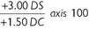
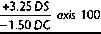
Which of the following represents the correct transposition of this lens prescription?
In Gullstrand’s schematic eye, what is the distance of the second nodal point behind the anterior corneal surface, in millimetres?
Regarding hypermetropia, which statement is LEAST likely to be correct?
Manifest hypermetropia is defined as the strongest concave lens correction accepted for clear distance vision.
Latent hypermetropia is masked by ciliary tone and involuntary accommodation.
Hypermetropia that can be overcome by accommodation is termed facultative.
Hypermetropia exceeding the amplitude of accommodation is termed absolute.
Which of the following does NOT represent an optical problem when correcting aphakia with spectacles?
Ring scotomas
Jack-in-the-box phenomenon
Heavy lenses
Barrel distortion
Regarding bifocal lenses, which statement is LEAST likely to be true?
Excessive prismatic effect at the near visual point can be particularly marked in high-powered lenses.
Prismatic jump can be minimised if the optical centres of the two lenses lie away from the junction of the distance and near portions. Split bifocals represent the earliest bifocal design.
Solid bifocals are those of single-piece construction.
Which of the following does NOT represent a stage within retinoscopy?
Illumination stage
Dark-adaptation stage
Reflex stage
Projection stage
Regarding the Javal–Schiøtz keratometer, which statement is MOST likely to be true?
The instrument uses an object of fixed size. Each mire is a small lantern with a clear window. Doubling of the image is achieved using a Porro prism.
When examining an astigmatic patient, the two images are displaced vertically in all except the two principal meridians of the cornea.
Regarding lenses for fundus examination, which statement is MOST likely to be true?
The Hruby lens is a powerful plano-convex lens.
The 90-dioptre lens provides more magnification than the 78-dioptre lens.
The panfunduscope contact lens forms a virtual, inverted image of the fundus.
The field of view of the 78-dioptre lens is less than that achieved by the panfunduscope contact lens.
What does the acronym ‘LASER’ stand for?
Laser Amplification by Stimulated Energy Radiation
Light Amplification by Stimulated Energy Radiation
Laser Amplification by Stimulated Emission of Radiation
Light Amplification by Stimulated Emission of Radiation
Regarding auto-refractors, which statement is LEAST likely to be true?
All but infrared light is filtered out.
The fixation target is designed to avoid accommodation by the patient.
The instrument detects the end point of refraction using an electronic focus detector.
The instrument performs well even in eyes with broad iridectomies.
Regarding slit lamp filters, which statement is LEAST likely to be correct?
Red light is scattered more than blue light.
The cobalt blue filter is used for applanation tonometry. The green (red-free) filter is useful for inspecting the vitreous. Blue light is of 465–490 nanometres.
16. 17.
Anatomy 1
Regarding walls of the orbit, which statement is MOST likely to be true?
The roof comprises the orbital plate of frontal bone and body of sphenoid.
The floor comprises the orbital plate of maxilla, orbital surface of zygomatic, and orbital process of palatine.
The lateral wall comprises the zygomatic bone and lesser wing of sphenoid.
The medial wall comprises the frontal process of maxilla, lacrimal bone, orbital plate of ethmoid, and greater wing of sphenoid.
18.
Regarding the eyelids, which statement is LEAST likely to be true?
The insertion of the aponeurotic fibres of the levator palpebrae superioris forms the upper eyelid sulcus.
The malar and nasojugal sulci may be present in older individuals. When the eye is closed, the upper eyelid normally covers the upper half of the cornea.
The superior palpebral sulcus divides each eyelid into an orbital and tarsal part.
19.
Regarding the orbicularis oculi muscle, which statement is LEAST likely to be true?
The orbicularis oculi muscle is supplied by temporal and zygomatic branches of the seventh cranial nerve.
The action of the palpebral portion is both voluntary and involuntary. The fibres of the palpebral portion sweep concentrically and laterally across the lids, behind the orbital septum.
The lacrimal portion draws the eyelids and papillae medially.
20.
Which nerve does NOT pass through the common tendinous ring?
Nasociliary
Abducent
Trochlear
Upper division of oculomotor
21.
Where does the optic nerve perforate the sclera, in millimetres (mm)?
3 mm medial and 1 mm above the posterior pole
1 mm medial and 3 mm above the posterior pole
3 mm lateral and 1 mm below the posterior pole
1 mm lateral and 3 mm below the posterior pole
22.
Regarding the cornea, which statement is LEAST likely to be true?
The central cornea is supplied with oxygen dissolved in the tear film. The peripheral cornea is supplied with oxygen by diffusion from the posterior ciliary blood vessels.
The long ciliary nerves provide sensory innervation to the cornea. Complete turnover of the surface epithelial cells is estimated to take 7 days.
23.
Regarding the pituitary fossa, which statement is MOST likely to be true?
Which of the following options represents the correct order of layers of the retina, from inner to outer layers?
Approximately how long is the intracranial portion of the optic nerve, in millimetres (mm)?
1 mm 5 mm 10 mm
25 mm
26. It is formed by an indentation in the ethmoid bone. It is anteriorly bound by the dorsum sellae. It is posteriorly bound by the tuberculum sellae. It is roofed by the diaphragma sella.
Which of the following represents the correct order of structures of the anterior chamber angle as seen on gonioscopy, from posterior to anterior?
Iris, ciliary body, scleral spur, trabecular meshwork, Schwalbe’s line
Iris, scleral spur, ciliary body, trabecular meshwork, Schwalbe’s line
Regarding classification of the extraocular muscles, which statement is LEAST likely to be true?
Type B muscle fibres possess a smaller diameter than type A muscle fibres.
Type C muscle fibres align both visual axes with fine local contractions.
Type B muscle fibres possess multiple end plates. Type A muscle fibres are mainly involved in smooth pursuit movements.
What is the secondary action of the superior oblique muscle?
Intorsion Extorsion
Abduction Adduction
Regarding the lateral geniculate nucleus, which statement is LEAST likely to be true?
It is located on the undersurface of the pulvinar of the thalamus. It receives the caudal termination of the lateral root of the optic tract. The contralateral eye sends visual information to layers 2, 3, and 5 of the lateral geniculate nucleus. Its inner two layers are magnocellular layers, while its outer four layers are parvocellular layers.
On what day of embryological development can the lens placode be identified?
Physiology 1
Physiology of the eye and vision
What is the approximate thickness of the precorneal tear film in micrometres (μm)?
3.4 μm
6.8 μm
13.4 μm
16.8 μm
Which of the following represents the unconventional outflow pathway of aqueous humour at the anterior chamber angle?
What shaped suture is formed by the lens fibres meeting posteriorly?
What is the resting membrane potential of a dark-adapted rod cell, in millivolts (mV)?
−40 mV
−70 mV
−100 mV
−130 mV
Regarding visual acuity, which statement is LEAST likely to be true?
Visual acuity testing measures an individual’s ability to discriminate two stimuli separated in space.
Bloch’s law assumes a monotonic decrease in perceived contrast with increased duration.
The Broca–Sulzer effect assumes a peak in perceived contrast with prolonged duration.
At 5° from the central fovea, visual acuity drops to 25% of foveal acuity.
36.
Regarding the pupillary light reflex, which statement is MOST likely to be true?
The pupillary light reflex varies the amount of retinal illumination in response to changes in lighting.
Pupil input from retinal ganglion cells leaves the optic tract in the brachium of the inferior colliculus.
Neurons in the olivary pretectal nucleus send crossed and uncrossed fibres via the posterior commissure to the Edinger–Westphal nucleus.
Postganglionic parasympathetic neurons pass from the ciliary ganglion via the long ciliary nerves to the iris sphincter muscle.
Deutan defects are most commonly associated with the absence of which gene?
OPN1DW
OPN1SW
OPN1LW
OPN1MW
General physiology
38.
Regarding action potentials, which statement is MOST likely to be true?
There is a positive membrane potential present during the resting stage.
During depolarisation, the nerve fibre membrane becomes permeable to sodium ions. The potassium channels close during the repolarisation stage. In smaller nerve fibres and many central nervous system neurons, the membrane potential overshoots above the zero level during depolarisation.
39.
Regarding cardiac arrhythmias, which statement is MOST likely to be true?
Tachycardia is defined as a heart rate faster than 90 beats per minute.
Second-degree type II atrioventricular (AV) block is characterised by a progressive prolongation of the PR interval until a ventricular beat is skipped.
Third-degree AV block typically features a fixed number of nonconducted P waves per QRS complex.
Atrial fibrillation reduces the efficiency of ventricular pumping by 20–30%.
40.
Regarding sensory receptors, which of the following does NOT cause receptor potentials?
Application of a chemical
Effects of electromagnetic radiation
Closure of ion channels
Mechanical deformation
41.
Which of the following hormones is NOT secreted by the anterior pituitary gland?
Prolactin
Oxytocin
Thyroid-stimulating hormone
Growth hormone
Biochemistry
42.
Regarding the nucleus, which statement is LEAST likely to be true?
The nuclear membrane contains receptors for ligands.
Heterochromatin is less packed than euchromatin.
Chromatin is the main nuclear component.
The nucleolus is the site of ribosomal RNA synthesis and substantial transcriptional activity.
43.
Regarding the biochemical properties of the vitreous, which statement is LEAST likely to be true?
Collagen type II is predominantly responsible for the gel structure of the vitreous body.
The central vitreous body possesses a higher concentration of hyaluronan and collagen as compared to the cortex.
Floaters become visible with age when the hyaluronan molecules degrade into smaller moieties while the collagen fibrils coagment, forming larger fibrils.
Opticin possesses a role in regulating fibril thickness within the vitreous body.
Pathology 1
General and ocular pathology
44.
Regarding acute inflammation, which statement is MOST likely to be correct?
Following transendothelial migration and extravasation, the movement of leucocytes is subsequently controlled by expression. Chemotactic agents are released from cytokines, complement components, leukotrienes, or pathogenic bacteria.
Phagocytosis involves opsonisation of bacteria by leucocytes followed by engulfment within complement components.
Histamine and leukotrienes decrease vascular permeability
45.
Regarding the healing and repair of ocular structures, which statement is LEAST likely to be correct?
Following trauma, reactive proliferation of the iris pigment epithelium may take place.
Scar tissue in the sclera is derived from scleral fibroblasts.
Axonal loss and demyelination occur following trauma to the optic nerve.
The limbus is the site at which the corneal epithelium regenerates.
Regarding cataractogenesis, which statement is LEAST likely to be true?
Lens crystallins break down to albuminoids. Amino acids are converted to adrenaline and melanin. There is reduced absorption of blue light.
Nuclear sclerosis and loss of zonule elasticity contribute to presbyopia.
46. 47.
Regarding Sjögren’s syndrome, which statement is LEAST likely to be true?
Individuals with primary Sjögren’s syndrome possess specific antibodies (anti-Rho, anti-La).
Xerostomia is a prevalent clinical feature.
Affected glands include the glands of Wolfring and Krause. There is a proliferation of goblet cells in the conjunctival epithelium.
48.
Regarding vitelliform dystrophy, which statement is LEAST likely to be true?
Classic fundoscopic findings include an egg-yolk lesion with satellite lesions.
Peripherin 2 is required for normal photoreceptor function.
Vitelliform macular dystrophy is caused by mutations in BEST1 and PRPH2.
There is an accumulation of lipofuscin in the outer nuclear layer and atrophy of the photoreceptor layer
49.
Which of the following stains is most useful for identifying macular corneal dystrophy?
Oil Red O
Masson trichrome
Alcian blue
Congo red
50.
Regarding herpes simplex keratitis, which statement is MOST likely to be true?
Type 1 herpes simplex virus can cause dendritic ulceration within the corneal endothelium.
Primary infection rarely occurs through the oral mucosa. Complications of herpes simplex infection of the eye include disciform keratitis and secondary lipid keratopathy. When reactivated, the virus can produce vesicle formation in the skin.
51.
Which of the following does NOT represent a common primary site for choroidal metastasis?
Prostate Breast Microbiology
52.
Approximately what proportion of total white cells is constituted by neutrophils, in normal individuals?
What is the MOST common cause of postoperative pseudophakic endophthalmitis?
Staphylococcus epidermidis
Escherichia coli
Propionibacterium acnes
Staphylococcus aureus
What type of parasite is Toxocara canis?
Trematode
Nematode
Cestode
Protozoan
Immunology
Which of the following is NOT a cell of the mononuclear phagocyte system?