Pediatric Emergency Radiology
Edited by Ann Dietrich Associate Editor
Gayathri Sreedher
Oxford University Press is a department of the University of Oxford. It furthers the University’s objective of excellence in research, scholarship, and education by publishing worldwide. Oxford is a registered trade mark of Oxford University Press in the UK and certain other countries.
Published in the United States of America by Oxford University Press 198 Madison Avenue, New York, NY 10016, United States of America.
© Oxford University Press 2023
All rights reserved. No part of this publication may be reproduced, stored in a retrieval system, or transmitted, in any form or by any means, without the prior permission in writing of Oxford University Press, or as expressly permitted by law, by license, or under terms agreed with the appropriate reproduction rights organization. Inquiries concerning reproduction outside the scope of the above should be sent to the Rights Department, Oxford University Press, at the address above.
You must not circulate this work in any other form and you must impose this same condition on any acquirer.
Library of Congress Cataloging-in-Publication Data
Names: Dietrich, Ann M., editor. | Sreedher, Gayathri, editor.
Title: Pediatric emergency radiology / editor, Ann Dietrich; associate editor, Gayathri Sreedher.
Description: New York, NY : Oxford University Press, [2023] | Includes bibliographical references and index.
Identifiers: LCCN 2022040656 (print) | LCCN 2022040657 (ebook) | ISBN 9780197628553 (paperback) | ISBN 9780197628577 (epub) | ISBN 9780197628584 (online)
Subjects: MESH: Emergencies | Child | Infant | Radiology | Pediatric Emergency Medicine | Case Reports
Classification: LCC RJ370 (print) | LCC RJ370 (ebook) | NLM WS 205 | DDC 618.92/0025—dc23/eng/20230103
LC record available at https://lccn.loc.gov/2022040656
LC ebook record available at https://lccn.loc.gov/2022040657
DOI: 10.1093/med/9780197628553.001.0001
This material is not intended to be, and should not be considered, a substitute for medical or other professional advice. Treatment for the conditions described in this material is highly dependent on the individual circumstances. And, while this material is designed to offer accurate information with respect to the subject matter covered and to be current as of the time it was written, research and knowledge about medical and health issues is constantly evolving and dose schedules for medications are being revised continually, with new side effects recognized and accounted for regularly. Readers must therefore always check the product information and clinical procedures with the most up-to-date published product information and data sheets provided by the manufacturers and the most recent codes of conduct and safety regulation. The publisher and the authors make no representations or warranties to readers, express or implied, as to the accuracy or completeness of this material. Without limiting the foregoing, the publisher and the authors make no representations or warranties as to the accuracy or efficacy of the drug dosages mentioned in the material. The authors and the publisher do not accept, and expressly disclaim, any responsibility for any liability, loss, or risk that may be claimed or incurred as a consequence of the use and/or application of any of the contents of this material.
9 8 7 6 5 4 3 2 1
Printed by Marquis, Canada
Contents Contributors vii
1. Walking and Wheezing 1
Rajesh Krishnamurthy and Beth Bubolz
2. Fast-Breathing Baby 9
Ajay K. Puri, Mantosh S. Rattan, and Melissa A. McGuire
3. Green Vomit . . . Ewwww!!! 25
Esther Ro, Yamini Jadcherla, and Maegan S. Reynolds
4. What’s That in the Diaper? 37
Maegan S. Reynolds, Yamini Jadcherla, and Esben Vogelius
5. My Baby Won’t Poop! 49
Yamini Jadcherla, Narendra Shet, and Maegan S. Reynolds
6. Breathing through a Straw 59
Gina Pizzitola, David Teng, and Peter Assaad
7. Drooling, Drooling, Drooling All the Way Home 67
Brooke Lampl, Nkeiruka Orajiaka, and Meika Eby
8. Burn, Baby, Burn 75
Cory Gotowka, Ailish Coblentz, and Michael Stoner
9. To Wheeze or Not to Wheeze 89
Linda Vachon and Emily Rose
10. I Thought They Fixed Me? 101
Gina Pizzitola, William Mak, and Rachelle Goldfisher
11. Weebly, Wobbly 109
Meika Eby, Nkeiruka Orajiaka, and Lauren May
12. Extreme Extremity 117
Waroot S. Nimjareansuk and Jane S. Kim
13. FOOSHING through the Snow . . . 127
David Fernandez, Bindu N. Setty, and Isabel A. Barata
14. Sentinel Injuries: It’s What’s Inside That Counts 137
Madeline Zito, Summit Shah, Isabel A. Barata, and Dana Kaplan
15. What a Pain in the Neck 151
Michael Sperandeo and Gayathri Sreedher
16. Torticollis in a Child with Down Syndrome 165
Kristy Williamson and Rachelle Goldfisher
17. Shouldn’t This Be Connected? 175
Joshua Rocker and Anna Thomas
18. Head First into the Pool 185
Aaron McAllister and Kristol Das
19. Pain in the Back 195
Deanna Margius, Sophia Gorgens, David Foster, and Shankar Srinivas Ganapathy
20. Why Is This Infant So Fussy? 205
Michelle Greene, Anna Thomas, and Berkeley Bennett
21. Ripping Apart My Heart 221
Thomas P. Conway, George C. Koberlein, and Francesca M. Bullaro
22. The Unlucky Hit! 231
McKenzie Montana, Robert L. Gates, and Zachary Burroughs
Index 247
Contributors
Peter Assaad, MD, MPH, MBA
Lurie Children’s Hospital Chicago, IL, USA
Isabel A. Barata, MS, MD, MBA Professor of Pediatrics and Emergency Medicine Department of Emergency Medicine
Donald and Barbara Zucker School of Medicine at Hofstra/Northwell
Ozone Park, NY, USA
Berkeley Bennett, MD, MS
Attending Physician
Department of Emergency
Nationwide Children’s Hospital, The Ohio State University Columbus, OH, USA
Beth Bubolz, MD
Assistant Professor Department of Pediatrics
The Ohio State University College of Medicine
Lewis Center, OH, USA
Francesca M. Bullaro, MD
Associate Trauma Medical Director, Assistant Professor
Pediatric Emergency Medicine
Cohen Children’s Medical Center Northwell Health
Garden City, NY, USA
Zachary Burroughs, MD
Clinical Assistant Professor
Emergency Medicine, Division of Pediatric Emergency Medicine
Prisma Health Upstate Greenville, SC, USA
Ailish Coblentz, MBBS, FRCPC
Assistant Professor and Paediatric
Radiologist
Department of Diagnostic Imaging
The Hospital for Sick Children, University of Toronto Toronto, ON, CA
Thomas P. Conway, DO Physician Fellow Department of Pediatric Emergency Medicine
Cohen Children’s Medical Center
Astoria, NY, USA
Kristol Das, MD Fellow
Department of Pediatric Emergency Medicine
Nationwide Children’s Hospital Columbus, OH, USA
Ann Dietrich, MD Professor of Pediatrics and Emergency Medicine, University of South Carolina College of Medicine
Department of Emergency Medicine
Division Chief Pediatric Emergency Medicine, PRISMA
Greenville, SC, USA
Meika Eby, MD, FAAP
Department of Pediatrics
Nationwide Children’s Hospital, The Ohio State University
Columbus, OH, USA
David Fernandez, MD
Chief Resident-Education
Department of Emergency Medicine
Northwell: Northshore-LIJ Hospital
Astoria, NY, USA
David Foster, MD, MS
Assistant Professor
Departments of Emergency Medicine and Pediatrics
Associate Director EM Residency Program, Northwell Health, Zucker School of Medicine Manhasset, NY, USA
Shankar Srinivas Ganapathy, MBBS, MD
Pediatric Radiologist, Associate Professor of Radiology at NEOMED
Department of Radiology
Akron Children’s Hospital Akron, OH, USA
Robert L. Gates, MD
Professor
Department of Surgery
University of South Carolina School of Medicine—Greenville
Greenville, SC, USA
Rachelle Goldfisher, MD
Associate Chief
Department of Pediatric Radiology
Northwell Health
Queens, NY, USA
Sophia Gorgens, MD
Resident
Department of Emergency Medicine
Zucker-Northwell North Shore/ Long Island Jewish Manhasset, NY, USA
Cory Gotowka, DO, MS
Pediatric Hospitalist
UPMC Hamot Erie, PA
Michelle Greene, DO Assistant Professor
Division of Emergency Medicine, Department of Pediatrics
The Ohio State University College of Medicine, and Nationwide Children’s Hospital, Columbus, OH Columbus, OH, USA
Yamini Jadcherla, MD
Pediatric Emergency Medicine Fellow
Department of Pediatric Emergency Medicine
Nationwide Children’s Hospital Galena, OH, USA
Dana Kaplan, MD
Director of Child Abuse and Neglect Department of Pediatrics
Staten Island University Hospital
Marlboro, NJ, USA
Jane S. Kim, MD
Assistant Professor of Pediatrics and Radiology Division of Diagnostic Imaging and Radiology
Children’s National Hospital
Kensington, MD, USA
George C. Koberlein, MD
Associate Professor Department of Radiology
Atrium Health at Wake Forest Baptist Winston-Salem, NC, USA
Rajesh Krishnamurthy, MD
Director, Cardiothoracic Imaging Department of Radiology
Nationwide Children’s Hospital Dublin, OH, USA
Brooke Lampl, DO, FAOCR
Staff Radiologist/Clinical Assistant Professor of Radiology Department of Diagnostic Radiology
Cleveland Clinic
Pepper Pike, OH, USA
William Mak, DO
Attending Physician Department of Pediatrics
Cohen Children’s Medical Center
Flushing, NY, USA
Deanna Margius, MD
Resident
Department of Emergency Medicine
Hofstra Northwell NS-LIJ
Manhasset, NY, USA
Lauren May, MD
Nemours Children’s Hospital, Delaware, Wilmington, DE
Clinical Assistant Professor Department of Radiology and Pediatrics
Sidney Kimmel School of Medicine
Thomas Jefferson University Philadelphia, PA
Aaron McAllister, MS, MD
Assistant Professor Department of Radiology
Nationwide Children’s Hospital Columbus, OH, USA
Melissa A. McGuire, MD
Assistant Professor and Associate Program Director of Northwell Health CCMC Pediatric Emergency Medicine Fellowship Department of Emergency Medicine Northwell Health, Cohen Children’s Medical Center and North Shore University Hospital Massapequa Park, NY, USA
McKenzie Montana, DO
Resident Physician Department of Pediatrics Prisma Health-Upstate
Greenville, SC, USA
Waroot S. Nimjareansuk, DO
Assistant Professor
Department of Orthopaedics and Sports Medicine
University of Florida
Minneola, FL, USA
Nkeiruka Orajiaka, MBBS, MPH
Pediatrician, Assistant Professor
Department of Emergency Medicine
Nationwide Children’s Hospital, The Ohio State University Columbus, OH, USA
Gina Pizzitola, MD, MS
Pediatric Emergency Medicine Fellow
Department of Pediatric Emergency Medicine
Cohen Children’s Medical Center, Northwell Health
Glen Oaks, NY, USA
Ajay K. Puri, MD Fellow, Montefiore Medical Center
Critical Care Medicine North Shore University Hospital
Forest Hills, NY, USA
Mantosh S. Rattan, MD
Staff Radiologist
Department of Radiology
Cleveland Clinic Foundation Cincinnati, OH, USA
Maegan S. Reynolds, MD
Assistant Professor of Emergency Medicine and Pediatrics Department of Emergency
Medicine The Ohio State University and Division of Pediatric Emergency Medicine Nationwide Children’s Fracture Columbus, OH, USA
Esther Ro, MD
Instructor of Radiology Department of Radiology
Northwestern University Feinberg School of Medicine
Wilmette, IL, USA
Joshua Rocker, MD
Associate Professor and Division Chief Department of Pediatric Emergency Medicine
Cohen Children’s Medical Center New Hyde Park, NY, USA
Emily Rose, MD
Associate Professor of Clinical Emergency Medicine (Educational Scholar) Department of Emergency Medicine
Keck School of Medicine of the University of Southern California Arcadia, CA, USA
Bindu N. Setty, MD
Clinical Associate Professor
Department of Radiology
Boston University Lexington, MA, USA
Summit Shah, MD, MPH
Pediatric Radiologist
Department of Radiology
Nationwide Children’s Hospital Columbus, OH, USA
Narendra Shet, MD
Associate Professor and Director of Body MR
Division of Diagnostic Imaging and Radiology
Children’s National Hospital
Ellicott City, MD, USA
Michael Sperandeo, MD
Attending Physician
Department of Emergency Medicine
Long Island Jewish Medical Center
Long Beach, NY, USA
Gayathri Sreedher, MBBS, MD
Staff Pediatric Radiologist
Department of Radiology
Akron Children’s Hospital Akron, OH, USA
Michael Stoner, MD
Associate Professor and Section Chief
Department of Pediatrics/Division of Emergency Medicine
The Ohio State University College of Medicine and Nationwide Children’s Hospital
Canal Winchester, OH, USA
David Teng, MD
Assistant Professor
Department of Pediatrics, Division of Emergency Medicine
Cohen Children’s Medical Center—Northwell Health
Dix Hills, NY, USA
Anna Thomas, MD
Clinical Associate Professor Department of Radiology
Children’s Hospital of Los Angeles, USC Keck School of Medicine
Redlands, CA, USA
Linda Vachon, MD
Associate Professor of Clinical Radiology
Department of Radiology
University of Southern California, Keck School of Medicine
Manhattan Beach, CA, USA
Esben Vogelius, MD
Radiologist
Department of Radiology
Cleveland Clinic
Moreland Hills, OH, USA
Kristy Williamson, MD
Associate Chief
Department of Pediatric Emergency
Cohen Children’s Medical Center
Garden City, NY, USA
Madeline Zito, DO, FAAP
Department of Pediatrics
Maimonides Children’s Hospital
Brooklyn, NY, USA
1 Walking and Wheezing
Rajesh Krishnamurthy and Beth Bubolz
Case Study
A 16-year-old male with no past medical history and a recent viral infection (not COVID) presents with 1 month of cough, shortness of breath, and wheezing with walking. He must frequently stop and rest when walking outside, has trouble going up the stairs, and has stopped participating in sports. He also has decreased appetite with associated nausea, vomiting, and abdominal pain off and on for a few weeks.
He was seen by his pediatrician 2 weeks ago for vague abdominal pain, nausea and diarrhea and was diagnosed with gastroenteritis and was discharged with ondansetron. Vital signs and physical examination at that visit were normal.
His cough is currently dry. Vital signs are: blood pressure 100/67mmHg; pulse 106 beats per minute; temperature 97.8°F (36.6°C); respiratory rate 20 breaths per minute; weight 62.4 kg; oxygen saturation (SpO2) 93%. He is well-appearing. Lungs are clear and his heart sounds are normal, with no murmur. Abdominal exam reveals no pain or masses, with a normal sized liver.
While completing the exam, he vomits, becomes pale, diaphoretic, and tachypneic.
What do you do now?
DISCUSSION
An adolescent with cough, shortness of breath, and wheezing has a differential that includes asthma, pneumothorax, pneumonia, achalasia, myocarditis, and congestive heart failure (HF).
Patients with asthma, a chronic inflammatory disease of the airways, typically have recurrent episodes of airflow obstruction resulting from edema, bronchospasm, and increased mucus production in the airways. Children frequently have associated seasonal allergies (allergic rhinitis) and eczema (atopic dermatitis), with these three conditions forming what is known as the atopic triad. This patient does not have a history of allergic diseases or wheezing and on physical exam is not currently wheezing.
Children have an increased risk of pneumothorax with a previous history of an emphysematous bleb, asthma (10%), and tobacco use (4%). An underlying congenital anomaly might also serve as a predisposing factor, particularly in a younger child. Young males with Marfan syndrome, the most common inherited disorder of connective tissue, are also more likely to have a spontaneous pneumothorax. Clinically, children usually present with the acute onset of pain at rest or during physical activity and, depending on the size of the pneumothorax, might have dyspnea and cough. Physical exam findings may reveal differences in air entry to the lungs; however, with a small pneumothorax, unequal breath sounds, hyperresonance with percussion, and asymmetric wall movements might be subtle or not yet present. This child does not have risk factors or findings associated with a pneumothorax.
Achalasia is a rare esophageal neurodegenerative disorder that may occur in the pediatric population. Achalasia may be associated with other conditions including Trisomy 21, congenital hypoventilation syndrome, glucocorticoid insufficiency, eosinophilic esophagitis, familial dysautonomia, Chagas’ disease, and achalasia, alacrima, and adrenocorticotropic hormone (ACTH) insensitivity (AAA) syndrome. Children afflicted with achalasia usually present with progressive dysphagia, vomiting, and weight loss. Recurrent pneumonia, nocturnal cough, aspiration, hoarseness, and feeding difficulties may occur in younger children. Although this patient has vomiting, he does not have progressive dysphagia, making this diagnosis unlikely.
Usually, acute myocarditis presents with preceding viral symptoms (about two-thirds of patients), HF symptoms, and a poorly functioning ventricle with or without dilation. Fulminant myocarditis may present with tachyarrhythmias and significant cardiac dysfunction. A history of a preceding viral prodrome is commonly present (two-thirds of patients); ventricular and atrial arrhythmias are also common (about 45%). Sudden cardiac death may also be a presentation of myocarditis. Complications of myocarditis may include a dilated cardiomyopathy or a pericardial effusion.
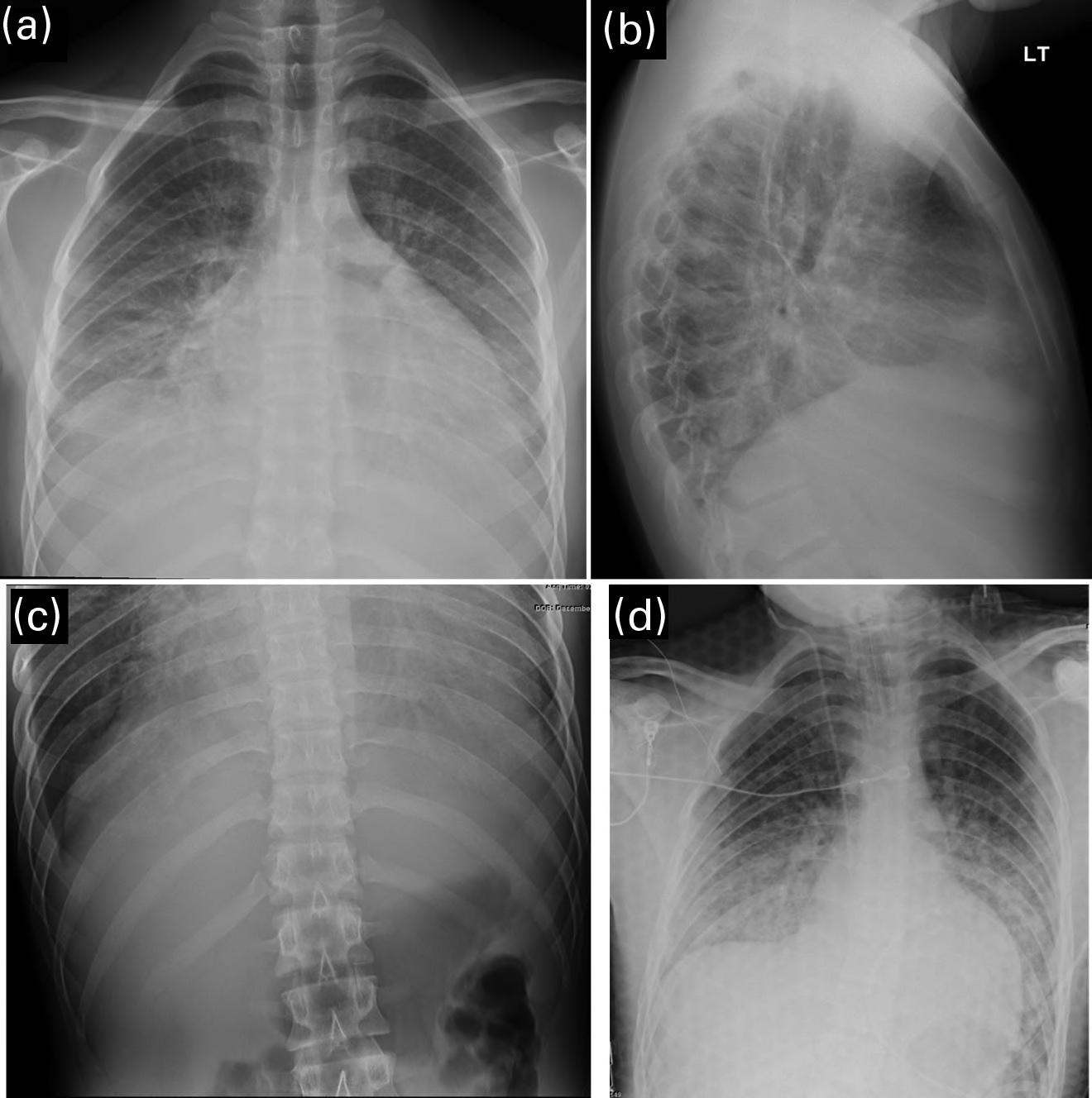
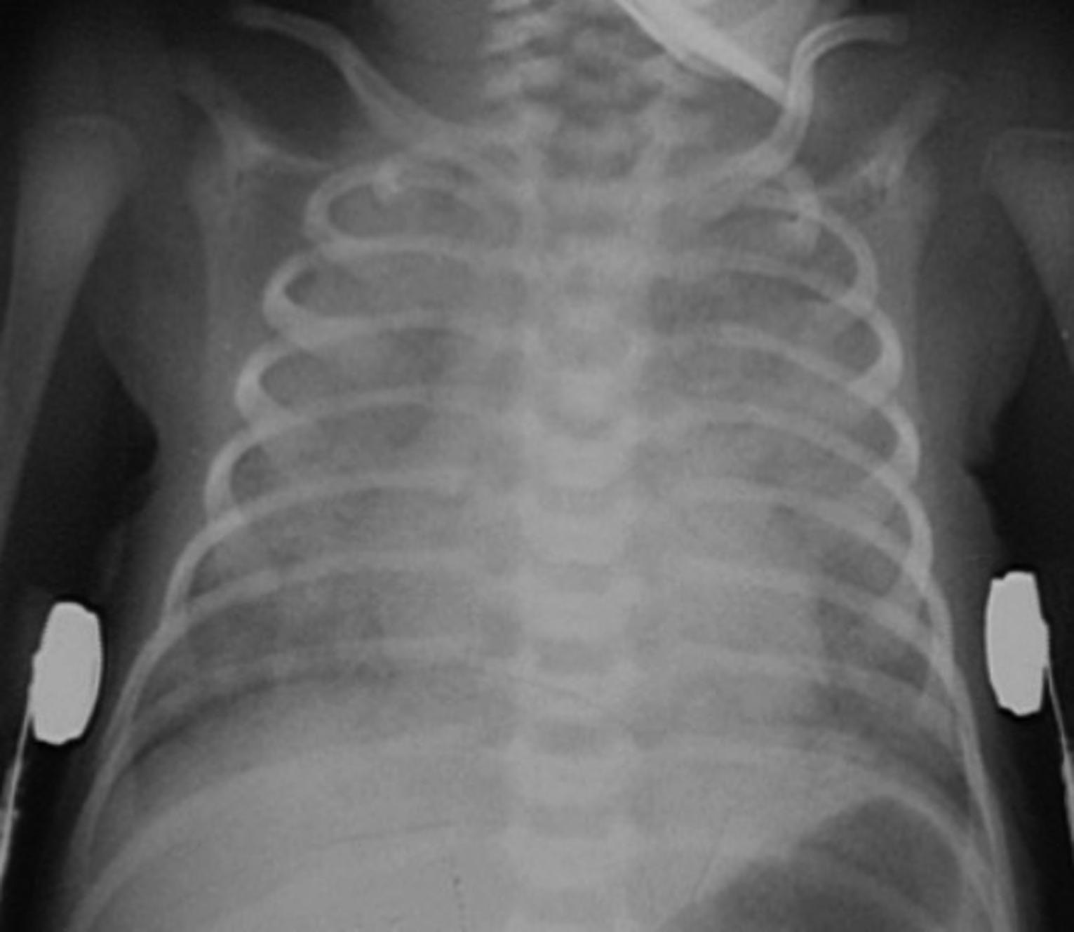
The clinicians for this patient started with a chest radiograph. The initial chest radiographic identified small pleural effusions and interlobular septal thickening, which were suggestive of interstitial pulmonary edema (see Figures 1.1a and 1.1b). Findings become more apparent when compared with a normal 16-year-old’s chest radiographs (Figures 1.2a and 1.2b). Because of the patient’s vomiting, an abdominal film was also obtained, which demonstrated significant cardiomegaly (see Figure 1.1c), thus leading to the diagnosis of dilated cardiomyopathy and/or pericardial effusion. On the chest radiograph, the reduced lung volumes and elevated hemidiaphragms masked the cardiomegaly, which was more apparent on the better penetrated abdominal radiograph.
Careful attention to the heart, mediastinum, airway, lungs, pleura, bones, and soft tissues is essential to accurately diagnose the cause of chest pain. A screening chest film for patients with chest pain has low sensitivity for structural cardiovascular lesions, such as myocarditis, dissection, or pulmonary infarction, but is helpful in the acute setting to diagnose complications of underlying cardiovascular conditions, such as HF, mediastinal hematoma, or pulmonary infarction. It is also helpful to exclude noncardiac causes of chest pain, including pneumonia, pneumothorax, rib fracture, or an aspirated foreign body (see normal chest radiograph, Figures 1.2a and 1.2b).
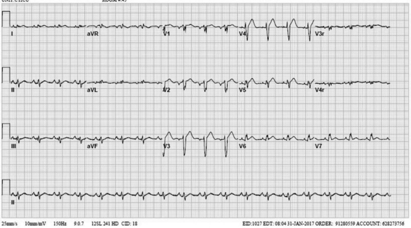
In addition, the physician ordered an electrocardiogram (ECG). An ECG with a chest radiograph is a great screening tool set for cardiomyopathy of any type. ECGs are particularly valuable in patients who present with symptoms that may be suspicious for cardiac involvement and include dyspnea, fatigue, shortness of breath, tachypnea, unexplained tachycardia, murmur, gallop, rub, vague abdominal pain, etc.
ECG demonstrates sinus tachycardia, low voltage, borderline long QT interval, and mild conduction delay in V1.
FIGURES 1.1 a, 1.1b, 1.1c, AND 1.1d . (a, b) PA and lateral views of the chest demonstrating low lung volumes, pulmonary vascular congestion, interstitial opacities in the lung bases, and bilateral trace pleural fluid; (c) the abdominal radiograph is better penetrated than the chest radiograph, and shows an enlarged cardiac silhouette; and (d) post-procedure chest radiograph showing decreased size of the cardiac silhouette after drainage of the pericardial effusion.
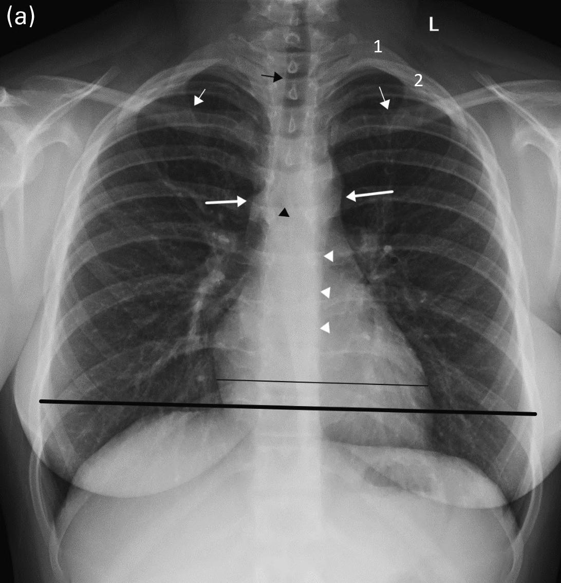

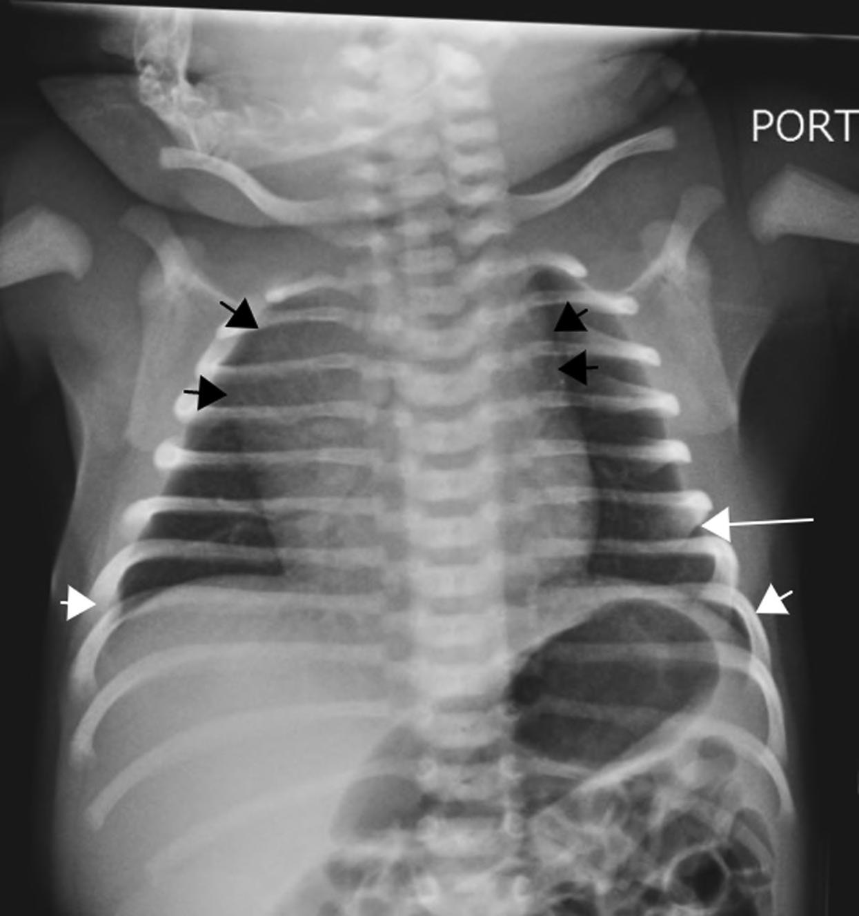
FIGURES 1.2 a AND 1.2 b PA and lateral views of the chest in a normal 16-year-old for comparison (different patient). Note the normal appearance of structures. Contour of descending aorta (white arrowheads), superior mediastinal width between superior vena cava on the right and aortic arch on the left (white thick arrows on right and left side), tracheal air column (black arrow), carina (black arrowhead), and the orientation of the clavicles (thin white arrows). In a properly positioned patient the spinous processes lie midway between the medial ends of the clavicles. The cardiothoracic ratio is measured as the ratio of the cardiac transverse diameter (black thin line) divided by the maximum chest transverse diameter (thick black line). The first and second ribs on the left side are numbered (1, 2).
In general, electrocardiographic findings may include sinus tachycardia, dysrhythmia, low voltage, widened QRS, or repolarization abnormalities, with atrial or ventricular dysrhythmias leading to sudden death. Pathologic Q waves may be seen, and were found in one study with parvovirus B19 myocarditis.
Other tests that may help facilitate a timely diagnosis include a bedside ultrasound (US) for anatomic and functional evaluation, as well as assessment for pericardial fluid. This patient had an echocardiogram that showed a pericardial effusion, which was subsequently drained. A repeat chest radiograph shows the heart following the procedure (see Figure 1.1d).
Other tests that may be considered include creatine kinase MB, troponin, and BNP. Troponin I and troponin T, although not a sensitive or specific marker of myocarditis, may be elevated in acute myocarditis. Higher troponin levels have been associated with the need for extracorporeal membrane oxygenation (ECMO) and mortality. BNP and NT-proBNP are generally related to HF, not myocarditis, and are typically elevated and are associated with cardiac dysfunction and acute HF.
The chief complaints of adolescents with dilated cardiomyopathy following myocarditis rarely involve the cardiovascular system. The clinical presentation is characterized by multiple encounters with the healthcare system for nonspecific symptoms, antecedent viral infection, nonspecific respiratory and gastrointestinal symptoms, and fatigue which may or may not be exertional. The key symptom which is often overlooked or discounted is vague abdominal pain accompanied by nausea with minimal vomiting.
Dilated cardiomyopathy is notoriously difficult to diagnose in pediatrics. Young children often have chief complaints referable to the respiratory system, and adolescents present with abdominal pain. Therefore, a high index of suspicion is necessary to make an accurate diagnosis.
CASE CONC l USIONS
In the current case, the patient’s final diagnosis was dilated cardiomyopathy and pericardial effusion following myocarditis. He was admitted to the hospital, and an LVAD (left ventricular assist device) was placed 3 days later. He underwent cardiac transplant about 2 months later but unfortunately died due to accelerated rejection and infection shortly thereafter.
k EY POINTS
• Adolescents with cardiomyopathy/HF often have persistent, vague abdominal pain with minimal nausea and multiple visits.
• Young children with dilated cardiomyopathy often have respiratory symptoms such as tachypnea or wheezing.
• Patients with cardiomyopathy/HF may only have subtle signs of their disease and may rapidly decompensate.
• Cardiomyopathy and HF may predispose patients to fatal arrhythmias.
• Cardiac involvement in viral illnesses is common and may often go unnoticed. It can, in rare cases, have substantial acute hemodynamic and clinical sequelae, including pericardial effusion, dilated cardiomyopathy, and HF.
TIPS FROM THE RADIO l OGIST
• Plain radiographs may be the first clue to the presence of a pericardial effusion, cardiomyopathy, or HF.
• Careful attention to the heart, mediastinum, airway, lungs, pleura, bones, and soft tissues on a pediatric chest x-ray is essential to accurately diagnose the cause of chest pain.
• Bedside US or an echocardiogram of the heart may reveal complications of myocarditis, including dilated cardiomyopathy and pericardial effusion.
Further Reading
1. Law YM, et al. Diagnosis and management of myocarditis in children: a scientific statement from the American Heart Association. Circulation. 2021;144:e123–e135. https://www.ahajournals.org/doi/10.1161/CIR.00000 0000 0001001
WA lk ING AND W HEE z ING
2 Fast-Breathing Baby
Ajay K. Puri, Mantosh S. Rattan, and Melissa A. McGuire
Case Study
A 7-day-old female patient is brought to your emergency department by ambulance for difficulty breathing. She was born via spontaneous vaginal delivery at 37 weeks gestation and had an uncomplicated hospital stay and was discharged at the end of day 2 post-delivery. Over the past day she began developing a cough and fever to 101°F, decreased feeding, and “fast breathing”. Her vital signs are significant for a heart rate of 180 beats per minute, a rectal temperature of 102.1°F, a blood pressure of 78/44mmHg, a respiratory rate of 70 breaths per minute, and an oxygen saturation of 85% on room air. On physical examination she appears lethargic. She is tachypneic with retractions bilaterally, slightly dry mucous membranes, and coarse breath sounds over the right lower lobe. Despite supplemental oxygen via nasal cannula at 6 liters per minute, her oxygen saturation only increases to 91%. Her chest radiograph (CXR) is seen in Figure 2.1.
What do you do now?
INTRODUCTION
Respiratory diseases are the leading conditions resulting in neonatal ICU admission in both preterm and term infants. Furthermore, respiratory distress of varying severity occurs in approximately 7% of neonates. As such, it is important for the emergency physician to be able to recognize and manage neonatal respiratory distress in its various presentations.
SIGNS AND SYMPTOMS
Signs of neonatal respiratory distress include:
• Tachypnea (respiratory rate >60 breaths per minute)
• Tachycardia (heart rate >160 beats per minute)
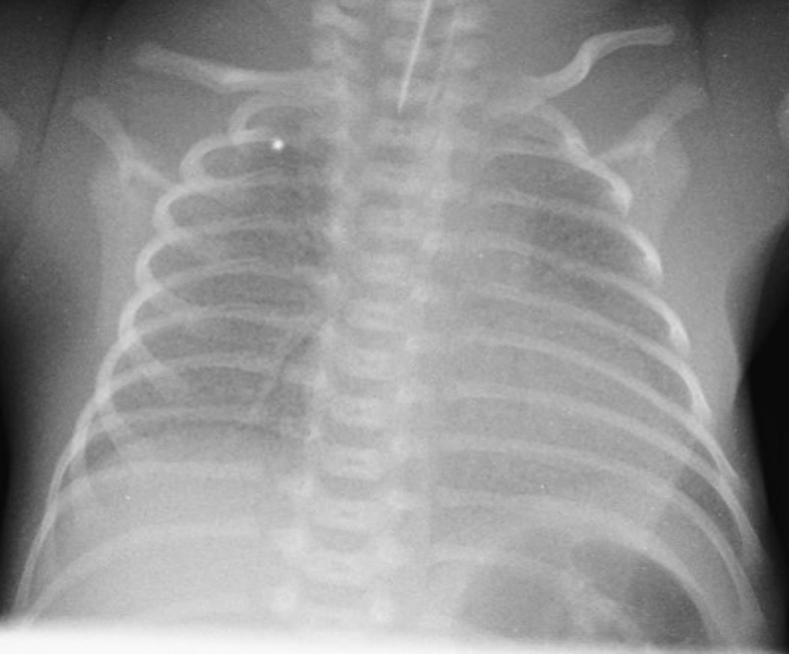
FIGURE 2.1. AP view of the chest demonstrates bilateral diffuse pulmonary air space opacification.
• Nasal flaring
• Grunting
• Chest wall retractions
• Cyanosis
• Apnea
Tachypnea is the most common presenting sign of a neonate in respiratory distress. Subtler symptoms include lethargy and poor feeding. Associated presenting signs may include hypothermia and hypoglycemia.
EMERGENCY MANAGEMENT
All neonates with respiratory distress should be placed on a continuous cardiac monitor and continuous pulse oximetry. Emergency management is directed at reversing hypoxia with oxygen supplementation and preventing or reversing respiratory acidosis by ensuring adequate ventilation. This may require support with humidified high flow nasal cannula (HHFNC) or with noninvasive positive pressure ventilation (NIPPV) such as continuous positive airway pressure (CPAP). Infants with evidence of upper airway obstruction with secretions should be suctioned beginning with a bulb-suction syringe. Patients with moderate to severe symptoms should have intravenous access obtained as they should be kept nil per os (NPO). Strong consideration to giving an initial bolus to replace losses already incurred should be made prior to starting maintenance intravenous fluids containing dextrose. A complete blood count, basic metabolic panel, a blood gas sample, and a CXR should also be performed to better determine the underlying pathology. There is a low threshold to begin antibiotic therapy in the ill-appearing infant with respiratory distress due to the difficulty in excluding bacterial infections.
Infants that are apneic, lose their airway protective reflexes such as a gag or cough, have continued respiratory failure despite NIPPV, or in whom chest compressions are started should be endotracheally intubated. Intubated patients will require contact with a tertiary care neonatal or pediatric intensive care team to assist with ventilator settings and/or for transfer to be arranged.
Neonatologists or pediatric intensivists should be involved early in the care of an ill neonate. A nonexhaustive list for when to involve a neonatologist for respiratory distress includes (adapted from Box 1 in Pramanik et al.):
• Inability to stabilize or ventilate an infant
• Requirement for vasopressors
• Cardiac disease suspected
• Meconium aspiration
• Sepsis with pneumonia
• Pneumothorax or pneumomediastinum.
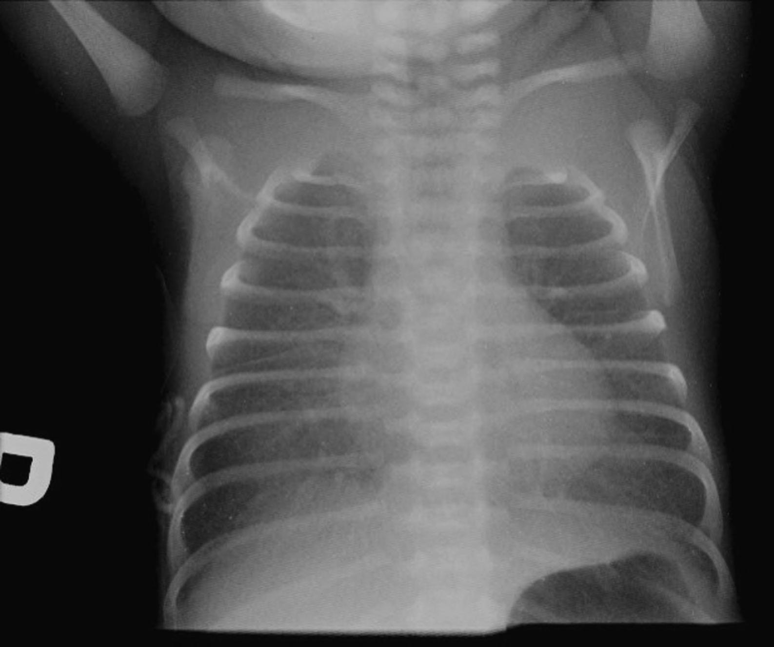
2.2. A normal neonatal chest radiograph. Note the sharp costophrenic angles (small white arrows), the aerated lung extending below the level of the 6th rib anteriorly (long white arrow), and the contour of a normal for age thymus expanding the superior mediastinum (small black arrows).
FIGURE
THE NORMA l NEONATA l CHEST RADIOGRAPH
The following should be evaluated systematically when approaching any neonatal CXR:
• Symmetric aeration—determined by visualization of the 6th rib anteriorly and 8th rib posteriorly
• Pulmonary vasculature—should be visible in the central 2/3 of the lungs
• Hemidiaphragm(s)—dome shaped with sharp costophrenic angles
• Cardiothoracic ratio can be up to 60% (0.6) in neonates. Cardiothoracic ratio is the ratio of maximal horizontal cardiac diameter to maximal horizontal thoracic diameter on a frontal CXR.
• Thymus gland
• Don’t forget to evaluate the upper abdomen, bones, and soft tissues!
COMMON CONDITIONS PRESENTING AS NEONATA l RESPIRATORY DISTRESS
Transient Tachypnea of the Newborn
Transient tachypnea of the newborn (TTN) is the most common etiology of respiratory distress in neonates. It occurs in up to 6 per 1000 term births and 10 per 1000 preterm births. TTN may begin as early as 2 hours after delivery and last up to 5 days. Risk factors include maternal asthma, maternal diabetes, maternal sedation, fetal distress, delivery prior to 39 weeks gestation, and cesarean delivery.
Pathophysiology of TTN originates from delayed clearance and resorption of alveolar fluid from the intrauterine environment. During the stress of labor, release of fetal prostaglandins and adrenaline aid in the absorption of lung fluid. Cesarean delivery is thought to bypass this mechanism, thus increasing the risk for developing TTN.
CXR reflects the underlying pathophysiology, retained fetal lung fluid. This is seen in Figure 2.3 with interstitial opacities ( thickening of the fissures and streaky opacities radiating from the hila), normal to increased lung volumes, and possibly small volume pleural fluid. A “prominent” cardiothymic silhouette may also be present. Blood gas measurements may
show hypoxemia with normocarbia or hypercarbia with a mild respiratory acidosis.
However, definitive diagnosis often can only be made in retrospect once symptoms improve after a period of minimal intervention. Until then, overarching workup should include all possibilities.
Treatment of TTN is supportive. TTN usually responds to oxygen supplementation but may require CPAP to distend alveoli and support resorption of excess lung fluid. Mechanical ventilation is rarely required.
Pneumonia
Neonatal pneumonia can be divided into two categories; early onset (≤ 7days) and late onset (> 7 days). Early onset (or congenital) pneumonia originates from transplacental infection or aspiration of infected amniotic fluid and usually presents within the first 72 hours of life. Group B
FIGURE 2.3. Neonatal chest radiograph with interstitial opacities and fissural thickening with normal to increase lung volumes suggestive of TTN.
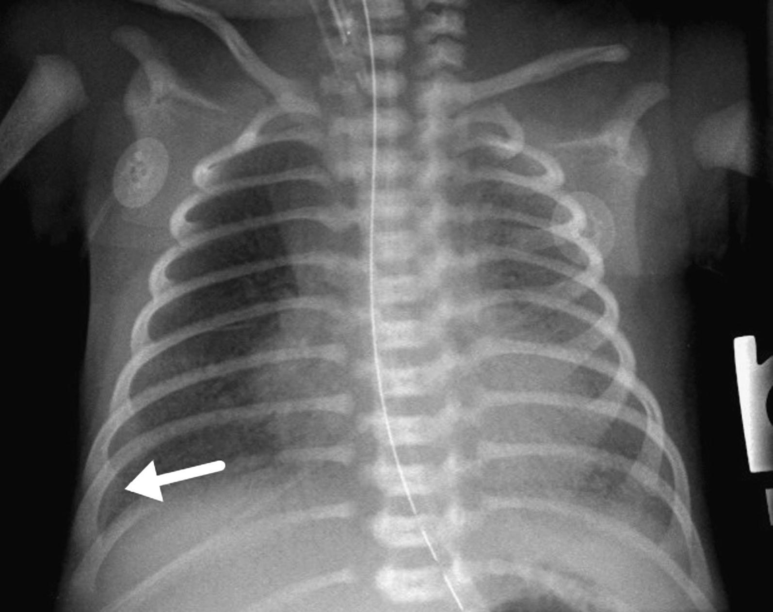
streptococcus is the most common causative organism. Late onset pneumonia usually occurs after discharge; or, in the hospital, it is commonly acquired from the neonatal unit or is associated with mechanical ventilation. Signs of neonatal pneumonia may mimic those of TTN and respiratory distress syndrome (RDS). Nonrespiratory signs include temperature instability, apnea, poor feeding, and lethargy. CXR can have protean imaging manifestations with radiographic findings potentially mimicking those seen in RDS, transient tachypnea of the newborn, and meconium aspiration. Isolated focal consolidation is rare with bilateral airspace disease most common, as seen in Figure 2.1. The presence of a pleural effusion can be a helpful distinguishing feature, being described in up to two-thirds of cases, such as in Figure 2.4.
Infants with suspected pneumonia require a full set of labs, including a complete blood count with a differential and blood cultures prior to initiating antibiotic therapy. It is worth noting that nearly all infants < 29 days
FIGURE 2.4. Neonatal pneumonia in a neonate with a small right pleural effusion (arrow) and diffuse left and right lower lobe hazy pulmonary opacification.
with a fever or hypothermia require a full septic workup, including a lumbar puncture.
Respiratory Distress Syndrome
RDS usually presents soon after birth and worsens over the following few hours. It is commonly observed in premature infants (< 37 weeks gestation) due to surfactant deficiency, with the risk of RDS decreasing as gestational age increases.
CXR reflects the underlying alveolar instability due to abnormal surface tension, demonstrating ground-glass/granular opacities and air bronchograms, as seen in Figure 2.5. Low lung volumes were classically described, but most neonates undergo imaging while under positive pressure support, often resulting in hyperinflation on the radiograph. Pleural effusions are not typical and may be an important clue to the possibility
FIGURE 2.5. AP chest radiograph in a premature neonate with diffuse granular opacities. Endotracheal tube is present. Findings typical of respiratory distress syndrome (RDS).
of neonatal pneumonia. Air leak phenomena are common complications. Blood gas measurements demonstrate respiratory acidosis, hypoxemia, and eventually metabolic acidosis.
The cornerstone of management of RDS is prevention with antenatal corticosteroids. Routine administration of antenatal corticosteroids to mothers with threatened preterm birth has revolutionized the management of RDS. Neonates with mild to moderate presentations may respond to the distending forces of CPAP; however, severe cases require endotracheal intubation. Exogenous surfactant is routinely administered to preterm infants requiring endotracheal intubation at birth to prevent RDS.
Pneumothorax
Spontaneous pneumothorax occurs in about 1%–2% of term infants. Risk factors include premature births, meconium aspiration, and RDS. This is likely due to their requirement for positive pressure ventilation which can lead to an air leak, causing the creation of a pneumothorax.
On physical examination, decreased breath sounds may be auscultated on the side of the pneumothorax. CXR demonstrates a lack of lung markings and lucency in the hemithorax with the pneumothorax. Decubitus films can better depict the findings as opposed to supine films, as seen in Figure 2.6. The neonate is lying in the left lateral decubitus position to reflect a right-sided pneumothorax.
Signs of a tension pneumothorax may include contralateral tracheal deviation, jugular venous distention, hypoxia, cyanosis, and/or hemodynamic instability. Tension pneumothoraces should be decompressed emergently.
Management of pneumothoraces depends on severity. If the patient is alert, has an oxygen saturation above 90%, and has normal vital signs, consult a pediatric surgeon or neonatologist to discuss placement of a pigtail catheter or chest tube. These may resolve spontaneously without intervention if the positive pressure that likely led to its creation is withdrawn. However, a chest tube should be placed immediately if the patient decompensates.
Meconium Aspiration Syndrome
Meconium aspiration syndrome (MAS) is usually a sequela of fetal distress during labor. Fetal distress may lead to passage of meconium. Fetal distress