Breast Disease Management: A Multidisciplinary Manual
James Harvey, Sue Down, Rachel Bright-Thomas, John Winstanley, and Hugh Bishop
Cardiovascular Disease in the Elderly: A Practical Manual
Rosaire Gray and Louise Pack
Dementia Care: A Practical Manual
Jonathan Waite, Rowan H Harwood, Ian R Morton, and David J Connelly
Diabetes Care: A Practical Manual (2nd Edition)
Rowan Hillson
Headache: A Practical Manual
David Kernick and Peter J Goadsby (eds)
Motor Neuron Disease: A Practical Manual (2nd Edition)
Kevin Talbot, Martin R Turner, Rachael Marsden, Jennifer Rolfe, and Alexander Thompson
Multiple Sclerosis Care: A Practical Manual
John Zajicek, Jennifer Freeman, and Bernadette Porter (eds)
Neuromuscular Disorders in the Adult: A Practical Manual
David Hilton-Jones, Jane Freebody, and Jane Stein
Preventive Cardiology: A Practical Manual
Catriona Jennings, Alison Mead, Jennifer Jones, Annie Holden, Susan Connolly, Kornelia Kotseva, and David Wood
Stroke Care: A Practical Manual (3rd Edition)
Rowan H Harwood, Farhad Umer Huwez, Paul Guyler, Sajid Alam, and Catherine Gaynor
Stroke Care: A practical manual
THIRD EDITION
Rowan H. Harwood
Consultant Physician
Nottingham University Hospitals, Nottingham, UK
Farhad Umer Huwez
Consultant Stroke Physician
Hyper-acute Stroke Unit, Royal London Hospital, London, UK
Paul Guyler
Clinical Director, East of England Regional Stroke Network; Consultant in Stroke Medicine, Mid and South Essex University Hospitals Group, Southend University Hospital, Essex, UK
Sajid Alam
Consultant in Stroke Medicine
East Suffolk and North Essex NHS Foundation Trust Ipswich Hospital, UK
Catherine Gaynor
Consultant Geriatrician
Nottingham University Hospitals NHS Trust, Nottingham, UK
Great Clarendon Street, Oxford, OX2 6DP, United Kingdom
Oxford University Press is a department of the University of Oxford. It furthers the University’s objective of excellence in research, scholarship, and education by publishing worldwide. Oxford is a registered trade mark of Oxford University Press in the UK and in certain other countries
The moral rights of the authors have been asserted
First Edition published in 2005
Second Edition published in 20
Third Edition published in 2023
All rights reserved. No part of this publication may be reproduced, stored in a retrieval system, or transmitted, in any form or by any means, without the prior permission in writing of Oxford University Press, or as expressly permitted by law, by licence or under terms agreed with the appropriate reprographics rights organization. Enquiries concerning reproduction outside the scope of the above should be sent to the Rights Department, Oxford University Press, at the address above
You must not circulate this work in any other form and you must impose this same condition on any acquirer
Published in the United States of America by Oxford University Press 98 Madison Avenue, New York, NY 006, United States of America
British Library Cataloguing in Publication Data
Data available
Library of Congress Control Number: 2022952038
ISBN 978–0–9–879656–5
DOI: 0.093/med/978098796565.00.000
Printed and bound by CPI Group (UK) Ltd, Croydon, CR0 4YY
Oxford University Press makes no representation, express or implied, that the drug dosages in this book are correct. Readers must therefore always check the product information and clinical procedures with the most up-to-date published product information and data sheets provided by the manufacturers and the most recent codes of conduct and safety regulations. The authors and the publishers do not accept responsibility or legal liability for any errors in the text or for the misuse or misapplication of material in this work. Except where otherwise stated, drug dosages and recommendations are for the non-pregnant adult who is not breast-feeding
Links to third party websites are provided by Oxford in good faith and for information only. Oxford disclaims any responsibility for the materials contained in any third party website referenced in this work.
Preface
‘Painting the Forth Bridge’ is a British analogy for a job that is never completed. When you finish, it is time to start again at the beginning. Medical books are similar. Stroke Medicine is driven by research, service development and quality improvement. Consequently, evidence, policy and practice are ever changing. A book of practical advice needs to keep pace. We therefore welcome the opportunity to offer the third edition of Stroke Care
Our text is quite direct and directive. You might even say dogmatic. If you are faced with a clinical problem, you need to know what do, not the details of academic debate. We are well aware of the need to justify bold assertions with high quality evidence, but the quality of the evidence we have available is variable. And even the highest quality evidence may not apply in every different context.
We try to capture the accumulated wisdom about how to do things that resides in experienced teams. We illustrate what we say with evidence where it is available, or where sensible extrapolations can be made. We present this in a fairly raw form in boxes scattered throughout the text. We confine references to these boxes. You might want to follow up the evidence for what we suggest, but that is not our main purpose.
We do not set out to compete with formal guidelines. Many sets of guidelines have been produced over recent years. It is a reasonable professional expectation that practitioners should be aware of them and their contents. Instead, we hope to add value through broadening the range of evidence that informs what we say and interpreting it in the face of everyday experience.
We follow a time-based sequence of chapters, which charts the journey of a stroke patient from diagnosis to outcome. We take a very broad view of what stroke care requires. We struggle most when working at the limits of our knowledge and experience, and in this book we push at the boundaries of the subject. For example, the quality of clinical decision making is topical and appears in postgraduate examinations. We spend large amounts of clinical time pondering difficult decisions. Therefore, we include a chapter on it. Many stroke patients die, so we include a chapter on end-of-life care. A well-functioning service requires ‘flow’, so we discuss how to discharge a patient. Advice on managing coma, pain or disturbed behaviour is not specific to stroke, but these are all issues which frequently arise in practice. Each chapter is fairly self-contained. There is some repetition, but we cross-reference where possible. Some issues arise early and persist, such as positioning, venous thrombosis prophylaxis and continence. The distinction between acute and rehabilitation care is clearly arbitrary. Secondary prevention starts early rather than at the end of the process.
Books are not a substitute for proper professional assessment and opinion. Evidence changes, interpretation varies with circumstances and from individual to individual, and different places have quite justifiably different ways of doing things. We have checked drug doses, but correct prescription remains the responsibility of the prescriber, who also needs to
take account of local policies or guidelines. On legal and ethical issues, we write from the perspective of the law and current practice in England and Wales but hope that the general principles will be of interest elsewhere. This book is written for people who look after stroke patients, in particular, doctors, nurses and therapists working in stroke units. A new staff member want to read it straight through, but this is mainly a book to refer back to. We thank the many colleagues who have generously contributed ideas, expertise and images. Most of all we thank our patients, who inspire what we do.
Symbols and abbreviations
ACA anterior cerebral artery
ACE angiotensin-converting enzyme
ACEI angiotensin-converting enzyme inhibitor
ACP advance care planning
ADC apparent diffusion coefficient
ADH antidiuretic hormone
ADL activities of daily living
AF atrial fibrillation
AFO ankle–foot orthosis
AHA/ASA American Heart Association/ American Stroke Association
AIM activate–initiate–monitor
AMT Abbreviated Mental Test
APTT activated partial thromboplastin time
ARB angiotensin receptor blocker
ASPECTS Alberta Stroke Program Early CT Score
AVM arteriovenous malformation
AVPU Alert, rousable to Voice, rousable to Pain, Unconscious
bd twice a day
BMA British Medical Association
BP blood pressure
CAM Confusion
Assessment Method
CAVPU Confused, Alert, rousable to Voice, rousable to Pain, Unconscious
CBF Cerebral Blood Flow
CBT Cognitive Behavioral Therapy
CBV Cerebral Blood Volume
CCB calcium channel blocker
CHD coronary heart disease
CI confidence interval
CNS central nervous system
COPD chronic obstructive pulmonary disease
CPR cardiopulmonary resuscitation
CPSP central post-stroke pain
CSF cerebrospinal fluid
CT computerized tomography
CTA computed tomography angiography/angiogram
CTP computed tomography perfusion
DIC disseminated intravascular coagulation
DNACPR do not attempt cardiopulmonary resuscitation
DOAC direct-acting oral anticoagulant
DSA digital subtraction angiography
DSM-5 Diagnostic and Statistical Manual of the American Psychiatric Association, fifth revision
DTI direct thrombin inhibitor
DVLA Driver and Vehicle Licensing Agency
DVT deep vein thrombosis
DWI diffusion-weighted imaging
ECG electrocardiogram
ECMO extracorporeal membrane oxygenation
ECST European Carotid Surgery Trial
EEG electroencephalography
eGFR estimated Glomerular Filtration Rate
EMG electromyography
ESD early supported discharge
ESR erythrocyte sedimentation rate
FAST Face, Arm, and Speech Test
FBC full blood count
FDP fibrin degradation product
FEES Fibreoptic Evaluation of Swallowing
FES functional electrical stimulation
FMD fibromuscular dysplasia
g gram
G&S group and screen/save
GCS Glasgow Coma Scale
GI gastrointestinal
GRE gradient echo
GTN glyceryl trinitrate
h hour
HASU Hyperacute Stroke Unit
Hb haemoglobin
HDL high-density lipoprotein
HMGCoA hydroxymethylglutarylcoenzyme A
HRT hormone replacement therapy
HU Hounsfield units
HVZ Herpes Varicella Zoster/ shingles
ICA internal carotid artery
ICD International Classification of Diseases
ICH intracerebral haemorrhage
ICU intensive Care Unit
IM intramuscular
INR international normalized ratio
IQCODE Informant Questionnaire on Cognitive Decline in the Elderly
IQR interquartile range
IPC Intermittent pneumatic compression
ITT intention to treat
ITU intensive therapy unit
IUCD intrauterine device
IV intravenous
kg kilogram
kJ kiloJoule
L litre
LACI Lacunar infarcts
LACS Lacunar stroke
LDL-C low-density lipoprotein-cholesterol
LMN lower motor neuron
LMWH low-molecular-weight heparin
LP lumbar puncture
MCA middle cerebral artery
MCI mild cognitive impairment
MI myocardial infarction
min minute
MMSE Mini-Mental State Examination
MoCA Montreal Cognitive Assessment
MR modified release
MRA magnetic resonance angiography
MRI magnetic resonance imaging
mRS modified Rankin Scale
MRSA methicillin-resistant Staphylococcus aureus
MTT Mean Transit Time
MUST Malnutrition Universal Screen Tool
NASCET North American Symptomatic Carotid Endarterectomy Trial
NG nasogastric
NHS National Health Service
NICE National Institute for Health and Care Excellence
NIHSS National Institutes of Health Stroke Scale
NINDS National Institute of Neurological Disorders and Stroke
NNT number needed to treat
O2 oxygen
OCSP Oxfordshire Community Stroke Project
od once daily
OR odds ratio
OT occupational therapist
PACI Partial Anterior Circulation Infarcts
PACS Partial Anterior Circulation Strokes
PCA posterior cerebral artery
PCC prothrombin complex concentrate
PEG percutaneous endoscopic gastrostomy
PFO patent foramen ovale
PIN personal identification number
PO by mouth
POCI Posterior Circulation Infarct
POCS Posterior circulation stroke
PR via the rectum
PROGRESS Perindopril Protection against Recurrent Stroke Study
PSCK9 proprotein convertase subtilisin/kexin type 9
PSV peak systolic velocity
PT physiotherapist/therapy
PT prothrombin time
PWI perfusion-weighted imaging
qds four times a day
RCP Royal College of Physicians
RCT randomized controlled trial
RIG radiologically guided gastrostomy
RR relative risk
RRR risk reduction ratio
s second
SAH subarachnoid haemorrhage
SaO2 arterial haemoglobin oxygen saturation
SC subcutaneous
SIADH syndrome of inappropriate secretion of antidiuretic hormone
SLE systemic lupus erythematosus
SLT Speech and Language Therapy
SNRI serotonin and norepinephrine reuptake inhibitor
SSRI selective serotonin reuptake inhibitor
SSS Scandinavian Stroke Score
TACI Total Anterior Circulation Infarcts
TACS Total Anterior Circulation Strokes
tds three times per day
TENS transcutaneous electrical nerve stimulation
TIA transient ischaemic attack
TOAST Trial of Org 072 in Acute stroke Treatment
TOF time of flight
rt-PA tissue plasminogen activator
TT thrombin time
TURP transurethral resection of the prostate
μg microgram
U&E urea and electrolytes
UK United Kingdom
UMN upper motor neuron
WFNS World Federation of Neurological Surgeons
WHO World Health Organization
Is it a stroke?
Presentation of stroke 2
What else might it be? 4
Face, Arm, and Speech Test (FAST) 0
Diagnosing stroke
Examination 2
Investigations 6
Clinical subtypes and pathology 8
Stroke in younger adults 24
Carotid and vertebral arterial dissection 26
Leukoaraiosis 28
Stroke in COVID-9 disease 29
Summary 3
a stroke?
Presentation of stroke
A diagnosis is an explanation, in biological terms, of a problem that a patient presents. An accurate diagnosis allows you to:
• give an explanation of what is going on to the patient and others
Stroke is a syndrome—a collection of symptoms and signs—which are usually obvious. The established WhO definition is: a rapidly developing episode of focal or global neurological dysfunction, lasting longer than 24 hours or leading to death, and of presumed vascular origin. (WhO Technical report Series, No. 469, 97)
This definition has limitations:
• Some patients who appear to have had a stroke have something other than cerebral infarction or haemorrhage (sometimes called ‘stroke mimics’).
• Neurological deficit progresses to some extent over the first 24 hours in about 25% of cases, and deterioration within the first week is common.
• It tells us nothing about the underlying pathology. More precise characterization of the type of stroke gives us clues about causes, treatment options, prognosis, and risk of recurrence.
• If re-perfusion therapies are considered for acute stroke, ‘time is brain’. Treatment must be delivered without delay, and no later than within 4.5 hours of symptom onset for thrombolysis. Work-up must therefore begin without waiting to see if the deficit will resolve spontaneously— although in the face of rapidly resolving symptoms, administering potentially dangerous treatment would be unwise.
• Some non-specific presentations (immobility, falls, confusion, or incontinence) may be due to vascular brain disease, amongst other things.
• Co-morbid conditions (especially in older people) can make diagnosis difficult.
• A number of cerebrovascular conditions fall outside the definition, including vascular dementia, silent infarction on brain imaging, and transient ischaemic attack (TIA).
• Subarachnoid haemorrhage fits the clinical definition for a stroke, but behaves and is managed as a separate entity.
An alternative definition of stroke is a type of brain injury caused by sudden interruption of blood flow This definition relies on the results of imaging, and includes some patients with transient symptoms. The most recent definitions combine pathology and presence or absence of neurological symptoms (Box .).
Box . Updated definition of stroke
Cerebral infarction and ischaemic stroke comprise brain, spinal cord, or retinal cell death based on a) imaging or other evidence of ischaemic injury in a defined vascular distribution or b) appropriate neurological symptoms persisting ≥ 24 hours or until death, without an alternative explanation. ‘Ischaemic stroke’ implies persisting neurological symptoms. ‘Silent infarction’ implies absence of detectable neurological signs.
Intracerebral haemorrhage is a focal collection of blood within the brain parenchyma or ventricular system, not caused by trauma, but including haemorrhage following cerebral infarction.
Subarachnoid haemorrhage is bleeding into the subarachnoid space (between the arachnoid and the pia mater of the brain or spinal cord).
Stroke due to intracerebral or subarachnoid haemorrhage implies rapidly developing neurological dysfunction.
Cerebral venous thrombosis can cause infarction or haemorrhage in the brain, spinal cord, or retina because of thrombosis of a cerebral venous structure.
Source: data from Sacco rL et al. Stroke 203 44:2064–89. DOI: 0.6/ STr.0b03e38296aeca.
What else might it be?
Transient ischaemic attack (TIA)
• See also Chapter 0 section: Neurovascular or TIA clinics, p. 3.
A TIA is an acute, focal, loss of cerebral function, or transient monocular blindness (amaurosis fugax), of presumed vascular origin, with the symptoms lasting less than 24 hours:
• Initially it is indistinguishable from a stroke.
• Most TIAs last less than an hour. It is difficult to define a lower limit to duration. Some descriptions say ‘seconds’, and 5% last less than a minute in published series, but it is difficult to imagine nerve cell failure due to ischaemia and recovery in much less than a minute. There can be problems with patients’ recall of the passage of time when anxious.
• Amaurosis fugax is a rapidly progressive loss of vision, or partial loss of vision, in one eye (often, but not exclusively, ‘like a curtain coming down’), coming on over a few seconds to a minute. After a variable time, usually seconds to a few minutes, it resolves with gradual recovery of vision over the whole visual field.
• hemiplegic migraine is excluded.
• The main difficulty is making an accurate diagnosis based only on the history, and the absence of examination or investigation findings that suggest another diagnosis. Considerable uncertainty may remain.
• The importance of TIA and minor stroke lies in their propensity to recur: 0% in a week, 20% in a month. A third of these recurrences are persisting, disabling, or fatal strokes. Patients with TIA and minor stroke should be offered immediate, thorough, and rapid investigation, and appropriate secondary prevention.
• risk factors, and prognosis for stroke recurrence or ischaemic heart disease, are identical for TIA and minor stroke, regardless of symptom duration. however, higher and lower risk situations can be defined for individuals according to what symptoms, risk factors, and investigation findings they have.
• About a quarter of patients with clinical TIA have an appropriate infarct on CT brain imaging, half on diffusion-weighted MrI, including most of those in whom symptoms last over an hour. however, imaging evidence of infarction does not change management.
• Transient dizziness, confusion, vertigo, double vision, syncope, and drop attacks should not be diagnosed as TIA in the absence of other neurological findings.
Other differential diagnoses of stroke
• From the perspective of hospital admissions, about 25% of patients referred with possible stroke have something else.
• Some uncertainty is inevitable, but experienced doctors are better at diagnosing (and ruling out) stroke than less experienced ones.
• Mimics are most likely to be referred as possible stroke where there is cognitive impairment, loss of consciousness, or seizure at onset; an inexact time of onset; an absence of focal neurological signs or symptoms; or an inability to classify the stroke to a typical location (e.g. using the OCSP classification).
• Important differential diagnoses are shown in Table .. Others that may arise include Bell’s palsy, multiple sclerosis, metabolic disturbances, intoxication, transient global amnesia, psychiatric illnesses, dementia, and Parkinson’s disease.
• Ask a neurologist’s opinion if you are struggling to explain the clinical features, or are considering some of the more difficult or rare diagnoses.
. Conditions that can cause stroke-syndrome
Diagnosis Key features
Old stroke, with increased weakness during intercurrent illness
Fits, with Todd’s paresis
Old neurological signs are often worse during intercurrent illnesses, especially infections, or appear to be so. Excluding a recurrent stroke is difficult, but rapid return to previous level of function is usual with appropriate treatment. Diffusion-weighted MrI is the best way to make (or rule out) a definite diagnosis of new stroke
Commonest cause for misdiagnosis of recurrent stroke. Clinical diagnosis, usually requiring an eyewitness. Consider ictal features (loss of consciousness, convulsions, tongue biting, incontinence) and post-ictal features (headache, sleepiness, confusion). Diffusion-weighted MrI is the best way to make (or rule out) a definite diagnosis of new stroke
Cerebral tumours, primary or secondary
CT/MrI scan diagnosis. There may be features of raised intracranial pressure (headache, vomiting, drowsiness, papilloedema). Onset is slower than stroke. A step-wise progression over days or weeks may occur, but only in 6 patients with a progressive course has a tumour. Onset may be sudden if there is bleeding into a tumour
hypoglycaemia
Subdural haematoma
Cerebral abscess
Almost always drug-induced, severe, hypoglycaemia. Usually rapidly reversible, but hemiplegia can persist 24 hours or more
CT/MrI scan diagnosis. If significant, it will cause drowsiness. Sometimes headache, confusion, hemiplegia, or aphasia. Features may fluctuate
CT/MrI scan diagnosis. Usually due to spread from sinuses or ear. Onset is sub-acute, but not always with prodromal infective symptoms. headache is usual. Later drowsiness, vomiting, delirium, and bradycardia. Aphasia, visual field defects, and facial weakness are more common than hemiplegia. Avoid lumbar puncture. Needs surgical drainage. 25% mortality, even if optimally treated
Encephalitis Occasionally confused with stroke: 5% have focal signs. Usually mild preceding febrile illness, headache, and drowsiness. Sometimes fits, confusion, and gradual-onset coma. Ophthalmoplegia, nystagmus, other cranial nerve, cerebellar, and sensory signs possible. Neck may not be stiff. CT scan may be normal. CSF usually abnormal. MrI can show specific or nonspecific abnormalities
(Continued )
Table
(‘stroke-mimics’)
Table . (Contd.)
Diagnosis Key features
Cerebral vasculitis
Venous thrombosis
Conversion disorder
Difficult to diagnose. Primary or secondary (to temporal arteritis, amphetamines, cocaine, SLE, infection). Can result in infarct or bleed. headache prominent, focal neurological deficits include cranial nerve palsies, or delirium. ESr can be raised, but this and other systemic markers are typically normal in primary CNS vasculitis. MrI and CSF abnormal. Check auto-antibodies. May need angiography or temporal artery /brain /meningeal biopsy. Treat underlying cause and/or high-dose steroids
Difficult to diagnose. Most have headache, half have raised intracranial pressure (nausea, papilloedema), some have focal neurological signs (hemiparesis or paraplegia) or fits. May be secondary to thrombophilia, trauma, infection, or post-partum. CSF is often abnormal (raised pressure, high protein, few red and white cells). CT may show hyperdensity of cortical veins or sinuses, filling defects with contrast (empty delta sign), infarction, disproportionate swelling, and haemorrhage. Mr or CT venography is diagnostic
Lack of cranial nerve findings, neurological findings in a nonvascular distribution, inconsistent examination
Features prompting caution include:
• headache (25% of patients with infarcts have a headache, usually mild)
• Pyrexia
• Malaise or prodromal illness
• gradual progression over days
• Features of raised intracranial pressure (headache, worst at night, on waking, and on coughing; drowsiness; vomiting; hypertension with bradycardia; papilloedema)
• Young age, or absence of vascular risk factors
• Unobtainable or uncertain history.
Some transient neurological conditions can mimic TIA. The most important are:
• Migraine. An aura, often a visual disturbance, starts in one homonymous hemi-field, usually develops over about 30 minutes, and lasts less than an hour. Visual phenomena include lights, halos, zigzag lines, scotomata, or hemianopias, which build up and may migrate across the entire visual field. Sensory symptoms or hemiparesis can develop with or after visual symptoms, and spread progressively across body parts over several minutes. Aphasia can occur. headache, often unilateral and throbbing, typically starts as the aura is resolving, and lasts 4–72 hours, often with nausea and photophobia. Aura may occur without headache, or during the headache, and may last > 24 hours. headache may precede the aura. Side may vary with attacks. Basilar territory symptoms are also possible (vertigo, ataxia, dysarthria).
• Fits. generalized seizures imply loss of consciousness. The patient is rigid and may become blue during the attack. May be followed by unilateral weakness (Todd’s paresis, lasting a few hours to a day or two).
Partial seizures start in clear consciousness, but may be secondarily generalized. They may be motor or sensory, with jerking or tingling that tends to build up and spread. Complex partial seizures comprise a disturbance of content of consciousness, with sensory hallucinations (smell or taste, remembered scenes or déjà vu, distorted perceptions of the world), and motor features such as chewing or organized motor activity like undressing. Aphasia may occur. 2% of patients with stroke have a seizure at onset, half generalized and half partial.
• Syncope presents with loss of consciousness and postural tone due to a sudden fall in cerebral blood flow. The patient is pale, sweaty, clammy, and floppy, and may jerk. Light-headedness may occur before syncope with dimming or loss of vision. A third have amnesia for the event.
• Transient global amnesia. Sudden onset. Loss of memory for new information (anterograde amnesia), may also have retrograde amnesia (past events). No loss of personal identity (patients know who they are), problem solving, language, or visuospatial orientation. Look healthy and repetitively ask the same questions. May have headache. good recovery, recurrence is rare.
Differential diagnosis of coma
• Stroke will sometimes result in sustained unconsciousness (especially when due to bleeding, very large infarcts, or some basilar artery territory strokes). Exclude other causes of coma (metabolic, infective), as some are treatable (Table .2).
• impairment of the brainstem reticular activating system (lesions of midbrain to mid-pons, or compression from trans-tentorial herniation due to supra- or infra-tentorial pressure).
• Large cerebral infarcts with oedema may increase intracranial pressure enough to impair cortical function bilaterally, or cause tentorial herniation.
Evaluation and treatment must be rapid, and must proceed together.
• Look for asymmetry—in tone, movement, and reflexes, and test brainstem function (pupillary light reflex, doll-eye manoeuvre, corneal and gag reflexes).
• The coma is probably metabolic in origin if the pupils are symmetrically reactive and there are no focal neurological signs.
• Coma developing over seconds to minutes suggests a cardiovascular, cerebrovascular or epileptic cause. Consider extradural or subdural haematoma, if there was recent trauma.
• Drug abuse is a cause of otherwise unexplained coma.
• Neurological clues help localization (Table .3). But remember that anticholinergic drugs and anoxia can produce large pupils; opiates and some metabolic disorders can give small pupils (usually reactive).
• Anyone in coma needs an immediate CT head scan—unless you are sure of the diagnosis, or that the patient would not have wanted intervention.
Is it a stroke?
Table .2 Differential diagnosis of coma
Cause Clues
Metabolic
hypoglycaemia
Diabetic ketoacidosis or hyperosmolar coma
hyper or hyponatraemia
glucometer
glucometer, acidosis, +/− ketonuria/ ketonaemia
Serum sodium
hypothermia/hyperthermia Temperature
hepatic, uraemic coma
Stigmata, flap, history, blood tests
Myxoedema coma/thyroid storm history, clinical state, thyroid function tests
Subdural or extradural haematoma history of trauma. CT scan. Lucid interval after injury
Table .2 (Contd.)
Cause Clues
Meningitis, encephalitis
hypertensive encephalopathy
Brain tumour, abscess
Fever, malaise, headache, neck and skin signs, CT/MrI, lumbar puncture
BP, fundi, headache, confusion, urinalysis, renal function
CT/MrI scan
Table .3 Localizing the cause of coma
Level Features
Infra-tentorial
Supratentorial (structural lesion)
Toxic-metabolic
Brainstem causes usually have the most obvious signs and are easiest to diagnose. Look for brainstem signs: Cranial nerve signs +/− long tract signs, divergent squint, pupillary and doll’s eye reflex loss
Asymmetrical long tract signs without brain stem signs (may be false localizing III, IV, or VI if mass effect or aneurysm), focal seizures, conjugate eye deviation
Confusion and drowsiness with few motor signs
Motor signs symmetrical
Pupillary responses preserved
Myoclonus, asterixis (flap), tremulousness, and seizures common
Acid-base imbalance
Psychogenic Eyes tight shut
Pupils reactive
Doll’s eye and caloric reflexes preserved
Motor tone normal or inconsistent resistance to movement
reflexes normal
EEg shows wakefulness
Face, Arm, and Speech Test (FAST)
FAST is a rapid test to screen for the possibility of stroke, designed for lay and pre-hospital use (Box .2). Any sudden-onset abnormality raises the possibility of stroke and medical assistance should be sought.
BE-FAST is an alternative version (https://bef ast.org). In addition to the Face, Arm, and Speech items, B stands for balance (sudden vertigo or loss of balance); E for eyes (loss of vision); T for terrible headache. Sensitivity is better than FAST, but specificity is worse.
Box .2 FAST—the Face, Arm, and Speech Test
A quick and easy test for possible stroke
FACE—Ask patient to smile. Do both sides of the face move the same?
ArM—Ask patient to lift both arms out in front of them and hold them there. Is one side weaker than the other?
SPEECh In simple conversation (how are you? What happened?) is speech slurred, hesitant, unintelligible, or completely absent?
If any of these is abnormal, the test is positive, and there is a strong possibility of a stroke. Sensitivity and specificity are both about 80%. That means about 80% of all patients with stroke are ‘FAST positive’, and about 80% of patients who have not had a stroke are ‘FAST negative’. Posterior circulation strokes are underdiagnosed.
UK Stroke Association. Source: data from kleindorfer DO, et al. Designing a message for public education regarding stroke: does FAST capture enough stroke? Stroke. 2007;38(0):2864–8.
Diagnosing stroke
You need a careful history. If the patient is unconscious, unable to communicate (e.g. aphasic) or confused, that is no excuse—ask someone else. If an informant is not immediately present, use the telephone. There may be old hospital case notes available, paper or electronic. Look at them, and briefly summarize useful information. You need to know:
• what happened, and what the current symptoms are
• the time of onset, and time-course of progression
• if it has happened before (previous stroke, TIA)
• past medical and drug history (prescription, over the counter and illicit—nasal decongestants, amphetamines, and cocaine can cause strokes)
• vascular risk factors
• previous functional, occupational, and cognitive ability (including driving)
• information useful for rehabilitation and discharge planning—type of accommodation, co-habitation (and the health of an often-elderly cohabitee), family, and other domestic support
• family history of stroke or thrombotic disease (occasionally gives a diagnostic clue, may also reveal previous knowledge, experiences, or expectations).
Some of this can be collected later on, if necessary. But admission is a good opportunity to be thorough.
history taking (and examination) is an inductive process. Use the information you gather to formulate hypotheses about what is going on, which you test with new questions. You want evidence that this is a stroke, and to rule in or rule out other diagnoses. You also want to put the new pathology in context by documenting co-morbid conditions and their disabling consequences.
Examination
General
A thorough general examination is required, because:
• the patient may be very ill, and require securing of the airway, breathing, and circulation before an adequate assessment can be made.
• the possibility of a condition mimicking or causing stroke (atrial fibrillation, malignancy, endocarditis).
• the importance of co-morbidity in a generally older population.
The cardio-vascular system is examined routinely, but the mental state and musculo-skeletal systems, in particular, are often overlooked. An admissions ward or Emergency Department is not always the best place to examine these properly.
Initially test cognition using simple orientation (person, place, and time) and short-term memory, or the 0-point abbreviated mental test score (AMT). Later on, use a more comprehensive test (such as Montreal
Cognitive Assessment or Addenbrooke’s Cognitive Examination, ACE, or the shorter 30-point mini-ACE).
Blood pressure may be raised (or very raised), but the ward record over the next hours, days, and weeks will give a better picture of ‘usual’ blood pressure. The pulse may be slowed in raised intracranial pressure, or irregular in AF. There may be periodic (Cheyne–Stokes) respiration, or evidence of chest infection. record oxygen saturation by pulse oximetry.
Neurological examination
Is directed at:
• identifying features which require special precautions (e.g. coma, dysphagia).
• defining a clinical stroke syndrome (localizing the lesion).
• quantifying neurological impairments as a baseline for subsequent improvements or deteriorations.
• raising suspicion of alternative, non-stroke, diagnoses.
In acute situations, where emergency treatments (thrombolysis, thrombectomy) are possible, a rapid neurological assessment must be performed. The National Institutes of health Stroke Scale (NIhSS) can be used to quantify neurological impairment. This comprises items, each scoring a specific neurological ability. For each item, a score of 0 indicates normal function, higher scores indicate increasing levels of impairment. Item scores are summed to calculate a total score between 0 (normal) and 42 (maximum impairment). In general, the higher the NIhSS score, the more severe the stroke, but this is only as a guide. Some symptoms can have life-changing consequences despite a low NIhSS; for example, isolated severe aphasia, or an isolated homonymous hemianopia which may preclude driving.
Subsequently, a standard neurological examination—cranial nerves, limb tone, power, reflexes, cerebellar function, and sensation—should be performed, but some aspects need emphasizing, and others need adapting. You cannot examine coordination in a paralysed limb, or assess subtle parietal lobe signs in a drowsy patient.
• At minimum in an unconscious, un-comprehending, or un-cooperative patient, and with a little ingenuity, you can record eye movements, facial weakness, limb tone and gross power, and usually reflexes.
• Level of consciousness. This is important for prognosis and immediate nursing care. Use the glasgow Coma Scale. Describe the responses for eye opening, motor, and voice as well as the total score. There is a clear problem in under-estimating level of consciousness in aphasia, but gCS is familiar and well-understood. The AVPU/CAVPU system ((Confused), Alert, rousable to Voice, rousable to Pain, Unconscious) is a valid alternative.
• Check for a stiff neck, and for evidence of head trauma
• Examine the fundi for papilloedema, retinopathy, or sub-hyaloid haemorrhage.
• If unconscious:
• check brainstem function pupillary reaction to light, doll’s eye movements, corneal reflexes, gag reflex
• The caloric reflex is sometimes useful, for example after cervical spine trauma:
• Check the tympanic membrane is intact and there is no wax, then inject 20 mL of ice-cold water into the ear canal
• Conjugate eye movement towards stimulated ear indicates that the midbrain/pons is intact
• Absent or dysconjugate response implies brainstem damage at the level of the pons or sedative drug intoxication.
• Loss of pupillary reaction to light implies a midbrain lesion. Pontine lesions can cause small but reactive pupils.
• Dysconjugate gaze indicates a palsy of cranial nerves III, IV, or VI (nuclei in the midbrain and pons) or their connections (medial longitudinal fasciculus), a false localizing sign in raised intracranial pressure, a mimic such as myasthenia gravis, or a congenital squint.
• Conjugate deviation of the eyes suggests either a frontal lobe infarct on the same side as the direction of gaze, the opposite frontal lobe if an ‘irritative’ lesion (tumour, haemorrhage), or a pontine lesion in the opposite lateral gaze centre.
• No eye movements at all indicates a pontine lesion (or a mimic such as guillain–Barre syndrome).
• Check the visual fields, upper and lower quadrants. Also, if possible, test for visual inattention (sensory extinction—inability to perceive a stimulus when a simultaneous stimulus is presented to the other visual field, in the absence of a visual field defect). Wiggling fingers are sufficient for the purpose, rather than coloured pin heads.
• record speech impairment: dysarthria, receptive aphasia, expressive aphasia. Test receptive ability (understanding, following commands) first using one-stage commands with non-verbal response (e.g. ‘close your eyes’, ‘touch your left ear’). If there is reasonable understanding, then test for expressive aphasia (spontaneous speech, naming, sequences such as counting, yes/no responses).
• Test swallowing with the patient sitting up, give small sips of water, and, from behind, feel for prompt laryngeal elevation. Observe for
delay, aspiration (choking or coughing), or ‘wetness’ of the voice. Tap water is more or less sterile. We produce a litre of saliva a day, which is normally swallowed, and which is far from sterile. Many hospitals have nurse-delivered swallow testing protocols, which should be followed.
• The presence or absence of the gag reflex tells you nothing about the safety of swallowing.
• Examine motor function:
• Examine power in the face, arm, and leg
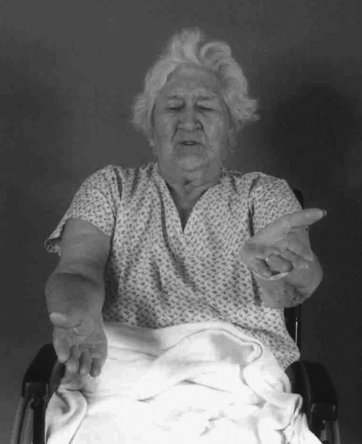
• ‘Pronator drift’ is a good test for subtle deficits—the downward drifting and pronation of hands held stretched out horizontally in front, with palms upwards and eyes closed (Figure .)
• Weakness follows a ‘pyramidal distribution’. Shoulder abduction, elbow extension, and wrist dorsiflexion will be weaker than corresponding flexor functions, and hip and knee flexion and foot dorsiflexion will be weaker than extensor functions.
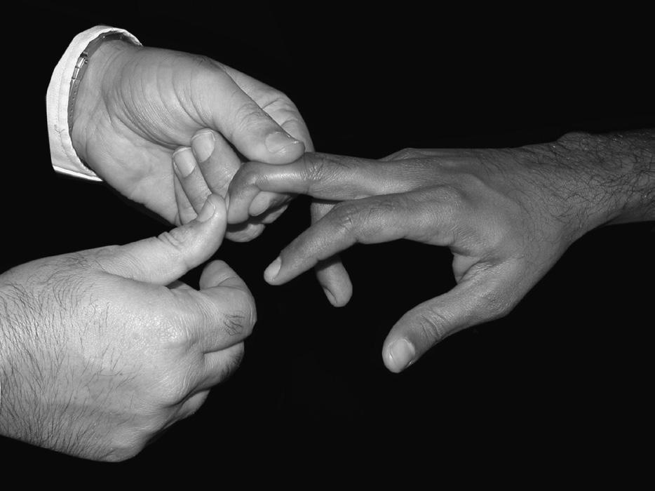
• Carefully test the limb tone and reflexes, especially in mild cases. If the reflexes are very brisk, try the pectoral jerks or hoffman’s reflex (thumb flexion when the terminal phalanx of the middle finger is flexed under tension then suddenly released with a ‘flick’), where asymmetry may be easier to detect (Figure .2).
• Test coordination, and gait if possible. If not, assess head and trunk control (sitting balance).
• Test sensation
• There may be spinothalamic sensory loss (temperature, pin prick/ pain).
• More useful are some ‘cortical sensory modalities’, often as part of a search for ‘cortical involvement’ when identifying a stroke syndrome
• stereognosis (identifying objects in the hand)
• graphaesthesia (identifying numbers traced on the hand)
• Test for sensory inattention (similar to visual inattention, using touch instead of visual stimuli).
• If possible, test for other cortical or parietal functions, including:
• neglect (Albert’s test—line cancellation, drawing a clock face, or double-headed daisy)
• sometimes specific dyscalculia (sums), dyslexia (reading) or dysgraphia (writing)
• Body image and proprioception can be assessed using the ‘thumbfinding test’ (affected arm supported in front, eyes closed, the patient is asked to find his thumb with his unaffected hand).
Some of these tests can wait for a few days. however, signs may resolve rapidly.
Fig. . Examining for pronator drift.
Fig. .2 Examining for hoffman’s reflex.
Investigations
• First check blood glucose with a portable glucometer.
• get a CT head scan (or MrI) as soon as possible after admission, unless the diagnosis is certain and the patient is moribund (which will be rare). A CT scan is primarily to diagnose or exclude bleeds and stroke mimics rather than to confirm infarction.
• CT scan should be immediate if thrombectomy or thrombolysis are possible, or if there is (or suspicion of):
• trauma
• cerebellar haematoma
• subarachnoid haemorrhage
• raised intracranial pressure
• undiagnosed coma, or deteriorating or fluctuating level of consciousness
• if the patient is on anticoagulants, has a known bleeding disorder, or needs anticoagulation (or anti-thrombotics, if a bleed is suspected).
• Blood count, electrolytes, including calcium, glucose, renal, liver, and thyroid function, C-reactive protein, or erythrocyte sedimentation rate (ESr) and urinalysis should be done routinely. Check coagulation if on anticoagulants, or if proposing them, or if the scan shows a bleed. Measure cholesterol if within 2 days of the stroke.
• Electrocardiogram in everyone.
• Ideally get an echocardiogram in potentially embolic (partial anterior and posterior circulation) strokes. But the call for echocardiography is high, and the diagnostic yield low. Local services may limit this to cases where there is other clinical or ECg evidence of heart disease. Younger patients with no other apparent cause for stroke should have transoesophageal or trans-thoracic echocardiography with bubble contrast to look for patent foramen ovale (Box .3).
• A chest x-ray is unlikely to be technically satisfactory. Don’t request routinely unless there are specific chest problems or signs you want to investigate (e.g. unexplained fever, or presumed aspiration pneumonia).
• Carotid duplex scan if anterior circulation stroke resulting in no more than minor disability, and the patient would be fit and willing to undergo carotid endarterectomy. Duplex scanning will also detect half of carotid dissections.
• CT or Mr angiography may be required to diagnose large vessel occlusion (suitable for thrombectomy), dissection, as a prelude to carotid endarterectomy, or to investigate intracranial bleeding (especially those aged under 50, or where initial imaging suggests the possibility of aneurysm, AVM, or tumour).
• CT or MrI perfusion imaging may be used to distinguish areas of salvageable ischaemia (‘penumbra’) from the parts which are infarcted or irretrievably destined to infarct (‘core’). This may determine suitability for thrombolysis and thrombectomy where the time of stroke onset is unknown (including whilst asleep).
• An interval MrI scan after 6–2 weeks (or longer, when the blood has fully resorbed and oedema has settled) may be required to exclude an underlying lesion after a bleed.
Box .3 Patent Foramen Ovale (PFO) closure following cryptogenic stroke
• PFO is present in 25% of the general population, and 40% of patients with ‘cryptogenic’ stroke. relative risks are 3–5, depending on age, or more in the presence of an atrial septal aneurysm
• Meta-analysis of 5 rCTs of device closure of PFO compared with medical therapy (antithrombotic or anticoagulant drugs) included 3440 patients, mean age 45 years, followed up over a mean of 4 years
• PFO closure was superior to medical therapy for the prevention of further strokes in appropriately selected patients with moderate to large shunts
• hazard ratio for patients who had PFO closure was:
• 0.32 (95% CI 0.–0.8) for recurrent stroke (0.6% vs .8% per year).
• 0.33 (95% CI 0.2–0.7) for recurrent stroke in patients with a large shunt (0.5% vs .6% per year).
• 0.90 (95% CI 0.5–.6) for recurrent stroke in patients with a small shunt.
• Presence or absence of an atrial septal aneurysm did not change effect size
• The risk of atrial fibrillation was increased with PFO closure, rr 4.7 (95% CI 2.2–0)
• The risk of recurrent stroke is low with medical secondary prevention (72/6 over 4 years). NNT for closure is 78 for all shunts and 96 for large shunts (per year). PFO closure has rare but severe complications
• Authors stress the importance of careful patient selection, that is, under 60, with full work-up for alternative aetiologies, and a duly informed and willing patient. Medical management is a reasonable option.
• The risk of Paradoxical Embolism (roPE) Score indicates the likelihood of PFO-related stroke in patients with cryptogenic stroke.
Source: data from handke M et al. N Engl J Med 2007; 357: 2262–8. DOI: 0.056/ NEJMoa07422; Ahmad Y et al. Eur Heart J 208; 39: 638–49. DOI: 0.093/eurheartj/ ehy2; Safouris A et al. Front. Neurol 2020; :434. DOI: 0.3389/fneur.2020.00434. Davis AP et al. JAMA Neurol 2022; 79:02–04. DOI:0.00/jamaneurol.2022.227
• Ambulatory ECg (5 days recording) detects new paroxysmal atrial fibrillation in up to 5%. Consider where stroke aetiology is not clear and cardiac embolism is suspected (e.g. cortical lesions in different vascular territories). Because episodes are often asymptomatic, patientactivated devices are less useful. Implantable loop recorders allow monitoring for up to 3 years (they are placed subcutaneously through a minimally invasive procedure and are easily removed).
• Additional tests may be required in younger stroke patients (< 50 years). See also section ‘Stroke in younger adults’, p. 24.
Clinical subtypes and pathology
Stroke is a mixed bag of pathologies. These include infarction, intra-cerebral, and subarachnoid bleeding. Infarction divides between large vessel disease, small end-artery (lacunar) disease, cardio-embolism, and rarer causes such as venous infarction, vasculitis, and infective endocarditis.
Intra-cerebral haemorrhage
See Table .4. Bleeds occur in the cerebral lobes, basal ganglia, thalamus, brainstem (especially pons), and cerebellum. Cytotoxic and vasogenic oedema forms around them over the next 2–4 days.
Acute bleeds have some characteristic clinical features:
• ‘apoplectic’ onset (sudden loss of consciousness)
• headache
Table .4 Pathology of intra-cerebral haemorrhage
Type Features
Charcot–
Bouchard microaneurysms
Lipohyalinosis, often associated with hypertension, causes weakness of the walls of small perforating arteries, usually to the basal ganglia, thalamus, or pons
Amyloid angiopathy Commonest cause of lobar haemorrhage in people aged over 60. Affects small arteries particularly in the meninges and superficial cortex. Arteries are weakened by fibrinoid degeneration, amyloid deposition, segmental dilatation, and micro-aneurysm formation. Affects men and women equally, especially those with dementia. resulting haematoma is usually superficial and lobar. Often recur
Berry aneurysms
Fusiform aneurysms
Arteriovenous malformations
Comprise the majority of intra-cranial aneurysms. Thin-walled saccular dilatation of the arteries, may be multi-loculated if large. Probably mostly acquired rather than congenital. Most are small, found in 2–5% of autopsies. Associated with age, hypertension, and atheroma. Found at distal end of the arteries, mainly at Circle of Willis: carotid tree 75%; basilar tree 0%; both 5%. rupture causes subarachnoid haemorrhage, but may extend into the brain substance or ventricles
Found on atheromatous large arteries (internal carotid, basilar) in older people, due to replacement of the muscular layer by fibrous tissue. A common site is the supra-clinoid segment of the internal carotid artery. A complication is compression of structures in the cavernous sinus wall
Consist of a mass of enlarged and tortuous vessels. Supplied by one or more large arteries. Drained by one or more large veins. They are congenital and may run in families. Present with recurrent headaches, epilepsy, subarachnoid or intracerebral haemorrhages. Commonest site is on the middle cerebral artery
Secondary haemorrhage
Due to anticoagulant therapy, thrombolytic therapy (e.g. for heart attack or ischaemic stroke), haemorrhagic disease, bleeding into tumours or mycotic aneurysms, or haemorrhagic transformation of an infarct
• vomiting
• stiff neck.
Unfortunately, these, and various scoring systems derived from them (such as the guy’s and Siriraj scores) are insufficiently accurate for clinical use. Small bleeds, and some bleeds seen very early (e.g. within 3 hours of onset) are clinically indistinguishable from infarcts.
An early CT scan is required to make the diagnosis. haematomas absorb over 0–30 days. If the scan is delayed longer than a week, a small bleed may have resolved on CT, although MrI can still detect haemoglobin breakdown products.
haematomas tend to enlarge over the first few hours after onset (25% enlarge in the first hour, 40% over the first day), especially when aspirin or anticoagulants have been taken. growth of the haematoma is associated with early neurological deterioration.
Cerebral
infarcts
Pathological mechanisms
Most patients have cerebral infarction due to arterial thrombosis or embolism. A good level of diagnostic acumen and clinical suspicion is needed to detect rare but treatable causes of infarction such as infective endocarditis (peripheral stigmata, new heart murmurs, raised inflammatory markers, positive blood cultures), cerebral vasculitis, thrombophilia, or venous infarction. If we are to direct further investigation and management logically, we need to know more than just that a stroke has occurred. Often, we can work out exactly why the stroke has occurred. Table .5 gives some different pathological mechanisms. In practice, however, up to 40% of causes remain undetermined despite comprehensive work-up.
Table .5 Pathology of cerebral infarction
Type Features
Cardiac emboli
Large vessel disease
About 20% of ischaemic strokes. half are due to atrial fibrillation. Other causes include mitral stenosis and prosthetic valves, mural thrombus after myocardial infarction, left ventricular aneurysm, dilated cardiomyopathy, atrial myxoma, patent foramen ovale with paradoxical embolism of venous thrombi. Typically results in a peripherally located, wedge-shaped infarcts, often becoming haemorrhagic. Can involve multiple arterial distributions
Atherosclerosis of aorta, common carotid, and internal carotid artery. Stenosis, plaque rupture and ulceration, platelet aggregation, and red cell thrombus formation, may cause occlusion or provide a source of emboli. Internal carotid artery clot may propagate into the middle cerebral artery. In cases of occlusion perfusion is dependent on collaterals from the Circle of Willis
Small vessel (lacunar) disease
Lipohyalinosis or micro-atheroma of small end arteries, associated with hypertension, diabetes mellitus, or hyper-lipidaemia