Preface
Anaesthesia for oral and maxillofacial surgery encompasses a wide range of procedures, treatments, and interventions for an array of pathologies affecting the teeth, mouth, and jaw. There is frequently overlap with other related surgical specialties such as otorhinolaryngology, neurosurgery, plastic surgery, and reconstructive surgery. Since the publication of the first edition of the Oxford Textbook of Anaesthesia for Oral and Maxillofacial Surgery in 2010, this subspecialty has continued to develop, with procedures and techniques evolving, new guidelines and standards being introduced, and new research evidence becoming available. This second edition has been completely updated by a new group of authors, adding an original, contemporary, and fresh
perspective on existing topics, with the addition of new chapters on human factors in anaesthesia and surgical complications, written from the surgeon’s perspective.
The safe perioperative care of patients undergoing oral and maxillofacial surgery necessitates a comprehensive knowledge and understanding of the specific challenges posed by this cohort, and their underlying pathologies and comorbidities, including a full appreciation of the implications of a ‘shared airway’. This textbook is primarily intended as a reference tool for anaesthetists of all grades, but will also be of interest to maxillofacial surgeons, anaesthetic practitioners, anaesthetic nurses, recovery and intensive care nurses, and operating department practitioners.
Abbreviations xv
Contributors xvii
1. Preoperative assessment 1
Roger H. Y. Ho and David M. H. Lam
Introduction 1
Range of procedures 1
Airway evaluation and planning 1
Pathology-specific considerations 2
Environmental considerations 4
Evaluation of comorbidities 4
Preoperative risk stratification 10
Perioperative management of medications 12
Conclusion 15
2. Difficult airway 19
Craig Lyons and Ellen P. O’Sullivan
Introduction 19
Airway assessment 19
The airway plan 20
Positioning 20
Preoxygenation 20
Neuromuscular blockade 20
Apnoeic oxygenation 21
Bag mask ventilation 21
Supraglottic airway devices 21
Direct laryngoscopy 21
Videolaryngoscopy 22
Awake tracheal intubation 22
Fibreoptic intubation 22
Awake videolaryngoscopy 24
Awake tracheostomy 24
Retrograde intubation 24
Blind nasal intubation 25
Cricothyroidotomy 25
Intubation for maxillomandibular fixation 25
Tracheal extubation 26
Human factors 27
Conclusion 27
3. Surgical airway 29
William P. L. Bradley and Gordon A. Chapman
Introduction 29
Anatomy 29
Elective tracheostomy 30
Percutaneous dilatational tracheostomy 30
Surgical tracheostomy 31
Complications of surgical and percutaneous tracheostomy 33
Submental tracheal intubation 33
Emergency front of neck airway 34
Use of ultrasound 36
Conversion techniques 37
Retrograde intubation 38
Jet oxygenation 39
Conclusion 40
Acknowledgements 40
4. Regional anaesthesia 43
Shona Love and Ian Bailes
Introduction 43
The face 43
Anaesthetizing the face 46
The scalp 47
Anaesthetizing the scalp 47
The nasal cavity 47
Anaesthetizing the nasal cavity 48
The mouth 49
Anaesthetizing the mouth 49
The pharynx 50
Anaesthetizing the pharynx 51
The larynx 51
Anaesthetizing the larynx 52
Awake tracheal intubation 52
Complications of regional anaesthesia 53
Conclusion 54
5. Imaging 57
Indu Mitra
Introduction 57
Imaging modalities 57
Trauma imaging 58
Dentition 59
Craniofacial anatomy 60
Craniofacial fractures 60
Infection 63
Tumours 63
Conclusion 64
6. Sedation 65
James W. D. Mann and Vivian Yuen
Introduction 65
Aims of sedation 65
Advantages of sedation 66
Potential complications of sedation 66
The ideal sedative agent 67
Pharmacology of sedative agents 67
Synergy of agents 70
Conduct of sedation 71
Conduct of surgery 72
Safety and guidelines 72
Preoperative assessment and preparation 73
Monitoring 74
Equipment availability 74
Training and personnel 74
Recovery and discharge 75
Conclusion 75
7. Hypotensive anaesthesia 77
Kimberley Hodge, Patrick A. Ward, and Michael G. Irwin
Introduction 77
Background 77
Physiology 77
Indications 78
Contraindications 78
Conduct of deliberate hypotensive anaesthesia 79
Pharmacological techniques for hypotensive anaesthesia 79
Non-pharmacological techniques to minimize blood loss 82
Conclusion 82
8. Dental anaesthesia 83
Jennifer Gosling and John Myatt
Introduction 83
Background and history 83
Preoperative assessment 84
Consent 84
Anaesthesia 86
Postoperative complications and follow-up 89
Sedation 89
Conclusion 90
9. Aesthetic surgery 93
Corina Lee
Introduction 93
Orofacial aesthetic surgery 93
Preoperative assessment 93
Anaesthesia for orofacial aesthetic procedures 94
Surgical requirements for orofacial aesthetic procedures 96
Postoperative priorities for orofacial aesthetic surgery 97
Considerations for specific orofacial surgical procedures 97
Considerations for non-surgical facial aesthetic procedures 102
Revision surgery 104
Acknowledgements 104
10. Orthognathic surgery 107
Patrick A. Ward and Michael G. Irwin
Introduction 107
Preoperative assessment, preparation, and planning 107
Preoperative consent and premedication 110
Intraoperative management 110
Tracheal extubation and emergence 117
Postoperative management 118
Specific orthognathic procedures 121
Conclusion 121
11. Paediatric surgery 123
Silky Wong and Theresa Wan- Chun Hui
Introduction 123
Preoperative evaluation 123
Premedication 123
Fasting 124
Identification of the difficult airway and its management 124
Induction of anaesthesia 125
Tracheal tube selection 125
Fluid management 126
Analgesia 126
Postoperative nausea and vomiting 126
The compromised airway in the postoperative period 126
Oromaxillofacial procedures 127
Craniofacial procedures 128
Orthognathic procedures 129
Anaesthesia for craniofacial and orthognathic procedures 130
Surgery for trauma 131
Minor oromaxillofacial procedures 131
Conclusion 132
12. Infection 135
Adam R. Duffen and David J. A. Vaughan
Introduction 135
Epidemiology 135
Pathophysiology 135
Microbiology 136
Antimicrobials 137
Antibiotic prophylaxis 138
Clinical assessment 138
Management of the uncomplicated patient 139
Management of the patient with serious complications 139
Tracheal extubation and postoperative management 142
Secondary infections 142
Conclusion 143
13. Trauma 145
Rebecca Thurairatnam and Fauzia Mir
Introduction 145
Epidemiology and aetiology 145
Classification of fractures 145
Associated injuries 147
Emergency airway management 147
Airway guidelines and maxillofacial trauma 149
Anaesthetic management of maxillofacial trauma 150
Conclusion 153
14. Burns and inhalational injury 155
Caroline A. R. Nicholas and Tim N. Vorster
Introduction 155
Epidemiology 155
The emergency department acute major burn admission 155
Resuscitation 156
Secondary survey 157
Inhalation injury 158
Systemic gaseous toxins 158
Thermal injury to the airway 159
Smoke injury to the lungs 160
Chemical burns 161
Other considerations in patients with airway burns 161
Prognosis in the airway burns patient 162
Critical care therapy 162
Immediate major burn debridement 162
General considerations in burns anaesthesia 162
Burns pharmacology 163
Long-term considerations 164
Conclusion 164
15. Malignancy 167
Michelle Gerstman, Orla J. Lacey, and Cyrus Kerawala
Introduction 167
Anatomical distribution of malignancy 167
Risk factors for malignancy 167
Types of surgical procedures 167
Treatment options 167
Anaesthetic assessment 169
Fourth National Audit Project 170
Difficult Airway Society 2015 guidelines 170
Awake airway management techniques 170
Oxygenation techniques 172
Airway devices 173
Anaesthesia and perioperative care 173
Tracheal extubation techniques 174
Range of procedures 175
Conclusion 177
16. Postoperative care and planning 179
Joshua H. Atkins, Christopher H. Rassekh, and Andrew Herlich
Introduction 179
Enhanced Recovery After Surgery 179
Postoperative levels of care and monitoring 180
Anaesthetic considerations 180
Oral hygiene 181
Tissue oedema 181
Cognitive recovery 181
Difficult airway identification 181
Analgesia 182
Obstructive sleep apnoea syndrome 183
Venous thromboprophylaxis 183
Perioperative nutrition 183
Neurological sequelae 184
Perioperative antimicrobials 184
Procedure-specific considerations 184
Conclusion 189
17. Surgical complications 191
Justin P. Curtin and Gene Lee
Introduction 191
Airway management 191
Extubation planning and postoperative airway
compromise 192
Return to theatre! 193
Bleeding 194
Infection 194
Other perioperative complications 194
Procedure-specific complications 195
Conclusion 198
18. Non-technical skills 201
Frances Lui
Introduction 201
Anaesthetic non-technical skills (ANTS) framework 201
Challenges of oromaxillofacial anaesthesia 201
Task management 202
Situation awareness 202
ANTS applied to situation awareness 203
Decision-making 204
ANTS applied to decision-making 204
Teamwork 205
ANTS applied to teamwork 205
Team communication 205
Team performance 206
Crisis resource management and simulation training 206
Cognitive aids 207
Continued path towards improving patient safety 207
Conclusion 207
19. Orofacial pain 211
Stanley Sau Ching Wong and Chi Wai Cheung
Introduction 211
Assessment for orofacial pain 211
Classification 212
Specific chronic orofacial pain disorders 213
Conclusion 220
Index 225
Abbreviations
2D two-dimensional
3D three-dimensional
ACC American College of Cardiology
ACEI angiotensin-converting enzyme inhibitor
AFOI awake fibreoptic intubation
AHA American Heart Association
ANTS anaesthetic non-technical skills
ARB angiotensin II receptor blocker
ASA American Society of Anesthesiologists
ASA-PS American Society of Anesthesiologists Physical Status
ATLS Advanced Trauma Life Support
AVL awake videolaryngoscopy
BMI body mass index
CABG coronary artery bypass grafting
CICO can’t intubate, can’t oxygenate
CICV can’t intubate, can’t ventilate
CPAP continuous positive airway pressure
CPET cardiopulmonary exercise testing
CRM crisis resource management
CSF cerebrospinal fluid
CT computed tomography
DAPT dual antiplatelet therapy
DAS Difficult Airway Society
DC/TMD Diagnostic Criteria for Temporomandibular Disorders
DNM descending necrotizing mediastinitis
DOAC direct oral anticoagulant
EEG electroencephalogram
eFONA emergency front of neck airway
ERAS Enhanced Recovery After Surgery
ESA European Society of Anaesthesiology
ESC European Society of Cardiology
FiO2 fraction of inspired oxygen
FONA front of neck airway
GCS Glasgow Coma Score
HFNO high-flow humidified nasal oxygen
IASP International Association for the Study of Pain
ICHD International Classification of Headache Disorders
ICU intensive care unit
IMF intermaxillary fixation
INR international normalized ratio
LMA laryngeal mask airway
MAP mean arterial pressure
MICA Myocardial Infarction and Cardiac Arrest
MRI magnetic resonance imaging
N2O nitrous oxide
NAP4 Fourth National Audit Project
NICE National Institute for Health and Care Excellence
NIHSS National Institutes of Health Stroke Scale
NMDA N-methyl-d-aspartate
NSAID non-steroidal anti-inflammatory drug
NSQIP National Surgical Quality Improvement Project
OMFS oromaxillofacial surgery/surgical
OSAS obstructive sleep apnoea syndrome
PACU post-anaesthesia care unit
PBM patient blood management
PCC prothrombin complex concentrate
PCI percutaneous coronary intervention
PEG percutaneous endoscopic gastrostomy
PHN post-herpetic neuralgia
PIFP persistent idiopathic facial pain
PONV postoperative nausea and vomiting
RAE Ring–Adair–Elwyn
RCRI Revised Cardiac Risk Index
SAD supraglottic airway device
SMAS superficial muscular aponeurotic system
SpO2 oxygen saturation
SSC Surgical Safety Checklist
TBSA total body surface area
THRIVE transnasal humidified rapid-insufflation ventilatory exchange
TIF tracheo-innominate artery fistula
TIVA total intravenous anaesthesia
TMJ temporomandibular joint
TORS transoral robotic surgery
VKA vitamin K antagonist
WHO World Health Organization
Contributors
Joshua H. Atkins, MD, PhD, CPE Associate Professor of Anesthesiology & Critical Care, Associate Professor of Otorhinolaryngology: Head and Neck Surgery (Secondary), Perelman School of Medicine at the University of Pennsylvania, Philadelphia, PA, USA
Ian Bailes, BMBCh MA (Oxon), FRCA Consultant Anaesthetist, Imperial College NHS Trust, London, UK
William P. L. Bradley, MBChB, FANZCA, MAICD Adjunct Clinical Associate Professor, Monash University, TAS & VIC State Airway Lead, Specialist Anaesthetist, Department of Anaesthesia and Perioperative Medicine, The Alfred, Melbourne, Victoria, Australia
Gordon A. Chapman, MBChB, FRCA, FANZCA, MD Consultant Anaesthetist, Royal Perth Hospital, Perth, Western Australia, Clinical Senior Lecturer, University of Western Australia, Australia
Chi Wai Cheung, MBBS (HK), MD (HKU), FHKCA, FHKAM (Anaesthesiology), FHKCA (Pain Med), Dip Pain Mgt (HKCA) Clinical Professor, Department of Anaesthesiology, University of Hong Kong; Honorary Consultant, Department of Anaesthesiology, Queen Mary Hospital and Grantham Hospital and Duchess of Kent Children’s Hospital, Chair of Specialty for Anaesthesiology and Adult Intensive Care Services Gleneagles Hospital, Chief of Service Department of Anaesthesiology HKU-Shenzhen Hospital, Hong Kong
Justin P. Curtin, FRACDS (OMS), FRCSEd Associate Professor in Oral and Maxillofacial Surgery, College of Medicine and Dentistry, James Cook University, Australia
Adam R. Duffen, MBBS, BSc, FRCA Consultant Anaesthetist, University Hospitals Bristol and Weston, UK
Michelle Gerstman, MBBS, FANZCA, MD Consultant Anaesthetist, Peter MacCallum Cancer Centre, Melbourne, Australia
Jennifer Gosling, MBBS, BSc, MA, FRCA, FFICM Senior Registrar in Anaesthesia, St. Mary’s Hospital Department of Anaesthesia, Imperial College Healthcare NHS Trust, London, UK
Andrew Herlich, DMD, MD, FAAP, FASA, FAAOMS (H) Professor Emeritus, Department of Anaesthesiology and Perioperative Medicine, University of Pittsburgh School of Medicine, Pittsburgh, PA, USA
Roger H. Y. Ho, MBBS, BSc, MRCP, FRCA, FHKCA, FHKAM (Anaesthesiology) Associate Consultant, Department of Anaesthesiology, Queen Mary Hospital and University of Hong Kong, Hong Kong
Kimberley Hodge, MBChB, FRCA, PGDip Med Ed Senior Registrar in Anaesthesia, St. Mary’s Hospital Department of Anaesthesia, Imperial College Healthcare NHS Trust, London, Squadron Leader, Royal Air Force, UK
Theresa Wan-Chun Hui, MBBS, FANZCA, FHKAM Consultant Anaesthetist, Department of Anaesthesiology, The Duchess of Kent Children’s Hospital, Hong Kong
Michael G. Irwin, MBChB, MD, FRCA, FCAI, FANZCA, FHKAM Daniel CK Yu Professor, Department of Anaesthesiology, University of Hong Kong, Hong Kong
Cyrus Kerawala, BDS (Hons), FDSRCS (Eng), MBBS (Hons), FRCS, FRCS (OMFS) Professor, Consultant Maxillofacial/Head & Neck Surgery, Royal Marsden Foundation Trust, London, UK
Orla J. Lacey, MBChB FRCA Consultant Anaesthetist, The Royal Marsden NHS Foundation Trust, London, UK
David M. H. Lam, MBChB, FHKCA, FHKAM (Anaesthesiology) Honorary Clinical Assistant Professor, Department of Anaesthesiology, University of Hong Kong, Hong Kong
Corina Lee, MBChB, FRCA, MRCP Consultant Anaesthetist, Chelsea & Westminster Hospital, London, UK
Gene Lee, MBBS, FRCA, FANZCA Consultant Anaesthetist, Department of Anaesthesia and Pain Management, Royal North Shore Hospital and St Vincent’s Hospital, Sydney, Australia
Shona Love, BSc (Med Sci), MBChB, MRCS, FRCA Consultant Anaesthetist, Imperial College NHS Trust, London, UK
Frances Lui, MBChB, FHKCA, FHKAM (Anaesthesiology), FANZCA Consultant Anaesthetist, Department of Anaesthesiology, Queen Mary Hospital, Honorary Clinical Associate Professor, University of Hong Kong, Hong Kong
Craig Lyons, MBBCh, BAO, FCAI Senior Registrar, Department of Anaesthesia and Intensive Care Medicine, Galway University Hospitals, Galway, Ireland
James W. D. Mann, MBChB, FRCA Consultant Anaesthetist, Great Western Hospital, Swindon, Wiltshire, UK
Fauzia Mir, MBBS, FRCA, EDICM, PGCert Med Ed Consultant Anaesthetist, St George’s Hospital, London, UK
Indu Mitra, MRCS, FRCR Consultant Radiologist, Department of Imaging, Chelsea & Westminster Hospital, London, UK
John Myatt, BSc, MBBS, FRCA Consultant Anaesthetist, Imperial College NHS Trust, London, UK
Caroline A. R. Nicholas, BSc (Hons Lon), MB BS, FRCA Consultant Anaesthetist, University Hospitals Sussex NHS Foundation Trust, UK
Ellen P. O’Sullivan, MBBCh, BAO, FRCA, FCAI Professor and Consultant in Anaesthesia, Department of Anaesthesiology and Intensive Care, St James’s Hospital, Dublin, Ireland
Christopher H. Rassekh, MD Professor of Otorhinolaryngology: Head and Neck Surgery, Perelman School of Medicine at the University of Pennsylvania, Philadelphia, PA, USA
Rebecca Thurairatnam, MBBS, BSc, FRCA Consultant Anaesthetist, Croydon University Hospital, London, UK
David J. A. Vaughan, MBBS, FRCA Consultant Anaesthetist, Northwick Park Hospital, Harrow, UK
Tim N. Vorster, BSc (Hons Lon), MBBS, FRCA Consultant Anaesthetist and Clinical Director Anaesthetics, Perioperative and Clinical Support Services, Queen Victoria Hospital, East Grinstead, UK
Patrick A. Ward, MBChB, BSc, FRCA Consultant Anaesthetist, St John’s Hospital, NHS Lothian, Scotland, UK
Silky Wong, MBBS, FHKCA, FHKAM (Anaesthesiology), FANZCA Associate Consultant in Anaesthesia, The Duchess of Kent Children’s Hospital, Hong Kong
Stanley Sau Ching Wong, MBBS (HK), MD (HKU), FHKCA, FHKAM (Anaesthesiology), FANZCA, FHKCA (Pain Med) Clinical Assistant Professor, University of Hong Kong, Hong Kong
Vivian Yuen, MD, MBBS, FANZCA, FHKCA, FHKAM Consultant Anaesthetist, Queen Mary Hospital, Honorary Assistant Clinical Professor, Department of
Preoperative assessment
Roger H. Y. Ho and David M. H. Lam
Introduction
Preoperative assessment for oromaxillofacial surgery (OMFS) can be particularly challenging. OMFS encompasses a wide range of procedures, some of which may overlap with ear, nose, and throat surgery; head & neck surgery; neurosurgery; and/or plastic surgery. The anaesthetist must have a good understanding of the extent and complexities of the surgery being undertaken in order to make an appropriate assessment and formulate an effective plan for anaesthesia and postoperative care. The setting in which these procedures are undertaken may be variable, ranging from the hospital operating theatre to the less familiar environment of the outpatient dental clinic, placing emphasis on the preoperative assessment in determining the most appropriate pathway. The indications for OMFS procedures frequently have anaesthetic implications, where concomitant injuries or associated syndromes/diseases must be considered in the anaesthetist’s preoperative evaluation—in particular, the potential for difficult airway management. As with all shared-airway surgery, sound preoperative assessment and planning is essential in ensuring patient safety, while optimizing surgical access and operating conditions.
Range of procedures
OMFS encompasses dentoalveolar procedures (such as general dentistry and wisdom teeth extraction), intraoral surgery, orthognathic surgery, facial cosmetic surgery, major reconstructive surgery with implants or local/distant microvascular free flaps, temporomandibular joint (TMJ) surgery, and craniofacial surgery. The goals of surgery can be broadly classified as functional restorative, structural support, and aesthetic reconstruction.
Airway evaluation and planning
Preoperative assessment of the airway for OMFS should include screening patients for the following:
• Predictors of difficulty in tracheal intubation (oral or nasal).
• Predictors of difficulty in ventilation/rescue oxygenation (via face mask or supraglottic airway device).
• Potential requirement for an awake airway management technique (and ability of patient to cooperate/consent for this approach).
• Ease of a front of neck airway.
In the elective setting, a detailed review of previous anaesthetic records, past medical history, and surgical history should be undertaken. In addition to routine examination of the airway, which includes assessment of mouth opening, dentition, thyromental distance, jaw protrusion, and neck mobility, the OMFS airway examination often necessitates assessment for nasotracheal intubation—screening for potential contraindications, such as nasopharyngeal carcinoma or previous cleft palate repair, and risk factors for epistaxis, such as altered coagulation or nasal polyps. The findings of recent imaging, such as computed tomography or magnetic resonance imaging, may provide additional information (including the specific location of any lesion and degree of airway compression, distortion, or obstruction). Other technologies, including virtual airway endoscopy and three-dimensional printed models, have also been described in the assessment and planning of complex airway management, though are less commonly utilized at present. Should the findings of bedside assessments and routine investigations remain inconclusive, awake nasendoscopy can often provide invaluable information. These assessments and investigations assist the anaesthetist in formulating an airway management strategy, which should comprise a primary airway plan, as well as airway rescue plan(s), in accordance with the UK National Audit Project 4 (NAP4).1 (Assessment and management of the difficult airway is discussed in greater detail in Chapter 2.)
In the emergency setting (e.g. oromaxillofacial trauma or infection), it is crucial to identify any signs or symptoms indicative of impending airway obstruction—stridor, dyspnoea, hoarseness, drooling, and/or lack of tongue protrusion, requiring urgent airway intervention. Management of the airway may be further complicated by limited mouth opening due to trismus, difficult front of neck access due to previous surgery/radiotherapy, infection, obesity, or the presence of a rigid cervical spinal collar, and/or an uncooperative/ combative patient.
A crucial aspect in formulating any airway management strategy is the involvement of the multidisciplinary team—prior discussion with the surgical team (in advance of anaesthesia) is essential to
ensure the respective requirements/priorities of each team are met. The anaesthetist may wish the surgeon to be present at induction of anaesthesia if there are concerns that a rescue surgical airway may be required. The position of the airway device/breathing circuit should be specifically discussed, so as not to obscure the surgical field or prevent assessment of bite occlusion (if required). Where significant movement of the head and neck is expected (particularly TMJ surgery), this should be emphasized by the surgical team to the anaesthetist, thus ensuring the airway device is secured appropriately to prevent accidental tracheal extubation. In some procedures, patency of the airway may be compromised postoperatively by bleeding or oedema, therefore a plan for tracheal extubation (technique, timing, and setting) must be agreed preoperatively (and modified as necessary). Clearly, if a tracheostomy, intermaxillary fixation device, or other significant intervention is planned, the anaesthetist and surgeon must have discussed them with each other, and with the patient.
Pathology-specific considerations
Trauma
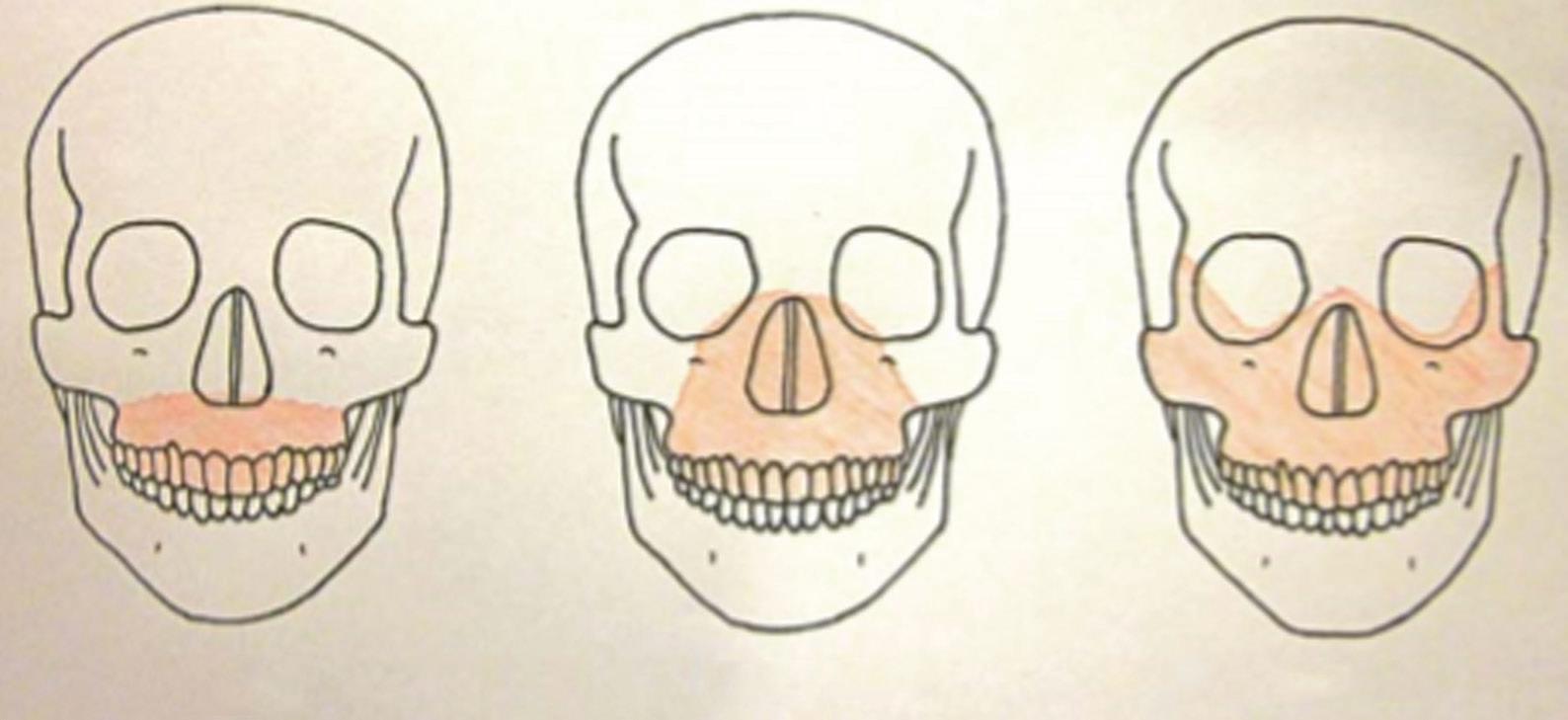
While the appearance of facial injuries can be distracting, it is critical to prioritize management of trauma patients according to clinical importance, following a systematic approach (as per Advanced Trauma Life Support principles). The goal of the airway assessment in the primary survey is to establish any immediate threat to patency, identifying any signs and symptoms of potential airway obstruction (e.g. tracheal deviation, subcutaneous emphysema, and/or marked soft tissue swelling). Traumatic injuries classically associated with airway obstruction include bilateral anterior mandibular fractures and the Le Fort III fracture (Fig. 1.1).
Airway management in the trauma setting should always be presumed to be difficult. Cervical spine movements are often restricted due to the application of a rigid cervical spine collar and/ or manual in-line stabilization. Mouth opening may be restricted by pain, muscle spasm, or mechanical obstruction (e.g. a tripod fracture of the zygomatic complex may interfere with movement of the coronoid process of the mandible). The patient may be combative/ unable to cooperate with an awake tracheal intubation technique,
and there may be the additional risk of aspiration of blood or broken teeth fragments. Having undertaken initial assessment and stabilization of the patient, the secondary survey should include an ‘AMPLE’ (Allergy, Medication, Past medical history, Last food and drink, Event) history, full body examination, and relevant investigations. In particular, the mechanism of injury and magnitude of energy transfer are important factors in guiding identification and assessment of injuries. Classification of facial fractures as lower, middle, and/or upper third may also be helpful in identifying concomitant injuries commonly associated at each level. (Management of the oromaxillofacial trauma patient is discussed in detail in Chapter 13.)
Infection
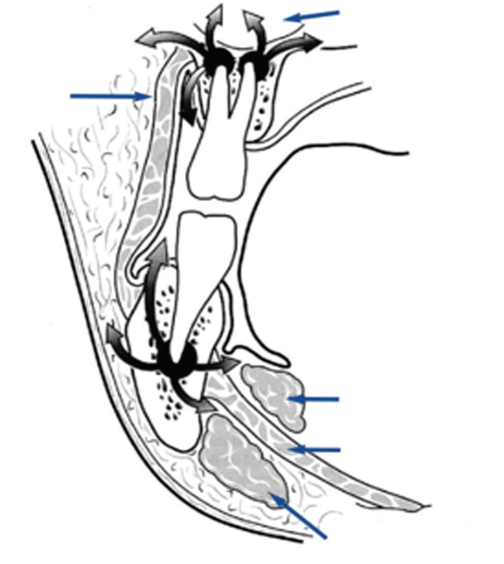
The most common cause of oromaxillofacial infection is impacted teeth, and the resulting dental abscess may cause significant facial swelling and trismus, affecting airway management (rarer causes of infection include tuberculosis, syphilis, and fungal or viral infection in immunocompromised states). Infection usually begins with dental decay and pulpitis, leading to perforation of the bone cortex, allowing the infection to spread to the subperiosteal region. In more severe cases, infection may spread along the fascial planes into the infratemporal fossa, subtemporalis, or even the cervical fascial plane (Fig. 1.2), forming a parapharyngeal abscess and even mediastinitis. The classic example of this is Ludwig’s angina, in which cellulitis extends over the entire floor of mouth, including both the submandibular and sublingual spaces. Preoperative evaluation of the airway reveals difficulty in swallowing secretions and an inability to protrude the tongue. Neck and/or pretracheal fascial involvement complicates the option of tracheostomy formation. In addition to assessing the extent of local spread, the systemic effects of bacteraemia/sepsis via haematogenous spread must be actively sought and promptly managed according to the Surviving Sepsis Campaign.3 (Management of oromaxillofacial infection is discussed in greater detail in Chapter 12.)
Congenital anomalies
Cleft lip and palate are among the most common congenital defects requiring surgery. In addition to the primary repair, patients often require subsequent procedures for lip aesthetics, closure of residual palatal defects, bone grafts, alignment of alveolar and dental defects,
Le Fort ILe Fort IILe Fort III
Fig. 1.1 The Le Fort classification of facial fractures.2
Maxillary sinus
Deep lobe of submandibular gland
Mylohyoid muscle
Superficial lobe of submandibular gland
jaw realignment, and correction of nasal deformity. Although cleft defects per se do not lead to upper airway obstruction and/or difficult airway management, coexisting structural or neuromuscular dysfunction may result in either/both. For example, in Pierre Robin sequence, the combination of micrognathia, glossoptosis, and cleft palate may cause airway obstruction relieved only by prone positioning or insertion of a nasopharyngeal airway, and in some cases necessitating tracheostomy formation. Crucially, beyond the airway examination, the anaesthetist must be aware of the multisystem nature of many of the syndromes associated with cleft anomalies, and make appropriate assessment of cardiac, renal, and skeletal systems as required (Table 1.1).
Malignancy
Airway management may be complicated by distortion, invasion, or compression of tissues by the tumour and/or progressive airway obstruction by the lesion itself. Previous cancer treatments/surgery may also contribute to airway management difficulties (Table 1.2).
Consideration should be given to the specific risk factors associated with head and neck cancer, including human papilloma virus, smoking, and alcohol use, and their associated comorbidities—in particular, chronic obstructive lung disease, ischaemic heart disease, alcoholic cardiomyopathy, liver cirrhosis, and alcohol dependence. There is a 10% risk of a synchronous primary cancer elsewhere within the aerodigestive tract, which should be sought through appropriate imaging. Malnutrition is common due to poor appetite, dysphagia, or side effects of chemotherapy and/or radiation mucositis, and dietician input is advocated to optimize nutritional support preoperatively in order to minimize the reduced wound healing, and increased risk of infection/other postoperative complications associated with it. (Management of the oromaxillofacial patient with malignancy is discussed in detail in Chapter 15.)
Table 1.1 Congenital syndromes associated with cleft lip and palate
Down syndrome
Pierre Robin sequence
Hemifacial microsomia
Treacher Collins syndrome
Velocardiofacial syndrome
Microstomia and macroglossia
Atlantoaxial subluxation and instability
Congenital cardiac disease
Micrognathia
Glossoptosis
Usually easier to intubate with age
Hemifacial and mandibular hypoplasia
Cervical spine abnormalities
Eye and ear malformations
Usually more difficult to intubate with age
Micrognathia and maxillary hypoplasia
Choanal atresia
Eye and ear malformations
Usually more difficult to intubate with age
Microcephaly
Microstomia, flat nasal bridge, velopharyngeal incompetence, tracheal and laryngeal anomalies
Congenital cardiac disease
Small ears, short stature
Stickler syndrome
Klippel–Feil syndrome
Free flap transfer
Micrognathia and flat face
Eye, ear, and joint abnormalities
Congenital cardiac disease
Webbed neck and fused cervical vertebrae
Congenital cardiac disease
Short stature
This surgical technique may be employed to provide tissue for reconstruction. Successful flap perfusion (and surgical outcome) relies upon optimization of perioperative haemodynamics (a relative hyperdynamic circulation with high cardiac output and vasodilation is desirable), thus preoperative assessment of cardiorespiratory reserve is crucial in establishing the appropriateness of the planned procedure. Contraindications might include sickle cell disease and untreated polycythaemia rubra vera due to their hypercoagulable states, and flap survival may be compromised in patients with active vasculitis or peripheral vascular disease. Prolonged preoperative fasting should be avoided in order to minimize any fluid deficits.
Table 1.2 Sequelae of oromaxillofacial and head and neck cancer treatments
Maxillectomy and craniofacial resection
Tongue, floor of mouth surgery
Laryngeal surgery
Neck dissection
Radiotherapy
Difficult facemask seal
Difficult nasotracheal intubation
TMJ pseudoankylosis
Trismus
Limited mandibular space
Fixed tongue
Laryngeal stenosis
Impaired swallowing
Aspiration risk
Damage to cranial nerves IX, X, XII
Vocal cord palsy
Impaired swallowing
Aspiration risk
Limited neck extension
TMJ ankylosis
Osteoradionecrosis of mandible
Carotid artery stenosis
Poor wound healing
Buccinator muscle
Fig. 1.2 Spread of odontogenic infection through the fascial planes.
(Management of OMFS patients for free flap reconstruction is discussed in greater detail in Chapter 15.)
Environmental considerations
Minor dental procedures are commonly performed under sedation or general anaesthesia in short-stay ambulatory care centres, and sometimes dental clinics. Accessibility to drugs and equipment, availability of postoperative recovery facilities, staff numbers, and staff training in airway management, resuscitation, and sedation may vary significantly between the settings. Appropriate patient selection through thorough preoperative assessment is crucial in ensuring patient safety in the remote site/day surgical environment. Patient suitability should be based upon assessment of comorbidities and fitness for day surgery, as well as determining the availability of an appropriate escort, geographical proximity, and access to emergency services if required (as described by the Society of Day Surgery4). The guidelines for the management of children referred for dental extraction under general anesthesia5 state that sedation or general anaesthesia is best delivered in the inpatient setting for patients considered to be of high anaesthetic risk, with significant comorbidities or complex dental problems (Table 1.3), where high dependency unit/intensive care is immediately available or may be arranged in advance. (Dental anaesthesia and sedation are covered in greater detail in Chapters 8 and 6, respectively.)
Evaluation of comorbidities
Obesity and obstructive sleep apnoea syndrome
Obesity is defined as a body mass index (BMI) ≥30 kg/m2; morbid obesity ≥35 kg/m2; and supermorbid obesity ≥50 kg/m2. There is an increasing number of patients with raised BMI presenting for OMFS, and the pathophysiological consequences of obesity pose significant challenges to the anaesthetist.
Obstructive sleep apnoea syndrome (OSAS) arises from periodic partial or complete collapse of the upper airway during sleep,
Table 1.3 Conditions for dental extraction in the inpatient (hospital) setting
High anaesthetic risk
Anatomically or functionally abnormal airway
Significant learning disabilities or behavioural abnormalities
Severe anxiety
Congenital syndromes associated with increased anaesthetic risk
History of adverse reaction to anaesthetic agents
History of complications occurring under general anaesthesia
Family history of significant problems occurring under general anaesthesia
Table 1.4 Classification of the severity of OSAS according to the American Academy of Sleeping Medicine Task Force
Apnoea–hypopnoea index
<5
5–15
15–30
>30
Significant comorbidities
Severe or poorly controlled asthma
Symptomatic cardiac disease, requiring treatment
Coagulopathy, anticoagulant therapy, or antiplatelet therapy
Impaired renal or hepatic function
Unstable metabolic or endocrine disorders
Significant neurological or neuromuscular disorders
Active systemic infection
Haemoglobinopathies
Abnormal BMI (<18.5 kg/m2 or >30 kg/m2)
Severity classification
Normal
Mild
Moderate
Severe
Sleep-related breathing disorders in adults: recommendations for syndrome definition and measurement techniques in clinical research. The Report of an American Academy of Sleep Medicine Task Force. Sleep. 1999;22(5):667–89.
resulting in decreased (hypopnoea) or complete cessation (apnoea) of airflow. The severity of OSAS is defined by the apnoea–hypopnoea index (the number of apnoea or hypopnoea events per hour) (Table 1.4).6 These recurrent episodes of hypoxia can lead to significant cardiopulmonary morbidity, such as pulmonary hypertension and cor pulmonale. Patients often have poor sleep quality due to the frequent arousal that occurs during their sleep cycle to restore airway patency,7 resulting in behavioural disturbances, such as daytime hypersomnolence and sexual dysfunction.
Patients with OSAS presenting for surgery are at increased risk of perioperative airway, respiratory, and cardiovascular complications,8 and these risks are further increased if the condition remains undiagnosed (and untreated) at the time of surgery.9 Respiratory complications (e.g. oxygen desaturation, acute respiratory failure, respiratory arrest, and aspiration pneumonia) are the most common, as the underlying pathophysiology of OSAS may be exacerbated by the depressant effects of anaesthetic medications upon respiratory drive, airway protective reflexes, and arousal responses.8 These problems may be further compounded in patients undergoing major OMFS procedures due to significant alterations in airway anatomy and postoperative oedema.
Obesity per se is not always associated with difficult tracheal intubation; however, many aspects of airway management may be more challenging. Thorough preoperative airway examination should be undertaken, seeking features suggestive of difficult airway management—in particular, a Mallampati score ≥3, a high Wilson score, and increased neck circumference. Often, a standard asleep tracheal intubation is both practical and safe (especially following the advent of videolaryngoscopy); however, a robust strategy for airway management must always be in place prior to induction of anaesthesia.
Obese patients are more prone to both restrictive and obstructive respiratory insufficiency—decreased chest wall and lung compliance results in a restrictive defect, while increased adipose tissue within the pharyngeal walls predisposes to airway collapse during normal breathing and contributes to the development of OSAS. Obese patients should therefore be routinely screened for OSAS preoperatively—including identification of any risk factors (Box 1.1),10 thorough physical examination, use of the STOP-BANG questionnaire (Table 1.5),8 and appropriate investigations such as electrocardiography, pulse oximetry, pulmonary function tests,11 and polysomnography.12 Abnormal spirometry is associated with increased postoperative complications.13 The STOP-BANG questionnaire is currently the most sensitive, specific, and best-validated screening questionnaire for OSAS. A score of 0–2 indicates ‘low risk’, 3–4 indicates ‘intermediate risk’, and a score of ≥5 indicates ‘high
Box 1.1 Predisposing conditions for obstructive sleep apnoea
• Obesity
• Age 40–70 yr
• Male gender
• Excess alcohol intake
• Smoking
• Pregnancy
• Low Physical activity
• Unemployment
• Neck circumference >40 cm
• Surgical patient
• Tonsillar and adenoidal hypertrophy
• Craniofacial abnormalities (e.g. Pierre Robin, Down’s syndrome)
• Neuromuscular disease
Source: Martinez G, Faber P. Obstructive Sleep apnoea. Continuing Education in Anaesthesia Critical Care & Pain. 2011; 11(1): 5–8.
risk’ for OSAS. Patients who have an intermediate-to-high risk score are at greater risk of perioperative complications14 and referral to a sleep specialist for formal evaluation and optimization is recommended. Strategies that may be considered include weight loss, the use of mandibular advancement devices, and non-invasive positive pressure ventilation.6 Utilization of continuous positive airway pressure/bi-level positive airway pressure devices may reduce the incidence of perioperative hypoxic events in obese patients, regardless of whether OSAS has been formally diagnosed.11
Early preoperative identification of OSAS allows initiation of non-invasive ventilation therapy (as indicated) prior to surgery, reducing the overall risk of perioperative complications. Prompt diagnosis also enables the appropriate preoperative planning to occur, influencing the conduct of all aspects of anaesthetic care, including the choice of anaesthetic medications, airway management, and nature of the postoperative care facility. Many patients with OSAS will also suffer from concomitant comorbidities that should be optimized preoperatively.
Obesity is associated with cardiovascular comorbidities such as hypertension, ischaemic heart disease, and arrhythmias. Patients with these conditions should be managed in accordance with the European Society of Cardiology (ESC)/European Society of Anaesthesiology (ESA) guidelines11 or American College of Cardiology (ACC)/American Heart Association (AHA) guidelines15 to achieve satisfactory control prior to any elective surgery.
Table 1.5 STOP-BANG questionnaire, utilized as a screening tool for
Snoring: do you snore loudly (louder than talking or loud enough to be heard through closed doors)?
Yes/No
Tired: do you often feel tired, fatigued, or sleepy during the daytime? Yes/No
Observed: has anyone observed you stopping breathing during your sleep? Yes/No
Blood Pressure: do you have high blood pressure or are you on treatment for high blood pressure? Yes/No
BMI: is your body mass index greater than 35 kg/m2? Yes/No
Age: are you over 50 years old? Yes/No
Neck circumference: is your neck circumference greater than 40 cm (16 inches)? Yes/No
Gender: are you male?
Yes/No
Hall A. Sleep physiology and the perioperative care of patients with sleep disorders. BJA Education. 2014;15(4):167–72.
Obesity is an independent risk factor for perioperative renal dysfunction,16 and although there is no evidence that any particular preoperative optimization strategy is effective in minimizing postoperative renal impairment, general protective strategies such as correcting preoperative anaemia, avoiding nephrotoxic medications, and maintaining adequate volume status should be employed.17
Obesity is also commonly associated with metabolic disorders such as diabetes, hyperlipidaemia, and fatty liver disease. Perioperative hyperglycaemia is associated with increased morbidity and mortality in patients undergoing non-cardiac surgery.18 The 2018 ESA guidelines recommend that laboratory testing to screen for diabetes should be carried out in obese patients prior to elective non-cardiac surgery (if the condition has not already been diagnosed).11 Glycaemic control should be optimized, with elective surgery delayed in order to achieve this.19
Obesity is also associated with anaemia, and deficiencies in various micronutrients such as vitamin D, ascorbic acid, and betacarotene.20 These deficits should be identified during preoperative assessment and corrected before elective surgery is undertaken (anaemia is discussed in detail later in this chapter).
Patients with OSAS should have their condition thoroughly reviewed at preassessment, and, if compliant with existing therapy, asymptomatic, and without significant cardiopulmonary sequelae, can be considered appropriate to proceed with surgery as planned without further investigation and/or treatment. The non-invasive ventilation device used by the patient should be brought into hospital on admission and its use continued throughout the perioperative period,11 unless specifically contraindicated (e.g. if there are surgical concerns following craniofacial surgery). However, patients who have poorly controlled OSAS (or who have developed secondary cardiopulmonary complications) should be referred for further assessment and optimization before elective surgery. In semi-elective/expedited surgery (e.g. cancer surgery), the decision to defer surgery for further investigation and/or treatment for OSAS should take an individualized approach, taking into consideration the urgency and risk of surgery, the severity of OSAS and other comorbidities, and the relative accessibility of proposed investigations/treatments. In urgent or emergency surgery, where preoperative optimization may be precluded, patients considered to be at high risk of OSAS should be presumed to have the condition and managed as such, with measures taken to minimize perioperative complications, including postoperative care in an appropriately staffed and monitored facility.
OSAS is not an absolute contraindication for day surgery, although patient selection is key—taking into consideration the extent of planned surgery, severity of OSAS, comorbidities, anaesthetic technique, postoperative analgesic requirements, and necessity for advanced postoperative monitoring and non-invasive ventilation.
Coronary artery disease and revascularization
Coronary artery disease is a risk factor for perioperative major adverse cardiovascular events.15 For elective surgery, perioperative cardiac assessment should proceed according to the stepwise approaches described in the ‘ESC/ESA guidelines on non-cardiac surgery: cardiovascular assessment and management’11 or the ‘ACC/AHA guideline on perioperative cardiovascular evaluation and management of patients undergoing non-
OSAS
Patient scheduled for surgery with known or risk factors for CAD* (Step 1)
Emergency
No
ACS† (Step 2)
No
Yes
Clinical risk stratification and proceed to surgery
*See Sections 2.2, 2.4, and 2.5 in the full-text CPG for recommendations for patients with symptomatic HP, VHD, or arrhythmias.
†See UA/NSTEMI and STEMI CPGs (Table 2).
Yes
Estimated perioperative risk of MACE based on combined clinical/surgical risk (Step 3)
Low risk (<1%) (Step 4)
No further testing (Class III: NB)
Proceed to surgery
Elevated risk (Step 5)
Evaluate and treat according to GDMT†
Excellent (>10 METs)
Moderate or greater (≥4 METs) functional capacity
Moderate/Good (≥4–10 METs)
No further testing (Class Ila)
Proceed to surgery
No or unknown
Poor OR unknown functional capacity (<4 METs): Will further testing impact decision making OR perioperative care? (Step 6)
Yes
No further testing (Class Ilb)
Pharmacologic stress testing (Class Ila)
If normal
No If abnormal
Proceed to surgery according to GDMT OR alternate strategies (noninvasive treatment, palliation) (Step 7)
Coronary revascularization according to existing CPGs (Class I)
Fig. 1.3 Step-wise approach to assessment of patients with pre-existing coronary artery disease (CAD) undergoing surgery.15 ACS, acute coronary syndrome; CABG, coronary artery bypass graft; CPG, clinical practice guideline; DASI, Duke Activity Status Index; GDMT, guideline-directed medical therapy; HF, heart failure; MACE, major adverse cardiac event; MET, metabolic equivalent; NB, no benefit; NSQIP, National Surgical Quality Improvement Program; PCI, percutaneous coronary intervention; RCRI, Revised Cardiac Risk Index; STEMI, ST-elevation myocardial infarction; UA/ NSTEMI, unstable angina/non-ST-elevation myocardial infarction; VHD, valvular heart disease.
Reproduced with permissions from Fleisher LA, Fleischmann KE, Auerbach AD, Barnason SA, Beckman JA, Bozkurt B, et al. 2014 ACC/AHA guideline on perioperative cardiovascular evaluation and management of patients undergoing noncardiac surgery: a report of the American College of Cardiology/American Heart Association Task Force on Practice Guidelines. Circulation. 2014;130(24):e278–333.
a low combined clinical and surgical risk (<1% risk of a major adverse cardiovascular event) can safely proceed to surgery without additional testing. Many OMFS procedures are considered to be of low surgical risk (e.g. superficial oral and periodontal surgery); however, the presence of ischaemic heart disease alone may elevate the overall risk of major adverse cardiovascular events to ≥1%.21 In such cases, assessment
of patients’ functional capacity is essential, and pharmacological stress testing (and subsequent coronary revascularization) may be indicated (Figs 1.3 and 1.4). Routine preoperative coronary angiography or prophylactic revascularization is not recommended to exclusively reduce perioperative cardiac events22 an approach supported by the recent Coronary-Artery Revascularisation Prophylaxis (CARP) trial,
One of active or unstable cardiac conditions (table 9)
Determine the risk of the surgical procedure (table 3)
Intermediate or high
Consider the functional capacity of the patient
In patients with a poor functional capacity consider the risk of the surgical procedure
High-risk surgery
Cardiac risk factors (table 4)
Consider non-invasive testing. Noninvasive testing can also be considered prior to any surgical procedure for patient counselling, change of peri-operative management in relation to type of surgery and anaesthesia technique.
Interpretation of non-invasive stress test results
risk surgery
Patient or surgical specific factors dictate the strategy, and do not allow further cardiac testing or treatment. The consultant provides recommendations on peri-operative medical management, surveillance for cardiac events and continuation of chronic cardiovascular medical therapy.
Treatment options should be discussed in a multidisciplinary team, involving all peri-operative care physicians as interventions might have implication on anaesthesiological and surgical care. For instance in the presence of unstable angina, depending on the outcome of this discussion, patients can proceed for coronary artery intervention, with the initiation of dual-anti platelet therapy if the index surgical procedure can be delayed, or directly for operation if delay is impossible with optimal medical therapy.
The consultant can identify risk factors and provide recommendations on lifestyle and medical therapy, according to the ESC Guidelines.
In patients with one or more clinical risk factors, preoperative baseline ECG may be considered to monitor changes during the peri-operative period.
In patients with known IHD or myocardial ischaemia, initiation of a titrated low-dose beta-blocker regimen may be considered before surgery .
In patients with heart failure and systolic dysfunction, ACEI should be considered before surgery.
In patients undergoing vascular surgery, initiation of statin therapy should be considered.
In addition to suggestions above:
In patients with one or more clinical risk factors, non-invasive stress testing may be considered.
In addition to suggestions above: Rest echocardiography and biomarkers may be considered for evaluation of LV function and obtaining prognostic information for peri-operative and late cardiac events
No/mild/ moderate stress-induced ischaemia
Bare-metal stent:
Balloon angioplasty:
Surgery can be performed >2 weeks after intervention with continuation of aspirin treatment
Surgery can be performed >4 weeks after intervention.
Dual antiplatelet therapy should be continued for at least 4 weeks.
An individualized peri-operative management is recommended considering the potential benefit of the proposed surgical procedure compared with the predicted adverse outcome, and the effect of medical therapy and/or coronary revascularization.
Surgery can be performed within 12 months after intervention for old-generation DES and within 6 months for new-generation DES.
Continuation or discontinuation of aspirin in patients previously treated with aspirin may be considered in the peri-operative period, and should be based on an individual decision that depends on the peri-operative bleeding risk weighed against the risk of thrombotic complications (see also Table 8).
Surgery
aTreatment should be initiated optimally between 30 days and at least 2 days before surgery and should be continued postoperatively aiming at target resting heart rate of 60–70 beats per minute and systolic blood pressure >100 mmHg. bFor strategy of anaesthesia and perioperative monitoring see appropriate sections.
ACEI = angiotensin converting enzyme inhibitor; CABG = coronary artery bypass graft; DES = drug-eluting stent; ECG = electrocardiogram; IHD = ischaemic heart disease; MET = metabolic equivalent.
Fig. 1.4 Assessment and treatment strategies in the management of patients with cardiac risk factors undergoing surgery.11
Kristensen SD, Knuuti J, Saraste A, Anker S, Botker HE, Hert SD, et al. 2014 ESC/ESA Guidelines on non-cardiac surgery: cardiovascular assessment and management: The Joint Task Force on non-cardiac surgery: cardiovascular assessment and management of the European Society of Cardiology (ESC) and the European Society of Anaesthesiology (ESA). Eur Heart J. 2014;35(35):2383–431. 2014;35(35):2383–431
CABG
Table 1.6 Recommendations from the 2014 ACC/AHA guidelines and the 2014 ESC/ESA guidelines regarding coronary revascularization and the timing of surgery following different revascularization modalities
Routine preoperative coronary angiography or revascularization prior to elective non- cardiac surgery
ACC/AHA guidance13
Not recommended unless revascularization is otherwise indicated according to existing guidance for stable coronary artery disease
ESC/ESA guidance9
Not recommended unless revascularization is otherwise indicated according to existing guidance for stable coronary artery disease
Recommended duration that elective surgery should be delayed following balloon angioplasty 14 days 14 days
Recommended duration that elective surgery should be delayed following bare-metal stent insertion 30 days
Recommended duration that elective surgery should be delayed following drug-eluting stent (DES) insertion
6 months for newer-generation DESs 12 months for conventional DESs
Minimum of 30 days, but ideally 3 months
6 months for newer-generation DESs 12 months for conventional DESs
Kristensen SD, Knuuti J, Saraste A, Anker S, Botker HE, Hert SD, et al. 2014 ESC/ESA Guidelines on non-cardiac surgery: cardiovascular assessment and management: The Joint Task Force on non-cardiac surgery: cardiovascular assessment and management of the European Society of Cardiology (ESC) and the European Society of Anaesthesiology (ESA). Eur Heart J. 2014;35(35):2383–431
Fleisher LA, Fleischmann KE, Auerbach AD, Barnason SA, Beckman JA, Bozkurt B, et al. 2014 ACC/AHA guideline on perioperative cardiovascular evaluation and management of patients undergoing noncardiac surgery: a report of the American College of Cardiology/American Heart Association Task Force on Practice Guidelines. Circulation. 2014;130(24):e278–333.
which showed no difference in perioperative or long-term cardiac outcomes with preoperative prophylactic coronary revascularization, even in the setting of high-risk surgery.23
If revascularization is indicated, the two principal options are percutaneous coronary intervention (PCI) and coronary artery bypass grafting (CABG). The choice of technique is beyond the scope of this chapter, but is dependent upon various factors, including the extent of the coronary artery disease. Following satisfactory CABG revascularization, the risk of subsequent perioperative myocardial ischaemia may be considered relatively low, but it is often recommended to postpone elective non-cardiac surgery for at least 3 months.24 The recommendations regarding perioperative risk and timing of surgery following PCI varies according to the particular intervention undertaken (Table 1.6)—for example, if surgery is time sensitive (required within weeks), then PCI with a bare-metal stent may be more appropriate than a drug-eluting stent to avoid the greater period of anticoagulation required for the latter (discussed in detail later in the chapter).
In the elective setting, patients with stable ischaemic heart disease (who do not fulfil the criteria for coronary revascularization) should still be referred to a cardiologist for optimization of medical therapy and to ensure long-term follow-up. In urgent or emergency surgery, where there is limited time for evaluation and/or optimization prior to surgery, all members of the multidisciplinary team involved in the patient’s care (anaesthetist, surgeon, cardiologist, intensivist, and/or haematologist), as well as the patient and their family, should discuss together the increased perioperative cardiovascular risk and the appropriateness of surgery. Specific measures to mitigate risk may be considered, such as performing the surgery at a centre where emergency coronary revascularization is available, and upgrading the level of perioperative haemodynamic monitoring and postoperative care facility. In extreme circumstances, where revascularization is absolutely necessary and surgery cannot be postponed, CABG may be undertaken as part of the proposed surgery.15 In the case of a patient with acute coronary syndrome presenting for elective surgery, priority should be given to the evaluation and management of acute coronary syndrome according to established guidelines, providing that the condition requiring elective surgery is not life-threatening.
Cardiac murmurs
Patients who present to preoperative assessment with a heart murmur may be classified into three subgroups:
• One or more isolated valvular lesions (involving aortic/pulmonary/tricuspid/mitral valves).
• Complex congenital heart disease with a combination of intra- or extracardiac defects.
• A flow murmur, where the cardiovascular system is otherwise physiologically and anatomically normal.
Patients with known or suspected valvular heart disease should be evaluated by formal echocardiography prior to elective OMFS, in order to identify the site(s) and severity of lesion(s), and to assess for any complications (unless echocardiography has been performed within 1 year and there has been no change in the patient’s clinical status).15 If a patient has a valvular lesion of sufficient severity (based on clinical and/or echocardiographic criteria) to warrant intervention (replacement or repair), then this should be undertaken before proceeding to any elective OMFS procedure of intermediate to high risk.15 However, if a valvular lesion is present but is of insufficient severity to warrant valvular intervention, elective surgery may still proceed, but consideration should be given to the use of invasive haemodynamic monitoring and postoperative intensive care for ongoing monitoring and/or organ support.15 For patients with uncorrected valvular lesions who require urgent or emergency surgery, where there is limited time for evaluation and optimization, surgery may proceed providing that there is multidisciplinary team involvement in the patient’s care, and the patient and their family are aware of the increased perioperative cardiovascular risk; the type and severity of the valvular lesion is known; the chosen anaesthetic approach is appropriate for the existing valvular lesion; and the appropriate level of perioperative cardiovascular monitoring and support is available.
Patients with corrected valvular lesions which are functioning well may be regarded as physiologically normal, and hence do not carry an increased cardiovascular risk during OMFS procedures unless there are other risk factors present. These patients are normally under regular review by a cardiologist and, as such, any subsequent deterioration in valvular or cardiac function will usually be detected early. Patients with prosthetic metallic valves will require an appropriate perioperative anticoagulation bridging regimen.
Patients with a history of complex congenital heart disease are generally considered to have an increased perioperative cardiovascular risk, but the precise degree of risk is highly variable because
it is dependent upon the nature of the pre-existing cardiac condition itself, the extent of previous surgical corrections, the presence of associated complications (e.g. heart failure, pulmonary hypertension, and dysrhythmias), the proposed surgical procedure, and the urgency of surgery. The ESC/ESA guideline11 and the 2014 ACC/ AHA guideline15 both recommend that preoperative evaluation and any subsequent elective non-cardiac surgery should be carried out by an expert multidisciplinary team at a specialist centre. In the case of urgent or emergency procedures where there is minimal time for evaluation and/or optimization prior to surgery, all clinicians involved in patient care and the patient/family should be made aware of the increased perioperative risk. If it is not practically possible for surgery to be performed at a specialist centre due to the urgency of the condition, expert advice should be sought from an affiliated specialist centre regarding the most appropriate perioperative optimization strategies.
Patients with a physiological flow murmur only are regarded as physiologically normal and do not carry an increased cardiovascular risk during OMFS procedures unless there are other risk factors present. The difficulty lies in determining if the murmur is, in fact, merely a functional flow murmur or if there is a pathological element to it. If other concerns are also identified during the preoperative assessment, then appropriate consideration should be given to further investigation of the murmur.
Arterial hypertension
Uncontrolled arterial hypertension is one of the most common reasons for deferring elective surgery.
Indeed, there is an association between hypertension and adverse perioperative cardiovascular complications, but only when the severity of arterial hypertension is grade 3 or above (Table 1.7).25,26 As such, the potential benefit of achieving improved blood pressure control in order to minimize cardiovascular complications has to be balanced against the considerable socioeconomic cost, psychological burden upon patients and their families, and the risk of disease progression associated with unnecessary postponement of operations.
The Association of Anaesthetists has published guidelines regarding the management of arterial hypertension in patients scheduled for elective non-cardiac surgery. Firstly, any abnormal blood
Optimal <120 and
Normal 120–129 and/or 80–84
High normal 130–139 and/or 85–89
Grade 1 hypertension 140–159 and/or 90–99
Grade 2 hypertension 160–179 and/or 100–109
Grade 3 hypertension ≥180 and/or ≥110
Isolated systolic hypertension ≥140 and <90
Williams B, Mancia G, Spiering W, Agabiti Rosei E, Azizi M, Burnier M, et al. 2018 ESC/ESH Guidelines for the management of arterial hypertension. Eur Heart J. 2018;39(33):3021–104.
Surgeon receives referral
Documented SBP <160 mmHg AND DBP <100 mmHg in past year?
Request measurement from GP
Lowest SBP <160 mmHg AND the lowest DBP <100 mmHg?
No No
Measures BP up to 3 times
Lowest SBP <180 mmHg AND the lowest DBP <110 mmHg?
Fig. 1.5 Stepwise approach to blood pressure assessment in patients referred for elective surgery.28 *The GP should be informed of blood pressure readings in excess of 140 mmHg systolic or 90 mmHg diastolic, so that the diagnosis of hypertension can be refuted or confirmed and investigated and treated as necessary. DBP, diastolic blood pressure; SBP, systolic blood pressure.
Reproduced with permissions from Hartle A, McCormack T, Carlisle J, Anderson S, Pichel A, Beckett N, et al. The measurement of adult blood pressure and management of hypertension before elective surgery: Joint Guidelines from the Association of Anaesthetists of Great Britain and Ireland and the British Hypertension Society. Anaesthesia. 2016;71(3):326-37.
pressure readings should be confirmed by multiple readings, with the use of ambulatory or home blood pressure monitoring if necessary.27 Grade 2 hypertension or below should be managed according to established guidelines for the treatment of hypertension (e.g. National Institute for Health and Care Excellence guidelines), but surgery may proceed as planned and regular antihypertensive medications continued through the perioperative period. However, if the systolic blood pressure is ≥180 mmHg or diastolic blood pressure ≥110 mmHg (i.e. at least grade 3 hypertension), the patient should be referred for further investigation and treatment prior to surgery (Fig. 1.5).28 In the case of urgent or emergency operations where there is limited opportunity for preoperative optimization, all those who are involved in the patient’s care, as well as the patients and their families, should be made aware of the increased perioperative risk. Specific measures to mitigate the risk should be considered, such as employing invasive haemodynamic monitoring, and providing an enhanced level of postoperative care.
Anaemia
Anaemia is defined by the World Health Organization as a haemoglobin level of <13 g/dL for males and <12 g/dL for females. Preoperative anaemia has been shown to be associated with increased morbidity and mortality in patients undergoing major non-cardiac surgery, as well as an increased risk of allogeneic blood transfusion perioperatively, which itself is associated with significant morbidity.29 This has led to the development of various perioperative blood conservation strategies and the concept of patient blood management (PBM). PBM involves a patient-centred, multidisciplinary, multimodal, and evidence-based approach to the management of perioperative anaemia. The primary aim of PBM is to avoid unnecessary blood transfusion thereby minimizing the associated morbidity, improving patient outcome, and minimizing resource utilization. This strategy is supported by the World
Table 1.7 Classification of blood pressure and grade of hypertension
Table 1.8 The three pillars of PBM: preoperative components
Pillar 1: detection and management of anaemia Pillar 2: minimization of bleeding and blood loss Pillar 3: management of anaemia and optimization of tolerance
Aim for assessment of anaemia 4–6 weeks before surgery
Identify, evaluate, and treat anaemia
Treat absolute or functional iron deficiency with oral or intravenous iron
Consider erythropoiesis stimulating agents if nutritional anaemia is excluded/treated
Refer for further evaluation as necessary
Identify and manage bleeding risk (past medical and family history)
Review medications (antiplatelets, anticoagulants)
Minimize iatrogenic blood loss
Procedure planning and rehearsal
Compare estimated blood loss with patient- specific tolerable blood loss
Assess and optimize patient’s physiological reserve, e.g. pulmonary and cardiac function
Formulate patient- specific management plan using appropriate blood conservation modalities
Au TH, Castillo S, Morrow L, Malesker M. Obstructive sleep apnea and continuous positive airway pressure: A primer for pharmacists2015. 30–3.
Health Organization and many of its principles have been endorsed and adopted internationally, including by the European Society of Anaesthesiology, the National Blood Transfusion Committee in the UK, the National Blood Authority in Australia, and the American Association of Blood Banks in the US. Central to the concept of PBM are the so-called three pillars of PBM (Table 1.8):
• Detection and management of anaemia to optimize red cell mass.
• Minimization of blood loss.
• Managing and optimizing patients’ tolerance to anaemia.30
Each pillar comprises preoperative, intraoperative, and postoperative components. Each pillar should be considered in turn during the preoperative assessment, with appropriate involvement of the multidisciplinary team (surgeon, anaesthetist, haematologist, and other relevant clinicians).
The first pillar of PBM in the preoperative phase includes all the strategies to detect and manage pre-existing anaemia, such that red cell mass is optimized at the time of surgery. Patients scheduled for major elective surgery (with an estimated blood loss of ≥500 mL)31 should undergo screening tests for anaemia 4–6 weeks before surgery.30 Any underlying cause should be identified and treated accordingly. Iron deficiency remains the most common cause of anaemia worldwide and patients with iron deficiency anaemia should be treated with oral iron therapy in the first instance. If the patient has failed oral iron therapy (insufficient response to treatment or intolerance), or if surgery is planned in <6 weeks, intravenous iron therapy is recommended as an alternative (as 3 months are required to replenish body iron stores fully). Other nutritional deficiencies should be treated with haematinics. The use of erythropoietinstimulating agents may be considered if nutritional deficiencies have been corrected or excluded30 the 2018 ESA guidelines suggest that these agents may be used in conjunction with conventional iron therapy to accelerate red cell mass production.12 Blood tests should be rechecked following a course of appropriate treatment.
The second pillar of PBM includes strategies to minimize bleeding and blood loss. In the preoperative phase, this should include a comprehensive review of the patient’s medical background, drug history, laboratory results, and physical examination findings in order to identify conditions which predispose to bleeding and coagulopathy. These conditions (e.g. congenital bleeding disorders) should be corrected and optimized where possible. Patients who are taking anticoagulant medications should undergo an assessment of the thromboembolic risk associated with cessation of anticoagulant therapy versus the risk
of perioperative bleeding associated with continued use. In some cases, a perioperative bridging regimen will be indicated. The proposed surgical procedure and technique should be carefully planned in advance by the surgical team with consideration to the risk of bleeding so that iatrogenic blood loss due to the procedure itself is minimized.
The third pillar of PBM includes strategies to manage and optimize the patient’s tolerance to anaemia. This refers to the ability of the patient’s cardiopulmonary reserve to compensate for a decrease in oxygen-carrying capacity via an increase in cardiac output. Hence, it is important to assess the functional reserve of the cardiopulmonary system during the preoperative assessment in accordance with the approaches outlined in the 2014 ESC/ESA guideline11 and 2014 ACC/AHA guideline.15
Every attempt should be made to correct anaemia to normal levels by implementing PBM principles prior to proceeding with elective surgery. Some of these principles may still be applicable for patients who require urgent or emergency surgery, though it may still be necessary to proceed despite suboptimal preoperative evaluation and optimization.
Preoperative risk stratification
Although low-risk procedures account for a significant proportion of OMFS caseload, in-hospital mortality has been reported to be around 3% for patients undergoing major head and neck operations within NHS England, while the overall mortality is around 1% for all maxillofacial and head and neck procedures in the UK. It is therefore important to identify patients at increased risk of perioperative morbidity and mortality to allow shared decision-making, informed consent, preoperative optimization, and modification of surgical pathways as required. While various risk stratification tools are available, the accuracy of risk prediction remains uncertain because OMFS accounted for only a small proportion of the surgical procedures included in the databases used during prediction tool development, and consequently there is a paucity of evidence supporting their validity in this particular field.
Prediction
of morbidity and mortality
American Society of Anesthesiologists Physical Status (ASA-PS) score
The ASA-PS classification system was first developed in 1941 as a statistical tool for retrospective analysis of hospital records and was
updated in 2014 to include BMI, alcohol intake, and smoking. An ASA class is assigned based upon the severity of systemic disease, if any, and the likelihood of survival without an operation being undertaken. The ASA-PS is widely adopted across many specialties, is simple to perform, and is validated for its association with perioperative morbidity and mortality. However, the risk specific to the planned surgical procedure is not taken into account. The ASA-PS is also reported to have poor inter-rater reliability, even among anaesthetists, due to the subjective nature of assessment.
Physiological and Operative Severity Score for the enUmeration of Mortality and Morbidity (POSSUM)
POSSUM was developed by Copeland et al. in 1991 from patients undergoing both elective and emergency gastrointestinal, hepatobiliary, urological, and vascular surgery. It uses 12 physiological and six surgical variables for the calculation of 30-day mortality. Portsmouth-POSSUM (P-POSSUM) is a newer model that requires the same input variables as POSSUM, but uses alternative equations for risk calculation. Both POSSUM and P-POSSUM are well validated in other surgical specialties; however, patients undergoing OMFS procedures were not included in the database. Therefore, the validity of these scores remains uncertain despite one small studying demonstrating reasonable discrimination.32 In addition to a number of subjective assessments of physiological variables, input of intraoperative findings is also required, which limits the applicability and practicality of these tools in the preoperative setting.
Surgical Outcome Risk Tool (SORT)
In 2014, Protopapa et al. developed SORT from post hoc analysis of data from the ‘Knowing the Risk’ report of the National Confidential Enquiry into Patient Outcome and Death (NCEPOD), which comprised 16,788 patients undergoing both elective and emergency inhospital surgical procedures. SORT requires the input of only six preoperative variables: ASA-PS, urgency of surgery, surgical specialty, severity of surgery, presence/absence of cancer, and age. It is simple to use, but it does not calculate morbidity and, to date, has not been validated for OMFS.
American College of Surgeons National Surgical Quality Improvement Project (NSQIP) surgical risk calculator
The NSQIP surgical risk calculator was developed in 2013, based on both inpatients and outpatients undergoing major surgical procedures in the private healthcare setting in the US. It requires input of 21 preoperative variables, such as age, sex, BMI, dyspnoea, previous myocardial infarction, and functional status. NSQIP has the largest data pool, consisting of 1.4 million patients, and predicts procedure-specific morbidity and mortality risks in addition to 14 other postoperative outcomes. However, the tool lacks validation in other populations, and the differing service sector must be taken into account.
Cardiopulmonary exercise testing (CPET)
CPET is the most sensitive and specific exercise capacity test for the purposes of preoperative risk stratification (compared with the shuttle test, 6-minute walk test, and the stair climb test). CPET allows assessment of the cardiopulmonary function by exercising patients on a bike ergometer through four key physiological stages (cardiodynamic, increased cellular respiration, steady state, and
incremental work phases), deriving anaerobic threshold, peak oxygen consumption (peak VO2), and ventilatory equivalents (VE/ VECO2) —all of which have been shown to be independent predictors of morbidity, mortality, and length of hospital stay. In addition to risk stratification, CPET can also direct preoperative optimization, identify undiagnosed pathology, and assess effectiveness of neoadjuvant cancer therapies. Its role in risk stratification and decision-making has been explored in a few small studies on patients with head and neck malignancy (with or without tissue reconstruction), and, although the findings are encouraging, further investigation is required.33,34 It should be noted that CPET carries a reported mortality of 2–4 in 100,000, and is therefore not suitable for all patients, and should be undertaken in an appropriately equipped facility by trained personnel, as recommended by the American Thoracic Society and the Perioperative Exercise Testing and Training Society.35
Prediction of cardiac complications
Lee’s Revised Cardiac Risk Index (RCRI)
Lee’s RCRI was developed in 1999 by Lee et al. from 4315 patients undergoing non-emergency operations, to predict the risk of developing cardiac complications after major non-cardiac operations (including myocardial infarction, pulmonary oedema, ventricular fibrillation, cardiac arrest, and complete heart block). It utilizes six independent factors: high-risk surgery (intraperitoneal, intrathoracic, or suprainguinal vascular procedures); ischaemic heart disease; history of congestive heart failure; cerebrovascular disease (transient ischaemic attack or cerebrovascular accident); diabetes mellitus, on insulin; and preoperative serum creatinine >176 µmol/L). It is simple to use and is highly recommended by consensus guidelines on preoperative evaluation for non-cardiac surgery, including those by the ESC/ESA11 and ACC/AHA.15 Only one independent predictor refers to the nature of the planned operation, and its applicability to patients undergoing emergency or minor procedures remains unclear.
NSQIP Myocardial Infarction and Cardiac Arrest (MICA)
NSQIP-MICA was developed in 2011 from a multicentre study of 211,410 patients. It uses five predictors of perioperative myocardial infarction or cardiac arrest: type of surgery, functional status, abnormal serum creatinine, ASA, and age. NSQIP-MICA outperforms the RCRI in discriminative power among patients undergoing vascular surgery and is recommended for cardiac risk stratification by the ESA, whose consensus view is that NSQIP-MICA and Lee’s RCRI are complementary in providing prognostic perspective in the decision-making process.
Biomarker assays
Preoperative blood levels of natriuretic peptides (brain natriuretic peptide or N-terminal pro-brain natriuretic peptide) and C-reactive protein have been shown to have incremental predictive value in several observational studies.36–38 However, there are no data to suggest that targeting these biomarkers for treatment and intervention will reduce the postoperative risk. As such, the ESA does not recommend routine preoperative biomarker sampling in all patients for risk stratification purposes, but suggests brain natriuretic peptide levels may be used to provide independent prognostic information on perioperative and late cardiac events in high-risk patients undergoing major surgery.