[FREE PDF sample] Mirroring brains: how we understand others from the inside giacomo rizzolatti eboo
Visit to download the full and correct content document: https://ebookmass.com/product/mirroring-brains-how-we-understand-others-from-theinside-giacomo-rizzolatti/
More products digital (pdf, epub, mobi) instant download maybe you interests ...
The Evolution of Biological Information: How Evolution Creates Complexity, from Viruses to Brains Christoph Adami
Great Clarendon Street, Oxford, OX2 6DP, United Kingdom
Oxford University Press is a department of the University of Oxford. It furthers the University’s objective of excellence in research, scholarship, and education by publishing worldwide. Oxford is a registered trade mark of Oxford University Press in the UK and in certain other countries
The translation of this work has been funded by SEPS Segretariato Europeo Per Le Pubblicazioni Scientifiche
Via Val d’Aposa 7 - 40123 Bologna - Italy seps@seps.it - www.seps.it
The moral rights of the authors have been asserted First Edition published in 2023
Impression: 1
All rights reserved. No part of this publication may be reproduced, stored in a retrieval system, or transmitted, in any form or by any means, without the prior permission in writing of Oxford University Press, or as expressly permitted by law, by licence or under terms agreed with the appropriate reprographics rights organization. Enquiries concerning reproduction outside the scope of the above should be sent to the Rights Department, Oxford University Press, at the address above
You must not circulate this work in any other form and you must impose this same condition on any acquirer
Published in the United States of America by Oxford University Press 198 Madison Avenue, New York, NY 10016, United States of America
British Library Cataloguing in Publication Data Data available
Library of Congress Control Number: 2022946251
ISBN 978–0–19–887170–5
DOI: 10.1093/oso/9780198871705.001.0001
Printed in the UK by Bell & Bain Ltd., Glasgow
Oxford University Press makes no representation, express or implied, that the drug dosages in this book are correct. Readers must therefore always check the product information and clinical procedures with the most up-to-date published product information and data sheets provided by the manufacturers and the most recent codes of conduct and safety regulations. The authors and the publishers do not accept responsibility or legal liability for any errors in the text or for the misuse or misapplication of material in this work. Except where otherwise stated, drug dosages and recommendations are for the non-pregnant adult who is not breast-feeding
Links to third party websites are provided by Oxford in good faith and for information only. Oxford disclaims any responsibility for the materials contained in any third party website referenced in this work.
PREFACE
Atthe end of the 1990s, Michael Arbib, mathematician and neuroscientist of the University of Southern California, dashed off a note to Giacomo Rizzolatti, ‘We need to get a move on and write a book about the mirror neurons, because in a couple of years they’ll be common knowledge and no one will be interested in them any more’. In the end, we wrote the book, although to be honest we took our time about it. So quel che fai. Il cervello che agisce e i neuroni specchio was published in Italian in 2006 with the English version following nine months later as Mirrors in the Brain: How our minds share actions and emotions. Contrary to Michael’s prediction, however, mirror neurons did not lose their appeal and indeed still continue to be of interest to specialists and the general public alike, as is borne out by the success of that first book.
Over a decade has passed, and mirror neurons are still intriguing, even more so than before. Today we know that the mirror property—that property whereby a neuron responds both when we ourselves are engaged in a given behaviour and when we observe someone else engaged in it—is not an oddity of a handful of neurons in a very small portion of the premotor cortex, but characterizes a goodly part of our primate brain. A huge number of studies have contributed to substantially increasing our knowledge of how these neurons work and the role they might play, so our understanding of them is now much richer and multifaceted. These studies were not limited to the human brain; mirror
neurons have also been identified in species very different from ours, such as marmosets, songbirds, rats, and bats. Given these developments, we decided to write another book together to present these new experimental findings and above all, explore their relative theoretical implications.
A main aim of this book is to show that the mirror property refers to a fundamental neuronal mechanism. Chapter 1 tackles this, describing the brain structures that currently are known to possess the mirror property and analysing the kind of mechanism they instantiate, so highlighting how mirror responses implicate a transformation of the sensory representations of the behaviour of others into the processes and representations that the observer would recruit if they were to behave in that self-same manner. As the anatomical and functional characteristics of the areas endowed with the mirror property can differ significantly from one brain structure to another, the incoming sensory representations as well as the outgoing processes and representations may vary. We will see that there are differences between the mirror responses recorded from the premotor and motor areas and, for example, those of the insula or the amygdala. In spite of these differences, however, all mirror responses embody a mechanism that transforms the processes and representations concerning a given behaviour we observe in other people into the processes and representations which are recruited when we produce that kind of behaviour ourselves. Given the nature and diffusion of the mirror responses, this mechanism appears to be a fundamental principle of the organization and functioning of the whole brain.
Chapter 2 focuses on actions, reviewing both the early and more recent studies. These suggest that mirror responses can involve a transformation of the sensory representations of the observed
actions into the motor representations of their action goals similar to the representations that the observer would recruit if they were to plan and execute actions of that kind. We will show that this transformation is not connected to the merely visual aspects of the action being observed, nor to a specific sensory modality, but rather depends primarily on the capacity of the observer to motorically represent possible action goals. This is substantiated by the fact that the more this capacity is developed, the greater the possibility to mirror these goals when the actions are observed rather than executed.
One of the most fundamental discoveries in this field is that the mirror mechanism is not the prerogative of the cerebral areas concerned with the motor representation of action; it is also present in certain brain structures such as the insula, the amygdala and the cingulate gyrus that are known for being involved in producing motor and visceral responses characteristic of specific emotions such as, for instance, disgust and fear. Chapter 3 highlights that observing a grimace of disgust, a look of fear or a sonorous laugh triggers visceromotor representations similar to the representations that the observer would recruit if they were to experience the same kind of emotions themselves.
In Chapter 4 we argue that mirror responses may also concern what Daniel Stern defined as vitality forms. These forms characterize the dynamics of an action or an emotional reaction; for example, we speak about an energetic handshake, a delicate caress, a violent outburst of rage, a hint of a smile. Research in this field is still in the early stages, but there is enough data to suggest that there are indeed areas, such as the dorso-central portion of the insula, equipped with the mirror property for action-related vitality forms. Hence observing an action being executed gently or
energetically prompts motor processes and representations that are similar to those the observer would recruit if they themselves were to execute that action with that particular vitality form.
Now that it has been ascertained that the mirror mechanism is present in various cerebral regions of different species and that, at least in the case of the primate brain, the transformations it entails can affect not only possible action goals but also emotional reactions and affective aspects, the question remains as to whether this mechanism has some form of cognitive function, and if so, what form this takes. Without a doubt, if not the most debated question of recent years, this is certainly one of the thorniest.
In the final two chapters, we develop and defend the theory that the mirror mechanism plays a distinctive role in understanding the actions, emotional reactions, and vitality forms we observe in others. In Chapter 5 we review the principal evidence that has emerged over the years in support of this theory, while in Chapter 6 we clarify the kind of understanding that mirror responses enable. In this regard, we have to distinguish between basic and full-blown understanding. While a basic understanding of an action can be reached by simply identifying its possible goal(s), a full-blown understanding requires a degree of knowledge of the states that motivated the execution of the action. The same holds true for emotions and vitality forms; it is one thing to identify the kind of emotion or the kind of vitality form displayed by an individual, it is quite another to account for why that individual, with their particular character, state of mind, sensitivity, and beliefs, experienced that kind of emotion or displayed that vitality form in that particular situation.
This allows us to take a first step towards clarifying in what sense the mirror mechanism plays a distinctive role in understanding
other people’s actions, emotions, and forms of vitality. In fact, we will argue and provide evidence that, all things being equal, mirror responses and their corresponding transformations are sufficient for a basic understanding of an action, emotional reaction, or a vitality form, while at the same time influencing the ability of the observer to judge them. This, of course, does not rule out the possibility that the actions, emotions, and vitality forms we observe in other people can be understood by processes and representations other than those evoked by mirror responses and their corresponding transformations. Indeed, drawing comparison with other forms of understanding will allow us to specify what kind of understanding of actions, emotions, and vitality forms we observe in other individuals can be attributed to mirror responses.
This leads us to introduce and discuss the notion of understanding from the inside, to which we have dedicated much thought in recent years and for which, in its initial form, we owe thanks to Marc Jeannerod. The phrase from the inside primarily refers to the fact the mirror mechanism would allow us to capture the ‘intrinsic components’ of an action we are observing, starting from its possible goals (Jeannerod, 2004). Mirror responses, in fact, would enable us to ‘go inside’ the observed action, by penetrating beyond the surface of its sensory aspects as we can capitalize on motor processes and representations similar to those we recruit when executing an action of that kind ourselves.
There is nothing strange or mysterious in all this. Anyone who is expert at activities involving particular motor skills, whether it be performing ballet, playing the piano, or playing basketball, is aware of having privileged access to the actions involved in these activities when they observe them being executed by others—and this even more so when the level of skills is similar to their own.
Not surprisingly, people who possess such skills understand the related actions more quickly and accurately than those who do not, as in understanding them, they deploy the same neuronal and representational resources they would use if they were actually executing the actions themselves. This is true to an even greater extent for emotions, as we will see when we discuss some emblematic cases of patients with lesions in brain structures with the mirror property.
The notion of understanding from the inside is susceptible to a second definition that in a certain sense integrates and expands the first. In fact, the understanding from the inside is not only characterized by the recruitment of representations usually involved in the carrying out of actions or emotional reactions, but also, and above all, by the fact that these representations can shape the experience of actions or emotions also when they are observed being performed or displayed by others. As we will see in Chapters 5 and 6, there is evidence to suggest that our experience of action or an emotion changes according to how it is represented motorically or viscerally. This applies not only to those actions and emotions we experience directly ourselves but also to those we observe in others. Assuming that this is indeed the case, understanding actions, emotions, and vitality forms of other people from the inside involves an experience of them, which, while undoubtedly differing from first-person action or emotion experience, will still share certain fundamental phenomenological aspects.
Apart from the obvious differences, executing an action or living an emotion and observing another person doing the same can be phenomenologically similar with respect to the kind of action or emotion in question. In some respects, what we experience when we observe others performing an action or living an emotion is
what we experience when we perform that kind of action or live that kind of emotion ourselves. Mirroring the actions or emotions we observe in others enables us to share processes and representations but also, and most importantly, experiences; probably this is what makes it so special.
ACKNOWLEDGEMENTS
There are books that owe their existence to the efforts of more than one person; this is one such, and not just because it has two authors. Much of the data and many of the ideas presented in this volume are the result of years of research, with the significant contributions of many friends and colleagues. It would be impossible to mention them all, but we are grateful to each and every one of them. A particular vote of thanks is due to Michael Arbib, Luca Bonini, Stephen A. Butterfill, Luigi Cattaneo, Gergely Csibra, Luciano Fadiga, Pier Francesco Ferrari, Leonardo Fogassi, Vittorio Gallese, Alvin A. Goldman, Pierre Jacob, Christian Keysers, James Kilner, Roger Lemon, Guy A. Orban, Jean Michel Roy, Barry C. Smith, for having helped us to reflect on the mirror mechanism and its role in understanding the actions and emotions of others. A special mention is due to Daniel Stern, with whom a few years ago we started to explore the processes underlying the representation of our own vitality forms and those of others. We would also like to thank the team of the Claudio Munari Center for Epilepsy Surgery of Milan’s Niguarda Hospital, in particular Ivana Maria Sartori and Giorgio Lo Russo, and Pietro Avanzini. A sincere thank you is due also to Fausto Caruana, especially for the section regarding disgust and laughter, and to Marzio Gerbella, who not only contributed a number of invaluable suggestions regarding the anatomical and functional characteristics of the various cerebral
networks endowed with the mirror property, but also many of the illustrations contained in this volume.
Finally, our heartfelt gratitude goes to the late Giulio Giorello for his constant and unwavering support and to Raffaello Cortina who particularly desired us to write this book; to Mariella Agostinelli for the grace and competence with which she monitored our progress, and Giorgio Catalano and Martina Scarpa for their invaluable editorial assistance and attention to detail.
ABBREVIATIONS
ACC Anterior cingulate cortex
AI Anterior insula
AIP Anterior Intraparietal area
aMCC Anterior middle cingulate cortex
cTBS Continuous thetaburst stimulation
DCI Dorso-central insula
EBA Extrastriate body area
EEG Electroencephalogram
ERP Event related potentials
FEF Frontal eye field
fMRI Functional magnetic resonance imaging
IPL Inferior parietal lobe
LIP Lateral intraparietal area
MI/F1 Primary motor cortex
MCC Middle cingulate cortex
MEG Magnetoencephalography
MEP Motor evoked potential
MT Medial temporal cortex
pACC Pregenual anterior cingulate cortex
PAG Periaqueductal gray
PCC Posterior cingulate cortex
PET Positron emission tomography
pMCC Posterior middle cingulate cortex
PMd/F2 Dorsal premotor cortex
PMv Ventral premotor cortex
Pre-SMA/F6 Pre-supplementary motor area
pSTS Posterior superior temporal sulcus
RSC Retrosplenial cingulate cortex
rTMS Repetitive transcranial magnetic stimulation
SII Secondary somatosensory cortex
sACC Subgenual anterior cingulate cortex
SMA/MII/F3 Supplementary motor area
STS Superior temporal sulcus
TMS Transcranial magnetic stimulation
TPJ Temporo-parietal junction
VIP Ventral intraparietal area
vPCC Ventral posterior cingulate cortex
CHAPTER 1
A MIRRORING BRAIN
Overthe years, several cerebral areas have been found to possess the mirror property. In this chapter we will map those areas and will show that far from characterizing a single brain network, this property can be considered a fundamental principle of the functioning of the entire nervous system. A neuron is said to be a mirror neuron if it fires both when an individual displays a kind of behaviour and also when they observe the same kind of behaviour being displayed by others. In the following pages, we will begin with the first studies carried out in the 1990s by a group of researchers at the University of Parma that led to the discovery of mirror neurons in the premotor cortex of the macaque. We will then focus on the subsequent research that revealed the presence of mirror neurons in other areas of the frontal, parietal, and prefrontal lobes. We will also review, albeit briefly, the vast literature that contributed to individuating human brain areas endowed with the mirror property not only in the frontal and the parietal lobes, but also in brain structures such as the insula, the amygdala, and the cingulate gyrus. Lastly, we will look at research done on other species such as marmosets, birds, rats, and bats, so as to offer an initial characterization of the mechanism underlying what can justifiably be called a mirroring brain.
For a long time it was thought that the agranular frontal cortex in the macaque brain, corresponding to Brodmann areas 4 and 6 and classically considered as the motor cortex, was composed of two functionally distinct areas: the primary motor cortex, characterized by a complete and fine-grained representation of movements, and the supplementary motor cortex in which the representation of movement is less defined.1
Today, mainly due to the work of Massimo Matelli and colleagues at the University of Parma, we know that the agranular frontal cortex is composed of seven anatomically separate areas, identified by the letter F (standing for Frontal) followed by an Arabic numeral from 1 to 7 (Matelli et al., 1985; Matelli et al., 1991; Belmalih et al., 2007). F1 refers to the primary motor cortex (sometimes indicated as MI) and corresponds roughly to Brodmann area 4. Brodmann area 6, on the other hand, is divided into three main regions (mesial, dorsal, and ventral), which in turn are divided into the rostral (anterior) and caudal (posterior) parts. The mesial region is composed of areas F3 (SMA) and F6 (pre-SMA), while the dorsal region (the premotor dorsal cortex, PMd) includes areas F2
1 Whoever is familiar with neurophysiological handbooks will certainly remember Clinton Woolsey’s famous simiunculus and Wilder Penfield’s equally well-known homunculus, mapped in the mid-1900s in monkeys and humans, respectively, by electrically stimulating the motor cortex with macroelectrodes positioned on the surface. Both identified two motor areas: the primary (MI/F1) and supplementary (SMA, sometimes also indicated as MII/F3) motor areas, with a complete representation of movement, in greater detail in MI and more roughly in SMA. For further reading on this subject, see Rizzolatti and Sinigaglia (2008).
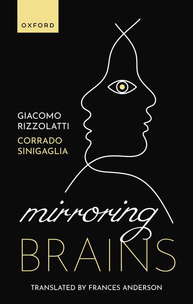
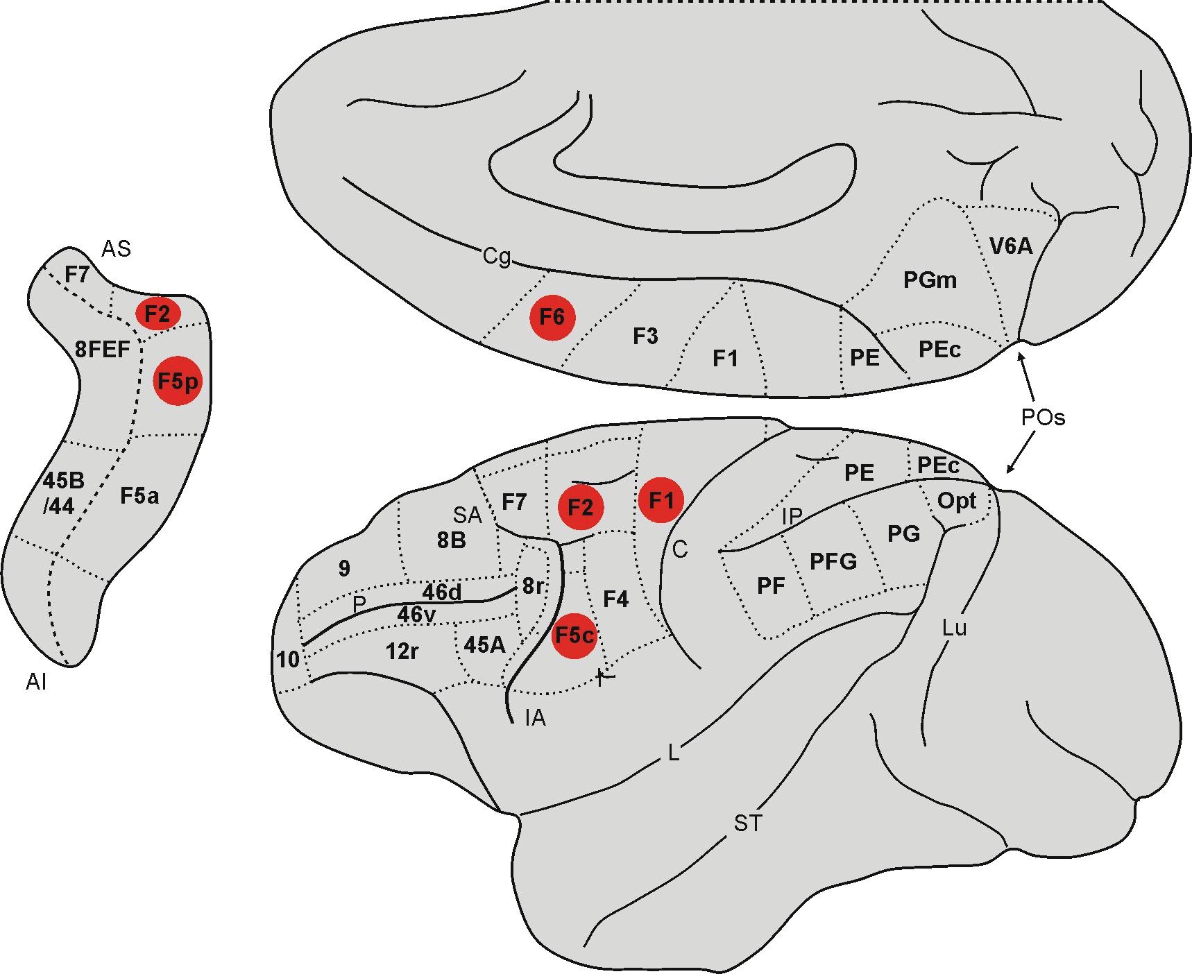
(whose proper denomination is PMd) and F7 (pre-PMd) and lastly, the ventral region (ventral premotor cortex, PMv), which consists of areas F4 and F5 (Fig. 1.1).
Since the mid-1980s, Giacomo Rizzolatti, Maurizio Gentilucci, and colleagues at the University of Parma have been systematically studying the functional properties of the motor areas, particularly area F5, registering the action potentials of single neurons (Gentilucci et al., 1988; Rizzolatti et al., 1988). The neuronal activity was recorded while the macaques were freely executing various actions, all present in their own motor repertoire, such as picking
Fig. 1.1 Mesial and lateral views of the macaque brain showing the anatomical and functional parcellation of the agranular frontal cortex.
Abbreviations: IA = inferior arcuate sulcus, C = central sulcus, Cg = cingulate sulcus, IP = intraparietal sulcus, L = lateral fissure or fissure of Silvius, Lu = lunate sulcus, P = principal sulcus, PO = parieto-occipital sulcus, SA = superior arcuate sulcus ST = superior temporal sulcus.
up a piece of food, holding it in their hand, lifting it to their mouth but also cracking a nut or swatting an object away. Sometimes the experimenters would lay out a variety of visual stimuli, pieces of food or objects of different shape, size, and orientation, placing them close to or far from the animal. They would also execute actions in front of the animal that were very similar to those the animal itself was accustomed to performing.
This approach turned out to be particularly successful, facilitating an ethological study of area F5 (but not only!) neuron responses, and the discovery of properties that would probably never have emerged if the neuronal activity had been recorded exclusively while the animal was executing predetermined and highly stereotypical movements. The first to be identified was a functional property that characterizes most F5 neurons: in many cases, their firing during the execution of a given action represents the goal of said action, and not simply this or that single movement somehow involved in the achievement of that goal (Rizzolatti et al., 1988). As we will see in Chapter 2 (pp. 40–48), this property is particularly important in the definition of the role and functions of motor processes and representations as we understand them today.
A second discovery was that a number of F5 neurons that fired while the macaque was executing an action such as grasping a small object, a sunflower seed for example, between the index finger and the thumb, also fired when the animal was presented with a piece of food or any other object that could be picked up with that kind of grip. This occurred regardless of whether the object was actually grasped or not (Rizzolatti et al., 1988; Murata et al., 1997). These neurons, characterized by congruent motor and visual responses, are known as canonical neurons.
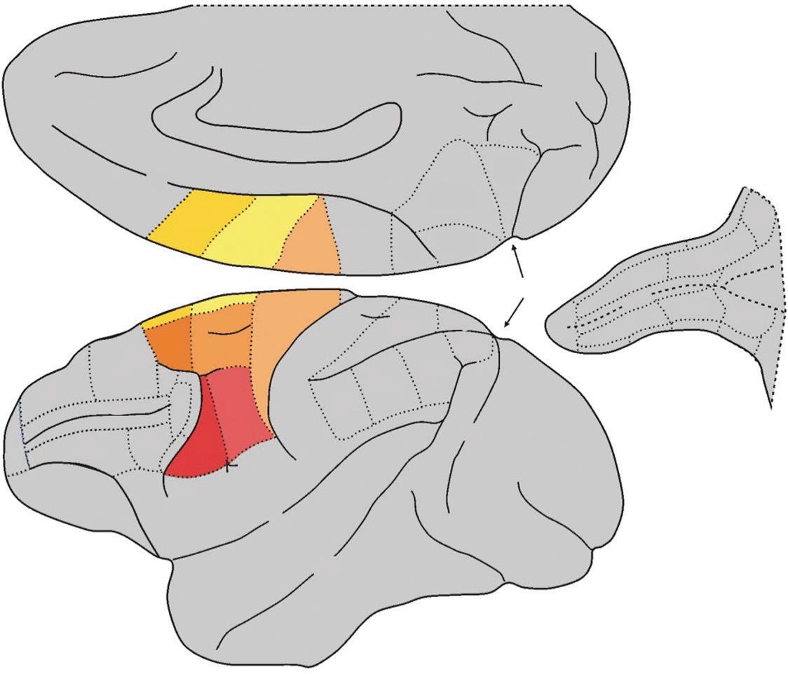
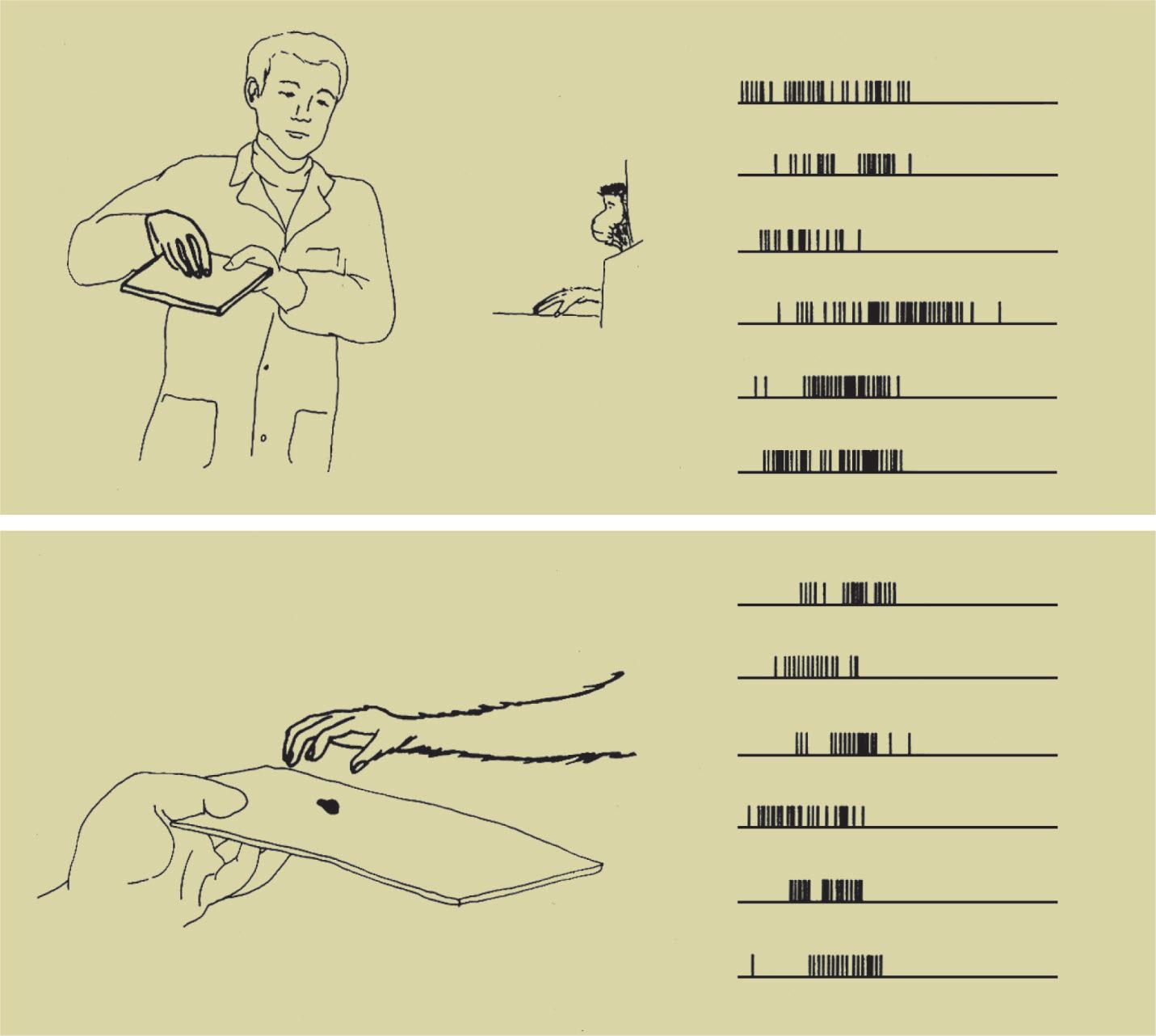
The third and most important discovery was made by a group of researchers led by Giacomo Rizzolatti, including Luciano Fadiga, Leonardo Fogassi, Vittorio Gallese and, in the early days, Giuseppe di Pellegrino: a number of F5 neurons that fired while the macaque was executing a specific action also fired when it saw the researcher executing the same kind of action, whereas the mere presentation of visual stimuli showing food or various objects failed to evoke a response (di Pellegrino et al., 1992; Gallese et al., 1996; Rizzolatti, Fadiga, Gallese et al., 1996). These neurons were given the name mirror neurons. Fig. 1.2 shows the activity of one such neuron: the lower panel (B) shows the discharges of the neuron while the macaque is grasping the food, whereas the upper panel (A) shows the discharges of the same neuron while the macaque observes the (A)
Fig. 1.2 Visual (A) and motor (B) responses of a classical F5 mirror neuron (di Pellegrino et al., 1992).
(B)
researcher executing the same kind of action. The congruence of the neuronal discharge profiles in the two conditions, respectively of executing the action and of observing it, is of particular importance.
Mirror neurons in the frontal lobe
Area F5 is located in the anterior portion of the premotor ventral cortex (PMv). Giuseppe Luppino and colleagues have shown that this area is not anatomically homogeneous, consisting as it does of three subareas that are cytoarchitectonically distinct, differing from each other in the organization and layout of the various types of cells: convexity F5 (F5c), posterior F5 (F5p), and anterior F5 (F5a) (Belmalih et al., 2009). Areas F5p and F5a are located on the posterior bank of the inferior arcuate sulcus, while area F5c is situated on the cortical convexity adjacent to the arcuate sulcus (Fig. 1.3). It is worthwhile mentioning that while areas F5p and F5c are similar from an anatomical and functional standpoint, area F5a is an area of transition between the typically granular prefrontal areas and the agranular motor areas.
This difference is also reflected in the correspondent cortical connections: in fact, while areas F5c and F5p are directly connected with area F1, area F5a has practically no direct connections with that area, being the only subarea of F5 to be connected with the ventrolateral prefrontal cortex (Matelli et al., 1986; Gerbella et al., 2011). On the basis of these early studies, it was thought that mirror neurons were located (almost) exclusively in area F5c and canonical neurons in area F5p. However, a later study (Bonini et al., 2014) showed that mirror and canonical neurons are to be found
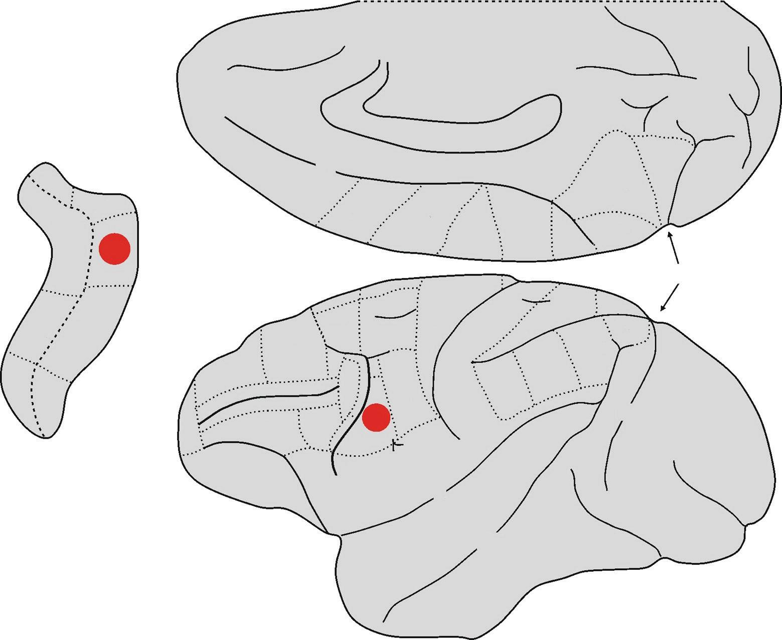
Fig. 1.3 Lateral and mesial views of the macaque brain. The hidden areas of the arcuate sulcus are shown on the left. The two sub-areas of F5, that according to currently available data have the mirror property, are highlighted in red.
Abbreviations: IA = inferior arcuate sulcus, C = central sulcus, Cg = cingulate sulcus, IP = intraparietal sulcus, L = lateral fissure or fissure of Silvius, Lu = lunate sulcus, P = principal sulcus, POs = parieto-occipital sulcus, SA = superior arcuate sulcus, ST = superior temporal sulcus.
in both areas, with a slight predominance of mirror neurons in area F5c and canonical neurons in area F5p (Fig. 1.3). Interestingly, this study also described a set of neurons with both canonical and mirror properties. Indeed, these neurons (known as canonicalmirror neurons) discharge while a given action is being executed on a target object with a specific shape and size, when someone is observed executing the action (mirror property) or again at the sight of a target object, with that specific shape and size (canonical property).
Roger Lemon and colleagues of University College London made an extremely interesting discovery: area F5p neurons that are part of the corticospinal (or pyramidal) tract may exhibit the mirror property. They demonstrated that approximately 50% of the neurons they recorded were modulated by the observation of other people’s actions (Kraskov et al., 2009). About one-third of the neurons found to have the mirror property was inhibited during observation of the actions executed by others (inhibitory mirror neurons), but fired while executing the same kind of action.
Lemon and colleagues recorded area F1 corticospinal neurons in a later study and discovered that in many cases their activities were modulated by observing actions executed by others (Fig. 1.4). As happens in the corticospinal tract that starts in area F5, in one part of these neurons the response increased when observing actions performed by others, but was inhibited in another part (Vigneswaran et al., 2013). Nevertheless, the mirror responses of the area F1 corticospinal neurons were much weaker than the homologous F5 neurons, which would explain why no significant mirror activity was found in area F1 in the initial recordings (Gallese et al., 1996; Fogassi et al., 2001).
Mirror neurons have also been described in the dorsal premotor cortex (PMd), in area F2 (Fig. 1.4). Paul Cisek and John Kalaska at the University of Montreal did in fact find F2 neurons that fired both when their macaques correctly guided a cursor to point to a target previously indicated between two possible options and when the animals observed the experimenter moving the cursor to the correct target. When the experimenter moved the cursor to the correct target, the macaques received some juice as a reward.
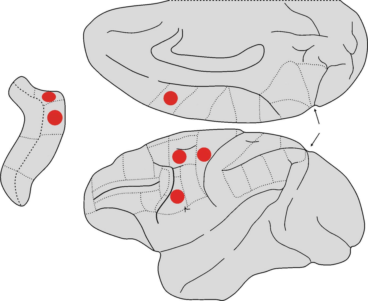
Fig. 1.4 Lateral and mesial views of the macaque brain. The frontal areas that have mirror properties (according to currently available data) are highlighted in red.
Abbreviations: IA = inferior arcuate sulcus, C = central sulcus, Cg = cingulate sulcus, IP = intraparietal sulcus, L = lateral fissure or fissure of Silvius, Lu = lunate sulcus, P = principal sulcus, POs = parieto-occipital sulcus, SA = superior arcuate sulcus, ST = superior temporal sulcus.
It was interesting to see that the neuronal activity was practically identical at both the individual and population levels in both conditions (Cisek & Kalaska, 2004). Later experiments using similar tasks identified mirror responses in area F2 as well as in area F1 (Tkach et al., 2007; Dushanova & Donoghue, 2010). Lastly, a recent study examined the activity of F2 neurons during the execution and observation of grasping actions similar to those typically used to investigate F5 mirror neurons. Not only did F2 neurons show a
percentage of mirror responses similar to that of F5 neurons, they also showed the same kind of congruence between motor and visual responses (Papadourakis & Raos, 2018).
Mirror neurons have also been discovered in area F6, which is located anteriorly to the supplementary motor area proper (area F3 or SMA) and for this reason is often indicated as pre-SMA (Matsuzaka et al., 1992; see Fig. 1.4). In spite of its fundamentally agranular structure, area F6 does show transitional characteristics towards the granular areas of the prefrontal lobe (Matelli et al., 1991). In addition, it does not project directly to area F1 as it is prevalently connected with the posterior premotor areas, and like area F5a, with the prefrontal cortex (Luppino et al., 1993). Masaki Isoda and colleagues at the Riken Institute recorded F6 neuron activity in two macaques; in their study, the two monkeys each had a button they had to push in turn, depending on the choice made by the other (Yoshida et al., 2011). In this study the authors identified three kinds of neurons: the so-called self-neurons that fired only while the action was being executed; the other-neurons that fired only when observing the other macaque executing the action; and, finally, the neurons that fired in both conditions, thus exhibiting the property that characterizes mirror neurons. Luca Bonini and colleagues at the University of Parma obtained similar results from a classic Go/NoGo experiment with macaques, consisting in reaching for and grasping different-sized objects or watching the hand of an experimenter, seated behind the animal, performing the same task (Livi et al., 2019). In this study too, the authors found neurons that fired only when the macaques themselves executed the action of reaching and grasping, others that fired only when they watched the experimenter doing the reaching and grasping, and those which fired in both conditions (mirror neurons).
Mirror neurons in the parietal lobe
A number of other single cell recordings have demonstrated that mirror neurons are also present in the inferior parietal lobe (IPL). The first studies recorded mirror neurons from the IPL convexity, in particular from the PF and PFG areas (Gallese et al., 2002; Fogassi et al., 2005; Rozzi et al., 2008), while subsequent studies found them in the anterior intraparietal area (AIP) (Pani et al., 2014; Maeda et al., 2015), where the presence of visuomotor neurons with functional characteristics not unlike the F5 canonical neurons had been previously reported (Murata et al., 2000; see Fig. 1.5).
For many years it was thought that the function of the posterior parietal lobe was mainly, if not exclusively, to associate and integrate sensory information into ‘percepts’ that could then be used when exploring the surrounding world, both as a guide to action and as a basis for categorization. This changed however when it was discovered that a significant portion of the posterior parietal areas is not only somatotopically organized, but also contains neurons with undeniably motor properties (Hyvärinen, 1981; Andersen, 1987; Mountcastle, 1995; Sakata et al., 1995). Today we know that the posterior parietal lobe is composed of a plurality of nodes connected to various centres, each with specific functional characteristics. For the purposes of this book, it is useful to remember that the area denominated as PFG has strong connections with the neighbouring areas of the inferior parietal lobule (PF, PG, AIP, and VIP), the adjacent parietal operculum, the insula, the premotor ventral areas (particularly area F5) and the prefrontal lobe, notably area 46. The AIP area is densely connected with the adjacent parietal areas (PF, PFG, PG, LIP, and VIP), vast sectors of the inferior temporal cortex, the insula, area F5, and the prefrontal