Professor Emeritus, Wayne University College of Nursing, Detroit, MI
OTHER VOLUMES IN THE SERIES
Pediatric Palliative Care, Edited by Lindsay B. Ragsdale & Elissa G. Miller Pain, Edited by Christopher M. Herndon
Respiratory Symptoms
Edited by Margaret L. Campbell, PhD, RN, FPCN
Professor Emeritus, Wayne University College of Nursing, Detroit, Michigan
Oxford University Press is a department of the University of Oxford. It furthers the University’s objective of excellence in research, scholarship, and education by publishing worldwide. Oxford is a registered trade mark of Oxford University Press in the UK and certain other countries.
Published in the United States of America by Oxford University Press 198 Madison Avenue, New York, NY 10016, United States of America.
All rights reserved. No part of this publication may be reproduced, stored in a retrieval system, or transmitted, in any form or by any means, without the prior permission in writing of Oxford University Press, or as expressly permitted by law, by license, or under terms agreed with the appropriate reproduction rights organization. Inquiries concerning reproduction outside the scope of the above should be sent to the Rights Department, Oxford University Press, at the address above.
You must not circulate this work in any other form and you must impose this same condition on any acquirer.
CIP data is on file at the Library of Congress
ISBN 978–0–19–009889–6
DOI: 10.1093/med/9780190098896.001.0001
This material is not intended to be, and should not be considered, a substitute for medical or other professional advice. Treatment for the conditions described in this material is highly dependent on the individual circumstances. And, while this material is designed to offer accurate information with respect to the subject matter covered and to be current as of the time it was written, research and knowledge about medical and health issues is constantly evolving and dose schedules for medications are being revised continually, with new side effects recognized and accounted for regularly. Readers must therefore always check the product information and clinical procedures with the most up-to-date published product information and data sheets provided by the manufacturers and the most recent codes of conduct and safety regulation. The publisher and the authors make no representations or warranties to readers, express or implied, as to the accuracy or completeness of this material. Without limiting the foregoing, the publisher and the authors make no representations or warranties as to the accuracy or efficacy of the drug dosages mentioned in the material. The authors and the publisher do not accept, and expressly disclaim, any responsibility for any liability, loss, or risk that may be claimed or incurred as a consequence of the use and/or application of any of the contents of this material.
Printed by Marquis Book Printing, Canada
List of Contributors vii
Introduction xi
1. Dyspnea Assessment 1
Margaret L. Campbell
2. Reducing Dyspnea by Optimizing Treatment of Chronic Obstructive Pulmonary Disease 7
Miranda Wilhelm and Jennifer Arnoldi
3. Treating Chronic Breathlessness in Severe Chronic Obstructive Pulmonary Disease 21
Lynn F. Reinke, Mary M. Roberts, and Tracy A. Smith
4. Dyspnea, Chronic Obstructive Pulmonary Disease, and Pulmonary Rehabilitation 31
DorAnne Donesky and Julie Howard
5. Treating Episodic Breathlessness 39
Yvonne Eisenmann and Steffen Simon
6. Reducing Episodic Dyspnea in Heart Failure 49
Beth B. Fahlberg and Ann S. Laramee
7. Dyspnea in Pediatric Congenital Heart Disease 61
Jennifer Wright and Jessica L. Spruit
8. Treating Chronic Dyspnea in Patients with Lung Cancer 69
Elizabeth A. Higgins, Susan Ezemenari, and Julia Arana West
9. Treating Dyspnea Through Reducing Malignant Pleural Effusion 77
Christine A. Crader
10. Treating Dyspnea in Lung Cancer with Noninvasive Ventilation 87
Vittoria Comellini and Stefano Nava
11. Palliative Care for Infants with Bronchopulmonary Dysplasia 95
Christine A. Fortney and Jodi A. Ulloa
12. Reducing Dyspnea by Treating Ascites 101
Habib A. Khan
13. Panting for Breath in End-Stage Dementia 109
Hermien W. Goderie-Plomp, Carole Parsons, David R. Mehr, and Jenny T. van der Steen
14. Last Days with Chronic Obstructive Pulmonary Disease 119
Margaret L. Campbell
15. Withdrawal of Invasive Mechanical Ventilation 125
Margaret L. Campbell
16. Palliative Sedation for Intractable Dyspnea 135
Patricia Bramati and Eduardo Bruera
17. Sialorrhea in Amyotrophic Lateral Sclerosis 145
Mark B. Bromberg
18. Death Rattle 153
Margaret L. Campbell
Index 159
Contributors
Jennifer Arnoldi, PharmD, BCPS
Clinical Associate Professor
Southern Illinois University
Edwardsville (SIUE) School of Pharmacy
Edwardsville, IL, USA
Patricia Bramati, MD
The University of Texas MD
Anderson Cancer Center
Houston, TX, USA
Mark B. Bromberg, MD, PhD
Department of Neurology University of Utah Salt Lake City, UT, USA
Eduardo Bruera, MD
The University of Texas MD Anderson Cancer Center
Houston, TX, USA
Margaret L. Campbell, PhD, RN, FPCN
Wayne State University, College of Nursing Detroit, MI, USA
Vittoria Comellini, MD
Respiratory and Critical Care Unit University Hospital St. Orsola-Malpighi
Bologna, Italy
Christine A. Crader, MD
Ascension Medical Group Internal Medicine Detroit, MI, USA
DorAnne Donesky, PhD, ANP-BC, ACHPN, ATSF Professor, School of Nursing
Touro University of California Vallejo, CA, USA
Yvonne Eisenmann, MD University of Cologne Faculty of Medicine and University Hospital Department of Palliative Medicine Cologne, Germany
Susan Ezemenari, MD Fellow, Palliative Medicine Division of Internal Medicine, Palliative Medicine and Geriatrics
Medical University of South Carolina Charleston, SC, USA
Beth B. Fahlberg, PhD, MN, RN University of Wisconsin Madison, WI, USA
Christine A. Fortney, PhD, RN
Assistant Professor
The Ohio State University College of Nursing
Martha S. Pitzer Center for Women, Children and Youth Columbus, OH, USA
Hermien W. Goderie-Plomp, MD, MSc, MSc
Elderly Care and Palliative Care
Physician
De Zellingen, Rotterdam, The Netherlands
Lecturer in Palliative Care
Leiden University Medical Center
Leiden, The Netherlands
Elizabeth A. Higgins, MD
Associate Professor of Internal Medicine
Division of Internal Medicine, Palliative Medicine and Geriatrics
Medical University of South Carolina Charleston, SC, USA
Julie Howard, RRT, TTS, CCM
COPD Case Manager
Adventist Health Rideout
Marysville, CA, USA
Habib A. Khan, MD
Johns Hopkins Medicine
Department of Palliative Medicine
Wayne State University Baltimore, MD, USA
Ann S. Laramee, MS, ANP-BC, ACNS-BC, CHFN, ACHPN, FHFSA
University of Vermont
Medical Center
Burlington, VT, USA
David R. Mehr, MD, MS
Professor Emeritus
Department of Family and Community Medicine
University of Missouri Columbia, MO, USA
Stefano Nava, MD
Department of Specialistic, Diagnostic and Experimental Medicine (DIMES), Alma Mater Studiorum University of Bologna Bologna, Italy
Carole Parsons, PhD, MPharm, MPSNI
Lecturer in Pharmacy Practice School of Pharmacy
Queen’s University Belfast Belfast, UK
Lynn F. Reinke, PhD, RN
Claire Dumke Ryberg, RN
Presidential Endowed Chair in End-of-Life/Palliative Care
University of Utah College of Nursing
Salt Lake City, UT, USA
Mary M. Roberts, MSN, RN
Department of Respiratory and Sleep Medicine, Westmead Hospital
Ludwig Engel Centre for Respiratory Research, Westmead Institute for Medical Research
The University of Sydney at Westmead Hospital
Westmead, New South Wales, Australia
Steffen Simon, MD Department of Palliative Medicine
University of Cologne
Faculty of Medicine and University Hospital Cologne, Germany
Tracy A. Smith, MD
Department of Respiratory and Sleep Medicine, Westmead Hospital
The University of Sydney at Westmead Hospital
Westmead, New South Wales, Australia
Jessica L. Spruit, DNP, CPNP-AC
Pediatric Nurse Practitioner
Stepping Stones Pediatric Palliative Care Program
University of Michigan Health System
Ann Arbor, MI, USA
Jodi A. Ulloa, DNP, APRN-CNP, NNP-BC
Assistant Professor of Clinical Practice
The Ohio State University College of Nursing
Martha S. Pitzer Center for Women, Children and Youth Columbus, OH, USA
Jenny T. van der Steen, MSc, PhD, FGSA
Associate Professor
Leiden University Medical Center, Department of Public Health and Primary Care Leiden, The Netherlands
Senior Researcher
Radboud University Medical Center, Department of Primary and Community Care
Nijmegen, The Netherlands
Julia Arana West, MD Fellow, Palliative Medicine
Division of Internal Medicine, Palliative Medicine and Geriatrics
Attending Physician, Department of Emergency Medicine
Medical University of South Carolina Charleston, SC, USA
Miranda Wilhelm, PharmD
Clinical Associate Professor
Southern Illinois University
Edwardsville (SIUE) School of Pharmacy
Edwardsville, IL, USA
x C ONTRIB u TORS
Jennifer Wright, MS, CPNP
Stepping Stones Pediatric
Palliative Care
Michigan Medicine
Ann Arbor, MI, USA
Introduction
Margaret L. Campbell
In this volume, nearly all the chapters relate to the complex symptom dyspnea across diagnoses, lifespan, and care settings. Other chapters relate to oral and pharyngeal secretions. These topics are addressed from a palliative care context.
Dyspnea, also known as breathlessness, has been defined as a person’s awareness of uncomfortable or distressing breathing. As this can only be known by the person, the term “respiratory distress” is used as the observed corollary relying on patient signs when the person is unable to report dyspnea, such as infants, young children, and adults with cognitive impairments, which may be acute or chronic.
Dyspnea develops when inspiratory effort, hypoxemia, and/or hypercarbia develops, which activates three redundant brain areas. In the cerebral cortex, the dyspneic person has an awareness of the change in breathing efficiency. The amygdala in the subcortical temporal lobe is activated when there is a threat to survival and produces a fear response. The pons in the brainstem reacts by activating compensatory accelerations of heart and respiratory rates and recruiting accessory muscles.
Assessment of dyspnea relies on self-report from as simple as a yesor-no response to the query “Are you short of breath?” to more complex numeric scales (0–10) or categorical scales (none, mild, moderate, or severe). For patients unable to report dyspnea, observation scales such as the Respiratory Distress Observation Scale may be used. High-risk patients should be assessed at every clinical encounter.
Dyspnea is one of the most difficult symptoms to experience and is also one of the most difficult to treat, as the evidence base for this symptom lags behind other prevalent symptoms such as pain or nausea, to name two. Dyspnea is prevalent in patients with cardiopulmonary disorders and cancer, and it escalates as death approaches. The development of dyspnea in chronic disease is a predictor of mortality.
Dyspnea may be acute when a reversible etiology presents such as pneumonia, pleural effusion, or ascites. It is chronic in an irreversible condition
such as chronic obstructive pulmonary disease (COPD), advanced heart failure, or congenital cardiac conditions. Episodic dyspnea may typify acute exacerbations in chronic conditions such as heart failure.
Treating dyspnea relies on a hierarchy of responses, beginning with treating underlying, reversible conditions such as infections, pleural effusions, volume overload, or ascites. Nonpharmacological treatments include pulmonary rehabilitation, noninvasive ventilation, balancing rest with activity, and optimal positioning. Pharmacological treatments include oxygen, bronchodilators, and opioids. In cases of refractory dyspnea, palliative sedation may be indicated.
Patients receiving invasive mechanical ventilation for respiratory failure may undergo ventilator withdrawal to afford a natural death. These patients are at very high risk for developing respiratory distress, which warrants careful attention to the processes to minimize distress.
Salivary secretions pose a significant problem for patients with bulbaronset amyotrophic lateral sclerosis characterized by difficulties swallowing. Treatment begins with anticholinergic medications and may include botulinum toxin injections or irradiation of salivary glands.
Pharyngeal secretions, also known as death rattle, develops in about half of dying patients in the last days of life. Controversies about whether medications are indicated or effective make up the evidence base. Ethical concerns about medicating the patient to assuage the listener have been raised.
The contributors to this volume have addressed all the treatments currently known for dyspnea, respiratory distress, and secretions with a case study approach.
1 Dyspnea Assessment
Margaret L. Campbell
Stella is a nursing home resident in the advanced stage of Alzheimer’s disease. Her goals of care are comfort-focused, with surrogate decisions previously made to withhold tube feedings, intubation, cardiopulmonary resuscitation, and hospitalization. She sleeps most of the day and night and is noncommunicative except for sounds of displeasure such as when being bathed or turned. Stella is at high risk for aspiration pneumonia; she is offered a soft, easy-to-swallow diet with foods such as pudding, scrambled eggs, oatmeal, and baby food.
On daily exam a change in her breathing is noted; it is highly likely that Stella has developed pneumonia, probably secondary to immobility or food or liquid aspiration. The clinical team cannot elicit a dyspnea assessment from Stella, and they know she is at risk for respiratory distress.
What do I do now?
WHAT IS DySPNEA?
Dyspnea is a person’s awareness of uncomfortable or distressing breathing that can only be known by self-report. In symptom care a patient’s selfreport has long been held as the gold standard for assessment. In a palliative care context dyspnea should be assessed at every patient encounter, for example in the hospital whenever vital signs are obtained, or at every outpatient clinic visit, or at every house call for home-bound patients. Since dyspnea escalates as death nears, the frequency of assessment should be increased. Several tools are available for measuring and trending dyspnea that are appropriate in palliative care.
unidimensional Assessment Tools
Responding to a symptom assessment requires several cognitive steps on the patient’s part: (1) ascertain their sensation, (2) pay attention to the clinician’s assessment instructions, (3) formulate a response, (4) communicate that response in some fashion, and (5) recall their previous report if trending is requested. When a patient is feeling poorly or is fatigued, these steps may pose difficulty and simpler tools are indicated.
The simplest measure is to ask for a “yes” or “no” response to the query “Are you short of breath?” or, in the case of the mechanically ventilated patient, “Are you getting enough air?” Knowing the presence or absence of dyspnea is helpful, but its intensity cannot be assessed with this approach.
A more complex tool is a numeric rating scale, usually anchored at 0 for no dyspnea and 10 for the worst possible dyspnea. Patients may need guidance on how to use numbers to characterize their symptom intensity. A variation to using numbers is to ask for a categorical ranking (none, mild, moderate, severe). Some patients may find these categorical rankings easier to understand and relate to their experience than a numeric rating.
Another variation that can be used to rate intensity is a visual analog scale, typically a 100-mm scale anchored at 0 and 100, with tick marks at every 10 points (Figure 1.1). The patient points to a line on the scale representing their experience and the clinician can convert that to a numeric score; this approach is useful in the critical care setting with patients who are mechanically ventilated and nonverbal. A vertical scale was preferred by patients with chronic obstructive pulmonary disease (COPD).1
Multidimensional Assessment Tools
Dyspnea is a multidimensional symptom with physical and affective attributes that are not captured by the unidimensional tools just described. These scales may be applied by patients with intact cognition who may be receiving palliative care concurrently with disease treatment. The Dyspnea-122 is a multidimensional scale composed of 12 descriptors that are scored on a four-item scale with the points summed to produce a total dyspnea score (Table 1.1). Subscales for physical and affective domains can be derived. The focal point for this assessment is “these days,” which captures the average experience of a chronic symptom.
The Multidimensional Profile (MDP)3 comprises 11 numeric rating scales (0–10) of different sensory and affective breathlessness sensations and a “forced choice” item requiring respondents to select which of five sensory qualities best describes their breathlessness. Individual item ratings are reported, and subdomain scores can be calculated. Where a single score is sought by users, the MDP-A1 item (0–10 rating of breathlessness unpleasantness) was recommended by the developers; a total summed score of all items is not recommended for the MDP. The user specifies the focal event
FIG u RE 1.1. Visual analog scale for dyspnea
TABLE 1.1. The Dyspnea-12
Item None Mild Moderate Severe
1. My breath does not go in all the way.
2. My breathing requires more work.
3. I feel short of breath.
4. I have difficulty catching my breath.
5. I cannot get enough air.
6. My breathing is uncomfortable.
7. My breathing is exhausting.
8. My breathing makes me feel depressed.
9. My breathing makes me feel miserable.
10. My breathing is distressing.
11. My breathing makes me agitated.
12. My breathing is irritating.
Reprinted with permission from J. Yorke.
(e.g., “after you climb three flights of stairs,” “last minute of breathing on the mouthpiece”) or timepoint (e.g., “right now”) for the MDP, and can choose to use only one or a few of the instrument scales, depending on the clinical purpose of assessment. Permission for use must be obtained from the author. The Modified Medical Research Council (MMRC) dyspnea scale4 stratifies dyspnea into one of four scores relative to function, with 0 signifying dyspnea only with strenuous exercise and 4 representing too dyspneic to leave the house or dress. The MMRC dyspnea scale is best used to establish baseline functional impairment related to respiratory disease.
Observation Scale
Patients who are unable to provide a symptom report but can experience the symptom are vulnerable to over- or under-treatment. Respiratory distress is the observed corollary to reported dyspnea relying on patient signs. The Respiratory Distress Observation Scale5 was developed for patients with advanced disease who are unable to self-report (Table 1.2). Eight variables are
TABLE 1.2. Respiratory Distress Observation Scale
Variable 0 points 1 point 2 points Total
Heart rate per minute <90 beats 90–109 beats ≥110 beats
Respiratory rate per minute ≤18 breaths 19–30 breaths >30 breaths
Restlessness: non-purposeful movements
Accessory muscle use: rise in clavicle during inspiration
Eyes wide open, facial muscles tense, brow furrowed, mouth open
scored from 0 to 2 points and the points are summed to yield a total intensity score ranging from 0 to 16. Reliability and validity have been established across diagnoses and settings of care; intensity cut-points are 0–2 = no distress, 3 = mild distress, 4–6 = moderate distress, and 7 and higher = severe distress.6,7
S u MMARy
Several unidimensional and multidimensional scales and an observation scale were described as suitable for assessment in patients receiving palliative
care. Selection of the optimal tool will depend on the patient’s cognitive abilities, which are influenced by disease trajectory, fatigue, sedation, mechanical ventilation, and underlying conditions such as dementia. Stella has a heart rate of 108 and respiratory rate of 22. She has slight restlessness and a slight rise in the clavicle, signifying accessory muscle use. There is no apparent paradoxical breathing, nor grunting, nor nasal flaring or a fearful facial expression. Thus, her RDOS score is 4, signifying moderate distress. The nurses elevated the head of her bed, placed a fan blowing on her cheek, and began immediate-release morphine elixir at 5 mg every 4 hours. Subsequent RDOS scores decreased to 2 or 3 with this regimen.
KE y POINTS TO REMEMBER
• Assessment is critical to optimizing treatment.
• Patient abilities will inform selection of an assessment tool or scale.
• Self-reported dyspnea presence and intensity is the gold standard.
• Respiratory distress signs are indicated for those who cannot report their experience.
References
1. Gift A. Validation of a vertical visual analogue scale as a measure of clinical dyspnea. Rehab Nurs. 1989;14:323–325.
2. Yorke J, Moosavi SH, Shuldham C, Jones PW. Quantification of dyspnoea using descriptors: development and initial testing of the Dyspnoea-12. Thorax. 2010;65(1):21–26.
3. Banzett RB, O’Donnell CR, Guilfoyle TE, et al. Multidimensional Dyspnea Profile: an instrument for clinical and laboratory research. Eur Respir J. 2015;45(6):1681–1691.
4. Mahler DA, Wells CK. Evaluation of clinical methods for rating dyspnea. Chest. 1988;93(3):580–586.
5. Campbell ML, Templin T, Walch J. A Respiratory Distress Observation Scale for patients unable to self-report dyspnea. J Palliat Med. 2010;13(3):285–290.
6. Campbell ML. Psychometric testing of a respiratory distress observation scale. J Palliat Med. 2008;11(1):44–50.
7. Campbell ML, Kero KK, Templin TN. Mild, moderate, and severe intensity cut-points for the Respiratory Distress Observation Scale. Heart Lung. 2017;46(1):14–17.
2 Reducing Dyspnea by Optimizing Treatment of Chronic Obstructive Pulmonary Disease
Miranda Wilhelm and Jennifer Arnoldi
JW, a 75-year old female, presents to the primary care clinic with a chief complaint of “This inhaler is not working.” The patient reports shortness of breath with activities of daily living but has not experienced an exacerbation or hospitalization or used an oral corticosteroid related to chronic obstructive pulmonary disease (COPD) for the last year. Relevant medical history includes 60 pack-year history of smoking cigarettes (1 pack per day for 60 years; quit “cold turkey” approximately 1 year ago), COPD for 5 years, osteoporosis for 10 years, hypertension for 20 years, and rheumatoid arthritis for 33 years. Current medications include tiotropium HandiHaler, albuterol hydrofluoroalkane (HFA), salmeterol Diskus (recently added), alendronate, lisinopril/hydrochlorothiazide, methotrexate and hydroxychloroquine. Recent records indicate that her forced expiratory volume in 1 second (FEV1) is 45% of predicted and her COPD Assessment Test (CAT) score is 18.
What do I do now?
The Global Initiative for Chronic Obstructive Lung Disease (GOLD) guidelines are updated and published on an annual basis. It is recommended that all healthcare providers incorporate this evidence-based guideline into their practice for the prevention, diagnosis, and treatment of COPD. Currently, the GOLD guidelines recommend short-acting bronchodilators, either a short-acting beta agonist (SABA) or a short-acting muscarinic antagonist (SAMA), for relief of acute symptoms in all patients. First-line maintenance pharmacotherapy includes the use of a long-acting bronchodilator such as a long-acting beta agonist (LABA) or a long-acting muscarinic antagonist (LAMA). Inhaled corticosteroids are considered the last option to reduce exacerbations (Table 2.1). It is important to continue these therapies through the end of life to manage symptoms and to maximize quality of life by minimizing dyspnea.
Selection of therapy for COPD should consider not only the patient’s disease characteristics such as symptoms, exacerbation history, and future risks, but also their physical capabilities, cognitive function, and other comorbidities. The patient’s previous experience with inhalation devices, their preferences, and insurance coverage of medication therapy should also be considered.
For established regimens, the patient’s adherence and their inhaler technique should be assessed. If the patient is not using the inhaler as prescribed or their technique is poor, this could reduce the actual and perceived efficacy of the medication. Therefore, selecting the device can be just as important as selecting the appropriate class of medications to use in COPD. Four inhalation devices—metered-dose inhalers (MDIs), dry powder inhalers (DPIs), soft mist inhalers (SMIs), and nebulizers—are used in COPD pharmacotherapy. Table 2.2 lists their advantages and disadvantages.
INHALATION DEVICES
MDIs
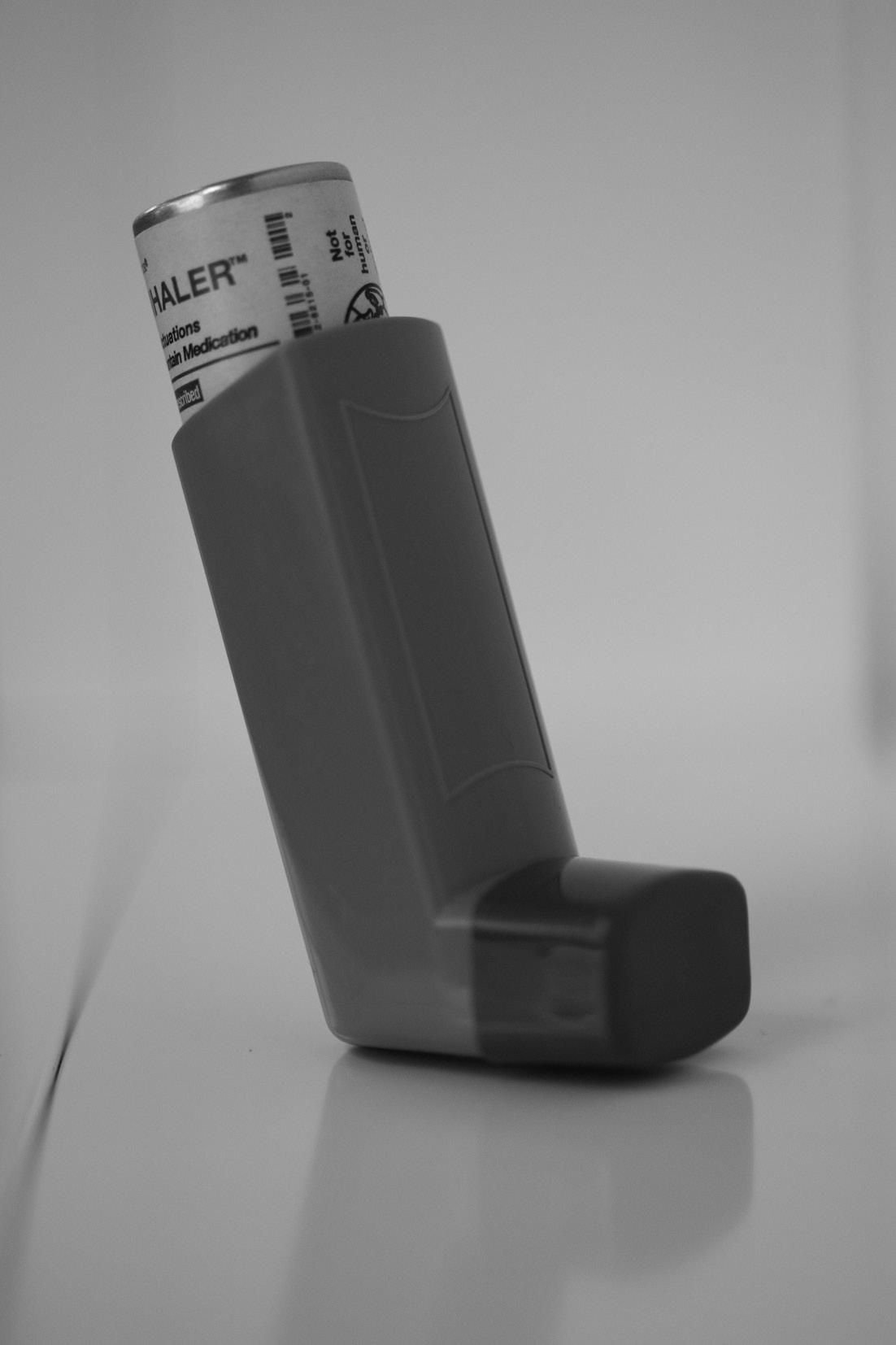
MDIs consist of a canister, a metering chamber, and an actuator with a mouthpiece (Figure 2.1). The canister contains a solution of the medication and a propellant. Shaking the device disperses the propellant into the medication solution, creating the pressure needed for aerosolization. As the canister is depressed, the patient must be ready to inhale the dose. Thus,
TABLE 2.1. Inhaled Medications Commonly used to Treat COPD
MDI use requires hand–breath coordination, and conditions that can decrease dexterity (e.g., rheumatoid arthritis, Parkinson’s disease) or cognitive performance may limit a patient’s ability to accurately use the inhaler. Due to the propellant contained in MDIs, failure to coordinate breath with dose activation can result in medication being deposited in the mouth and/or throat or loss of drug into the air. Holding chamber/spacer devices, when combined with MDIs, may improve patient technique in terms of hand–breath coordination but still require some manual dexterity to manipulate and are bulky to handle.
TABLE 2.2. Advantages and Disadvantages of Inhalation Devices
Inhalation device
MDI
DPI
SMI
Jet nebulizer
Advantages
• Convenient/portable
• Multidose devices
• Short administration time
• Counter indicates doses remaining
• Convenient/portable
• Single-dose and multidose devices
• Short administration time
• Counter indicates doses remaining
• Breath-actuated
• Convenient/portable
• Multidose devices
• Short administration time
• Counter indicates doses remaining
• Hand–breath coordination not required
• Minimal dexterity needed
Disadvantages
• Hand–breath coordination required
• Multiple steps
• Requires priming
• Throat deposition, leading to adverse drug events
• Requires moderate to high inspiratory flow rate
• May require priming
• Throat deposition, leading to adverse drug events
• Hand–breath coordination required
• Requires assembly
• Multiple steps
• Requires priming
• Temperature change of medication during use (cool)
• Limited portability
• Power source or battery pack needed
• Requires assembly
• Lengthy administration time (10–15 minutes)
• Requires frequent cleaning
• Not all medications available in this dosage form
• Generates noise
• Single dose
TABLE 2.2. Continued
Inhalation device Advantages
Ultrasonic nebulizer
• Portable
• Quiet
• Hand–breath coordination not required
• Minimal dexterity needed
Disadvantages
• Temperature change of medication during use (heat)
• Power source or battery pack needed
• Requires assembly
• Administration time (5 minutes)
• Requires frequent cleaning
• Not all medications available in this dosage form
• Single dose
Vibrating mesh nebulizer
• Portable
• Quiet
• Hand–breath coordination not required
• Minimal dexterity needed
• No temperature change of medication during use
• Expensive
• Power source or battery pack needed
• Requires assembly
• Administration time (5 minutes)
• Requires frequent cleaning
• Not all medications available in this dosage form
• Single dose
Asking the patient to demonstrate their technique with the inhaler may allow the provider to identify any potential errors in use that could prevent the medication from being effective. Common errors encountered in MDI use that could easily be identified through patient observation include failing to remove the device’s mouthpiece cover before use; not sealing the mouth around the mouthpiece when actuating the dose; lack of coordination between activating the dose and inhaling; and inhaling via the nose rather than the mouth.
DPIs
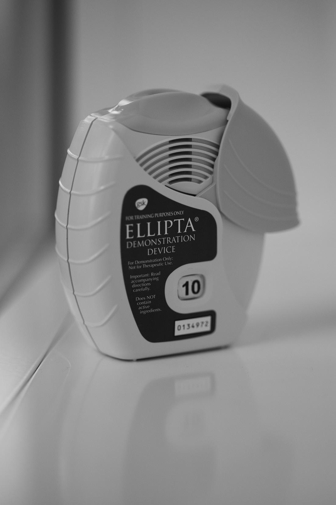
DPIs include a medication reservoir, an air inlet area, and a mouthpiece (Figures 2.2 and 2.3). The medication is in a dry powder dosage form that is drawn from the device using the patient’s own breath. This may simplify the hand–breath coordination required by MDIs; however, the lack of propellant in these devices requires the patient to generate a sufficient inspiratory flow rate to inhale the entire dose of the medication. Patients
FIG u RE 2.1. Metered-dose inhaler
Photo by Katherine Newman, PharmD.
with advanced COPD may experience disease-related airflow limitation, leading to difficulties in taking a breath with sufficient force and volume to inhale the full dose of the medication. One complicating factor is that DPIs are manufactured with some degree of variability in design, including the turbulence and resistance of the product and device. Due to this, the patient’s ability to use one type of DPI does not predict success with a DPI containing a different medication or administration device. A patient’s peak
FIG u RE 2.2. Dry powder inhaler: Ellipta
Photo by Katherine Newman, PharmD.
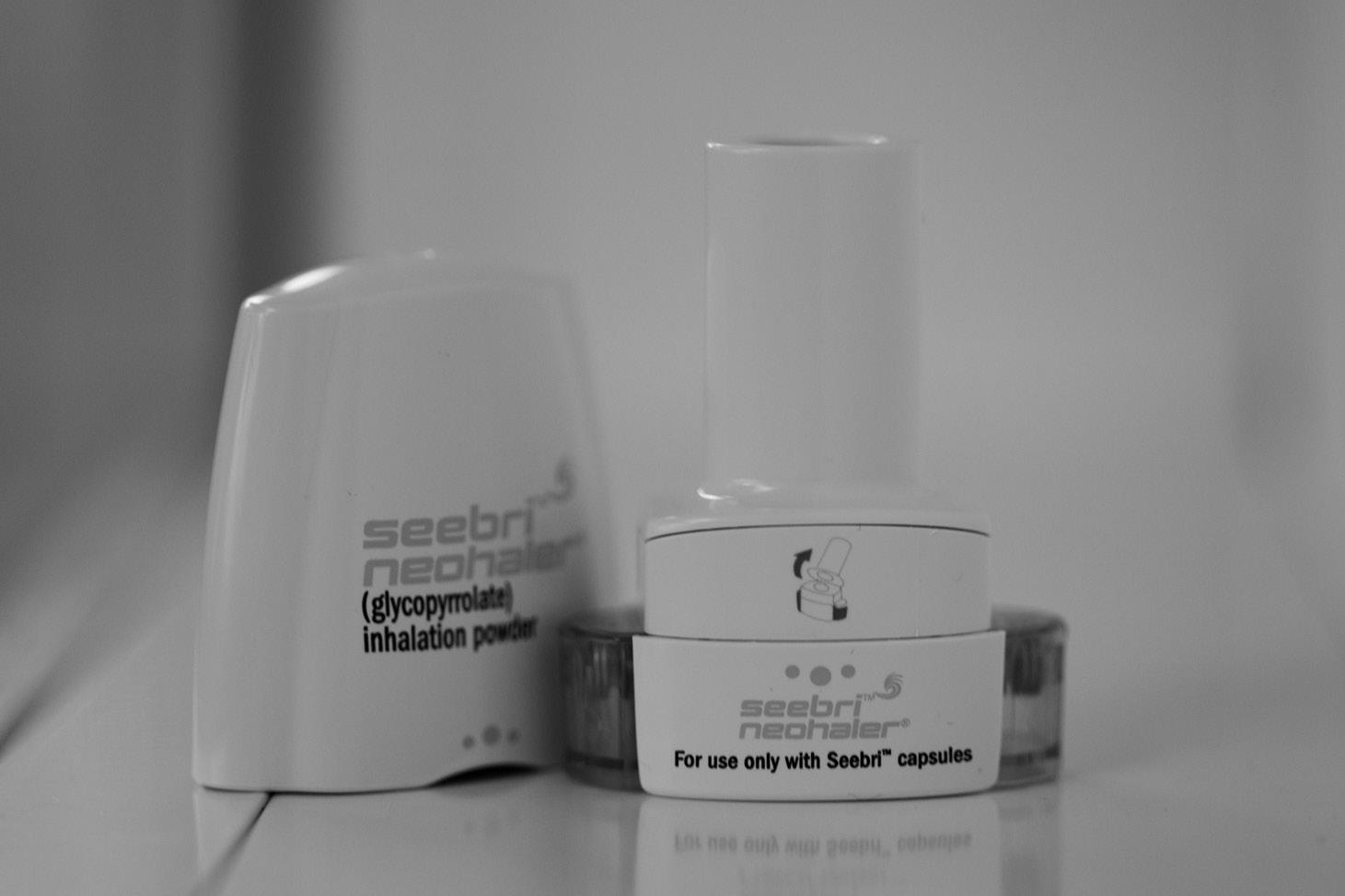
inspiratory flow rate (PIFR) can be obtained using a handheld inspiratory flow meter with an adjustable dial to simulate internal resistances of dry powder devices. For optimal DPI use, the patient should be able to attain at least 60 L/min; a PIFR less than 30 L/min is not likely to be sufficient. All patients receiving a DPI should be educated to avoid breathing into the device or getting it wet. Each device contains unique instructions about breathing in (e.g., quickly and deeply; a long, steady, deep breath; inhaling deeply and forcefully; taking two breaths from the same dosage). Simple instructions could be given as “breathe in hard and fast.” For DPIs, it is critical to consider the mechanism by which the dose is prepared for use; while the patient’s breath is the driving force to disperse and deliver the medication, each device uses a different approach to allow this to happen. For some devices, the dose may be prepared by opening the mouthpiece cover with or without the need to activate an additional lever (e.g., Diskus, Ellipta). For others (e.g., HandiHaler, Neohaler), the patient needs to open a capsule from a blister pack, place the capsule into the device, and pierce the capsule before inhaling. These device differences should also be
FIG u RE 2.3. Dry powder inhaler: Neohaler
Photo by Katherine Newman, PharmD.
considered when selecting the device (i.e., whether the patient has the dexterity required to manipulate the capsule or the cognitive ability to follow dosing instructions) and when evaluating the patient’s inhaler technique. Each step of the device’s usage process creates the potential for user error.
Finally, patients using DPIs should be educated that once the dose has been prepared for inhalation they should not tip, shake, or otherwise disturb the contents inside the inhaler. If they do, they risk losing some of the medication prior to use.
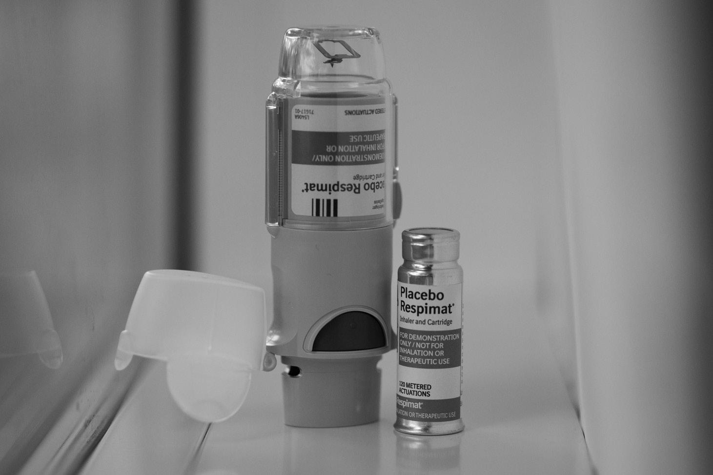
SMIs
SMIs do not use a propellant; rather, a liquid formulation delivers medication as a fine mist aerosol for inhalation. The unique nozzle delivers medication at a slower velocity (1.5 seconds) compared to MDIs (100–400 milliseconds). SMIs were designed to make medication delivery easier than with MDIs, although some hand–breath coordination is still required. In addition, the devices require assembly before use, which may require some degree of manual dexterity and/or cognition (Figure 2.4).