Review of the Electrical Conduction System of the Heart
TABLE 4–1
Summary of EKG Waveforms and Correlating Cardiac Events
TABLE 5–3
This page intentionally left blank
Understanding EKGs
A Practical Approach
FOURTH EDITION
BRENDA M. BEASLEY
MS, RN, EMT-Paramedic
Publisher: Julie Levin Alexander
Publisher’s Assistant: Regina Bruno
Editor-in-Chief: Marlene McHugh Pratt
Senior Acquisitions Editor: Sladjana Repic
Senior Managing Editor for Development: Lois Berlowitz
Editorial Assistant: Kelly Clark
Director of Marketing: David Gesell
Marketing Manager: Brian Hoehl
Marketing Specialist: Michael Sirinides
Marketing Assistant: Crystal Gonzalez
Managing Editor for Production: Patrick Walsh
Production Project Manager: Debbie Ryan
Editorial Project Manger: Patricia Guiterrez
Media Project Manager: Lorena Cerisano
Art Director: Jayne Conte
Cover Designer: Suzanne Behnke
Cover Image: Brenda M. Beasley
Composition: Aptara®, Inc.
Printer/Binder: Courier/Kendalville
Cover Printer: LeHigh-Phoenix Color/Hagerstown
Credits and acknowledgments borrowed from other sources and reproduced, with permission, in this textbook appear on the appropriate page within text.
Notice: The author and the publisher of this book have taken care to make certain that the information given is correct and compatible with the standards generally accepted at the time of publication. Nevertheless, as new information becomes available, changes in treatment and in the use of equipment and procedures become necessary. The reader is advised to carefully consult the instruction and information material included in each piece of equipment or device before administration. Students are warned that the use of any techniques must be authorized by their medical advisor, where appropriate, in accordance with local laws and regulations. The author and the publisher disclaim any liability, loss, injury, or damage incurred as a consequence, directly or indirectly, of the use and application of any of the contents of this book.
Many of the designations by manufacturers and sellers to distinguish their products are claimed as trademarks. Where those designations appear in this book, and the publisher was aware of a trademark claim, the designations have been printed in initial caps or all caps.
Library of Congress Cataloging-in-Publication Data
Beasley, Brenda M.
Understanding EKGs : a practical approach / Brenda M. Beasley, MS, RN, EMT-Paramedic.—Fourth edition.
pages cm
Includes index.
ISBN-13: 978-0-13-314772-8
ISBN-10: 0-13-314772-X
1. Electrocardiography. I. Title.
RC683.5.E5B378 2014 616.1'207547—dc23 2013011016
10 9 8 7 6 5 4 3 2
ISBN 13: 978-0-13-314772-8
ISBN 10: 0-13-314772-X
Dedication
This book is dedicated to my precious children, David & Kathy Beasley and Paul & Melanie Skvarek, and my grandchildren, Lauren, Jonathan, Mariah, Emilio, Jessica, Will, Malachi, Nehemiah, Casey and Nicole. “You may not be from my body, but you will always be a part of my heart. I love you all very much.” and to Michael C. West, MS, RN, EMT-P
Your assistance, support, encouragement, and constancy were invaluable to me throughout this revision process, and I am very grateful for, and blessed by, your friendship, and to the memory of my parents, Mr. and Mrs. Jack Messer—my role models for life.
This page intentionally left blank
This page intentionally left blank
Foreword xi
Preface xiii
Acknowledgments xv
About the Author xvii
1
The Anatomy of the Heart: Structure 1
Objectives 1
Introduction 1
Anatomy of the Heart 1
Location, Size, and Shape of the Heart 2
Layers of the Heart 3
Valves of the Heart 4
Arteries, Veins, and Capillaries 6
Pulmonary Circulation 9
Systemic Circulation 10
Summary 10
Key Points to Remember 11
Review Questions 11
2
Cardiovascular Physiology: Function 13
Objectives 13
Introduction 13
Blood Flow Through the Heart 13
Cardiac Cycle 14
Stroke Volume 15
Cardiac Output 15
Autonomic Nervous System 16
Receptors and Neurotransmitters 17
Summary 18
Key Points to Remember 19
Review Questions 19
3
Basic Electrophysiology 21
Objectives 21
Introduction 21
Basic Cell Groups 21
Primary Cardiac Cell Characteristics 22
Major Electrolytes That Affect Cardiac Function 22
Movement of Ions 23
Cardiac Depolarization 23
Cardiac Repolarization 24
Refractory Periods 24
Summary 25
Key Points to Remember 25
Review Questions 26
4
The Electrical Conduction System 28
Objectives 28
Introduction 28
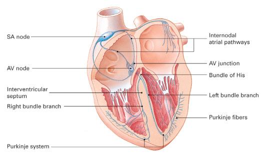
Sinoatrial Node 28
Internodal Pathways 29
Bachmann’s Bundle, Wenckebach’s Bundle, and Thorel’s Pathway 29
Atrioventricular Node 30
Atrioventricular Junction 30
Bundle of His 30
Bundle Branches 30
Purkinje’s Network 31
Summary 32
Key Points to Remember 32
Review Questions 32
5
The Electrocardiogram 34
Objectives 34
Introduction 34
The Electrocardiogram 34
Electrical Basis of the EKG 34
EKG Leads 35
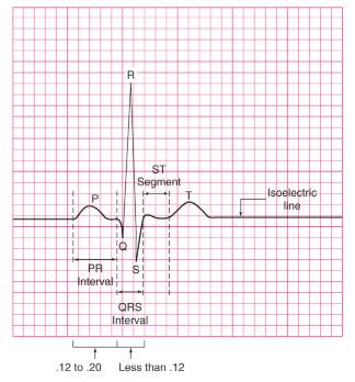
EKG Graph Paper 37
EKG Waveforms 39
Summary 43
Key Points to Remember 43
Review Questions 44
6
Interpretation of an EKG Strip 46
Objectives 46
Introduction 46
General Rules 46
The Five-Step Approach 47
ST Segment 51
The T Wave 52
The U Wave 52
Artifact 52
Summary 53
Key Points to Remember 53
Review Questions 54
7
Introducing the Sinus Rhythms 55
Objectives 55
Introduction 55
Origin of the Sinus Rhythms 55
Components of the Electrical Conduction System of the Heart 55
Normal Sinus Rhythm 56
Sinus Bradycardia Rhythm 57
Sinus Tachycardia Rhythm 58
Sinus Dysrhythmia 59
Sinus Arrest Rhythm 60
Clinical Significance of Sinus Rhythms 61
Summary 63
Key Points to Remember 63
Review Questions 63
Review Strips 64
8
Introducing the Atrial Rhythms 68
Objectives 68
Introduction 68
Origin of the Atrial Rhythms 68
Review: Components of the Electrical Conduction System of the Heart 68
Wandering Atrial Pacemaker Rhythm 69
Premature Atrial Contractions (Complexes) 71
Reentry Dysrhythmias 73
Atrial Flutter Rhythm 74
Atrial Fibrillation Rhythm 75
Supraventricular Tachycardia Rhythms 77
Wolff-Parkinson-White Syndrome 79
Clinical Significance of Atrial Rhythms 79
Summary 81
Key Points to Remember 81
Review Questions 81
Review Strips 82
9
Introducing the Junctional Rhythms 86
Objectives 86
Introduction 86
Origin of Junctional Rhythms 86
Review of the Electrical Conduction System 86
P Waves 86
Premature Junctional Contractions 87
Junctional Escape Rhythms 88
Accelerated Junctional Rhythms 89
Junctional Tachycardia Rhythms 91
Clinical Significance of Junctional Rhythms 92
Summary 93
Key Points to Remember 93
Review Questions 93
Review Strips 94
10
Introducing the Ventricular Rhythms 98
Objectives 98
Introduction 98
Review of the Heart’s Electrical Conduction System 98
Origin of Ventricular Rhythms 98
Premature Ventricular Complexes (Contractions) 99
Idioventricular Rhythm 103
Accelerated Idioventricular Rhythm 104
Ventricular Tachycardia 105
Torsades de Pointes 106
Ventricular Fibrillation 107
Asystole or Ventricular Asystole 109
Pulseless Electrical Activity 111
Clinical Significance of Ventricular Dysrhythmias 111
Clinical Significance of the Heart Block Rhythms 128
Summary 129
Key Points to Remember 129
Review Questions 129
Review Strips 130
12
Introducing the Pacemaker Rhythms 134
Objectives 134
Introduction 134
Temporary Pacing 134
Permanent Pacing 135
Indications for Pacing 137
Rules for Interpretation of Pacemaker Rhythms 138
Common Problems Associated with Pacemakers 139
Clinical Significance of Pacemaker Rhythms 140
Summary 140
Key Points to Remember 140
Review Questions 141
Review Strips 142
13
Assessment and Treatment of the Patient with Cardiac Emergencies 145
Objectives 145
Introduction 145
Chest Pain 145
Angina Pectoris 147
Acute Myocardial Infarction 148
Heart Failure 151
Cardiac Tamponade 154
Cardiogenic Shock 155
Summary 156
Key Points to Remember 157
Review Questions 157
14
More Review Questions 160
15
Review EKG Strips 171
Introduction 171
Appendix 1
Answers to Review Questions: Chapters 1–14 205
Chapter 1 205
Chapter 2 205
Chapter 3 205
Chapter 4 205
Chapter 5 205
Chapter 6 205
Chapter 7 205
Chapter 8 206
Chapter 9 207
Chapter 10 207
Chapter 11 208
Chapter 12 209
Chapter 13 210
Chapter 14 210
Appendix 2
Answers to Review Strips: Chapter 15 211
Glossary 224 Index 229
This page intentionally left blank
Foreword
Dr. Willis D. Israel, who was my mentor and dear friend, has now departed this earth for a better place. His invaluable advice and wisdom were indispensable to me throughout my life, both personally and professionally. As a tribute to him, for always being there for me and for expecting and demanding the very best of me, the Foreword that was written by him for the original manuscript of this book is included in this edition.
When advanced life-support training for paramedical personnel was still considered questionable by most of my physician colleagues, I became involved in teaching advanced cardiac life support (ACLS). It soon became apparent that emergency medical technicians—in those days very frequently volunteer and/or part-time workers in the field—were frequently the most enthusiastic and responsive students of ACLS. Their eagerness to learn and to provide the whole range of prehospital care has proved to be a huge factor in patient survival.
I loved every moment I taught ACLS (as well as BCLS, emergency medical technician training, and advanced trauma life support)—whether to physicians, nurses, or paramedical personnel. Indeed, the teachers of these basic and extremely important concepts have impacted all areas of current medical care. Many of those with whom I taught ACLS became my close and greatly cherished friends. One of these is Brenda Messer Beasley, MS, RN, EMT-P, with whom I shared in teaching the first EMT course ever offered in rural Randolph County, Alabama. From the initial class session, I saw that Ms. Beasley had an extraordinary ability to render a complicated concept in its most basic form of expression. Though she had this gift for rendering the complex in simple terms, she never allowed the importance of what she was teaching to be lost, and she always stressed the awareness of and evaluation of the patient.
Ms. Beasley brings to this text on EKG interpretation the same ability to simplify the complex for the health care professional. Real meaning is surely more valuable than the easy “information overload” encountered when we deal with real patients in a medical emergency.
After I had taught with Ms. Beasley, she made a career change from nursing to full-time EMT training, still in our same basic geographical area. It became fun and rewarding as a practicing small-town family physician to be aware of prehospital care that had been rendered by students of this teacher. Their expertise was (is) impressive, as was their attention to the care of and the state of the patient. Certainly, any physician’s ability to treat, and any patient’s ultimate well-being, depends greatly on that initial prehospital care.
An appropriate text on EKG interpretation can only deepen the perception and understanding of the health care professional; at the same time, this text seems to teach and reteach the basic concept from every situation: “First, look at your patient, and continue to look at your patient.”
I am honored to welcome this book to the plentiful material available on the heart, its functions, the circulatory system and its signals of dysfunction and illness.
Willis D. Israel, MD Wedowee,
AL
This page intentionally left blank
Preface
This informative and simple approach to EKG analysis continues in this, the fourth edition of this textbook. Based on the fact that cardiology and basic EKG interpretation are integral parts of most primary and allied health-related curricula, I originally wrote this book to assist the novice student in his or her understanding of basic EKG interpretation, and that purpose remains undaunted. This book is intended for the health care provider at the initial level(s) of understanding of cardiovascular anatomy, physiology, and rhythm strip interpretation. The categories of students who will benefit from this text include prehospital care providers, medical students, cardiac care monitor techs, ACLS candidates, nursing professionals, physician assistants, respiratory therapy students, and cardiac technology students.
This EKG book consists of 15 chapters designed to provide the user with a basic practical, yet comprehensive, approach to the skill of EKG interpretation. The strategy of this manuscript has centered on producing a useful guide to the understanding of abnormal heart rhythms, that is, dysrhythmias, for the health care provider in his or her provision of optimum patient care. In order to afford the instructor and the student the opportunity to work in a reasonable order through the technical information, the material has been presented in such a manner as to achieve understanding of each chapter prior to proceeding to the next chapter. The content is presented in short, succinct chapters in order to facilitate comprehension of each concept in a building-block format. Although the terms dysrhythmia and arrhythmia are synonymous, the term dysrhythmia is used throughout this book because I consider it to be the more correct and accurate description of the material presented.
In this revised fourth edition of the book, each chapter still contains a section of multiple-choice items to be used for self-assessment and review. The book includes expanded graphics, as well as rhythm strip examples with answers based on the five-step approach, review strips, key points to remember, chapter summary, and end-of-chapter questions to afford the student a comprehensive mastery of the material. Answers to review questions and review strips are provided in the appendices at the back of the book. Also included in this fourth edition of the book is a chapter dedicated to the assessment and management of the patient with cardiovascular emergencies.
An overview of major updates and additions to this revision may be helpful to you. These changes are summarized in the “What’s New” list below.
WHAT’S NEW IN THE FOURTH EDITION
◾ Information has been updated to reflect current standards of care.
◾ Added and enhanced Chapter objectives and marginal glossary terms.
◾ Numerous review strip answers based on the five-step approach have been added.
◾ The feature Key Points to Remember has been enhanced and revised at the end of each chapter.
◾ End-of-Chapter Review Questions have been updated and revised.
◾ Instructor and student resources are available online at Brady’s Resource Central Web site.
INSTRUCTOR RESOURCES
This Web site contains an array of instructor resources in one location. To access the Instructor Resource Center, visit www.pearsonhighered.com and follow registration/log-in instructions for Instructors. Your Brady sales representative can offer further assistance. Once you are logged onto the site, you’ll find the following teaching resources.
◾ PowerPoint Slides-updated and revised
◾ Lesson Plans
◾ Test Bank with more than 280 questions
It is my hope that you will find this book to be beneficial to your knowledge and comprehension of basic EKG interpretation. Your suggestions and comments are always welcome. Brenda M. Beasley, RN, MS, EMT-Paramedic Department Chair, Allied Health/EMS Program Director (Retired) Calhoun Community College E-mail address: bjm18@aol.com
Acknowledgments
Just as the first three editions of this textbook were created from the challenges that I have experienced and the lessons I have learned throughout the past 35 years as a nurse, paramedic and an EMS educator, this revision was written to enhance the chapter content and, where appropriate, replace some of the content.
I have learned that the publication of textbooks involves many key people. I would like to acknowledge all the individuals who were instrumental, each in their own special way, in making the textbook revision possible. I also offer my sincere appreciation to the talented team members at Brady/Pearson Health Science who have led me through the revision process with expert advice, encouragement, and support. Thanks especially to Julie Alexander, Marlene Pratt, Sladjana Repic, Lois Berlowitz, Jonathan Cheung, Patrick Walsh, Patty Gutierrez, Debbie Ryan, and Brian Hoehl. My first editor at Brady, Judy Streger, has continued her support and encouragement throughout the years. Her belief in me has never waivered, and for that I am truly grateful.
My former EMS program faculty, both at Southern Union Community College and at Calhoun Community College (too many names to mention here, but they know who they are!), have all been my strength and inspiration to strive for excellence in EMS education. The reviewers of this book, whose names follow, offered important perspective. Their comments and suggestions have been very valuable.
Mike West has continued to be my champion and has worked closely with me throughout this revision process. His encouragement and constancy were very critical to me, and I wish to thank him for the long hours he spent working with me on this revision, as well as for his valuable assistance in gathering and incorporating the new changes and additions to the review questions and EKG strips.
The memory of my mother and my father, who taught my sisters and me to believe first in God, then in our family, and ultimately in ourselves; they always provided me with unconditional love, acceptance, and a solid foundation upon which to build a life.
It is my belief that every novice author is, at some point, inspired by other authors, and colleagues who transition to mentors, role models, and precious friends. In my particular case, there were many. I especially appreciate the support and friendship of the following EMS colleagues: Walt Stoy, Joe Mistovich, Greg Margolis, Jeff Lindsey, Dwayne Clayden, Bryan Bledsoe, Baxter Larmon, and Dan Limmer.
And last, but not least, I gratefully acknowledge my family, friends, and colleagues, for they are the true “contributors” to this product. Their contributions include love, patience, support, encouragement, and acceptance of my erratic schedule during the text revision.
REVIEWERS
I wish to thank the following reviewers for providing invaluable feedback and suggestions during the revision of this text:
James “Bud” Adams, AAS, NREMT-P Instructor of Emergency Medicine College of Southern Nevada Las Vegas, NV
John L Beckman, AA, BS, FF/EMT-P EMS Instructor Addison Fire Protection District Addison, IL
Deborah K. Drummonds, RN, MN, CCRN, CEN
Assistant Professor School of Nursing and Health Sciences Abraham Baldwin Agricultural College Tifton, GA
Fidel O. Garcia, EMT-P
President/Owner Professional EMS Education, LLC Grand Junction, CO
Stephanie Morrison, RN, BSN
ACLS Instructor Thomas Hospital Fairhope, AL
David Pierce, BA, NREMT-P
EMS Faculty Century College White Bear Lake, MN
Shari Turner, M.Ed., EMT-P Palm Beach Gardens, FL
Randy Williams, NREMT-P
EMS Programs Coordinator Bainbridge College Bainbridge, GA
I also wish to thank the following professionals who reviewed earlier editions of Understanding EKGs: A Practical Approach:
John L. Beckman, AA, BS, FF/EMT-P Instructor
Affiliated with Addison Fire Protection District
Fire Science Instructor, Technology Center of DuPage Addison, IL
Mark Branon, BS, NREMT-P
EMS Program Director Calhoun Community College Decatur, AL
Art Breault, RN, EMT-Paramedic
Niskayuna Fire District No. 1 Albany Medical Center Hospital—Department of Emergency Medicine Albany, NY
Benjamin J. Camp, MD, FACEP
Chief of Staff Tanner Health System Emergency Services Carrollton, GA
Greg Charma, AAS, NREMT-P
Paramedic/Assistant EMS Training Director
Tucson Fire Department Pima Community College Tucson, AZ
Marilyn Ermish, NREMT-P
American Medical Response Cheyenne, WY
Jason Ferguson, BPA, NREMT-P
EMS Programs Head Central Virginia Community College Lynchburg, VA
Scott Garrett
Director of Education Upstate EMS Council Greenville, SC
Craig H. Jacobus, D.C.; NREMT-P; EMSI
Links 4 Life Metro Community College ALS
Affiliates Lincoln Medical Education Partnership (LMEP) Schuyler, NE
Scott Jones
Director—Paramedic Academy Victor Valley College Victorville, CA
Maryla Kathryn Lee, RN, BSN, MSN Calhoun Community College Decatur, AL
Jeff McDonald
Coordinator, Emergency Medical Services
Program Tarrant Count College—Northeast Campus Hurst, TX
Mike McEvoy, PhD, EMT-P, RN, CCRN
Saratoga County EMS Coordinator Clinical Associate Professor in Critical Care Medicine
Albany Medical College Waterford, NY
Steve McGraw
Assistant Professor The George Washington University School of Medicine and Health Sciences Washington, DC
Regina Pearson, EMT-P
Clinical Coordinator/Instructor, Emergency Medical Technology Jackson State Community College Jackson, TN
Jonathan Smith, NREMT-P
Lead Paramedic Instructor Chattahoochee Technical College Acworth, GA
Thomas Y. Smith, Sr. Fire Science/EMS Program Director West Georgia Technical College LaGrange, GA
Scott R. Snyder, BS, NREMT-P.
Primary EMT Instructor San Francisco Paramedic Association San Francisco, CA
Andrew E. Spain
Assistant Manager, Emergency Services University of Missouri Health Care EMS Education Columbia, MO
Carl Voskamp, LP
Coordinator of EMS and Firefighting Programs
The Victoria College Victoria, TX
Jim Williams, CCEMT-P, NREMT-P
Training Officer, Medical Center EMS Bowling Green, KY
About the Author
Brenda Messer Beasley is a paramedic and a registered nurse. She earned her bachelor’s and master’s degree, as well as additional postgraduate studies, from the University of Alabama and from Lorenz University, respectively. Following graduation from nursing school at the University of Alabama, Ms. Beasley was employed as a nurse in the Emergency Department of University Hospital in Birmingham. She remained in emergency nursing for the next 10 years, with a 2-year hiatus when she became certified as a critical care neurosurgical nurse and worked in an NICU at Carraway Medical Center.
Ms. Beasley was working as an ER nurse in 1978 when she was asked to teach a basic EMT course. With a great deal of reticence yet strong encouragement from the chief of staff at the local hospital, she concurred. She immediately developed a passion for quality EMS education, and the rest, as they say, is history. For the next 30 years, she served as an EMS educator in the state of Alabama, and in 2001 was named Department Chair of Allied Health at Calhoun Community College in Decatur, Alabama. She held that position until her retirement in 2007.
Ms. Beasley was an affiliate faculty member of the American Heart Association’s Emergency Cardiac Care Program for 25 years. Other professional activities include BTLS affiliate faculty and board of directors of the National Association of EMS Educators, and she serves on the medical advisory boards of Action Training Systems and Southern Ambulance Transport.
In 1999, Ms. Beasley published her first book on EKG interpretation and has subsequently authored other texts for Brady/Pearson Health Science. She resides in Wedowee, Alabama, (with her beloved pet, Kitty Boo) where she serves as vice chair of the local hospital board. Ms. Beasley is actively involved in the First United Methodist Church of Wedowee and currently serves as the Governance Committee Chair for the National Association of EMS Educators.
This page intentionally left blank
The Anatomy of the Heart: Structure
INTRODUCTION
A thorough understanding of the structure of the heart provides the student with a foundation upon which to build the knowledge of basic dysrhythmia interpretation. Therefore, the focus of this chapter will be to provide you, the student, with a simple yet comprehensive understanding of cardiac anatomy. After you have mastered the knowledge of basic cardiac anatomy (structure), you will be prepared to move to Chapter 2, which addresses the basic physiology (function) of the heart.
ANATOMY OF THE HEART
First you must realize that the heart is a muscle. Although we don’t think of exercising our heart muscle when we go to the gym, the fact is that your heart muscle (myocardium) is constantly in the “exercise mode.” At times of rest, the exercise is more sedate. Think, however, of the vigor with which your heart muscle must exercise when you walk (or run) up six flights of stairs! Now as you feel your heart pumping, you can easily understand that your heart muscle is indeed exercising.
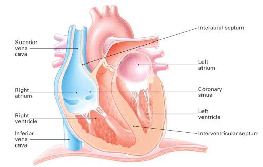
We often hear the heart referred to as a “two-sided pump,” and this analogy works well in our understanding of the basics of cardiac anatomy. Indeed, we can visualize this pump as having a right side and a left side. On each side of the pump, there is an upper chamber of the heart, which is referred to as the atrium (atria, plural), and a lower chamber of the heart known as the ventricle . In all, there are four hollow chambers in the normal heart. Again, the two upper chambers of the heart are called atria; the two lower chambers are called ventricles.
Separating the upper chambers is the interatrial septum. The lower, inferior chambers are separated by the interventricular septum. (See ■ Figure 1–1.) Externally, the atrioventricular groove, known as the coronary sulcus, surrounds the outside of the heart and divides the atria from the ventricles. The anterior and posterior interventricular grooves separate the ventricles externally. The muscle fibers of the ventricles are continuous, as are the atrial muscle fibers.
The two upper chambers of the heart are located at the base, or top, of the heart; the lower chambers are located at the bottom, or apex, of the heart. The upper chambers of the heart are thin walled and receive blood as it returns to the heart. The lower chambers of the heart have thicker walls and pump blood away from the heart, throughout the systemic circulation and to the myocardium.
Objectives
Upon completion of this chapter, the student will be able to:
■ Describe the chambers of the heart
a. Atria
b. Ventricles
■ Identify the location, size, and shape of the heart
■ Given a diagram, be able to name the layers of the heart
■ Given a diagram, be able to name the valves of the heart
■ Describe the structure and function of the blood vessels
a. Arteries
b. Veins
c. Capillaries
■ Discuss the concept of pulmonary circulation
■ Explain the concept of systemic circulation
atrium upper chamber of the heart
ventricle lower chamber of the heart
coronary sulcus the atrioventricular groove that surrounds the outside of the heart and divides the atria from the ventricles
mediastinum the central section of the thorax (chest cavity)
Right atrium
Right
Interatrial septum
Left atrium
Left ventricle
Interventricular septum
LOCATION, SIZE, AND SHAPE OF THE HEART
It is important for you to learn and understand the location of the heart in that the effectiveness of one of our most basic and most important skills—CPR—depends upon a reasonable knowledge of this position. In addition, the proper placement of electrodes to record an electrocardiogram, which is discussed in Chapter 5, depends upon the proper understanding of the location of the heart.
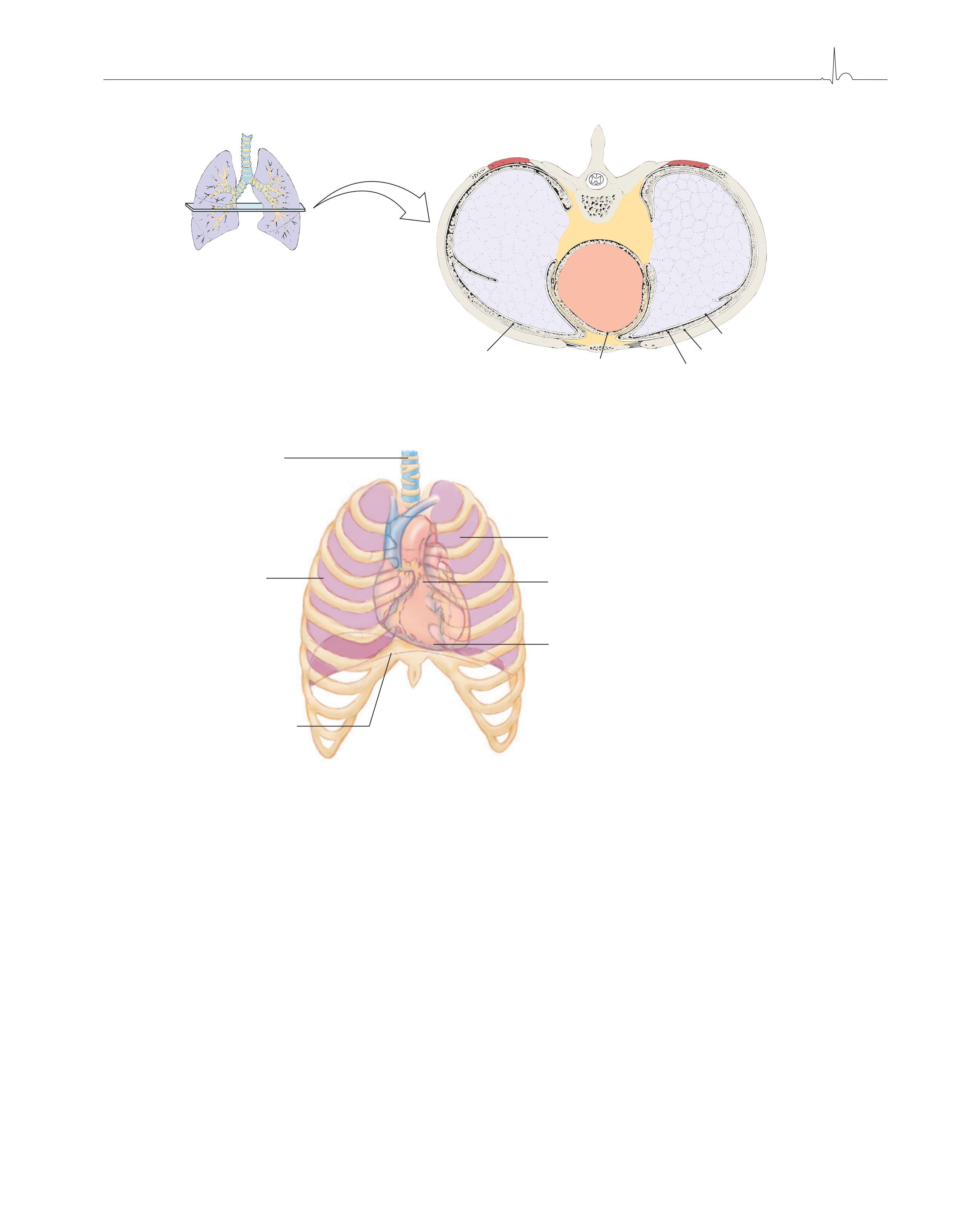
The central section of the thorax (chest cavity) is called the mediastinum. It is in this area that the heart and its large vessels are housed, lying in front of the spinal column, behind the sternum, and between the lungs. (See ■ Figure 1–2.) Also located in the mediastinum are the trachea, esophagus, thymus, lymph nodes, and other structures and tissues.
FIGURE 1–1. The chambers of the heart
FIGURE 1–2. Position and orientation of the heart within the mediastinum
Right Lung
Right pleural cavity
FIGURE 1–3. Anatomical relationships in the thoracic cavity
Mediastinum
Pericardial cavity Heart
Left Lung Visceral pleura
Parietal pleura
Left pleural cavity
FIGURE 1–4. Location of the heart within the chest
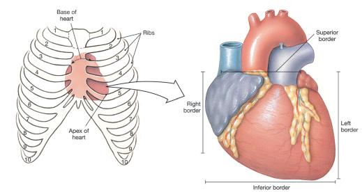
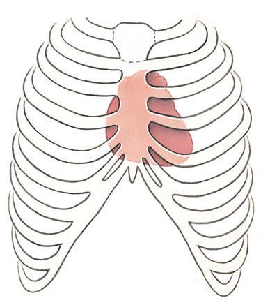
When thinking of the heart muscle in terms of its mass, one should realize that twothirds of the heart muscle lies to the left of the midline. The apex of the heart lies just above the diaphragm. The base of the heart lies at approximately the level of the third rib. (See ■ Figures 1–2 and 1–4.)
The exact size of the heart varies somewhat among individuals, but on average it is approximately 5 inches (in.), or 12 centimeters (cm), in length and 3 in., or 7.5 cm, wide. The shape of the heart is somewhat conelike. It is appropriate to visualize the heart as approximately the size of the owner’s closed fist. (See ■ Figure 1–3.)
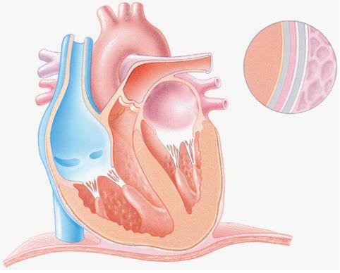
LAYERS OF THE HEART
Pericardium
Surrounding the heart is a closed, two-layered sac referred to as the pericardium, or pericardial sac. In direct contact with the pleura is the outer layer or parietal pericardium. (See ■ Figure 1–5.) This layer consists of tough, inelastic fibrous connective tissue and serves
pericardium closed, two-layered sac that surrounds the heart
Trachea
Right lung
pericarditis an inflammation of the serous pericardium
1–5.
epicardium the smooth outer surface of the heart
myocardium the thick middle layer of the heart composed primarily of cardiac muscle cells and responsible for the heart’s ability to contract
endocardium the innermost layer of the heart; composed of thin connective tissue
Myocardium
Epicardium
Pericardial cavity
Lung
Parietal pericardium
Pleural cavity
Pleura
Endocardium
Diaphragm
to prevent overdistention of the heart. The thin, serous inner layer of the pericardium is called the visceral pericardium and is contiguous with the epicardium, which surrounds the heart. The serous pericardium is considered a part of the heart and is continuous with the epicardium.
A space filled with a scant amount of fluid (approximately 10 to 50 cubic centimeters [cc]) separates the two pericardial layers. This fluid, by acting as a lubricant, helps to reduce friction as the heart moves within the pericardial sac.
An inflammation of the serous pericardium is called pericarditis. Although the cause of this disease is frequently unknown, it may result from infection or disease of the connective tissue. Pericarditis can cause severe pain, which may be confused with or mistaken for the pain of a myocardial infarction. This can make physical assessment of the patient a real challenge for the clinician.
An excess accumulation of fluid in the pericardial sac is called cardiac tamponade. This condition is an extreme emergency and must be detected and treated expeditiously. (See Chapter 13 for discussion.)
The heart wall
Three primary layers of tissue comprise the heart wall. (See Figure 1–5.) This specialized cardiac muscle tissue is unique to the heart. The epicardium accounts for the smooth outer surface of the heart. The main coronary arteries are located on the surface of the epicardium. The thick middle layer of the heart is called the myocardium and is the thickest of the three layers of the heart wall. The myocardium is composed primarily of cardiac muscle cells and is responsible for the heart’s ability to contract. The innermost layer, the endocardium, is composed of endothelial tissue. This area requires a constant and uninterrupted supply of oxygen and is subject to ischemia (decreased supply of oxygenated blood). This smooth inner surface of the heart and heart valves serves to allow blood to flow more easily throughout the heart.
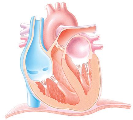
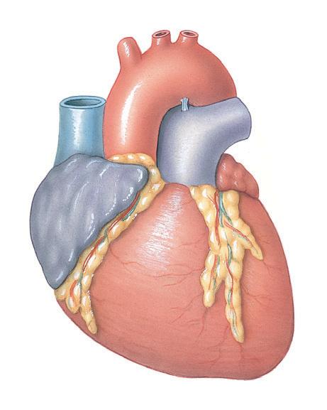
VALVES OF THE HEART
The four valves of the heart allow blood to flow in only one direction. (See ■ Figure 1–6.) There are two sets of valves, the atrioventricular valves and the semilunar valves.