Infection—Basic Concepts
Humanity has but three great enemies: fever, famine and war; of these by far the greatest, by far the most terrible, is fever.
— Sir William Osler, 1896*
When Sir William Osler, the great physician/humanist, wrote these words, fever (infection) was indeed the scourge of the world. Tuberculosis and other forms of pulmonary infection were the leading causes of premature death among the well to do and the less fortunate. The terror was due to the fact that, although some of the causes of infection were being discovered, little could be done to prevent or alter the course of disease. In the 20th century, advances in public sanitation and the development of vaccines and antimicrobial agents changed this (Figure 1–1), but only for the nations that could afford these interventions. As we move through the second decade of the 21st century, the world is divided into countries in which heart attacks, cancer, and stroke have surpassed infection as causes of premature death and those in which infection is still the leader. A new uneasiness that is part evolutionary, part discovery, and part diabolic has taken hold. Infectious agents once conquered have shown resistance to established therapy, such as multiresistant Mycobacterium tuberculosis, and diseases, such as acquired immunodeficiency syndrome (AIDS), have emerged. The spectrum of infection has widened, with discoveries that organisms earlier thought to be harmless can cause disease under certain circumstances. Who could have guessed that Helicobacter pylori, not even mentioned in the first edition of this book (1984), would be the major cause of gastric and duodenal ulcers and an officially declared carcinogen? Finally, bioterrorist forces have unearthed two previously controlled infectious diseases—anthrax and smallpox—and threatened their distribution as agents of biological warfare. For students of medicine, understanding the fundamental basis of infectious diseases has more relevance than ever.
BACKGROUND
The science of medical microbiology dates back to the pioneering studies of Pasteur and Koch, who isolated specific agents and proved that they could cause disease by introducing the experimental method. The methods they developed lead to the first golden age of microbiology (1875-1910), when many bacterial diseases and the organisms responsible for them were defined. These efforts, combined with work begun by Semmelweis and Lister, which showed how these diseases spread, led to the great advances in public health that initiated the decline in disease and death. In the first half of the 20th century, scientists studied the structure, physiology, and genetics of microbes in detail and began to answer
*Osler W. JAMA. 1896;26:999.
FIGURE 1–1. Death rates for infectious disease in the United States in the 20th century. Note the steady decline in death rates related to the introduction of public health, immunization, and antimicrobial interventions.
Microbes are small
Most play benign roles in the environment
Products of microbes contribute to the atmosphere
Public health departments established
pandemic Chlorination of water
Diphtheria immunization (1940)
Penicillin usage (1945)
Polio vaccine
Haemophllus influenzae conjugate vaccine (1990)
questions relating to the links between specific microbial properties and disease. By the end of the 20th century, the sciences of molecular biology, genetics, genomics, and proteomics extended these insights to the molecular level. Genetic advances have reached the point at which it is possible to know not only the genes involved but also to understand how they are regulated. The discoveries of penicillin by Fleming in 1929 and of sulfonamides by Domagk in 1935 opened the way to great developments in chemotherapy. These gradually extended from bacterial diseases to fungal, parasitic, and finally viral infections. Almost as quickly, virtually all categories of infectious agents developed resistance to all categories of antimicrobial agents to counter these chemotherapeutic agents.
INFECTIOUS AGENTS: THE MICROBIAL WORLD
Microbiology is a science defined by smallness. Its creation was made possible by the invention of the microscope (Gr. micro, small + skop, to look, see), which allowed visualization of structures too small to see with the naked eye. This definition of microbiology as the study of microscopic living forms still holds if one can accept that some organisms can live only in other cells (eg, all viruses and some bacteria) and that others include macroscopic forms in their life cycle (eg, fungal molds, parasitic worms). The relative sizes of some microorganisms are shown in Figure 1–2.
Microorganisms are responsible for much of the breakdown and natural recycling of organic material in the environment. Some synthesize nitrogen-containing compounds that contribute to the nutrition of living things that lack this ability; others (oceanic algae) contribute to the atmosphere by producing oxygen through photosynthesis. Because microorganisms have an astounding range of metabolic and energy-yielding abilities, some can exist under conditions that are lethal to other life forms. For example, some bacteria can oxidize inorganic compounds such as sulfur and ammonium ions to generate energy. Others can survive and multiply in hot springs at temperatures higher than 75°C.
Some microbial species have adapted to a symbiotic relationship with higher forms of life. For example, bacteria that can fix atmospheric nitrogen colonize root systems of legumes and of a few trees, such as alders, and provide the plants with their nitrogen requirements. When these plants die or are plowed under, the fertility of the soil is enhanced by nitrogenous compounds originally derived from the metabolism of the bacteria. Ruminants can use grasses as their prime source of nutrition, because the abundant flora of anaerobic bacteria in the rumen break down cellulose and other plant compounds to usable carbohydrates and amino acids and synthesize essential nutrients including some amino acids and vitamins. These few examples illustrate the protean nature of microbial life and their essential place in our ecosystem.
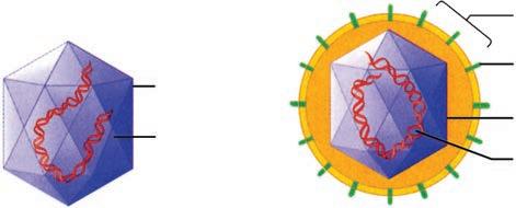
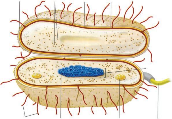
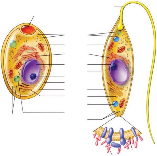
The major classes of microorganisms in terms of ascending size and complexity are viruses, bacteria, fungi, and parasites. Parasites exist as single or multicellular structures with the same compartmentalized eukaryotic cell plan of our own cells including a nucleus and cytoplasmic organelles like mitochondria. Fungi are also eukaryotic, but have a rigid external wall that makes them seem more like plants than animals. Bacteria also have a cell wall, but with a cell plan called “prokaryotic” that lacks the organelles of eukaryotic cells. Viruses are not cells at all. They have a genome and some structural elements, but must take over the machinery of another living cell (eukaryotic or prokaryotic) to replicate. The four classes of infectious agents are summarized in Table 1–1, and generic examples of each are shown in Figure 1–3.
VIRUSES
Viruses are strict intracellular parasites of other living cells, not only of mammalian and plant cells, but also of simple unicellular organisms, including bacteria (the bacteriophages). Viruses are simple forms of replicating, biologically active particles that carry genetic information in either DNA or RNA molecules. Most mature viruses have a protein coat over their nucleic acid and, sometimes, a lipid surface membrane derived from the cell they infect. Because viruses lack the protein-synthesizing enzymes and structural apparatus necessary for their own replication, they bear essentially no resemblance to a true eukaryotic or prokaryotic cell.
Viruses replicate by using their own genes to direct the metabolic activities of the cell they infect to bring about the synthesis and reassembly of their component parts. A cell infected with a single viral particle may, thus, yield thousands of viral particles, which can
TABLE 1–1 Features of Infectious Agents
aParasitic cysts have cell walls.
bA few bacteria grow only within cells.
cThe life cycle of some parasites includes intracellular multiplication.
FIGURE 1–2. Relative size of microorganisms.
Increasing complexity: viruses → bacteria → fungi → parasites
Viruses contain little more than DNA or RNA
FIGURE 1–3. Infectious agents. A. Virus. B. Bacterium. C. Fungus. D. Parasite. (Reproduced with permission from Willey JM: Prescott, Harley, & Klein’s Microbiology, 7th edition. McGraw-Hill, 2008.)
Capsid
Nucleic acid
Naked virus
Envelope
Spike
Capsid
Nucleic acid
Enveloped virus A
CapsuleRibosomesCell wall
Plasma membrane
Chromosome (DNA)
Bud scar
(Yeast) Cell C
Mitochondrion
Endoplasmic reticulum
Nucleus
Pellicle
Nucleolus
Cell wall
Cell membrane
Golgi apparatus
Water vacuole
Storage vacuole
Centrioles
Inclusion body
Flagellum
Cell membrane
Cell D
Nucleoid
Fimbriae
Glycocalyx
Centrioles
Flagellum
Ribosomes
Fungal
Protozoan
be assembled almost simultaneously under the direction of the viral nucleic acid. Infection of other cells by the newly formed viruses occurs either by seeding from or lysis of the infected cells. Sometimes, viral and cell reproduction proceed simultaneously without cell death, although cell physiology may be affected. The close association of the virus with the cell sometimes results in the integration of viral nucleic acid into the functional nucleic acid of the cell, producing a latent infection that can be transmitted intact to the progeny of the cell.
BACTERIA
Bacteria are the smallest (0.1-10 μm) independently living cells. They have a cytoplasmic membrane surrounded by a cell wall; a unique interwoven polymer called peptidoglycan makes the wall rigid. The simple prokaryotic cell plan includes no mitochondria, lysosomes, endoplasmic reticulum, or other organelles (Table 1–2). In fact, most bacteria are approximately the size of mitochondria. Their cytoplasm contains only ribosomes and a single, double-stranded DNA chromosome. Bacteria have no nucleus, but all the chemical elements of nucleic acid and protein synthesis are present. Although their nutritional requirements vary greatly, most bacteria are free living if given an appropriate energy source. Tiny metabolic factories, they divide by binary fission and can be grown in artificial culture, often in less than 1 day. The Archaea are similar to bacteria but evolutionarily distinct. They are prokaryotic, but differ in the chemical structure of their cell walls and other features. The Archaea (archebacteria) can live in environments humans consider hostile (eg, hot springs, high salt areas) but are not associated with disease.
FUNGI
Fungi exist in either yeast or mold forms. The smallest of yeasts are similar in size to bacteria, but most are larger (2-12 μm) and multiply by budding. Molds form tubular extensions called hyphae, which, when linked together in a branched network, form the fuzzy structure seen on neglected bread slices. Fungi are eukaryotic, and both yeasts and molds have a rigid external cell wall composed of their own unique polymers, called glucan, mannan, and chitin. Their genome may exist in a diploid or haploid state and replicate by meiosis or simple mitosis. Most fungi are free living and widely distributed in nature. Generally, fungi grow more slowly than bacteria, although their growth rates sometimes overlap.
TABLE 1–2 Distinc tive Features of Prokaryotic and Eukaryotic Cells
CELL COMPONENT PROKARYOTES
Nucleus No membrane, single circular chromosome
EUKARYOTES
Membrane bounded, a number of individual chromosomes
Extrachromosomal DNA Often present in form of plasmid(s) In organelles
Organelles in cytoplasm None
Cytoplasmic membrane Contains enzymes of respiration; active secretion of enzymes; site of phospholipid and DNA synthesis
Cell wall Rigid layer of peptidoglycan (absent in Mycoplasma)
Replication is by control of the host cell metabolic machinery
Some integrate into the genome
Mitochondria (and chloroplasts in photosynthetic organisms)
Semipermeable layer not possessing functions of prokaryotic membrane
No peptidoglycan (in some cases cellulose present)
Sterols Absent (except in Mycoplasma) Usually present
Ribosomes
70 S in cytoplasm
80 S in cytoplasmic reticulum
Smallest living cells
Prokaryotic cell plan lacks nucleus and organelles
Yeasts and molds are surrounded by cell wall
Range from tiny amoebas to meter-long worms
PARASITES
Parasites are the most diverse of all microorganisms. They range from unicellular amoebas of 10 to 12 μm to multicellular tapeworms 1 m long. The individual cell plan is eukaryotic, but organisms such as worms are highly differentiated and have their own organ systems. Most worms have a microscopic egg or larval stage, and part of their life cycle may involve multiple vertebrate and invertebrate hosts. Most parasites are free living, but some depend on combinations of animal, arthropod, or crustacean hosts for their survival.
THE HUMAN MICROBIOTA
Before moving on to discuss how, when, and where the previously mentioned agents cause human disease, we should note that the presence of microbes on or in humans is not, by itself, abnormal. In fact, from shortly after birth on, it is universal; we harbor 10 times the number of microbial cells than human cells. This population formerly called the normal flora is now referred to as our microbiota or microbiome. These microorganisms, which are overwhelmingly bacteria, are frequently found colonizing various body sites in healthy individuals. The constituents and numbers of the microbiota vary in different areas of the body and, sometimes, at different ages and physiologic states. Their names are mostly unfamiliar because they have not (yet) been associated with disease. They comprise microorganisms whose morphologic, physiologic, and genetic properties allow them to colonize and multiply under the conditions that exist in particular sites, to coexist with other colonizing organisms, and to inhibit competing intruders. Thus, each accessible area of the body presents a particular ecologic niche, colonization of which requires a particular set of properties of the colonizing microbe.
Flora may stay for short or extended periods
If pathogens are involved, the relationship is called the carrier state
Organisms of the microbiota may have a symbiotic relationship that benefits the host or may simply live as commensals with a neutral relationship to the host. A parasitic relationship that injures the host would not be considered “normal,” but, in most instances, not enough is known about the organism–host interactions to make such distinctions. Like houseguests, the members of the normal flora may stay for highly variable periods. Residents are strains that have an established niche at one of the many body sites, which they occupy indefinitely. Transients are acquired from the environment and establish themselves briefly, but they tend to be excluded by competition from residents or by the host’s innate or immune defense mechanisms. The term carrier state is used when organisms known to be potentially pathogenic are involved, although its implication of risk is not always justified. For example, Streptococcus pneumoniae, a cause of pneumonia, and Neisseria meningitidis, a cause of meningitis, may be isolated from the throat of 5% to 40% of healthy people. Whether these bacteria represent transient flora, resident flora, or carrier state is largely semantic. The possibility that their presence could be the prelude to disease is presently impossible to determine in advance.
It is important for students of medical microbiology and infectious disease to understand the role of the microbiota because of its significance both as a defense mechanism against infection and as a source of potentially pathogenic organisms. In addition, it is important for physicians to know the typical composition of the microbiota at various sites to avoid confusion when interpreting laboratory culture results. The following excerpt indicates that the English poet W.H. Auden understood the need for balance between the microbiota and its host. He was influenced by an article in Scientific American about the flora of the skin.
On this day tradition allots to taking stock of our lives, my greetings to all of you, Yeasts, Bacteria, Viruses, Aerobics and Anaerobics:
A Very Happy New Year
to all for whom my ectoderm is as Middle Earth to me.
For creatures your size I offer a free choice of habitat, so settle yourselves in the zone that suits you best, in the pools
of my pores or the tropical forests of arm-pit and crotch, in the deserts of my fore-arms, or the cool woods of my scalp.
Build colonies: I will supply adequate warmth and moisture,
ORIGIN AND NATURE
the sebum and lipids you need, on condition you never do me annoy with your presence, but behave as good guests should, not rioting into acne or athlete’s-foot or a boil.
—W.H. Auden, “A New Year Greeting”
The healthy fetus is sterile until the birth membranes rupture. During and after birth, the infant is exposed to the flora of the mother’s vagina and to other organisms in the environment. During the infant’s first few days of life, the microbiota reflects chance exposure to organisms that can colonize particular sites in the absence of competitors. Subsequently, as the infant is exposed to a broader range of organisms, those best adapted to colonize particular sites become predominant. Thereafter, the flora generally resembles that of other individuals in the same age group and cultural milieu.
Local physiologic and ecologic conditions determine the microbial makeup of the flora. These conditions are sometimes highly complex, differing from site to site, and sometimes with age. Conditions include the amounts and types of nutrients available, pH, oxidation–reduction potentials, and resistance to local antibacterial substances such as bile and lysozyme. Many bacteria have adhesin-mediated affinity for receptors on specific types of epithelial cells; this facilitates colonization and multiplication and prevents removal by the flushing effects of surface fluids and peristalsis. Various microbial interactions also determine their relative prevalence in the flora. These interactions include competition for nutrients and inhibition by the metabolic products of other organisms.
MICROBIOTA AT DIFFERENT SITES
At any one time, the microbiota of a single person contains hundreds if not thousands of species of microorganisms, mostly bacteria. The major members known to be important in preventing or causing disease, as well as those that may be confused with etiologic agents of local infections, are summarized in Table 1–3 and are described in greater detail in subsequent chapters. The Human Microbiome Project is an ongoing effort to bring this information together.
● Blood, Body Fluids, and Tissues
In health, the blood, body fluids, and tissues are sterile. Occasional organisms may be displaced across epithelial barriers as a result of trauma or during childbirth; they may be briefly recoverable from the bloodstream before they are filtered out in the pulmonary capillaries or removed by cells of the reticuloendothelial system. Such transient bacteremia may be the source of infection when structures such as damaged heart valves and foreign bodies (prostheses) are in the bloodstream.
● Skin
The skin surface provides a dry, slightly acidic, aerobic environment. It plays host to an abundant flora that varies according to the presence of its appendages (hair, nails) and the activity of sebaceous and sweat glands. The flora is more abundant on moist skin areas (axillae, perineum, and between toes). Staphylococci and members of the Propionibacterium genus occur all over the skin, and facultative diphtheroids (corynebacteria) are found in moist areas. Propionibacteria are slim, anaerobic, or microaerophilic gram-positive rods that grow in subsurface sebum and break down skin lipids to fatty acids. Thus, they are most numerous in the ducts of hair follicles and of the sebaceous glands that drain into them. Even with antiseptic scrubbing, it is difficult to eliminate bacteria from skin sites, particularly those bearing pilosebaceous units. Organisms of the skin flora are resistant to
Initial flora is acquired during and immediately after birth
Physiologic conditions such as local pH influence colonization
Adherence factors counteract mechanical flushing
Ability to compete for nutrients is an advantage
Tissues and body fluids such as blood are sterile in health
Transient bacteremia can result from trauma
Propionibacteria and staphylococci are dominant bacteria
Skin flora is not easily removed
Conjunctiva resembles skin
Oropharynx has streptococci and Neisseria H pylori turned out to be a stomach pathogen
Small intestinal flora is scanty but increases toward lower ileum
TABLE 1–3
BODY SITE
Predominant and Potentially Pathogenic Microbiota of Various Body Sites
POTENTIAL PATHOGENS (CARRIER)
Blood None
Tissues
Skin
Adult colonic flora is abundant and predominantly anaerobic
Diet affects species composition
None
Staphylococcus aureus
Mouth Candida albicans
Nasopharynx
Streptococcus pneumoniae, Neisseria meningitidis, Haemophilus influenzae, group A streptococci, Staphylococcus aureus (anterior nares)
Stomach None
Small intestine None
Colon Bacteroides fragilis, E coli, Pseudomonas, Candida, Clostridium (C perfringens, C difficile)
Vagina
Prepubertal and postmenopausal
Candida albicans
LOW VIRULENCE (RESIDENT)
Nonea
None
Propionibacterium, Corynebacterium (diphtheroids), coagulase-negative staphylococci
Neisseria spp., viridans streptococci, Moraxella, Peptostreptococcus
Neisseria spp., viridans streptococci, Moraxella, Peptostreptococcus
Streptococci, Peptostreptococcus, others from mouth
Scanty, variable
Eubacterium, Lactobacillus, Bacteroides, Fusobacterium, Enterobacteriaceae, Enterococcus, Clostridium
Diphtheroids, staphylococci, Enterobacteriaceae
Childbearing Group B streptococci, C albicans Lactobacillus, streptococci
aOrganisms such as viridans streptococci may be transiently present after disruption of a mucosal site.
the bactericidal effects of skin lipids and fatty acids, which inhibit or kill many extraneous bacteria. The conjunctivae have a very scanty flora derived from the skin flora. The low bacterial count is maintained by the high lysozyme content of lachrymal secretions and by the flushing effect of tears.
● Intestinal Tract
The mouth and pharynx contain large numbers of facultative and anaerobic bacteria. Different species of streptococci predominate on the buccal and tongue mucosa because of different specific adherence characteristics. Gram-negative diplococci of the genus Neisseria and coccobacillary Moraxella make up the balance of the most commonly isolated organisms. Strict anaerobes and microaerophilic organisms of the oral cavity have their niches in the depths of the gingival crevices surrounding the teeth and in sites such as tonsillar crypts, where anaerobic conditions can develop readily.
The total number of organisms in the oral cavity is very high, and it varies from site to site. Saliva usually contains a mixed flora of about 108 organisms per milliliter, derived mostly from the various epithelial colonization sites. The genera include Actinomyces, Bacteroides, Prevotella, Streptococcus, and others. The stomach contains few, if any, resident organisms in health because of the lethal action of gastric hydrochloric acid and peptic enzymes on bacteria. One species, H pylori, long thought to be a common resident is now known to be the primary cause of ulcers. The small intestine has a scanty resident flora, except in the lower ileum, where it begins to resemble that of the colon.
The colon carries the most abundant and diverse microbiota in the body. In the adult, feces are 25% or more bacteria by weight (about 1010 organisms per gram). More than 90% are anaerobes, predominantly members of the genera Bacteroides, Fusobacterium, Eubacterium, and Clostridium. The remainder of the flora is composed of facultative organisms such as Escherichia coli, enterococci, yeasts, and numerous other species. There are considerable
differences in adult flora depending on the diet of the host. Those whose diets include substantial amounts of meat have more Bacteroides and other anaerobic gram-negative rods in their stools than those on a predominantly vegetable or fish diet. Recent studies have suggested the composition of the colonic microbiota could play a role in obesity.
● Respiratory Tract
The external 1 cm of the anterior nares has a flora similar to that of the skin. This is the primary site of carriage of a major pathogen, Staphylococcus aureus. Approximately 25% to 30% of healthy people carry this organism as either resident or transient flora at any given time. The nasopharynx has a flora similar to that of the mouth; however, it is often the site of carriage of potentially pathogenic organisms such as pneumococci, meningococci, and Haemophilus species.
The respiratory tract below the level of the larynx is protected in health by the action of the epithelial cilia and by the movement of the mucociliary blanket; thus, only transient inhaled organisms are encountered in the trachea and larger bronchi. The accessory sinuses are normally sterile and are protected in a similar fashion, as is the middle ear by the epithelium of the eustachian tubes.
● Genitourinary Tract
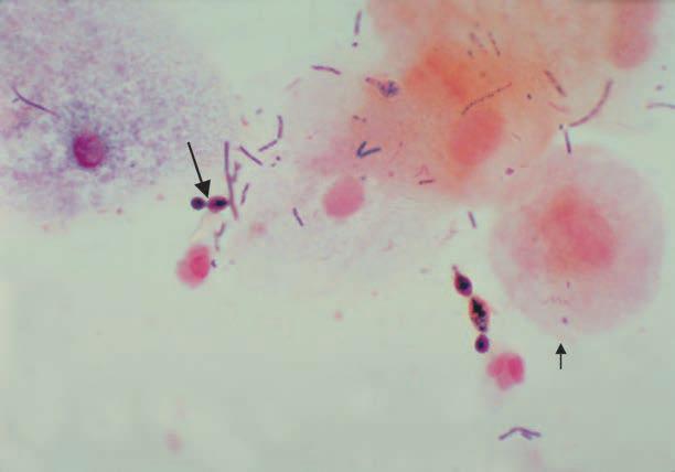
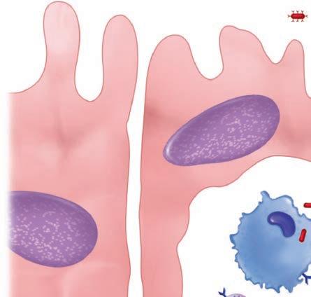
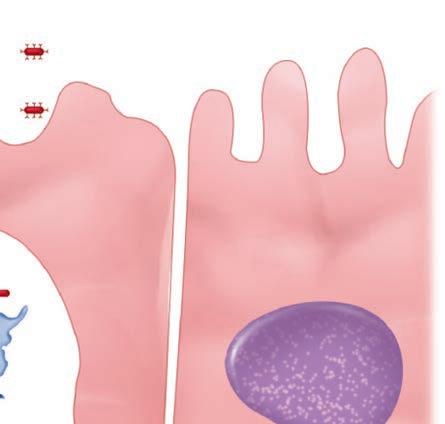
The urinary tract is sterile in health above the distal 1 cm of the urethra, which has a scanty flora derived from the perineum. Thus, in health, the urine in the bladder, ureters, and renal pelvis is sterile. The vagina has a flora that varies according to hormonal influences at different ages. Before puberty and after menopause, it is mixed, nonspecific, and relatively scanty, and it contains organisms derived from the flora of the skin and colon. During the childbearing years, it is composed predominantly of anaerobic and microaerophilic members of the genus Lactobacillus, with smaller numbers of anaerobic gram-negative rods, gram-positive cocci, and yeasts (Figure 1–4) that can survive under the acidic conditions produced by the lactobacilli. These conditions develop because glycogen is deposited in vaginal epithelial cells under the influence of estrogenic hormones and metabolized to lactic acid by lactobacilli. This process results in a vaginal pH of 4 to 5, which is optimal for growth and survival of the lactobacilli, but inhibits many other organisms.
Bacterial Vaginosis
Bacterial vaginosis (BV) is a long known and unfortunately common syndrome which is still poorly understood. Its dominant feature is an uncomfortable vaginal discharge with a “fishy” odor which contains epithelial cells coated with bacteria (clue cells). This change is associated with a shift in the vaginal microbiota away from the acidic Lactobacillus flora to one with a higher pH and a greater mixture of species including more anaerobes. Over the years several of these newcomers have been tagged as the cause of BV, particularly
S aureus is carried in anterior nares
Lower tract is protected by mucociliary action
Bladder and upper urinary tract are sterile
Hormonal changes affect the vaginal flora
Use of epithelial glycogen by lactobacilli produces low pH
BV is associated with a shift in vaginal microbiota
FIGURE 1–4. Vaginal flora. Vaginal Gram smear showing budding yeast (long arrow), epithelial cells (short arrow) and a mixture of other bacterial morphologies. The long gram-positive rods are most likely lactobacilli. [Redrawn from Centers for Disease Control and Prevention (CDC).]
Flora that reach sterile sites may cause disease
Virulence factors increase the opportunity for invasion
Mouth flora plays a major role in dental caries
Gardnerella vaginalis and Mobiluncus. The BV situation appears to be more complex than this, involving complex interactions of the vaginal microbiota.
ROLES IN HEALTH AND DISEASE
● Opportunistic Infection
Many species among the microbiota are opportunists in that they can cause infection when they reach protected areas of the body in sufficient numbers. For example, certain strains of E coli can reach the urinary bladder by ascending the urethra and cause acute urinary tract infection. Perforation of the colon from a ruptured diverticulum or a penetrating abdominal wound releases feces into the peritoneal cavity; this contamination may be followed by peritonitis or intraabdominal abscesses caused by members of the flora which have virulence factors allowing them to exploit this situation. There are now examples of the microbiota supplying a step in the pathogenesis of a classic pathogen. Attachment of Neisseria gonorrhoeae to the cervix has been shown to be enhanced when an enzyme produced by the cervicovaginal microbiota unmasks a crucial receptor (see Chapter 30). Caries and periodontal disease are caused by organisms that are members of the oral microbiota (see Chapter 41).
● Exclusionary Effect
Competing with pathogens has a protective effect
Antibiotic therapy may provide a competitive advantage for pathogens
Balancing the prospect of opportunistic infection is the tendency of the resident microbiota to produce conditions that compete with extraneous newcomers who happen to be pathogens and thus reduce their ability to establish a niche in the host. The microbiota in the colon of the breastfed infant produces an environment inimical to colonization by enteric pathogens, as does a vaginal flora dominated by lactobacilli. The benefit of this exclusionary effect has been demonstrated by what happens when it is removed. Antibiotic therapy, particularly with broad-spectrum agents, may so alter the microbiota of the gastrointestinal tract that antibiotic-resistant organisms multiply in the ecologic vacuum. Under these conditions, the spore-forming Clostridium difficile has a selective advantage that allows it to survive, proliferate, and produce a toxic colitis.
● Priming of Immune System
Sterile animals have little immunity to microbial infection
Low exposure correlates with asthma risk
Organisms of the microbiota play an important role in the development of immunologic competence. Animals delivered and raised under completely aseptic conditions (“sterile” or gnotobiotic animals) have a poorly developed reticuloendothelial system, low serum levels of immunoglobulins, and lack antibodies to antigens that often confer a degree of protection against pathogens. There is evidence of immunologic differences between children who are raised under usual conditions and those whose exposure to diverse flora is minimized. Some studies have found a higher incidence of immunopathologic states, such as asthma in the more isolated children.
PROMOTING A GOOD MICROBIOTA
Intestinal lactobacilli may protect against diarrheal agents
The field of probiotics is based on the notion that we can manipulate the microbiota by promoting colonization with “good” bacteria. Elie Metchnikoff originally suggested this in his observation that the longevity of Bulgarian peasants was attributable to their consumption of large amounts of yogurt; the live lactobacilli in the yogurt presumably replaced the colonic flora to the general benefit of their health. This notion persists today in capsules containing freeze-dried lactobacilli sold by the sizable probiotics industry and by promotion of the health benefit of natural (unpasteurized) yogurt, which contains live lactobacilli. Because these lactobacilli are adapted to food and not the intestine, they are unlikely to persist, much less replace, the typical microbiota of the adult colon. In some clinical studies, administration of preparations containing a particular strain of Lactobacillus (Lactobacillus rhamnosus strain GG, LGG) has been shown to reduce the duration of rotavirus diarrhea in children. The use of similar preparations to prevent relapses of antibiotic-associated diarrhea caused by C difficile has shown little success but fecal transplant (a whole new microbiota) has blocked recurrences of pseudomembranous colitis, the most serious form of this disease.
Research into the role of the microbiota in health and disease is one of the most exciting topics in science. This is by no means limited to topics related to infectious disease. Currently the most active areas involve mechanisms of obesity, autoimmune disorders (arthritis, asthma) and more subjective subjects like human cravings. Much of the work involves the interactions between multiple species many of which can only be detected by genomic methods. Obviously, it is going to take considerable time to sort these relationships out.
INFECTIOUS DISEASE
Of the thousands of species of viruses, bacteria, fungi, and parasites, only a tiny portion is involved in disease of any kind. These are called pathogens. There are plant pathogens, animal pathogens, and fish pathogens, as well as the subject of this book, human pathogens. Among pathogens, there are degrees of potency called virulence, which sometimes makes drawing the dividing line between benign and virulent microorganisms difficult. Pathogens are associated with disease with varying frequency and severity. Yersinia pestis, the cause of plague, causes fulminant disease and death in 50% to 75% of persons who come in contact with it. Therefore, it is highly virulent. Understanding the basis of these differences in virulence is a fundamental goal of this book. The better students of medicine understand how a pathogen causes disease, the better they will be prepared to intervene and help their patients. For any pathogen, the basic aspects of how it interacts with the host to produce disease can be expressed in terms of its epidemiology, pathogenesis, and immunity. Usually, our knowledge of one or more of these topics is incomplete. It is the task of the physician to relate these topics to the clinical aspects of disease and be prepared for new developments which clarify, or in some cases, alter them. We do not know everything, and not all of what we believe we know is correct.
EPIDEMIOLOGY
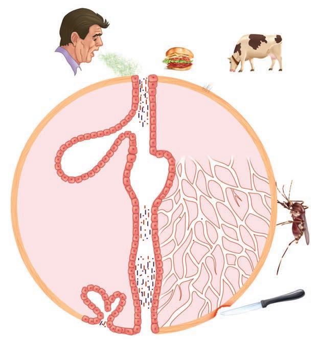
Epidemiology is the “who, what, when, and where” of infectious diseases. The power of the science of epidemiology was first demonstrated by Semmelweis, who by careful analysis of statistical data alone determined how streptococcal puerperal fever is transmitted. He even devised a means to prevent transmission (handwashing) decades before the organism itself (Streptococcus pyogenes) was discovered. Since then, each organism has built its own profile of vital statistics. Some agents are transmitted by air, some by food, and others by insects; many spread by the person-to-person route. Figure 1–5 presents some of the variables in this regard. Some agents occur worldwide, and others only in certain geographic locations or ecologic circumstances. Knowing how an organism gains access to its victim and spreads is crucial to understanding the disease. It is also essential in discovering the emergence of “new” diseases, whether they are truly new (HIV/AIDS) or just recently discovered (Legionnaires disease). Solving mysterious outbreaks or recognizing new epidemiologic patterns have often pointed the way to the isolation of new agents.
Epidemic spread and disease are facilitated by malnutrition, poor socioeconomic conditions, natural disasters, and hygienic inadequacy. Epidemics, caused by the introduction of new organisms of unusual virulence, often result in high morbidity and mortality rates. We are currently witnessing a new and extended Zika virus pandemic, but the prospect of recurrence of old pandemic infections (influenza, cholera) remains. Modern times and technology have introduced new wrinkles to epidemiologic spread. Air travel has allowed diseases to leap continents even when they have very short incubation periods. The efficiency of the food industry has sometimes backfired when the distributed products are contaminated with infectious agents. The outbreaks of hamburger-associated E coli O157:H7 bloody diarrhea and hemolytic uremic syndrome are an example. The nature of massive meat-packing facilities allowed organisms from infected cattle on isolated farms to be mixed with other meat and distributed rapidly and widely. By the time outbreaks were recognized, cases of disease were widespread, and tons of meat had to be recalled. In simpler times, local outbreaks from the same source might have been detected and contained more quickly.
Of course, the most ominous and uncertain epidemiologic threat of these times is not amplification of natural transmission but the specter of unnatural, deliberate spread. Anthrax is a disease uncommonly transmitted by direct contact with animals or animal
Pathogens are rare
Virulence varies greatly
Each agent has its own mode of spread
Poor socioeconomic conditions foster infection
Modern society may facilitate spread
FIGURE 1–5. Infection overview. The sources and potential sites of infection are shown. Infection may be endogenous from the internal flora or exogenous from the sources shown around the outside.
Respiratory tract
Anthrax and smallpox are new bioterrorism threats
Alimentary tract
injury
products. Under natural conditions, it produces a nasty, but not usually life-threatening, ulcer. The inhalation of human-produced aerosols of anthrax spores could produce a lethal pneumonia on a massive scale. Smallpox is the only disease officially eradicated from the world. It took place sufficiently long ago that most of the population has never been exposed or immunized and is, thus, vulnerable to its reintroduction. We do not know whether infectious bioterrorism will work on the scale contemplated by its perpetrators; however, in the case of anthrax, we do know that sophisticated systems have been designed to attempt it. We hope never to learn whether bioterrorism will work on a large scale.
PATHOGENESIS
Pathogenicity is multifactorial
Pathogens have molecules that bind to host cells
Invasion requires adaptation to new environments
When a potential pathogen reaches its host, features of the organism determine whether or not disease ensues. The primary reason pathogens are so few in relation to the microbial world is that being successful at producing disease is a very complicated process. Multiple features, called virulence factors, are required to persist, cause disease, and escape to repeat the cycle. The variations are many, but the mechanisms used by many pathogens have now been dissected at the molecular level.
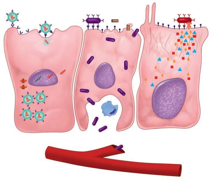
The first step for any pathogen is to attach and persist at whatever site it gains access. This usually involves specialized surface molecules or structures that correspond to receptors on human cells. Because human cells were not designed to receive the microorganisms, the pathogens are often exploiting some molecule important for some other essential function of the cell. For some toxin-producing pathogens, this attachment alone may be enough to produce disease. For most pathogens, it just allows them to persist long enough to proceed to the next stage—invasion into or beyond the surface mucosal cells. For viruses, invasion of cells is essential, because they cannot replicate on their own. Invading pathogens must also be able to adapt to a new milieu. For example, the nutrients and ionic environment of the cell surface differs from that inside the cell or in the submucosa. Some of the steps in pathogenesis at the cellular level are illustrated in Figure 1–6.
Skin
Capillary
Scratch,
Viral attachment
Viral DNA/RNA
Virus-directed synthesis
New viral particles
Viral receptorsViral entry
Pore-forming toxin
Bloodstream invasion
FIGURE 1–6. Infection cellular view. Left. A virus is attaching to the cell surface but can replicate only within the cell. Middle. A bacterial cell attaches to the surface, invades, and spreads through the cell to the bloodstream. Right. A bacterial cell attaches and injects proteins into the cell. The cell is disrupted while the organism remains on the surface.
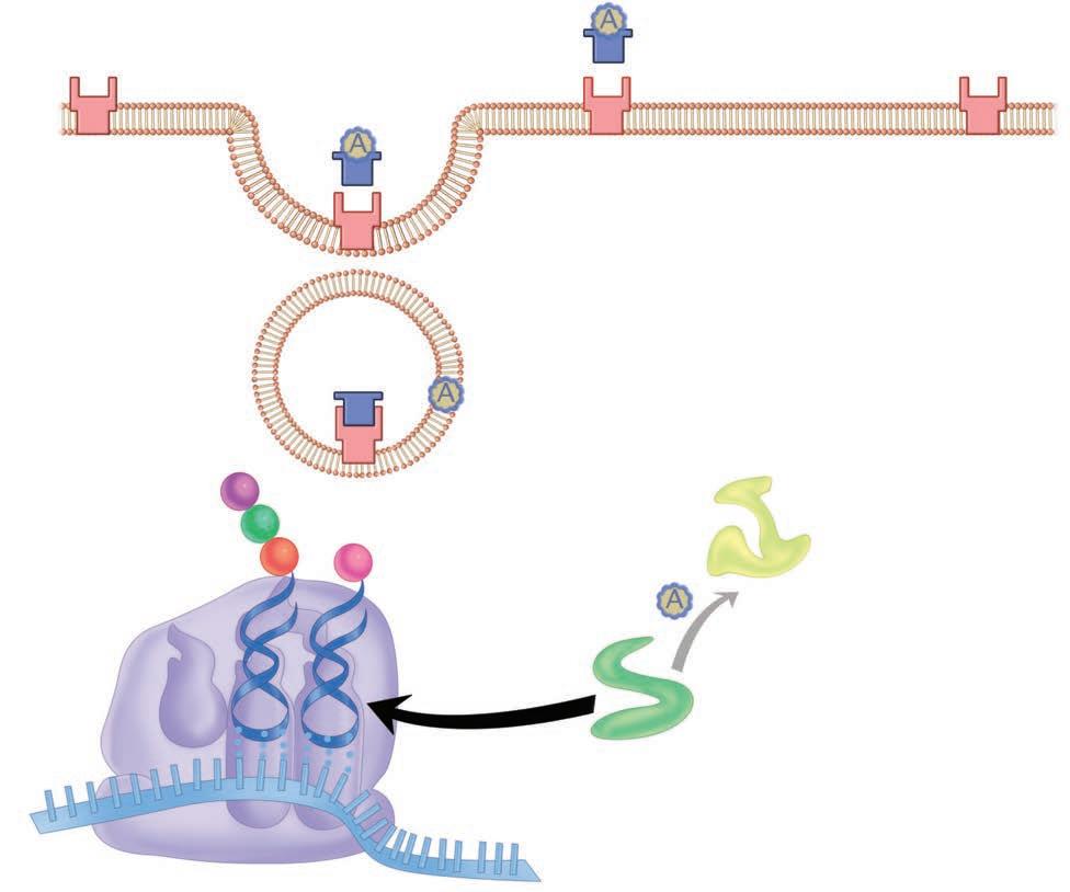
Persistence and even invasion do not necessarily translate immediately to disease. The invading organisms must disrupt function in some way. For some, the inflammatory response they stimulate is enough. For example, a lung alveolus filled with neutrophils responding to the presence of S pneumoniae loses its ability to exchange oxygen. The longer a pathogen can survive in the face of the host response, the greater the compromise in host function. Most pathogens do more than this. Destruction of host cells through the production of digestive enzymes, toxins, or intracellular multiplication is among the more common mechanisms. Other pathogens operate by altering the function of a cell without injury. Diphtheria is caused by a bacterial toxin that blocks protein synthesis inside the host cell. Details of the molecular mechanism for this action are illustrated in Figure 1–7. Some viruses cause the insertion of molecules in the host cell membrane, which cause other host cells to attack it. The variations are diverse and fascinating.
IMMUNITY
Although the science of immunology is beyond the scope of this book, understanding the immune response to infection (see Chapter 2) is an important part of appreciating pathogenic mechanisms. In fact, one of the most important virulence attributes any pathogen can have is an ability to neutralize the immune response to it in some way. Some pathogens attack the immune effector cells, and others undergo changes that evade the immune response. The old observation that there seems to be no immunity to gonorrhea turns out to be an example of the latter mechanism. Neisseria gonorrhoeae, the causative agent of gonorrhea, undergoes antigenic variation of important surface structures so rapidly that antibodies directed against the bacteria become irrelevant. For each pathogen, the primary interest is whether there is natural immunity and, if so, whether it is based on cell-mediated (TH1, CMI) or humoral (TH2, antibody) mechanisms.
Injection secretion system Secreted proteins
Cytoskeleton alterations
Inflammation alone can result in injury
Cells may be destroyed or their function altered
Evading the immune response is a major feature of virulence
Receptor for toxin
Receptor-mediated endocytosis
Antibody or cell-mediated mechanisms may be protective
FIGURE 1–7. Action of diphtheria toxin, molecular view. The toxin-binding (B) portion attaches to the cell membrane, and the complete molecule enters the cell. In the cell, the A subunit dissociates and catalyzes a reaction that ADP-ribosylates (ADPR) and, thus, inactivates elongation factor 2 (EF-2). This factor is essential for ribosomal reactions at the acceptor and donor sites, which transfer triplet code from messenger RNA (mRNA) to amino acid sequences via transfer RNA (tRNA). Inactivation of EF-2 stops building of the polypeptide chain.
Humoral and CMI responses are broadly stimulated with most infections, but the specific response to a particular molecular structure is usually dominant in mediating immunity to reinfection. For example, the repeated nature of strep throat (group A streptococcus) in childhood is not due to antigenic variation as described for gonorrhea. The antigen against which protective antibodies are directed (M protein) is stable, but naturally exists in more than 80 types. Each type requires its own specific antibody. Knowing the molecule against which the protective immune response is directed is particularly important for devising preventive vaccines.
CLINICAL ASPECTS OF INFECTIOUS DISEASE
● Manifestations
Fever, pain, and swelling are the universal signs of infection. Beyond this, the particular organs involved and the speed of the process dominate the signs and symptoms of disease.
Ribosome
Cough, diarrhea, and mental confusion represent disruption of three different body systems. On the basis of clinical experience, physicians have become familiar with the range of behavior of the major pathogens. However, signs and symptoms overlap considerably. Skilled physicians use this knowledge to begin a deductive process leading to a list of suspected pathogens and a strategy to make a specific diagnosis and provide patient care. Through the probability assessment, an understanding of how the diseases work is a distinct advantage in making the correct decisions.
● Diagnosis
A major difference between infectious and other diseases is that the probabilities just described can be specifically resolved, often overnight. Most microorganisms can be isolated from the patient, grown in artificial culture, and identified. Others can be seen microscopically or detected by measuring the specific immune response to the pathogen. Preferred modalities for diagnosis of each agent have been developed and are available in clinic, hospital, and public health laboratories all over the world. Empiric diagnosis made on the basis of clinical findings can be confirmed and the treatment plan modified accordingly. New methods which detect molecular or genomic markers of the agent are now realizing their potential for rapid, specific diagnosis.
● Treatment
Over the past 80 years, therapeutic tools of remarkable potency and specificity have become available for the treatment of bacterial infections. These include all the antibiotics and an array of synthetic chemicals that kill or inhibit the infecting organism, but have minimal or acceptable toxicity for the host. Antibacterial agents exploit the structural and metabolic differences between microbial and human eukaryotic cells to provide the selectivity necessary for good antimicrobial therapy. Penicillin, for example, interferes with the synthesis of the bacterial cell wall, a structure that has no analog in human cells. There are fewer antifungal and antiprotozoal agents because the eukaryotic cells of the host and those of the parasite have metabolic and structural similarities. Nevertheless, hosts and parasites do have some significant differences, and effective therapeutic agents have been discovered or developed to exploit them.
Specific therapeutic attack on viral disease has posed more complex problems, because of the intimate involvement of viral replication with the metabolic and replicative activities of the cell. However, recent advances in molecular virology have identified specific viral targets that can be attacked. Scientists have developed successful antiviral agents, including those that interfere with the liberation of viral nucleic acid from its protective protein coat or with the processes of viral nucleic acid synthesis and replication. The successful development of new agents for human immunodeficiency virus has involved targeting enzymes coded by the virus genome.
The success of the “antibiotic era” has been clouded by the development of resistance by the organisms. The mechanisms involved are varied but, most often, involve a mutational alteration in the enzyme, ribosome site, or other target against which the antimicrobial is directed. In some instances, organisms acquire new enzymes or block entry of the antimicrobial to the cell. Many bacteria produce enzymes that directly inactivate antibiotics. To make the situation worse, the genes involved are readily spread by promiscuous genetic mechanisms. New agents that are initially effective against resistant strains have been developed, but resistance by new mechanisms usually follows. The battle is by no means lost, but has become a never-ending policing action.
● Prevention
The goal of the scientific study of any disease is its prevention. In the case of infectious diseases, this has involved public health measures and immunization. The public health measures depend on knowledge of transmission mechanisms and on interfering with them. Water disinfection, food preparation, insect control, handwashing, and a myriad of other measures prevent humans from coming in contact with infections agents. Immunization relies on knowledge of immune mechanisms and designing vaccines that stimulate protective immunity.
Body system(s) involved dictate clinical findings
Disease-causing microbes can be grown and identified
Antibiotics are directed at structures of bacteria not present in host
Antivirals target unique virus-coded enzymes
Resistance complicates therapy
Mechanisms include mutation and inactivation
Public health and immunization are primary preventive measures
Attenuated strains stimulate immunity
Live vaccines rarely cause disease
Immunization follows two major strategies—live vaccines and inactivated vaccines. The former uses live organisms that have been modified (attenuated) so they do not produce disease, but still stimulate a protective immune response. Such vaccines have been effective, but carry the risk that the vaccine strain itself may cause disease. This event has been observed with the live oral polio vaccine. Although this rarely occurs, it has caused a shift back to the original Salk inactivated vaccine. This issue has reemerged with a debate over strategies for the use of smallpox immunization to protect against bioterrorism. This vaccine uses vaccinia virus, a cousin of smallpox, and its potential to produce disease on its own has been recognized since its original use by Jenner in 1798. Serious disease would be expected primarily in immunocompromised individuals (eg, from cancer chemotherapy or AIDS), who represent a significantly larger part of the population than when smallpox immunization was stopped in the 1970s. Could immunization cause more disease than it prevents? Despite the claims of those who oppose the use of all vaccines as “unnatural” the risk/benefit ratio of all currently licensed vaccines is greatly on the positive side.
Purified components are safe vaccines
Vaccines can be genetically engineered
The safest immunization strategy is the use of organisms that have been killed or, better yet, killed and purified to contain only the immunizing component. This approach requires much better knowledge of pathogenesis and immune mechanisms. Vaccines for meningitis use the polysaccharide capsule of the bacterium, and vaccines for diphtheria and tetanus use only a formalin-inactivated protein toxin. Pertussis (whooping cough) immunization has undergone a transition in this regard. The original killed whole-cell vaccine was effective, but caused a significant incidence of side effects. A purified vaccine containing pertussis toxin and a few surface components has reduced side effects, but its efficacy compared with the previous vaccine is now in question.
The newest approaches for vaccines require neither live organisms nor killed, purified ones. As the entire genomes of more and more pathogens are being reported, an entirely genetic strategy is emerging. Armed with knowledge of molecular pathogenesis and immunity and the tools of genomics and proteomics, scientists can now synthesize an immunogenic protein without ever growing the organism itself. Such an idea would have astonished even the great microbiologists of the last two centuries.
SUMMARY
Infectious diseases remain as important and fascinating as ever. Where else do we find the emergence of new diseases, together with improved understanding of the old ones? At a time when the revolution in molecular biology and genetics has brought us to the threshold of new and novel means of infection control, the perpetrators of bioterrorism threaten us with diseases we have already conquered. Meeting this challenge requires a secure knowledge of the pathogenic organisms and how they produce disease, as well as an understanding of the clinical aspects of these diseases. In the collective judgment of the authors, this book presents the principles and facts required for students of medicine to understand the most important infectious diseases.