Download full Hurwitz clinical pediatric dermatology e book: a textbook of skin disorders of childho
Hurwitz Clinical Pediatric Dermatology
E Book: A Textbook of Skin Disorders of Childhood and Adolescence 5th Edition, (Ebook PDF)
Visit to download the full and correct content document: https://ebookmass.com/product/hurwitz-clinical-pediatric-dermatology-e-book-a-textbo ok-of-skin-disorders-of-childhood-and-adolescence-5th-edition-ebook-pdf/
More products digital (pdf, epub, mobi) instant download maybe you interests ...
Paller and Mancini - Hurwitz Clinical Pediatric
Dermatology: A Textbook of Skin Disorders of Childhood & Adolescence 6th Edition Amy S. Paller
An Overview of Dermatologic Diagnosis and Procedures
Accurate diagnosis of cutaneous disease in infants and children is a systematic process that requires careful inspection, evaluation, and some knowledge of dermatologic terminology and morphology to develop a prioritized differential diagnosis. The manifestations of skin disorders in infants and young children often vary from those of the same diseases in older children and adults. The diagnosis may be obscured, for example, by different reaction patterns or a tendency toward easier blister formation. In addition, therapeutic dosages and regimens often differ from those of adults, with medications prescribed on a “per kilogram” (/kg) basis and with liquid formulations. Nevertheless, the same basic principles that are used to detect disorders affecting viscera apply to the detection of skin disorders. An adequate history should be obtained, a thorough physical examination performed, and, whenever possible the clinical impression verified by appropriate laboratory studies. The easy visibility of skin lesions all too often results in a cursory examination and hasty diagnosis. Instead, the entire skin should be examined routinely and carefully, including the hair, scalp, nails, oral mucosa, anogenital regions, palms, and soles, because visible findings often hold clues to the final diagnosis.
The examination should be conducted in a well-lit room. Initial viewing of the patient at a distance establishes the overall status of the patient and allows recognition of distribution patterns and clues to the appropriate final diagnosis. This initial evaluation is followed by careful scrutiny of primary and subsequent secondary lesions in an effort to discern the characteristic features of the disorder. Although not always diagnostic, the morphology and configuration of cutaneous lesions are of considerable importance to the classification and diagnosis of cutaneous disease. A lack of understanding of dermatologic terminology commonly poses a barrier to the description of cutaneous disorders by clinicians who are not dermatologists. Accordingly, a review of dermatologic terms is included here (Table 1-1). The many examples to show primary and secondary skin lesions refer to specific figures in the text that follows.
Configuration of Lesions
A number of dermatologic entities assume annular, circinate, or ring shapes and are interpreted as ringworm or superficial fungal infections. Although tinea is a common annular dermatosis of childhood, there are multiple other disorders that must be included in the differential diagnosis of ringed lesions including pityriasis rosea, seborrheic dermatitis, nummular eczema, lupus erythematosus, granuloma annulare, psoriasis, erythema multiforme, erythema annulare centrifugum, erythema migrans, secondary syphilis, sarcoidosis, urticaria, pityriasis alba, tinea versicolor, lupus vulgaris, drug eruptions, and cutaneous T-cell lymphoma.
The terms arciform and arcuate refer to lesions that assume arc-like configurations. Arciform lesions may be seen in erythema multiforme, urticaria, pityriasis rosea, and bullous dermatosis of childhood.
Lesions that tend to merge are said to be confluent. Confluence of lesions is seen, for example, in childhood exanthems, Rhus dermatitis, erythema multiforme, tina versicolor and urticaria.
Lesions localized to a dermatome supplied by one or more dorsal ganglia are referred to as dermatomal. Herpes zoster classically occurs in a dermatomal distribution.
Discoid is used to describe lesions that are solid, moderately raised, and disc-shaped. The term has largely been applied to discoid lupus erythematosus, in which the discoid lesions usually show atrophy and dyspigmentation.
Discrete lesions are individual lesions that tend to remain separated and distinct. Eczematoid and eczematous are adjectives relating to eczema and suggest inflammation with a tendency to thickening, oozing, vesiculation, and/or crusting.
Grouping and clustering are characteristic of vesicles of herpes simplex or herpes zoster, insect bites, lymphangioma circumscriptum, contact dermatitis, and bullous dermatosis of childhood.
Guttate or drop-like lesions are characteristic of flares of psoriasis in children and adolescents that follow an acute upper respiratory tract infection, usually streptococcal.
Gyrate refers to twisted, coiled, or spiral-like lesions, as may be seen in patients with urticaria and erythema annulare centrifugum.
Iris or target-like lesions are concentric ringed lesions characteristic of erythema multiforme. The classic “targets” in this condition are composed of a central dusky erythematous papule or vesicle, a peripheral ring of pallor, and then an outer bright red ring.
Keratosis refers to circumscribed patches of horny thickening, as seen in seborrheic or actinic keratoses, keratosis pilaris, and keratosis follicularis (Darier disease). Keratotic is an adjective pertaining to keratosis and commonly refers to the epidermal thickening seen in chronic dermatitis and callus formation.
The Koebner phenomenon or isomorphic response refers to the appearance of lesions along a site of injury. The linear lesions of warts and molluscum contagiosum, for example, occur from autoinoculation of virus from scratching; those of Rhus dermatitis (poison ivy) result from the spread of the plant’s oleoresin. Other examples of disorders that show a Koebner phenomenon are psoriasis, lichen planus, lichen nitidus, pityriasis rubra pilaris, and keratosis follicularis (Darier disease).
Lesions in a linear or band-like configuration appear in the form of a line or stripe and may be seen in epidermal nevi, Conradi syndrome, linear morphea, lichen striatus, striae, Rhus dermatitis, deep mycoses (sporotrichosis or coccidioidomycosis), incontinentia pigmenti, pigment mosaicism, porokeratosis of Mibelli, or factitial dermatitis. In certain genetic and inflammatory disorders, such linear configurations represent the lines of Blaschko, which trace various clones of embryonic cells and, as such, represent a form of cutaneous mosaicism. This configuration presents as a linear pattern on the extremities, wavy or S-shaped on the lateral trunk, V-shaped on the central trunk, and varied patterns on the face and scalp.
Moniliform refers to a banded or necklace-like appearance. This is seen in monilethrix, a hair deformity characterized by beaded nodularities along the hair shaft.
Multiform refers to disorders in which more than one variety or shape of cutaneous lesions occurs. This configuration is seen in patients with erythema multiforme, early Henoch–Schönlein purpura, and polymorphous light eruption.
Nummular means coin-shaped and is usually used to describe nummular dermatitis.
Polycyclic refers to oval lesions containing more than one ring, as commonly is seen in patients with urticaria.
A reticulated or net-like pattern may be seen in erythema ab igne, livedo reticularis, cutis marmorata, cutis marmorata telangiectatica congenita, and lesions of confluent and reticulated papillomatosis.
Serpiginous describes the shape or spread of lesions in a serpentine or snake-like configuration, particularly those of cutaneous larva migrans (creeping eruption) and elastosis perforans serpiginosa.
Umbilicated lesions are centrally depressed or shaped like an umbilicus or navel. Examples include lesions of molluscum contagiosum, varicella, vaccinia, variola, herpes zoster, and Kaposi varicelliform eruption.
Text continued on p. 7
Table
Lesion Description
PRIMARY LESIONS
Illustration
Examples
The term primary refers to the most representative, but not necessarily the earliest, lesions; it is distinguished from the cutaneous features of secondary changes such as excoriation, eczematization, infection, or results of previous therapy.
Macule Flat, circumscribed change of the skin. It may be of any size, although this term is often used for lesions <1 cm. A macule may appear as an area of hypopigmentation or as an area of increased coloration, most commonly brown (hyperpigmented) or red (usually a vascular abnormality). It is usually round but may be oval or irregular; it may be distinct or may fade into the surrounding area.
Patch Flat, circumscribed lesion with color change that is >1 cm in size.
Ephelides; lentigo (see Fig. 11-41); flat nevus (see Fig. 9-1); and tinea versicolor (see Fig. 17-35).
Mongolian spot (see Fig. 11-57); port wine stain (see Fig. 12-57); nevus depigmentosus (see Fig. 11-22); larger café-au-lait spot (see Fig. 11-43); and areas of vitiligo (see Figs. 11-1 though 11-10).
Papule Circumscribed, nonvesicular, nonpustular, elevated lesion that measures <1 cm in diameter. The greatest mass is above the surface of the skin. When viewed in profile it may be flat-topped, dome-shaped, acuminate (tapering to a point), digitate (finger-like), smooth, eroded, or ulcerated. It may be covered by scales, crusts, or a combination of secondary features.
Plaque Broad, elevated, disk-shaped lesion that occupies an area of >1 cm. It is commonly formed by a confluence of papules.
Elevated nevus (see Fig. 9-4); verruca (see Fig. 15-17); molluscum contagiosum (see Fig. 15-40); perioral dermatitis (see Fig. 8-20); and individual lesions of lichen planus (see Fig. 4-43).
Psoriasis (see Fig. 4-4); lichen simplex chronicus (neurodermatitis) (see Fig. 3-37); granuloma annulare (see Fig. 9-58); nevus sebaceus (see Figs. 9-41 through 9-44); and lesions of lichen planus (see Fig. 4-45).
Table 1-1 Glossary of Dermatologic Terms (Continued)
Lesion Description
Nodule Circumscribed, elevated, usually solid lesion that measures 0.5 to 2 cm in diameter. It involves the dermis and may extend into the subcutaneous tissue with its greatest mass below the surface of the skin.
Tumor Deeper circumscribed solid lesion of the skin or subcutaneous tissue that measures >2 cm in diameter. It may be benign or malignant.
Illustration
Examples
Erythema nodosum (see Figs. 20-44 and 20-45); pilomatricoma (see Fig. 9-48); subcutaneous granuloma annulare (see Fig. 9-60); and nodular scabies (see Fig. 18-9).
Deep hemangioma (see Fig. 12-7) and plexiform neurofibroma (Fig. 11-50).
Wheal Distinctive type of elevated lesion characterized by local, superficial, transient edema. White to pink or pale red, compressible, and evanescent, they often disappear within a period of hours. They vary in size and shape.
Darier sign of mastocytosis (see Fig. 9-52); urticarial vasculitis (see Fig. 21-14); and various forms of urticaria (see Fig. 20-2).
Vesicle Sharply circumscribed, elevated, fluidcontaining lesion that measures ≤1 cm in diameter.
Herpes simplex (see Figs. 2-47, 15-10 and 15-11); hand-foot-and-mouth disease (see Fig. 16-30); pompholyx (see Fig. 3-41); varicella (see Fig. 16-1); and contact dermatitis (see Fig. 3-57).
Continued on following page
Lesion Description
Bulla Larger, circumscribed, elevated, fluidcontaining lesion that measures >1 cm in diameter.
Pustule Circumscribed elevation <1 cm in diameter that contains a purulent exudate. It may be infectious or sterile.
Illustration
Abscess Circumscribed, elevated lesion >1 cm in diameter, often with a deeper component and filled with purulent material.
OTHER PRIMARY LESIONS
Comedone Plugged secretion of horny material retained within a pilosebaceous follicle. It may be flesh colored (as in closed comedone or whitehead) or slightly raised brown or black (as in open comedone or blackhead). Closed comedones, in contrast to open comedones, may be difficult to visualize. They appear as pale, slightly elevated, small papules without a clinically visible orifice.
Burrow Linear lesion produced by tunneling of an animal parasite in the stratum corneum.
Telangiectasia Persistent dilation of superficial venules, capillaries, or arterioles of the skin.
Examples
Blistering distal dactylitis (see Fig. 14-21); bullous pemphigoid (see Fig. 13-26); chronic bullous disease of childhood (see Fig. 13-29); bullous systemic lupus erythematosus (see Fig. 13-32); and epidermolysis bullosa (see Fig. 13-4).
Folliculitis (see Fig. 14-11); transient neonatal pustular melanosis (see Fig. 2-17); pustular psoriasis (see Fig. 4-22); and infantile acropustulosis (see Fig. 2-19).
Staphylococcal abscess (in a neonate, see Fig. 2-5; in a patient with hyperimmunoglobulinemia E, see Fig. 3-35).
Acne comedones (see Figs. 8-3 and 8-4) and nevus comedonicus (see Fig. 9-45).
Scabies (see Fig. 18-3) and cutaneous larva migrans (creeping eruption, see Fig. 18-39).
Spider angioma (see Fig. 12-86); periungual lesion of dermatomyositis (see Fig. 22-25); and Goltz syndrome (see Fig. 6-15).
Table 1-1 Glossary of Dermatologic Terms (Continued)
Lesion Description
SECONDARY LESIONS
Illustration
Examples
Secondary lesions represent evolutionary changes that occur later in the course of the cutaneous disorder. Although helpful in dermatologic diagnosis, they do not offer the same degree of diagnostic aid as that afforded by primary lesions of a cutaneous disorder.
Crust Dried remains of serum, blood, pus, or exudate overlying areas of lost or damaged epidermis. Crust is yellow when formed by dried serum, green or yellowish-green when formed by purulent exudate, and dark red or brown when formed by bloody exudative serum.
Herpes simplex (see Fig. 15-4); weeping eczematous dermatitis (see Fig. 3-1); and dried honey-colored lesions of impetigo (see Fig. 14-2).
Scale Formed by an accumulation of compact desquamating layers of stratum corneum as a result of abnormal keratinization and exfoliation of cornified keratinocytes.
Seborrheic dermatitis (greasy and yellowish, see Figs. 3-2 and 3-39); psoriasis (silvery and mica-like, see Fig. 4-1); pityriasis alba (fine and barely visible, see Fig. 3-33); and lamellar ichthyosis (large and adherent, see Fig. 5-10).
Fissure Dry or moist, linear, often painful cleavage in the cutaneous surface that results from marked drying and long-standing inflammation, thickening, and loss of elasticity of the integument.
Angular cheilitis (see Fig. 17-40) and dermatitis on the plantar aspect of the foot (see Fig. 3-61).
Erosion Moist, slightly depressed vesicular or bullous lesions in which part or all of the epidermis has been lost. Because erosions do not extend into the underlying dermis or subcutaneous tissue, healing occurs without subsequent scar formation.
Herpes simplex (see Figs. 3-28 and 15-1); epidermolytic ichthyosis in a neonate (see Fig. 5-4); and superficial forms of epidermolysis bullosa (see Fig. 13-7).
Continued on following page
Table 1-1 Glossary of Dermatologic Terms (Continued)
Lesion Description
Excoriation Traumatized or abraded (usually selfinduced) superficial loss of skin caused by scratching, rubbing, or scrubbing of the cutaneous surface.
Ulcer Necrosis of the epidermis and part or all of the dermis and/or the underlying subcutaneous tissue.
Illustration
Examples
Atopic dermatitis (see Fig. 3-9) and acne excoriée (see Fig. 8-19).
Pyoderma gangrenosum (see Fig. 25-27) and ulcerated hemangioma of infancy (see Figs. 12-15 and 12-26).
Atrophy Cutaneous changes that result in depression of the epidermis, dermis, or both. Epidermal atrophy is characterized by thin, almost translucent epidermis, a loss of the normal skin markings, and wrinkling when subjected to lateral pressure or pinching of the affected area. In dermal atrophy the skin is depressed.
Lichenification Thickening of the epidermis with associated exaggeration of skin markings. Lichenification results from chronic scratching or rubbing of a pruritic lesion.
Anetoderma (see Fig. 22-53); morphea (see Figs. 22-42 though 22-49); steroidinduced atrophy (see Fig. 3-32); and Goltz syndrome (see Fig. 6-17).
Atopic dermatitis (see Fig. 3-8); chronic contact dermatitis (see Fig. 3-53); and lichen simplex chronicus (see Fig. 3-37).
Lesion Description
Scar A permanent fibrotic skin change that develops after damage to the dermis. Initially pink or violaceous, scars are permanent, white, shiny, and sclerotic as the color fades. Although fresh scars often are hypertrophic, they usually contract during the subsequent 6 to 12 months and become less apparent. Hypertrophic scars must be differentiated from keloids, which represent an exaggerated response to skin injury. Keloids are pink, smooth, and rubbery and are often traversed by telangiectatic vessels. They tend to increase in size long after healing has taken place and can be differentiated from hypertrophic scars by the fact that the surface of a keloidal scar tends to extend beyond the area of the original wound.
Illustration
Universal (universalis) implies widespread disorders affecting the entire skin, as in alopecia universalis.
Zosteriform describes a linear arrangement along a nerve, as typified by lesions of herpes zoster, although herpes simplex infection can also manifest in a zosteriform distribution.
Distribution and Morphologic Patterns of Common Skin Disorders
The regional distribution and morphologic configuration of cutaneous lesions are often helpful in dermatologic diagnosis.
Acneiform lesions are those having the form of acne, and an acneiform distribution refers to lesions primarily seen on the face, neck, chest, upper arms, shoulders, and back (see Figs. 8-3 through 8-13).
Sites of predilection of atopic dermatitis include the face, trunk, and extremities in young children; the antecubital and popliteal fossae are the most common sites in older children and adolescents (see Figs. 3-1 through 3-12).
The lesions of erythema multiforme may be widespread but have a distinct predilection for the hands and feet (particularly the palms and soles) (see Figs. 20-33 through 20-37).
Lesions of herpes simplex may appear anywhere on the body but have a distinct predisposition for the areas about the lips, face, and genitalia (see Figs. 15-1 through 15-12). Herpes zoster generally has a dermatomal or nerve-like distribution and is usually but not necessarily unilateral (see Figs. 15-13 and 15-14). More than 75% of cases occur between the second thoracic and second lumbar vertebrae. The fifth cranial nerve commonly is involved, and only rarely are lesions seen below the elbows or knees.
Lichen planus often affects the limbs (see Figs. 4-43 through 4-51). Favorite sites include the lower extremities, the flexor surface of the wrists, the buccal mucosa, the trunk, and the genitalia.
The lesions of lupus erythematosus most commonly localize to the bridge of the nose, malar eminences, scalp, and ears, although they may be widespread (see Figs. 22-3 and 22-6). Patches tend to spread at the border and clear in the center with atrophy, scarring, dyspigmentation, and telangiectases. The malar or butterfly rash is neither specific for nor the most common sign of lupus erythematosus; telangiectasia without the accompanying features of erythema, scaling, or atrophy is never a marker of this disorder other than in neonatal lupus.
Molluscum contagiosum is a common viral disorder characterized by dome-shaped skin-colored to erythematous papules, often with a central white core or umbilication (see Figs. 15-35 through 15-45). These papules most often localize to the trunk and axillary areas.
Examples
Keloid (see Fig. 9-83); healed areas of recessive dystrophic epidermolysis bullosa (see Fig. 13-17); acne scarring (see Fig. 8-8), congenital erosive and vesicular dermatosis with reticulated supple scarring (see Fig. 2-21); and amniocentesis scars (see Fig. 2-4).
Although molluscum lesions can be found anywhere, the scalp, palms, and soles are rare sites of involvement.
Photodermatoses are cutaneous disorders caused or precipitated by exposure to light. Areas of predilection include the face, ears, anterior “V” of the neck and upper chest, the dorsal aspect of the forearms and hands, and exposed areas of the legs. The shaded regions of the upper eyelids, subnasal, and submental regions tend to be spared. The major photosensitivity disorders are lupus erythematosus, dermatomyositis, polymorphous light eruption, drug photosensitization, and porphyria (see Chapter 19).
Photosensitive reactions cannot be distinguished on a clinical basis from lesions of photocontact allergic conditions. They may reflect internal as well as external photoallergens and may simulate contact dermatitis from airborne sensitizers. Lupus erythematosus can be differentiated by the presence of atrophy, scarring, hyperpigmentation or hypopigmentation, and the presence of periungual telangiectases. Dermatomyositis with swelling and erythema of the cheeks and eyelids should be differentiated from allergic contact dermatitis by the heliotrope hue and other associated changes, particularly those of the fingers (periungual telangiectases and Gottron papules).
Pityriasis rosea begins as a solitary round or oval scaling lesion known as the herald patch in 70% to 80% of cases, which may be annular and is often misdiagnosed as tinea corporis (see Figs. 4-38 through 4-41). After an interval of days to 2 weeks, affected individuals develop a generalized symmetrical eruption that involves mainly the trunk and proximal limbs. The clue to diagnosis is the distribution of lesions, with the long axis of these oval lesions parallel to the lines of cleavage in what has been termed a Christmas-tree pattern. A common variant, inverse pityriasis rosea, often localizes in the inguinal region, but the parallel nature of the long axis of lesions remains characteristic.
Psoriasis classically consists of round, erythematous, wellmarginated plaques with a rich red hue covered by a characteristic grayish or silvery-white mica-like (micaceous) scale that on removal may result in pinpoint bleeding (Auspitz sign) (see Figs. 4-1 through 4-10). Although exceptions occur, lesions generally are seen in a bilaterally symmetric pattern with a predilection for the elbows, knees, scalp, and lumbosacral, perianal, and genital regions. Nail involvement, a valuable diagnostic sign, is characterized by pitting of the nail plate, discoloration, separation of the nail from the nailbed (onycholysis), and an accumulation of subungual scale (subungual hyperkeratosis). A characteristic feature of this disorder is the Koebner or isomorphic response in which new lesions appear at sites of local injury.
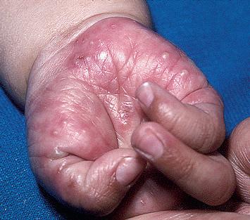
Scabies is an itchy disorder in which lesions are characteristically distributed on the wrists and hands (particularly the interdigital webs), forearms, genitalia, areolae, and buttocks in older children and
adolescents (see Figs. 18-1 through 18-11). Other family members may be similarly affected or complain of itching. In infants and young children, the diagnosis is often overlooked because the distribution typically involves the palms, soles, and often the head and neck. Obliteration of demonstrable primary lesions (burrows) because of vigorous hygienic measures, excoriation, crusting, eczematization, and secondary infection is particularly common in infants.
Seborrheic dermatitis is an erythematous, scaly or crusting eruption that characteristically occurs on the scalp, face, and postauricular, presternal, and intertriginous areas (see Figs. 3-38 and 3-39). The classic lesions are dull, pinkish-yellow, or salmon colored with fairly sharp borders and overlying yellowish greasy scale. Morphologic and topographic variants occur in many combinations and with varying degrees of severity from mild involvement of the scalp with occasional blepharitis to generalized, occasionally severe erythematous scaling eruptions. The differential diagnosis may include atopic dermatitis, psoriasis, various forms of diaper dermatitis, Langerhans cell histiocytosis, scabies, tinea corporis or capitis, pityriasis alba, contact dermatitis, Darier disease, and lupus erythematosus.
Warts are common viral cutaneous lesions characterized by the appearance of skin-colored small papules of several morphologic types (see Figs. 15-16 through 15-33). They may be elevated or flat lesions and tend to appear in areas of trauma, particularly the dorsal surface of the face, hands, periungual areas, elbows, knees, feet, and genital or perianal areas. Close examination may reveal capillaries appearing as punctate dots scattered on the surface.
Changes in Skin Color
The color of skin lesions commonly assists in making the diagnosis. Common disorders of brown hyperpigmentation include postinflammatory hyperpigmentation, pigmented and epidermal nevi, café-aulait spots, lentigines, incontinentia pigmenti, fixed drug eruption, photodermatitis and phytophotodermatitis, melasma, acanthosis nigricans, and Addison disease. Blue coloration is seen in mongolian spots, blue nevi, nevus of Ito and nevus of Ota, and cutaneous neuroblastomas. Cysts, deep hemangiomas, and pilomatricomas often show a subtle blue color, whereas the blue of venous malformations and glomuvenous malformations is often a more intense, dark blue. Yellowish discoloration of the skin is common in infants, related to the presence of carotene derived from excessive ingestion of foods, particularly yellow vegetables containing carotenoid pigments. Jaundice may be distinguished from carotenemia by scleral icterus. Localized yellow lesions may represent juvenile xanthogranulomas, nevus sebaceous, xanthomas, or mastocytomas. Red lesions are usually vascular in origin, such as superficial hemangiomas, spider telangiectases, and nevus flammeus (capillary malformations), or inflammatory, such as the scaling lesions of atopic dermatitis or psoriasis.
Localized lesions with decreased pigmentation may be hypopigmented (decreased pigmentation) or depigmented (totally devoid of pigmentation); Wood lamp examination may help to differentiate depigmented lesions, which fluoresce a bright white, from hypopigmented lesions. Localized depigmented lesions may be seen in vitiligo, Vogt–Koyanagi syndrome, halo nevi, chemical depigmentation, piebaldism, and Waardenburg syndrome. Hypopigmented lesions are more typical of postinflammatory hypopigmentation, pityriasis alba, tinea versicolor, leprosy, nevus achromicus, tuberous sclerosis, and the hypopigmented streaks of pigment mosaicism. A generalized decrease in pigmentation can be seen in patients with albinism, untreated phenylketonuria, and Menkes syndrome. The skin of patients with Chédiak–Higashi and Griscelli syndromes takes on a dull silvery sheen and may show decreased pigmentation.
Racial Variations in the Skin and Hair
The skin of African-American and other darker-skinned children varies in several ways from that of lighter-skinned children based on genetic background and customs.1,2 The erythema of inflamed black skin may be difficult to see and likely accounts for the purportedly decreased incidence of macular viral exanthems such as erythema
infectiosum. Erythema in African-American children commonly has a purplish tinge that can be confusing to unwary observers. The skin lesions in several inflammatory disorders such as in atopic dermatitis, pityriasis rosea, and syphilis commonly show a follicular pattern in African-American children.
Postinflammatory hypopigmentation and hyperpigmentation occur readily and are more obvious in darker-skinned persons, regardless of racial origin. Pityriasis alba and tinea versicolor are more commonly reported in darker skin types, perhaps because of the easy visibility of the hypopigmented lesions in marked contrast to uninvolved surrounding skin. Lichen nitidus is more apparent and reportedly more common in African-American individuals; lichen planus is reported to be more severe, leaving dark postinflammatory hyperpigmentation. Vitiligo is particularly distressing to patients with darker skin types, whether African-American or Asian, because of the easy visibility in contrast with surrounding skin.
Although darker skin may burn, in general sunburn and chronic sun-induced diseases of adults such as actinic keratosis and carcinomas of the skin induced by ultraviolet light exposure (e.g., squamous cell carcinoma, keratoacanthoma, basal cell carcinoma, and melanoma) have an extremely low incidence in African-Americans and Hispanics. Congenital melanocytic nevi also tend to have a lower tendency to transform to malignancy in darker-skinned individuals. Café-au-lait spots are more numerous and seen more often in AfricanAmericans, although the presence of six or more should still raise suspicion about neurofibromatosis. Dermatosis papulosa nigra commonly develop in adolescents, especially female, of African descent. Mongolian spots occur more often in persons of African or Asian descent. Physiologic variants in children with darker skin include increased pigmentation of the gums and tongue, pigmented streaks in the nails, and Voight–Futcher lines, lines of pigmentary demarcation between the posterolateral and lighter anteromedial skin on the extremities.
Qualities of hair may also differ among individuals of different races. African-American hair tends to tangle when dry and becomes matted when wet. As a result of its naturally curly or spiral nature, pseudofolliculitis barbae is more common in African-Americans than in other groups. Tinea capitis is particularly common in prepubertal African-Americans; the tendency to use oils because of hair dryness and poor manageability may obscure the scaling of tinea capitis. Pediculosis capitis, in contrast, is relatively uncommon in this population, possibly related to the diameter and shape of the hair shaft. Prolonged continuous traction on hairs may result in traction alopecia, particularly with the common practice of making tight corn row braids. The use of other hair grooming techniques such as chemical straighteners, application of hot oils, and use of hot combs increases the risk of hair breakage and permanent alopecia. Frequent and liberal use of greasy lubricants and pomades produces a comedonal and sometimes papulopustular form of acne (pomade acne).
Keloids form more often in individuals of African descent, often as a complication of a form of inflammatory acne, including nodulocystic acne and acne keloidalis nuchae. Other skin disorders reportedly seen more commonly are transient neonatal pustular melanosis, infantile acropustulosis, impetigo, papular urticaria, sickle-cell ulcers, sarcoidosis, and dissecting cellulitis of the scalp. Atopic dermatitis and Kawasaki disease have both been reported most often in children of Asian descent.
Procedures to Aid in Diagnosis
BETTER VISUALIZATION
Although most lesions are diagnosed by clinical inspection, several techniques are used to aid in diagnosis. The Wood lamp (black light) is an ultraviolet A (UVA)-emitting device with a peak emission of 365 nm. With the room completely dark and the light held approximately 10 cm from the skin, the examiner can see: (1) more subtle differences in pigmentation and the bright whiteness of vitiligo lesions based on the strong absorbance of the light by melanin; and (2) characteristic fluorescence of organisms such as the pink-orange fluorescence of urine in porphyria (see Chapter 19), the coral red fluorescence of erythrasma, the yellow-orange fluorescence of tinea versicolor, the
green fluorescence of ectothrix types of tinea capitis (e.g., Microsporum) (see Chapter 17), and sometimes pseudomonas infection. Falsepositive assessments can result from detection of other fluorescent objects such as lint, threads, scales, and ointments.
Magnification using a lens or lighted devices such as the otoscope or ophthalmoscope can be used to more easily visualize lesions such as nailfold capillaries, especially after swabbing the skin with alcohol or applying a drop of oil. Dermoscopy (also known as dermatoscopy or epiluminescence microscopy) refers to examination of the skin with a dermatoscope, a handheld magnifier with an embedded light source. Dermoscopy provides more than just magnification, because it allows the viewer to visualize dermal diagnostic clues. In pediatric patients dermoscopy can be particularly useful for reassurance regarding the benign nature of pigmented nevi, visualization of vascular lesions, and hair disorders ranging from shaft defects to alopecia areata.1–3 Finally, diascopy involves pressing a glass microscope slide firmly over a lesion and watching for changes in appearance. Purpura, which does not blanch with diascopy because the erythrocytes have leaked into tissue, can be distinguished from erythema from vasodilation, which blanches because the pressure from the slides forces the erythrocytes to move out of the compressed vessels. The yellow-brown (“apple jelly”) color of granulomatous lesions (e.g., granuloma annulares, sarcoidosis) persists during diascopy, and the constricted blood vessels of nevus anemicus do not refill when the slides are lifted after diascopy (as do the surrounding normal areas).
Several diagnostic techniques involve procedures to obtain scales or discharge (by scraping or swabbing) for analysis. Scraping can be performed with a sterile surgical or Fomon blade. A Cytobrush4 or moistened swab5 can be used for obtaining scales and broken hairs for fungal cultures and may be less frightening for young children (see Chapter 17). Vesicular lesions can be scraped for Tzanck smears and obtaining epidermal material for direct fluorescent analysis and viral (primarily herpes) cultures or to show the cellular content such as eosinophils in the vesicular lesions of incontinentia pigmenti. Potential scabies lesions, especially burrows, can be dotted with mineral oil and scraped vigorously for microscopic analysis, which may reveal live mites, eggs, or feces (see Chapter 18). When looking for superficial fungi, both potassium hydroxide (KOH) wet-mount preparations and cultures are often performed, although KOH examination should be performed in a Clinical Laboratory Improvement Amendments (CLIA)-approved setting (see Chapter 17). For skin lesions the blade or Cytobrush should scrape the active lesional border. For possible tinea capitis it is important to obtain broken (infected) hairs and scales. The Cytobrush technique has been shown to be more effective than scraping,3 and vigorously rubbing with a moistened cotton swab (either with tap water or the Culturette transport medium) before inoculation into fungal culture medium is well-tolerated, easy, and reliable.4 Nail scrapings and subungual debris can also be obtained for evaluation; nail clippings can be sent for histopathologic evaluation with special stains to demonstrate fungal elements.
Hair plucks tend to be traumatic for children and often cause hair shaft distortion, but gentle-traction hair pulling yields hair that is appropriate for determining whether alopecia areata is still active (hair-pull test) and for microscopic evaluation of the telogen bulbs of telogen effluvium and the distorted bulb and ruffled cuticle of loose anagen syndrome (see Chapter 7). Cutting the hair shafts may suffice for seeking hair shaft abnormalities via a microscopic trichogram (which may require polarizing light such as to detect trichothiodystrophy) and detecting nits of pediculosis versus hair casts (see Chapter 18).
Patch testing is key to determining or confirming triggers of delayedtype hypersensitivity reactions in children with allergic contact dermatitis (see Chapter 3). Round aluminum (Finn) chambers are taped to the back for 48 hours, and reactions are detected immediately after removal and generally twice thereafter to capture late reactivity. Although a ready-to-apply system is available (TRUE test), expanded testing is often necessary to comprehensively evaluate possible triggers and is usually best performed by dermatologists who have expertise in patch testing more comprehensively.
Although swabs of mucosae and of purulent skin material are appropriate for microbial cultures, obtaining biopsy material for special stains and cultures of suspected deep fungal or mycobacterial
infections is better for pathogen detection (see Therapeutic Procedures section). Biopsies are also important for making a diagnosis based on routine histopathologic, immunofluorescent, and/or immunohistochemical evaluation. For example, immunofluorescent testing is used to delineate the level of cleavage and absent skin proteins in epidermolysis bullosa (see Chapter 13), as well as to define the immune deposits and patterning in immunobullous disorders (see Chapter 13) and Henoch–Schönlein purpura (see Chapter 21); in contrast, immunohistochemistry is important for confirming the diagnosis of Langerhans cells in histiocytosis (see Chapter 10) and a variety of cutaneous lymphoproliferative disorders. Clinicopathologic correlation is important, however, and the pathologic result should be questioned (or repeated) if not consistent with clinical findings.
Therapeutic Procedures
The most common therapeutic procedures in pediatric dermatology are: (1) treatment of warts with cryotherapy; (2) treatment of molluscum with cantharidin or curettage; (3) lesional biopsy or excision; and (4) laser therapy. These techniques should only be performed by trained, experienced practitioners. Phototherapy with ultraviolet B (UVB) light, narrow-band UVB, and UVA light is used occasionally in children and is discussed in Chapter 19.
Cryotherapy involves the application of liquid nitrogen to lesional skin, which causes direct injury. It is most commonly used for warts (see Chapter 15) but can be selectively applied to keloids and molluscum contagiosum. Although spray delivery is possible, application with a cotton swab that is adapted with extra cotton to fit the size of the lesion allows better retention of the liquid nitrogen, provides better avoidance of nonlesional skin, and is less frightening for young children. More pedunculated lesions (or filiform warts) can be treated by grasping the lesion with a forceps and freezing the forceps near the tip rather than the lesion directly. Generally freezing is performed until there is a white ring around the lesion, often with two to three freezethaw cycles. Cryotherapy is painful and as a result is generally reserved for children 8 years of age and older. Alternative cryotherapy agents that contain dimethyl ether or chlorodifluoromethane achieve temperatures considerably lower than liquid nitrogen and are not as effective. Potential complications include hypopigmentation and atrophic scarring.
Cantharidin is an extract from the blister beetle, Cantharis vesicatoria, that leads to epidermal vesiculation after application to molluscum contagiosum lesions (see Chapter 15). It is applied precisely to the lesion using a wooden applicator, should not subsequently be occluded, and is rinsed off after 2 to 6 hours. Because the extent of blistering cannot be controlled (with some children developing extensive blisters and others virtually none, even with the same bottle of cantharidin and applicator), lesions near the eyes, on mucosae, and in occluded areas should not be treated with cantharidin. Blistering occurs in 24 to 48 hours, and crusting clears within about a week.
Curettage is a scraping technique used most commonly after topical anesthetic application for physical removal of molluscum contagiosum, especially for larger lesions for which cantharidin is less effective. Curettage can also be used after electrodesiccation (with a hyfrecator) to remove the desiccated tissue, most commonly for removal of a pyogenic granuloma (see Chapter 12). Most pediatric dermatologists avoid use of curettage in younger patients given the associated discomfort.
Biopsies and excisions are performed in pediatric patients as intervention, not just for diagnosis. The decision to remove a lesion therapeutically should be based on the indication and urgency for removal, the age and maturity of the pediatric patient, the location, and the expected cosmetic result. Careful explanation of the procedure to the parent(s) and child is important to allay concerns and manage expectations. If possible, the area to be biopsied or excised can be treated initially with a topical anesthetic cream (such as 4% lidocaine or 2.5% lidocaine/2.5% prilocaine) under a clear occlusive film to minimize any discomfort associated with subsequent injection of deeper anesthesia. Buffering the lidocaine with sodium bicarbonate and use of a 30-gauge needle also help to decrease the pain of injection; once buffered, lidocaine with epinephrine must either be kept refrigerated or
discarded after a week because of accelerated epinephrine degradation. Regional nerve blocks can be used selectively for larger excisions or cryotherapy. Distraction techniques such as conversation, listening to music, or watching a video can also allay fear at almost any age. Punch biopsy is most useful for removing lesions under 6 mm in diameter. For larger lesions and in cosmetically sensitive areas, an elliptical excision is preferred. Elliptical excisions ideally have their long axis following skin lines to minimize tension on the wound and to optimize the ultimate cosmetic appearance of the scar. Shave biopsies are appropriate for the superficial removal of skin tags (acrochordons) and more protuberant small nevi that are cosmetically problematic but can be followed by lesional regrowth and should not be performed if there is any concern about lesional atypia or malignancy. Surgical wounds of 4 mm or larger in diameter should be closed with suture; wounds which are 3 mm or less can be left to heal via secondary intention after hemostasis, although suturing of any lesion often gives a better cosmetic result. Although octylcyanoacrylates such as Dermabond are appropriate for closure of lacerations, the cosmetic result of their use in elective procedures may be suboptimal and is generally not recommended. Deep sutures are often required to close the deeper space of larger/deeper wounds (e.g., >6 mm in diameter) using buried absorbable suture materials. Although a variety of methods are available for closing at the surface, interrupted or running subcuticular suturing with nonabsorbable suture material is most often used. Steri-Strips are often used to further protect the wound from dehiscence.
The most common complications of biopsies and surgical excisions are wound infection, dehiscence, postoperative bleeding or hematoma (especially on the scalp), and contact dermatitis, especially to adhesives and topical antibiotics. Parents should be given clear, written postoperative instructions about keeping dressings in place (and the wound completely dry) for the first 48 hours, appropriate wound care thereafter, limitation in physical activity (generally 4 weeks without sports or gym if an excision), managing potential complications, and when to have sutures removed (typically 7 days for the face and 10 to 14 days on the body and extremities).
Light amplifications by stimulated emission of radiation (lasers) produce intense light energy at a specific wavelength that can be emitted as a pulse or continuous wave to target tissue components for destruction. After absorption of the light, heat is generated and the target tissue is selectively destroyed. This process of selective destruction has been called selective photothermolysis and carries the benefit of destruction of the target chromophores (substances that absorb specific wavelengths of light) with minimal damage to surrounding tissues.5
By far the most common laser utilized in children is the pulsed-dye laser (PDL; wavelength 585 to 595 nm), which targets hemoglobin and is used for a variety of vascular lesions including capillary malformations (port wine stains, salmon patches), macular (flat) infantile hemangiomas, ulcerated hemangiomas (in which case it helps speed reepithelialization), spider telangiectasias, and even small pyogenic
granulomas.6 PDL has also been utilized (with more variable response) for inflammatory linear verrucous epidermal nevus, erythematous striae, warts, and even some inflammatory dermatoses such as psoriasis and eczema.
The response of a port wine stain to PDL therapy is variable and may depend on the depth of the dermal capillaries, location of the stain (i.e., central facial stains classically respond less to PDL therapy than lesions on the forehead or peripheral face), size of the stain, and age at the time treatment is initiated. Sequential treatment sessions are often necessary (generally at 4- to 8-week intervals), and multiple treatments may be necessary to achieve significant improvement.7 Port wine stains located on the extremities tend to require more treatments than those located elsewhere.8
Other lasers utilized in pediatric patients include neodymium : yttrium aluminum garnet (Nd : YAG; 1064 nm), alexandrite (755 nm), diode (810 nm), Q-switched ruby (694 nm), and intensed pulsed light (555 to 950 nm) lasers, which have shown variable benefit in port wine stains, venous malformations, deeper hemangiomas, and pigmented lesions (mongolian spots, nevus of Ota, Becker melanosis).5,9 The xenon-chloride excimer laser (308 nm) provides a wavelength similar to narrow-band UVB therapy, with the advantage of being able to selectively treat a more targeted area of the skin. It has been demonstrated useful in psoriasis, vitiligo, and pityriasis alba.10–12
References
1. Haliasos EC, Kerner M, Jaimes-Lopez N, et al. Dermoscopy for the pediatric dermatologist. Part I. Dermoscopy of pediatric infectious and inflammatory skin lesions and hair disorders. Pediatr Dermatol 2013;30:163–71.
2. Haliasos EC, Kerner M, Jaimes N, et al. Dermoscopy for the pediatric dermatologist. Part II. Dermoscopy of genetic syndromes with cutaneous manifestations and pediatric vascular lesions. Pediatr Dermatol 2013;30:172–81.
3. Haliasos EC, Kerner M, Jaimes N, et al. Dermoscopy for the pediatric dermatologist. Part III. Dermoscopy of melanocytic lesions. Pediatr Dermatol 2013;30: 281–93.
4. Bonifaz A, Isa-Isa R, Araiza J, et al. Cytobrush-culture method to diagnose tinea capitis. Mycopathologia 2007;163(6):309–13.
5. Cordisco MR. An update on lasers in children. Curr Opin Pediatr 2009;21: 499–504.
6. Craig LM, Alster TS. Vascular skin lesions in children: a review of laser surgical and medical treatments. Dermatol Surg 2013;39:1137–46.
7. Alster TS, Wilson F. Treatment of port-wine stains with the flashlamp-pumped pulsed dye laser: extended clinical experience in children and adults. Ann Plast Surg 1994;32:478–84.
9. Franca K, Chacon A, Ledon J, et al. Lasers for cutaneous congenital vascular lesions: a comprehensive overview and update. Lasers Med Sci 2013;28: 1197–204.
10. Mudigonda T, Dabade TS, Feldman SR. A review of protocols for 308 nm excimer laser phototherapy in psoriasis. J Drugs Dermatol 2012;11(1):92–7.
11. Cho S, Zheng Z, Park YK, Roh MR. The 308-nm excimer laser: a promising device for the treatment of childhood vitiligo. Photodermatol Photoimmunol Photomed 2011;27(1):24–9.
12. Al-Mutairi N, Hadad AA. Efficacy of 308-nm xenon chloride excimer laser in pityriasis alba. Dermatol Surg 2012;38(4):604–9.
Cutaneous Disorders of the Newborn 2
Neonatal Skin
The skin of the infant dif fers from that of an adult in that it is thinner (40% to 60%), is less hair y, and has a weaker attachment between the epidermis and dermis.1 In addition, the body surface area-to-weight ratio of an infant is up to five times that of an adult. The infant is therefore at a significantly increased risk for skin injur y, percutaneous absorption, and skin-associated infection. Premature infants bor n before 32 to 34 weeks’ estimated gestational age may have problems associated with an immature stratum cor neum (the most superficial cell layer in the epidermis), including an increase in transepidermal water loss (TEWL). This increased TEWL may result in morbidity because of dehydration, electrolyte imbalance, and thermal instability. Interestingly, in the majority of premature infants an acceleration of skin maturation occur s after bir th such that most develop intact barrier function by 2 to 3 weeks of life.2 However, in extremely low-birthweight infants, this process may take up to 4 to 8 weeks.3 In light of the elevated TEWL levels seen in premature infants, a variety of studies have evaluated the use of occlusive dressings or topical emollients in an ef fort to improve compromised bar rier function.4–7 The risk of percutaneous toxicity from topically applied substances is increased in infants, especially those bor n prematurely.8,9 Percutaneous absorption is known to occur through two major pathways: (1) through the cells of the stratum cor neum and the epidermal malpighian layer (the transepidermal route) and (2) through the hair follicle–sebaceous gland component (the transappendageal route). Increased neonatal percutaneous absorption may be the result of the increased skin surface area-to-weight ratio as well as the stratum corneum immaturity seen in premature neonates. Although transdermal delivery methods may be distinctly advantageous in cer tain settings, extreme caution must be exercised in the application of topical substances to the skin of infants, given the risk of systemic absorption and potential toxicity. Table 2-1 lists some compounds repor ted in association with percutaneous toxicity in infants and children.
SKIN CARE OF THE NEWBORN
The skin of the newbor n is covered with a g rayish-white, g reasy material termed vernix caseosa The vernix represents a physiologic protective covering derived par tially from secretions of the sebaceous glands and in par t as a decomposition product of the infant’s epidermis. Vernix contains protein, lipids, and water and provides water-binding free amino acids that facilitate the adaptation from amniotic fluid immersion in utero to the dr y ambient postnatal state.10 Although its function is not completely under stood, it may act as a natural protectant cream to “waterproof” the fetus in utero, where it is submerged in the amniotic fluid.11 Some studies suggest that vernix be left on as a protective coating for the newbor n skin and that it be allowed to come of f by itself with successive changes of clothing (generally within the fir st few weeks of life). It has been suggested that vernixbased topical creams may be ef fective in augmenting stratum cor neum repair and maturation in infants and could play a role in the treatment of epidermal wounds.12
The skin acts as a protective organ. Any break in its integ rity therefore af fords an oppor tunity for initiation of infection. The impor tance of skin care in the newbor n is compounded by several factors:
1. The infant does not have protective skin flora at bir th.
2. The infant has at least one and possibly two open surgical wounds (the umbilicus and circumcision site).
3. The infant is exposed to fomites and per sonnel that potentially harbor a variety of infectious agents.
Skin care should involve gentle cleansing with a nontoxic, nonabrasive neutral material. During the 1950s, the use of hexachlorophenecontaining compounds became routine for the skin care of newbor ns as prophylaxis against Staphylococcus aureus infection. In 1971 and 1972, however, the use of hexachlorophene preparations as skin cleansers for newbor ns was restricted because of studies demonstrating vacuolization in the central ner vous system (CNS) of infants and laboratory animals after prolonged application of these preparations.13 At the minimum, neonatal skin care should include gentle removal of blood from the face and head, and meconium from the perianal area, by gentle rinsing with water. Ideally, vernix caseosa should be removed from the face only, allowing the remaining vernix to come of f by itself However, the common standard of care is for gentle dr ying and wiping of the newbor n’s entire skin surface, which is most desirable from a thermoregulatory standpoint. For the remainder of the infant’s stay in the hospital nursery, the buttocks and perianal regions should be cleansed with water and cotton or a gentle cloth. A mild soap with water rinsing may also be used at diaper changes if desired.
There is no single method of umbilical-cord care that has been proven to limit colonization and disease Several methods include local application of isopropyl alcohol, triple dye (an aqueous solution of brilliant g reen, proflavine, and gentian violet), and antimicrobial agents such as bacitracin or silver-sulfadiazine cream. The routine use of povidone-iodine should be discouraged, given the risk of iodine absorption and transient hypothyroxinemia or hypothyroidism. A safer alter native is a chlorhexidine-containing product.14
Physiologic Phenomena of the Newborn
Neonatal dermatology, by definition, encompasses the spectrum of cutaneous disorder s that arise during the fir st 4 weeks of life. Many such conditions are transient, appearing in the fir st few days to weeks of life only to disappear shor tly thereafter The appreciation of normal phenomena and their dif ferentiation from the more significant cutaneous disorder s of the newbor n is critical for the general physician, obstetrician, and pediatrician, as well as for the pediatric dermatologist.
At bir th, the skin of the full-term infant is normally soft, smooth, and velvety. Desquamation of neonatal skin generally takes place 24 to 36 hour s after delivery and may not be complete until the third week of life. Desquamation at bir th is an abnormal phenomenon and is indicative of postmaturity, intrauterine anoxia, or congenital ichthyosis.
The skin at bir th has a purplish-red color that is most pronounced over the extremities. Except for the hands, feet, and lips, where the transition is g radual, this quickly changes to a pink hue In many infants, a purplish discoloration of the hands, feet, and lips occur s during periods of cr ying, breath holding, or chilling This normal phenomenon, termed acrocyanosis, appears to be associated with an increased tone of peripheral ar terioles, which in tur n creates vasospasm, secondar y dilation, and pooling of blood in the venous plexuses, resulting in a cyanotic appearance to the involved areas of the skin. The intensity of cyanosis depends on the deg ree of oxygen loss and the depth, siz e, and fullness of the involved venous plexus. Acrocyanosis, a normal physiologic phenomenon, should not be confused with true cyanosis.
Table 2-1 Reported Hazards of Percutaneous Absorption in Infants and Children
Elevated blood levels of immunosuppressive medication
Ulceration of mucous membranes, skin necrosis, vomiting, diarrhea
Uremia
Reprinted with permission from Bree AF, Siegfried EC. Neonatal skin care and toxicology. In: Eichenfield LF, Frieden IJ, Esterly NB, editors. Textbook of neonatal dermatology, second ed. London: Saunders Elsevier; 2008. p. 59–72.
CUTIS MARMORATA
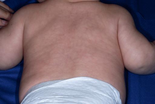
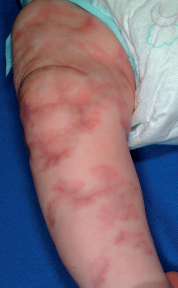
Cutis marmorata is a normal reticulated bluish mottling of the skin seen on the trunk and extremities of infants and young children (Fig. 2-1). This phenomenon, a physiologic response to chilling with resultant dilation of capillaries and small venules, usually disappears as the infant is rewarmed. Although a tendency for cutis marmorata may persist for several weeks or months, this disorder bear s no medical significance and treatment generally is unnecessar y. In some children cutis marmorata may tend to recur until early childhood, and in patients with Down syndrome, trisomy 18, and the Cor nelia de Lange syndrome, this reticulated marbling pattern may be per sistent. When the changes are per sistent (even with rewarming) and are deep violaceous in color, cutis marmorata telangiectatica congenita (Fig 2-2; see also Chapter 12) should be considered. In some infants a white negative pattern of cutis marmorata (cutis marmorata alba) may be created by a transient hypertonia of the deep vasculature. Cutis marmorata alba is also a transitory disorder and appears to have no clinical significance.
HARLEQUIN COLOR CHANGE
Harlequin color change, not to be confused with harlequin ichthyosis (see Chapter 5), is occasionally obser ved in full-term infants but usually occur s in premature infants. It occur s when the infant is lying on his or her side and consists of reddening of one-half of the body with simultaneous blanching of the other half. Attacks develop suddenly and may per sist for 30 seconds to 20 minutes. The side that lies uppermost is paler, and a clear line of demarcation runs along the midline of the body. At times, this line of demarcation may be
incomplete; when attacks are mild, areas of the face and genitalia may not be involved.
This phenomenon appears to be related to immaturity of hypothalamic center s that control the tone of peripheral blood vessels and has been obser ved in infants with severe intracranial injur y as well as in infants who appear to be otherwise perfectly normal. Although the peak frequency of attacks of harlequin color change generally occur s between the second and fifth days of life, attacks may occur anywhere
Figure 2-1 Cutis marmorata. Reticulate bluish mottling that resolves with rewarming.
Figure 2-2 Cutis marmorata telangiectatica congenita. Violaceous, reticulate patches with subtle atrophy. These changes did not resolve with rewarming and were associated with mild ipsilateral limb hypoplasia.
from the fir st few hour s to as late as the second or the third week of life.15
BRONZE BABY SYNDROME
Bronze baby syndrome is a term used to describe infants who develop a grayish-brown discoloration of the skin, serum, and urine while undergoing phototherapy for hyperbilirubinemia. Although the exact source of the pigment causing the discoloration is not clear, the syndrome usually begins 1 to 7 days after the initiation of phototherapy, resolves g radually over a period of several weeks after phototherapy is discontinued, and appears to be related to a combination of photoisomers of bilirubin or biliverdin or a photoproduct of copper-porphyrin metabolism.16–18 Infants who develop bronz e baby syndrome usually have modified liver function, par ticularly cholestasis, of various origins.19 Although not all babies with cholestasis develop bronz e baby syndrome during phototherapy, those that do should be investigated for underlying liver disease.20 The disorder should be dif ferentiated from neonatal jaundice, cyanosis associated with neonatal pulmonar y disorders or congenital hear t disease, an unusual progressive hyperpigmentation (universal-acquired melanosis, the “carbon baby” syndrome),21 and chloramphenicol intoxication (the “gray baby” syndrome), which is a disorder in infants with immature liver function who are unable to conjugate chloramphenicol and is characterized by elevated serum chloramphenicol levels, progressive cyanosis, abdominal distention, hypothermia, vomiting, ir regular respiration, and vasomotor collapse.22 A distinctive purpuric eruption on exposed skin has also been described in newbor ns receiving phototherapy and is possibly related to a transient increase in circulating porphyrins.23 This condition, however, is unlikely to be confused with bronz e baby syndrome.
Cephalohematoma
A cephalohematoma is a subperiosteal hematoma overlying the calvarium. These lesions are more common after prolonged labor,
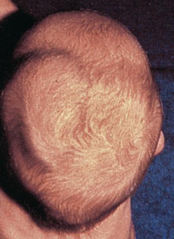
instrument-assisted deliveries, and abnormal presentations. They usually develop over the fir st hour s of life and present as subcutaneous swellings in the scalp They do not cross the midline (Fig 2-3), because they are limited to one cranial bone, which helps to distinguish them from caput succedaneum (see the next paragraph). Occasionally, a cephalohematoma may occur over a linear skull fracture. Other potentially associated complications include calcification (that may persist radiographically for years), hyperbilirubinemia, and infection. Although infected lesions (which are rare) may require aspiration,24 most lesions require no therapy with spontaneous resorption and resolution occur ring over several months.
Caput Succedaneum
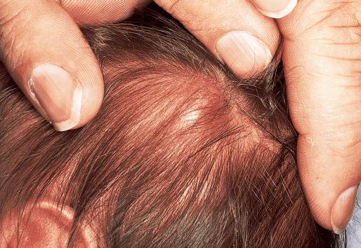
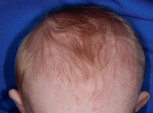
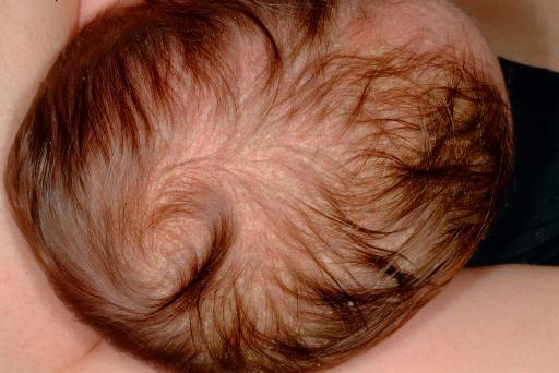
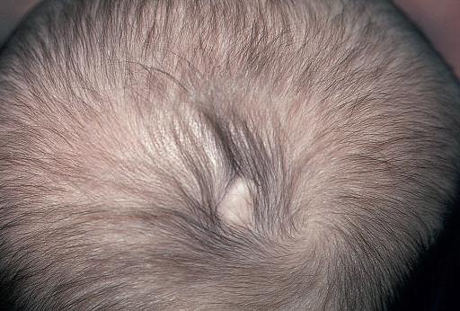
Caput succedaneum is a localiz ed edema of the newbor n scalp related to the mechanical forces involved in par turition. It is probably related to venous congestion and edema secondar y to cer vical and uterine pressure, and as such is more common with prolonged par turition and seen most often in primig ravidas. Caput succedaneum presents as a boggy scalp mass and may result in varying deg rees of bruising and necrosis in addition to the edema, at times with tissue loss. In distinction to cephalohematoma, caput succedaneum lesions often cross the midline. These lesions tend to resolve spontaneously over 48 hour s, and treatment is generally unnecessar y. One possible complication in cases of severe caput succedaneum is permanent alopecia. Halo scalp ring refers to an annular alopecia that presents in a circumferential ring around the scalp in infants with a histor y of caput.25 It represents a pressure necrosis phenomenon, and the hair loss may be transient or, occasionally, permanent.
Complications from Fetal and Neonatal Diagnostic Procedures
Fetal complications associated with invasive prenatal diagnostic procedures include cutaneous puncture marks, scar s or lacerations, exsanguination, ocular trauma, blindness, subdural hemor rhage, pneumothorax, cardiac tamponade, splenic laceration, porencephalic cysts, ar teriovenous or ileocutaneous fistulas, digital loss (in 1.7% of newbor ns whose mother s had undergone early chorionic villus
Figure 2-3 Cephalohematoma. Note the sharp demarcation at the midline.
sampling), musculoskeletal trauma, disruption of tendons or ligaments, and occasionally gangrene. Cutaneous puncture marks, which occur in 1% to 3% of newbor ns whose mother s have undergone amniocentesis, may be seen as single or multiple 1- to 6-mm pits or dimples on any cutaneous surface of the newbor n (Fig 2-4).26,27
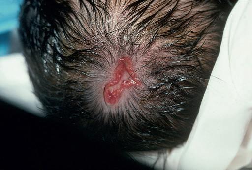
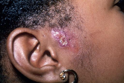
Fetal scalp monitoring can result in infection, bleeding, or fontanel puncture, and prenatal vacuum extraction can produce a localiz ed area of edema, ecchymosis, or localiz ed alopecia. The incidence of scalp electrode infection varies from 0.3% to 5.0%, and although local sterile abscesses account for the majority of adverse sequelae, S. aureus or Gram-negative infections, cellulitis, tissue necrosis, subgaleal abscess, osteomyelitis, necrotizing fasciitis, and neonatal herpes simplex infections may also occur as complications of this procedure (Fig. 2-5).28–30 It is not unusual for new parents to be under the false impression that fetal scalp electrodes are the cause of aplasia cutis congenita (ACC; see later in this chapter).
Scalp injuries sustained during the bir th process tend to be minor and include lacerations, erosions, and ecchymoses. Injuries of the scalp and face occur in approximately 16% of vacuum-assisted deliveries and in 17% of forceps-assisted deliveries.31
Transcutaneous oxygen monitoring (application of heated electrodes to the skin for continuous detection of tissue oxygenation) and pulse oximetry may also result in er ythema, tissue necrosis, and fir stor second-deg ree bur ns. Although lesions associated with transcutaneous oxygen monitoring generally resolve within 48 to 60 hour s,
persistent atrophic hyperpigmented craters may at times be seen as a complication. Frequent (every 2 to 4 hour s) changing of electrode sites and reduction of the temperature of the electrodes to 43° C, however, can lessen the likelihood of this complication.32,33
Anetoderma of prematurity refers to macular depressions or outpouchings of skin associated with loss of dermal elastic tissue seen in premature infants. Reports suggest that these cutaneous lesions may correlate with placement of electrocardiographic or other monitoring electrodes or leads.34,35
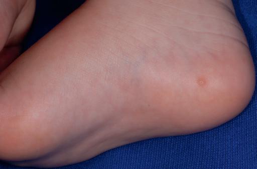
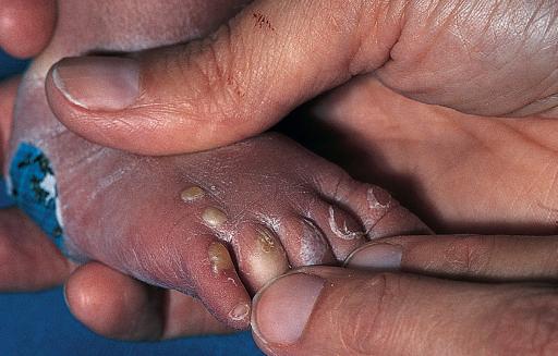
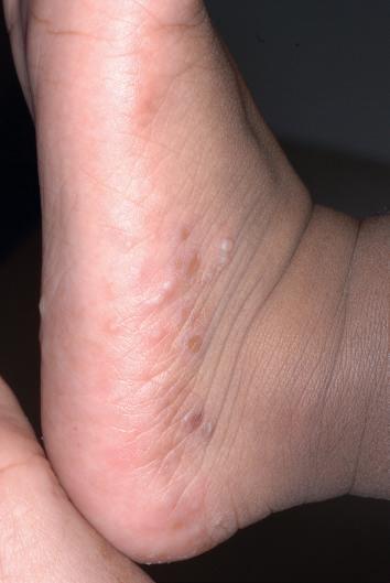
Calcinosis cutis may occur on the scalp or chest of infants or children at sites of electroencephalograph or electrocardiograph electrode placement, as a result of diagnostic heel sticks performed during the neonatal period, or after intramuscular or intravenous administration of calcium chloride or calcium gluconate for the treatment of neonatal hypocalcemia. Seen primarily in high-risk infants who receive repeated heel sticks for blood chemistr y determinations, calcified nodules usually begin as small depressions on the heels. With time, generally after 4 to 12 months, tiny yellow or white papules appear (Fig. 2-6), g radually enlarge to form nodular deposits, mig rate to the cutaneous surface, extrude their contents, and generally disappear spontaneously by the time the child reaches 18 to 30 months of age. Although calcified heel nodules are usually asymptomatic, children may at times show signs of discomfor t with standing or wearing shoes. In such instances, gentle cr yosurgery and curettage can be both diagnostic and therapeutic. Calcinosis cutis after electroencephalography or electrocardiography is more likely to be seen in infants and young children or individuals where the skin has been abraded and usually disappears spontaneously within 2 to 6 months. It can be avoided by the use of an electrode paste that does not contain calcium chloride, and like calcified heel sticks, they may be treated by gentle cr yosurgery and curettage.36,37
Abnormalities of Subcutaneous Tissue
Skin turgor is generally normal during the fir st few hour s of life. As normal physiologic dehydration occur s during the fir st 3 or 4 days of life (up to 10% of bir th weight), the skin generally becomes loose and wrinkled. Subcutaneous fat is normally quite adequate at bir th and increases until about 9 months of age, thus accounting for the traditional chubby appearance of the healthy newbor n. A decrease or absence of this normal panniculus is abnormal and suggests the possibility of prematurity, postmaturity, or placental insufficiency.
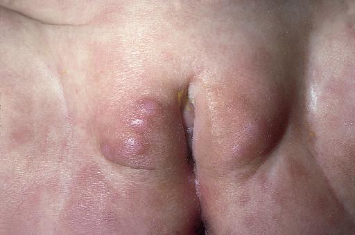
Sclerema neonatorum and subcutaneous fat necrosis (SCFN) are two disorder s that af fect the subcutaneous fat of the newbor n. Although there is considerable diagnostic confusion between these two entities, there are several distinguishing features that enable a clinical dif ferentiation (Table 2-2). Sclerema neonatorum seems to occur significantly less often than SCFN
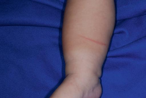
Figure 2-4 Amniocentesis scars. Multiple depressed scars on the thigh of an infant born to a mother who had amniocentesis during pregnancy. (Courtesy of Lester Schwartz, MD.)
Figure 2-5 Staphylococcal scalp abscess. Fluctuant, erythematous nodule on the scalp of this 9-day-old infant as a complication of intrauterine fetal monitoring.
Figure 2-6 Heel stick calcinosis. Firm, pink to yellow papule on the medial plantar heel in an infant who had multiple heel sticks as a newborn.
Table 2-2 Features of Sclerema Neonatorum and Subcutaneous Fat Necrosis
Sclerema Neonatorum
Premature infants
Serious underlying disease (sepsis, cardiopulmonary disease, diarrhea, or dehydration)
Wax-like hardening of skin and subcutaneous tissue
Whole body except palms and soles
Poor prognosis; high mortality
SCLEREMA NEONATORUM
Subcutaneous Fat Necrosis
Full-term or postmature infants
Healthy newborns; may have history of perinatal asphyxia or difficult delivery
Circumscribed, indurated, erythematous nodules and plaques
Buttocks, thighs, arms, face, shoulders
Excellent prognosis; treat associated hypercalcemia, if present
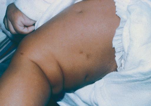
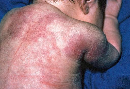
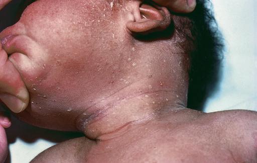
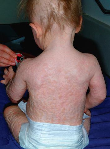
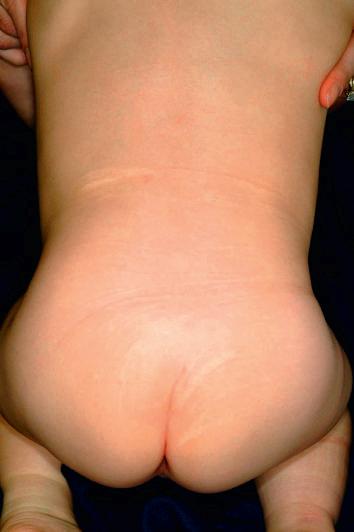
Figure 2-7 Subcutaneous fat necrosis. Indurated, erythematous plaques on the shoulders and back of this 1-week-old boy.
Sclerema neonatorum is a dif fuse, rapidly spreading, wax-like hardening of the skin and subcutaneous tissue that occur s in premature or debilitated infants during the fir st few weeks of life. The disorder, usually associated with a serious underlying condition such as sepsis or other infection, congenital hear t disease, respiratory distress, diarrhea, or dehydration, is characterized by a dif fuse nonpitting woody induration of the involved tissues. The process is symmetrical, usually starting on the legs and buttocks, and may progress to involve all areas except the palms, soles, and genitalia.38 As the disorder spreads, the skin becomes cold, yellowish-white, mottled, stony hard, and cadaverlike. The limbs become immobile, and the face acquires a fixed masklike expression. Infants with this disorder become sluggish, feed poorly, show clinical signs of shock, and in a high percentage of cases die
Although the etiology of this disorder is unknown, it appears to represent a nonspecific sign of severe illness rather than a primar y disease. Infants with this disorder are characteristically small or premature, debilitated, weak, cyanotic, and lethargic In 25% of cases the mothers are ill at the time of delivery. Exposure to cold, hypothermia, peripheral chilling with vascular collapse, and an increase in the ratio of saturated to unsaturated fatty acids in the triglyceride fraction of the subcutaneous tissue (because of a defect in fatty acid mobilization) have been hypothesiz ed as possible causes for this disorder but lack confirmation.39
The histopathologic findings of sclerema neonatorum consist of edema and thickening of the connective tissue bands around the fat lobules. Although necrosis and cr ystallization of the subcutaneous tissue have been described, these findings are more characteristically seen in lesions of SCFN
The prognosis of sclerema neonatorum is poor, and mor tality occurs in 50% to 75% of af fected infants. In a series of 51 infants with sclerema neonatorum in a special-care nursery within a Bangladeshi hospital, the fatality rate was 98%.40 In infants who sur vive, the cutaneous findings resolve without residual sequelae There is no specific therapy, although steroids and exchange transfusion have been used.38
SUBCUTANEOUS FAT NECROSIS
Subcutaneous fat necrosis (SCFN) is a benign, self-limited disease that affects apparently healthy, full-term newbor ns and young infants. It is characterized by sharply circumscribed, indurated, and nodular areas of fat necrosis (Fig 2-7). The etiology of this disorder remains unknown but appears to be related to perinatal trauma, asphyxia, hypothermia, and in some instances, hypercalcemia.41,42 Although the mechanism of hypercalcemia in SCFN is not known, it has been attributed to aberrations in vitamin D or parathyroid homeostasis. Birth asphyxia and meconium aspiration seem to be commonly associated. In one large series, 10 out of 11 infants with SCFN had been delivered via emergency cesarean section for fetal distress, and nine of the 11 had meconium staining of the amniotic fluid.43 The relation-
ship between SCFN, maternal diabetes, and cesarean section, if any, is unclear. SCFN after ice-bag application for treatment of supraventricular tachycardia has been repor ted,44 and it has also been obser ved after selective head or generalized cooling for hypoxic–ischemic encephalopathy.45,46
The onset of SCFN is generally during the fir st few days to weeks of life. Lesions appear as single or multiple localiz ed, sharply circumscribed, usually painless areas of induration. Occasionally the af fected areas may be tender, and infants may be uncomfor table and cr y vigorously when they are handled. Lesions vary from small er ythematous, indurated nodules to large plaques, and sites of predilection include the cheeks, back, buttocks, arms, and thighs. Many lesions have an uneven lobulated surface with an elevated margin separating it from the sur rounding normal tissue. Histologic examination of SCFN reveals larger-than-usual fat lobules and an extensive inflammatory infiltrate, needle-shaped clefts within fat cells, necrosis, and calcification. Magnetic resonance imaging (MRI) reveals decreased T1 and increased T2 signal intensity in af fected areas.47
The prognosis for SCFN is excellent. Although lesions may develop extensive deposits of calcium, which may liquefy, drain, and heal with scarring, most areas undergo spontaneous resolution within several weeks to months. Hypercalcemia is a rare association, and infants with this finding may require low calcium intake, restriction of vitamin D, and/or systemic cor ticosteroid therapy. Etidronate therapy has been repor ted for treatment of recalcitrant SCFN-associated hypercalcemia.48 Infants should be monitored for several months after delivery, because the onset of hypercalcemia can be delayed for several months.43,49 Other rare systemic complications may include thrombocytopenia, hypoglycemia, and hypertriglyceridemia, all of which tend to be mild and/or self-limited.
Miscellaneous Cutaneous Disorders
MILIARIA
Differentiation of the epidermis and its appendages, par ticularly in the premature infant, is often incomplete at bir th. As a result of this immaturity, a high incidence of sweat-retention phenomena may be seen in the newbor n. Miliaria, a common neonatal dermatosis caused by sweat retention, is characterized by a vesicular eruption with subsequent maceration and obstruction of the eccrine ducts. The pathophysiologic events that lead to this disorder are keratinous plugging of eccrine ducts and the escape of eccrine sweat into the skin below the level of obstruction (see Chapter 8).
Virtually all infants develop miliaria under appropriate conditions. There are two principal forms of this disorder:
1. Miliaria cr ystallina (sudamina), which consists of clear superficial pinpoint vesicles without an inflammatory areola;
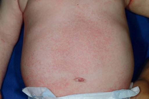
Figure 2-8 Miliaria rubra. Multiple, erythematous, pinpoint macules and papules in an infant with atopic dermatitis who was being treated with overapplication of greasy emollients.
2. Miliaria rubra (prickly heat), representing a deeper level of sweat gland obstruction and characterized by small discrete er ythematous papules, vesicles, or papulovesicles (Fig. 2-8).
The incidence of miliaria is g reatest in the fir st few weeks of life owing to the relative immaturity of the eccrine ducts, which favors poral closure and sweat retention. A pustular form of miliaria rubra has been obser ved in association with pseudohypoaldosteronism during salt-losing crises.50
Therapy for miliaria is directed toward avoidance of excessive heat and humidity. Light-weight cotton clothing, cool baths, and air conditioning are helpful in the management and prevention of this disorder. Avoidance of emollient overapplication (i.e., in infants with atopic dermatitis) should also be recommended, especially in warm, humid climates or in the winter when infants are bundled under heavy clothing.
MILIA
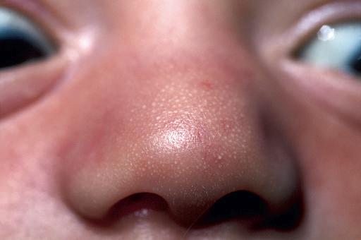
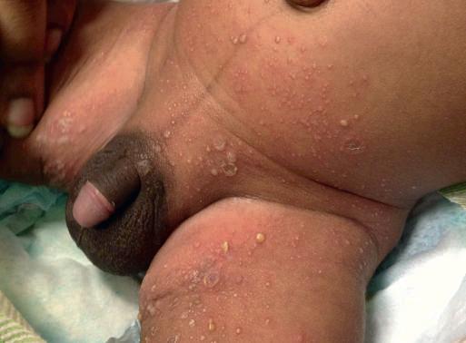
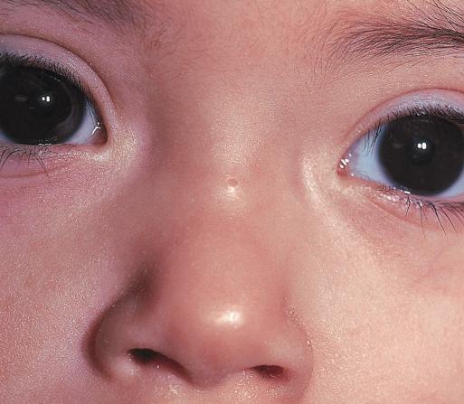
Milia, small retention cysts, commonly occur on the face of newbor ns. Seen in 40% to 50% of infants, they result from retention of keratin within the dermis. They appear as tiny 1- to 2-mm pearly white or yellow papules. Particularly prominent on the cheeks, nose, chin, and forehead, they may be few or numerous and are often g rouped (Fig. 2-9). Lesions may occasionally occur on the upper trunk, limbs, penis, or mucous membranes. Although milia of the newbor n may per sist into the second or third month, they usually disappear spontaneously during the fir st 3 or 4 weeks of life and accordingly require no therapy. Persistent milia in an unusual or widespread distribution, par ticularly when seen in association with other defects, may be a manifestation
2-10 Sebaceous gland hyperplasia. Yellow-white, pinpoint papules on the nasal tip of this 2-day-old boy.
of hereditar y trichodysplasia (Marie-Unna hypotrichosis), dystrophic forms of epidermolysis bullosa, Baz ex or Rombo syndromes, or the oral-facial-digital syndrome, type I.
BOHN NODULES AND EPSTEIN PEARLS
Discrete, 2- to 3-mm round, pearly white or yellow, freely movable elevations at the gum margins or midline of the hard palate (termed Bohn nodules and Epstein pearls, respectively) are seen in up to 85% of newborns. Clinically and histologically the counterpart of facial milia, they disappear spontaneously, usually within a few weeks of life, and require no therapy.
SEBACEOUS GLAND HYPERPLASIA
Sebaceous gland hyperplasia represents a physiologic phenomenon of the newbor n manifested as multiple, yellow to flesh-colored tiny papules that occur on the nose, cheeks, and upper lips of full-term infants (Fig 2-10). A manifestation of maternal androgen stimulation, these papules represent a temporary disorder that resolves spontaneously, generally within the fir st few weeks of life.
ACNE NEONATORUM
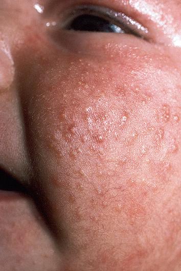
Occasionally infants develop a facial eruption that resembles acne vulgaris as seen in adolescents (Fig 2-11). Although the etiology of this disorder is not clearly defined, it appears to develop as a result of hormonal stimulation of sebaceous glands that have not yet involuted to their childhood state of immaturity. In mild cases of acne neonatorum, therapy is often unnecessar y; daily cleansing with soap and water may be all that is required. Occasionally, mild keratolytic agents or topical antibiotics may be helpful (see Chapter 8). Unusually severe or recalcitrant cases of acne neonatorum warrant investigation for underlying androgen excess.
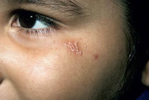
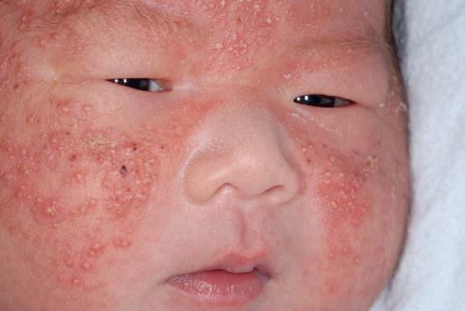
A facial acneiform eruption in infants has been associated with the saprophytic Malassezia species and has been termed neonatal cephalic pustulosis (see Chapter 8). Lesions consist of pinpoint papules, papulopustules, or larger pustules, and they are located on the cheeks, chin, and forehead (Fig. 2-12). A cor relation may exist between the clinical severity of lesions and the colonization with this fungal saprophyte.51,52 In these infants, topical antifungal agents may lead to more rapid resolution of lesions.
ERYTHEMA TOXICUM NEONATORUM
Erythema toxicum neonatorum (ETN), also known as toxic erythema of the newborn, is an idiopathic, asymptomatic, benign, self-limiting, cutaneous eruption in full-term newbor ns. Lesions consist of er ythematous macules, papules, and pustules (Fig 2-13), or a combination of these, and may occur anywhere on the body, especially the forehead, face, trunk, and extremities. The fact that these lesions
Figure 2-9 Milia. Clustered, small, white papules on the lateral cheek.
Figure
large
(which histologically reveal follicular-centered eosinophils) often tend to spare the palms and soles may be explained by the absence of pilosebaceous follicles in these areas.
ETN often initially appears as a blotchy, macular er ythema that then develops firm, 1- to 3-mm, pale yellow or white papules and pustules. The er ythematous macules are ir regular or splotchy in appearance, varying from a few millimeter s to several centimeter s in diameter. They may be seen in sharp contrast to the sur rounding unaf fected skin, may blend into a sur rounding er ythema, or may progress to a confluent eruption.
Although ETN appears most commonly during the fir st 3 to 4 days of life, it has been seen at bir th and may be noted as late as 10 days of age.53 Exacerbations and remissions may occur during the fir st 2 weeks of life, and the duration of individual lesions varies from a few hours to several days. The etiology of ETN remains obscure. One study suggested that it represents an immune response to microbial colonization of the skin at the hair follicle 54 ETN incidence data are variable. Some author s repor t an incidence as low as 4.5%; other s repor t incidences varying from 31% to 70% of newbor ns.55 Two large prospective series of skin findings in newbor ns (one in the United States and
2-13 Erythema toxicum neonatorum. Blotchy, erythematous macules and edematous papules.
one in Turkey) found incidences of 7% and 13.1%, respectively.56,57 The incidence of ETN clearly appears to increase with increasing gestational age of the infant.58 No sexual or racial predisposition has been noted.
ETN is usually diagnosed clinically. Skin biopsy, which is rarely necessary, reveals a characteristic accumulation of eosinophils within the pilosebaceous apparatus. The diagnosis can be rapidly dif ferentiated from other newbor n pustular conditions by cytologic examination of a pustule smear that with Wright or Giemsa staining reveals a predominance of eosinophils. Af fected infants may have a peripheral eosinophilia. Although the eosinophilic response has led some obser vers to attribute the etiology of this disorder to a hypersensitivity reaction, specific allergens have never been implicated or confirmed. Since er ythema toxicum is a benign, self-limiting, asymptomatic disorder, no therapy is indicated. Occasionally, however, it may be confused with other pustular eruptions of the neonatal period, including transient neonatal pustular melanosis (TNPM), milia, miliaria, and congenital infections including candidiasis, herpes simplex, or bacterial processes. Of these, the congenital infections are the most important diagnostic considerations because of the implications for possible systemic involvement. Table 2-3 lists the dif ferential diagnosis of the newbor n with vesicles or pustules.
EOSINOPHILIC PUSTULAR FOLLICULITIS
Eosinophilic pustular folliculitis (EPF) is an idiopathic dermatosis that occurs in both adults and infants. When it occur s in neonates or young infants, it may be clinically confused with other vesiculopustular disorder s. Lesions consist of follicular pustules, most commonly occurring on the scalp and the extremities (Fig. 2-15). They tend to recur in crops, in a similar fashion to acropustulosis of infancy (see below), and some suggest that these conditions may be related.59,60 As opposed to the adult form of EPF, the infancy-associated type does not reveal lesions g rouped in an annular ar rangement. EPF tends to present before 14 months of age in the majority of patients.61 Histologic evaluation reveals an eosinophilic, follicular, inflammatory infiltrate, and peripheral eosinophilia may be present. EPF of infancy appears to be distinct from classic (adult) and human immunodeficiency virus (HIV)-associated EPF, although an infant with HIV and EPF has been repor ted.62 Impor tantly, infantile EPF may occasionally be the presenting sign of hyperimmunoglobulinemia E syndrome (HIES) (see Chapter 3). Treatment for EPF is symptomatic, including topical cor ticosteroids and antihistamines, with eventual spontaneous resolution by 3 years of age in the majority of patients.61 Topical tacrolimus may be useful in patients who are unresponsive to topical corticosteroids.63
IMPETIGO NEONATORUM
Impetigo in newbor ns may occur as early as the second or third day or as late as the second week of life. It usually presents as a superficial
Figure 2-11 Acne neonatorum. Erythematous papules and papulopustules on the cheek.
Figure 2-12 Neonatal cephalic pustulosis. This 2-day-old male had numerous small and
pustules on the forehead, cheeks, and chin. They cleared rapidly over 1 week with ketoconazole cream.
Figure
Table
2-3 Differential Diagnosis of Vesicles or Pustules in a Newborn
Clinical Disorder
Acrodermatitis enteropathica
Acropustulosis of infancy
Behçet syndrome
Eosinophilic folliculitis
Epidermolysis bullosa
Erythema toxicum neonatorum
Incontinentia pigmenti
Infectious
Bacterial
Group A or B streptococci
Staphylococcus aureus
Listeria monocytogenes
Pseudomonas aeruginosa
Other Gram-negatives
Fungal
Candidiasis
Viral
Herpes simplex
Varicella zoster
Cytomegalovirus
Spirochetal
Syphilis
Langerhans cell histiocytosis
Miliaria
Neonatal Behçet syndrome
Neonatal cephalic pustulosis
Pustular psoriasis
Comments
Periorificial erosive dermatitis common
Recurrent crops of acral pustules
Oral and genital ulcers; may have cutaneous papules, vesicles, and pustules (Fig. 2-14)
Scalp and extremities most common sites
Trauma-induced blistering; bullae and erosions
Blotchy erythema, evanescent
XLD; linear and whorled patterns; may be vesicles, as well as warty lesions (hypopigmentation and hyperpigmentation occur later)
Superficial blisters rupture easily
Palms and soles involved; nail changes often present
3 types: SEM, CNS, disseminated
Blueberry muffin lesions more common
Red macules, papules; palm and sole scaling
Crusting, erosions, palms and soles, LAD
Especially intertriginous, occluded sites; crystallina type presents with clear vesicles without erythema; rubra type presents with red papules and papulopustules
Acneiform disorder, presenting with numerous pustules on the cheeks, forehead, chin; may respond to topical antifungal agents
Scabies Crusting, burrows; palms and soles usually involved
Transient neonatal pustular melanosis
Urticaria pigmentosa
Vesiculopustular eruption of transient myeloproliferative disorder
Mainly affects black skin; peripheral collarettes; pigment persists for months
Stroking leads to urtication (Darier sign)
Vesicles and pustules (face > elsewhere); usually in setting of trisomy 21
CNS, Central nervous system; LAD, lymphadenopathy; SEM, skin-eyes-mouth; XLD, X-linked dominant.
Figure 2-14 Behçet syndrome. Shallow ulcerations on the scrotum, foreskin, and glans penis of an infant male with oral erosions and the human leukocyte antigen (HLA)-B51 group genotype. Note the associated papulopustular lesions on the medial thighs and buttocks, another characteristic feature of Behçet syndrome.
Figure 2-15 Eosinophilic pustular folliculitis. Erythematous papules and pustules on the scalp of an infant female, who was subsequently diagnosed with hyperimmunoglobulinemia E syndrome.
multiple
Note some superficial erosions with peripheral collarettes of scale. The culture was positive for S. aureus
vesicular, pustular, or bullous lesion on an er ythematous base Vesicles and bullae are easily denuded, leaving a red, raw, and moist surface, usually without crust formation. Blister s are often wrinkled, contain some fluid, and are easily denuded. Lesions tend to occur on moist or opposing surfaces of the skin, as in the diaper area, g roin, axillae, and neck folds. S. aureus pustulosis (or neonatal pustulosis) is a characteristic manifestation of cutaneous S. aureus infection in the neonate or infant. Patients have small pustules on an er ythematous base (Fig 2-16), often distributed in the diaper region. The lesions denude easily upon swabbing, and culture is positive for S. aureus Streptococci may occasionally be causative. In term or late preterm neonates with localized involvement and without fever or systemic symptoms, evaluation for serious bacterial illness is generally not required, and treatment in the outpatient setting is often sufficient.64 However, a complete blood cell count and blood culture is advisable given the rare association with bacteremia.
The term pemphigus neonatorum is an archaic misnomer occasionally applied to superficial bullous lesions of severe impetigo widely distributed over the surface of the body. However, a transient neonatal form of pemphigus vulgaris does exist and is caused by transplacental passage of antibodies from a mother with the same disease (see Chapter 13).
SUCKING BLISTERS
Sucking blister s, presumed to be induced by vigorous sucking on the affected par t in utero, are seen in up to 0.5% of normal newbor ns as 0.5- to 2-cm oval bullae or erosions on the dor sal aspect of the fingers, thumbs, wrists, lips, or radial aspect of the forearms. These lesions, which must be dif ferentiated from bullous impetigo, epidermolysis bullosa, and herpes neonatorum, resolve rapidly and without sequelae
TRANSIENT NEONATAL PUSTULAR MELANOSIS
Transient neonatal pustular melanosis (TNPM) is a benign selflimiting disorder of unknown etiology characterized by superficial vesiculopustular lesions that rupture easily and evolve into hyperpigmented macules (Fig 2-17). This disorder is seen in fewer than 1% of newborns65 and occur s most commonly in infants with black skin. Lesions begin as superficial sterile pustules (Fig 2-18) that rupture easily to leave a collarette of fine, white scale around a small hyperpigmented macule. Although the distribution may be dif fuse, common areas of involvement include the inferior chin, forehead, neck, lower back, and shins. Rarely, vesicles that do not progress to pigmented macules may be detected on the scalp, palms, and soles.
Wright-stained smear s of the pustules of TNPM, in contrast to lesions of ETN, demonstrate variable numbers of neutrophils, few or
2-17 Transient neonatal pustular melanosis. Papules and papulopustules that rupture to leave a collarette of fine scales and eventual hyperpigmentation.
no eosinophils, and cellular debris. Histopathologic evaluation is usually unnecessar y.
TNPM is a benign disorder without associated systemic manifestations, and therapy is unnecessar y. The pustular lesions usually disappear within 24–48 h, leaving behind hyperpigmented macules that fade g radually, usually over several weeks to months. Occasionally, newborns may have solely the hyperpigmented macules, in which case it is presumed that the pustular phase occur red (and resolved) in utero.
ACROPUSTULOSIS OF INFANCY
Acropustulosis of infancy, also known as infantile acropustulosis (IA), is an idiopathic pustular disorder with onset usually between bir th and 2 years of age. It is characterized by recur rent, pruritic, vesiculopustular lesions that recur every few weeks to months. The lesions begin as pinpoint er ythematous papules and enlarge into wellcircumscribed discrete pustules.66 They are concentrated on the palms (Fig. 2-19) and soles (Fig 2-20) and appear in lesser numbers on the dorsal aspect of the hands, feet, wrists, and ankles. Occasional lesions may occur on the face and scalp
The dif ferential diagnosis of IA includes scabies, dyshidrotic ecz ema, pustular psoriasis, ETN, TNPM, impetigo, and subcor neal pustular dermatosis. However, the characteristic presentation and cour se of IA is usually distinctive enough to render a clinical diagnosis. A smear of pustule contents (or histologic evaluation) reveals large numbers of neutrophils and occasionally eosinophils.66–69 Although the etiology of IA remains unclear, several author s have noted a possible
Figure 2-16 Neonatal Staphylococcus aureus pustulosis with
pustules in the diaper region.
Figure
(Courtesy of Nancy B. Esterly, MD.)
Figure 2-18 Transient neonatal pustular melanosis. Tense pustules and collarettes of scale at sites of older lesions.
Figure 2-19 Acropustulosis of infancy. Multiple tense erythematous papules and pustules on the palm of this 4-month-old girl.
Figure 2-20 Acropustulosis of infancy. Tense pustules, some of which have ruptured, on the plantar and lateral surfaces of the foot of a 14-month-old female.
association with preceding scabies infestation.70–72 IA appears to be common in inter nationally adopted children.73
Patients with IA experience fewer and less intense flares of their lesions with time, and the entire process usually subsides within 2 to 3 years. Pruritus, however, may be severe early in the cour se, making therapy desirable. Possible associations include ir ritability, sleeplessness, excoriation, and secondar y bacterial infection. Systemic antihistamines, usually in high doses, may relieve pruritus. High-potency topical cor ticosteroids are quite ef fective for this condition,70 and given the limited distribution of lesions, the epidermal thickness at af fected (acral) sites, and the periodicity of flares, concer ns regarding systemic absorption of these medications should be minimal. Dapsone has long been a recommended therapy for severe cases, but the riskto-benefit ratio of this agent is not generally justified in patients with IA.
CONGENITAL EROSIVE AND VESICULAR DERMATOSIS
Congenital erosive and vesicular dermatosis healing with reticulated supple scar ring is an uncommon disorder characterized by erosive and
Figure 2-21 Congenital erosive and vesicular dermatosis healing with reticulated supple scarring. There is generalized, supple, reticulated scarring. Note also the associated scalp alopecia.
bullous lesions that, as the name implies, are present at bir th and heal with characteristic scar ring. Although its cause is unknown, it appears to represent a nonhereditar y intrauterine event such as infection or amniotic adhesions, or perhaps an unusual healing defect of immature skin. The disorder generally involves skin of the trunk, extremities, scalp, face, and occasionally the tongue, with sparing of the palms and soles.
Congenital erosive and vesicular dermatosis occur s most often in premature infants and presents with extensive cutaneous ulcerations and intact vesicles that develop crusting and then heal during the fir st month of life. Occasionally, blistering may continue to occur beyond infancy.74,75 Generalized, supple, reticulated scar s occur with alter nating elevated and depressed areas (Fig 2-21). Up to 75% of the cutaneous surface may be involved, and the skin lesions have been described as having depressed hypopigmented regions alter nating with normal to hyperpigmented z ones.76,77 Scar s on the trunk and head, which often have a cobblestone-like appearance, may be oriented along the cutaneous lines of cleavage; on the limbs they tend to follow the long axes of the extremities.77–79 Facial involvement was present in roughly 50% of published cases in one review 80 Although the eyebrows are usually normal, alopecia may be noted on the scalp Nails may be absent or hypoplastic, and af fected areas on the tongue may manifest scarring and absence of papillae. Dentition is usually normal. Hyperthermia, especially in warm weather or after exertion, is common and although sweating is absent in scar red areas, compensatory hyperhidrosis in normal-appearing skin may be noted. Chronic conjunctivitis is a major continuing problem for these patients, and cor neal scar ring may occur 74,76 Some patients have also been found to have neurologic defects, including mental and motor retardation, hemiparesis, microcephaly, pachygyria, cerebral palsy, and seizures.76,80
SEBORRHEIC DERMATITIS
Seborrheic dermatitis is a common, self-limiting condition of the scalp, face, ear s, trunk, and inter triginous areas characterized by greasy scaling, redness, fissuring, and occasional weeping. It appears to be related to the sebaceous glands and has a predilection for so-called “seborrheic” areas where the density of these glands is high. It usually presents in infants with a scaly dermatitis of the scalp termed cradle cap (Fig 2-22) and may spread over the face, including the forehead, ear s, eyebrows, and nose Other areas of involvement include the inter triginous z ones, umbilicus, and anogenital region
Figure 2-22 Seborrheic dermatitis of the scalp (cradle cap). Erythema and greasy yellow scales involving the scalp of an infant male who also had similar changes in the eyebrows.
Figure 2-23 Seborrheic dermatitis. Erythema of the medial thighs, suprapubic region and buttocks. This presentation can be difficult to distinguish from infantile psoriasis, but the scaling tends to be more mild and the response to topical anti-inflammatory therapy more brisk. The whitish debris in this child represents the barrier cream that his parents had applied rather than scaling.
(Fig. 2-23). (For a more detailed discussion of sebor rheic dermatitis and its therapy, see Chapter 3.)
LEINER DISEASE
The term Leiner disease refers to a shared phenotype for a number of nutritional and immunologic disorder s characterized by severe seborrheic dermatitis with exfoliation, failure to thrive, and diar rhea. The disorder may occur during the fir st week of life but generally star ts around 2 to 4 months of age. Patients are par ticularly prone to recurrent yeast and Gram-negative infections. Among disorder s that may show this phenotype are: deficiency or dysfunction of complement, Bruton agammaglobulinemia, severe combined immunodeficiency, and HIES.81–85
DIAPER DERMATITIS
Diaper dermatitis is perhaps the most common cutaneous disorder of infancy and early childhood. The term is used to describe an acute
Box 2-1 Differential Diagnosis of Diaper Dermatitis
inflammatory skin reaction in the areas covered by the diaper. The incidence of diaper dermatitis is estimated to be between 7% and 35%, with a peak incidence at 9 to 12 months of age.86–88
The term diaper rash is commonly used as a diagnosis, as though the diverse dermatoses that may af fect this region constitute a single clinical entity. In actuality, diaper dermatitis is not a specific diagnosis and is best viewed as a variable-symptom complex initiated by a combination of factors, the most significant being prolonged contact with urine and feces, skin maceration, and, in many cases, secondar y infection with bacteria or Candida albicans Although diaper dermatitis may often be no more than a minor nuisance, eruptions in this area may not only progress to secondar y infection and ulceration but may become complicated by other superimposed cutaneous disorder s or represent a manifestation of a more serious disease.
The three most common types of diaper dermatitis are chafing dermatitis, ir ritant contact dermatitis, and diaper candidiasis. However, the dif ferential diagnosis of diaper dermatitis is broad (Box 2-1). In patients in whom a response to therapy is slow or absent, alter native diagnoses should be considered and appropriate diagnostic evaluations performed. The following sections contain a brief discussion of several potential causes of diaper dermatitis. Many of these entities are discussed in more detail in other chapters.
Chafing Dermatitis
The most prevalent form of diaper dermatitis is the chafing or frictional dermatitis that af fects most infants at some time Generally present on areas where friction is the most pronounced (the inner surfaces of the thighs, the genitalia, buttocks, and the abdomen), the eruption presents as mild redness and scaling and tends to wax and wane quickly. This form responds quickly to frequent diaper changes and good diaper hygiene.
Irritant Contact Dermatitis
Irritant contact diaper dermatitis usually involves the convex surfaces of the buttocks, the vulva, the perineal area, the lower abdomen, and the proximal thighs, with sparing of the inter triginous creases (Fig. 2-24). The disorder may be attributable to contact with proteolytic enzymes in stool and ir ritant chemicals such as soaps, detergents, and topical preparations. Other significant factors appear to be excessive heat, moisture, and sweat retention associated with the warm local environment produced by the diaper.
2-24 Irritant contact diaper dermatitis. Erythema of the vulva, buttocks, and medial thighs. The inguinal creases were relatively spared.
The etiology of ir ritant contact diaper dermatitis is multifactorial, and past hypotheses have included potential roles for ammonia, bacteria and bacterial products, and urine pH. In 1921 when Cooke demonstrated that an aerobic Gram-positive bacillus (Bacillus ammoniagenes) was capable of liberating ammonia from urea, this organism was pinpointed as the etiologic agent of most diaper dermatoses.89 More recent studies, however, have refuted the role of urea-splitting bacteria in the etiology of this disorder and incriminate a combination of wetness, frictional damage, imper vious diaper coverings, and increase in skin pH. It is suggested that urinar y wetness increases the permeability of the skin to ir ritants as well as the pH of the diaper environment, thus intensifying the activities of the fecal proteases and lipases, the major ir ritants responsible for this disorder 90,91
Several technological innovations in the design of disposable diapers and other diapering products have aimed to reduce moisture and ir ritancy in this environment, thus decreasing the risk of ir ritant dermatitis. The introduction of absorbent gelling materials into diaper technology was one such breakthrough and has been shown to result in less diaper dermatitis than conventional cellulose-core disposable diapers.92 Other recent innovations include nonir ritating disposable diaper wipes and diapers designed to deliver petrolatum-based formulations to the skin.93
A blistering, erosive contact dermatitis has been obser ved after oral ingestion (either intentional or accidental) of senna-containing laxatives.94,95 Patients typically have with well-demarcated, diamondshaped eroded plaques with desquamation that in some cases initially could be mistaken for scalding bur ns. Prolonged contact with stool (i.e., via overnight wearing of the diaper) is often repor ted.
Allergic Contact Dermatitis
Although not traditionally considered a common cause for diaper dermatitis, allergic contact dermatitis has received increasing attention in the literature in recent years (see Chapter 3). Potential associations to consider include chemical constituents of the diaper (rubber additives, rubber accelerator compounds, adhesive resins), topically applied diaper products such as emollients and “butt balms” (emulsifiers), and baby wipes (fragrances and preser vatives).96,97 Disper se dyes, which are used to impar t color to synthetic fabrics, can also be contact sensitiz ers and are found in some disposable diapers.
Diaper Candidiasis
Candidal (monilial) diaper dermatitis is a commonly overlooked disorder and should be suspected whenever a diaper rash fails to respond to usual therapeutic measures. Cutaneous candidiasis is a possible sequela of systemic antibiotic therapy and should be considered in any diaper dermatitis that develops during or shor tly after antibiotic administration.98
Candidal diaper dermatitis presents as a widespread, beefy-red er ythema on the buttocks, lower abdomen, and inner aspects of the
2-25 Diaper candidiasis. Beefy-red, erythematous plaques with multiple red satellite papules and papulopustules.
2-26 Oral candidiasis (thrush). Gray-white, cheesy patches and plaques of the
thighs. Characteristic features include a raised edge, sharp marginalization with white scales at the border, and pinpoint pustulovesicular satellite lesions (the diagnostic hallmark) (Fig. 2-25). Although cutaneous candidiasis commonly occur s in association with oral thrush (Fig. 2-26), the oral mucosa may be uninvolved. Infants harbor C. albicans in the lower intestine, and it is from this focus that infected feces present the primar y source for candidal diaper eruptions. If necessar y, the diagnosis of candidal diaper dermatitis may be confirmed by microscopic examination of a potassium hydroxide preparation of skin scrapings, which reveals egg-shaped budding yeasts and hyphae or pseudohyphae. Growth of yeast on Sabouraud medium implanted with skin scrapings can also confirm the diagnosis, usually within 48 to 72 hour s.
Seborrheic Dermatitis
Seborrheic dermatitis of the diaper area may be recognized by the characteristic salmon-colored, g reasy plaques with a yellowish scale and a predilection for inter triginous areas (see above). Coincident involvement of the scalp, face, neck, and postauricular and flexural areas helps to establish the diagnosis. Sebor rheic dermatitis of the diaper region may be difficult to distinguish from psoriasis.
Figure
Figure
Figure
buccal mucosa, tongue, and gingiva.
Figure 2-27 Psoriasis (diaper). Sharply demarcated, erythematous, scaly plaques involving the genitals and suprapubic region in this infant male.
Figure 2-28 Jacquet dermatitis. Severe diaper area erythema with ulcerated papules and islands of reepithelialization.
Psoriasis
Psoriasis of the diaper area must also be considered in per sistent diaper eruptions that fail to respond to otherwise seemingly adequate therapy (Fig. 2-27). The sharp demarcation of lesions suggests diaper area psoriasis, but the typical scaling of psoriasis may be obscured because of the moisture of the diaper region. The presence of nail changes and red, well-marginated plaques with silvery mica-like scales on the trunk, face, axillae, umbilicus, or scalp may help confirm this diagnosis (see Chapter 4), although af fected infants may have involvement limited to the diaper area.
Intertrigo
Intertrigo (see Chapter 17) is a common skin eruption in the diaper area, par ticularly in hot weather or when infants are overdressed. It usually involves the inguinal creases, the intergluteal area, and the thigh creases (especially in chubby babies), and presents as bright red erythema often with a mild white-yellow exudate. Nondiapered areas of involvement include the anterior neck fold and the axillae
Jacquet Dermatitis
The term Jacquet dermatitis is used to describe a severe erosive diaper eruption with ulcerated papules or nodules (Fig 2-28). In male infants, erosion and crusting of the glans penis and urinar y meatus may result in painful or difficult urination.
Figure 2-29 Acrodermatitis enteropathica. Eroded, erythematous patches and plaques in this 4-month-old boy with zinc deficiency. Note the associated balanoposthitis.
Perianal Pseudoverrucous Papules and Nodules
This is an eruption composed of verrucous (wart-like) papules has been obser ved to occur in children with incontinence of stool or urine These patients have verrucous papules and nodules of the perianal and suprapubic regions, possibly representing a distinct reaction to severe ir ritant diaper dermatitis. Reported patients had a histor y of delayed ileoanal anastomosis for Hir schsprung disease, encopresis, or urinary incontinence 99–101 The impor tance of this diagnosis lies in differentiating it from condylomata acuminata or other more serious dermatoses.
Acrodermatitis Enteropathica
Acrodermatitis enteropathica, a disorder of zinc deficiency, may mimic a severe ir ritant contact dermatitis in the diaper area (see Chapter 24). Patients have a periorificial erosive dermatitis that is often most accentuated in the diaper region (Fig 2-29) but also may involve the perioral face. Er ythema and pustules may involve inter triginous or acral sites, and diar rhea, failure to thrive, and alopecia are commonly present.
Langerhans Cell Histiocytosis
Lesions of Langerhans cell histiocytosis (LCH; see Chapter 10) may also have a predilection for the diaper area. This eruption, which often presents in a sebor rheic dermatitis-like fashion, classically involves the groin, axillae, and retroauricular scalp Palms and soles may also be involved. Characteristic lesions consist of yellowish to red-brown papules, often with concomitant erosive or purpuric qualities (Fig 2-30). LCH should be considered in any infant with a recalcitrant or hemorrhagic sebor rheic dermatitis-like eruption and/or flexural papules with discrete erosions. Lymphadenopathy is common, and multiorgan involvement (especially bones, liver, lung, mucosa, and middle ear) is possible Skin biopsy with special stains for Langerhans cells is diagnostic.
Treatment of Diaper Dermatitis
Before any consideration for therapy of diaper dermatitis, the appropriate etiology must be identified. Educating parents that diaper dermatitis is often recur rent is vital in an ef fort to prevent perceived management failure. The primar y goals in preventing and treating diaper dermatitis include keeping the skin dr y, protected, and infection-free.102
The primar y goal in ir ritant or chafing dermatitis is to keep the area as clean and dr y as possible. Frequent diaper changes, gentle cleansing with a moistened soft cloth or fragrance-free diaper wipe, exposure to air whenever possible, and the judicious use of topical therapy may be sufficient in most cases. Zinc oxide and petrolatum-based formulations tend to be most ef fective in forming a bar rier to fur ther skin contact with urine and feces. These products should be applied at every diaper
Figure 2-30 Langerhans cell histiocytosis. Red-brown, purpuric eroded papules in a 3-month-old male. Note intertrigo-like erythema of the inguinal creases with superficial erosions.
change when acute dermatitis is present. Parents should be taught that cleansing the diaper area is necessar y only when stool is present, as overwashing in itself can lead to ir ritation. A low-potency, nonfluorinated topical cor ticosteroid (i.e., 1% hydrocortisone) applied two to three times daily is appropriate until improvement is noted. Stronger steroids and combination antifungal-corticosteroid preparations should be avoided, given risks of local cutaneous side ef fects and, more importantly, systemic absorption because of increased skin penetration from occlusion ef fect.
Secondarily infected (bacterial) dermatitis should be treated with the appropriate systemic antibiotic Candidal infection requires the use of a topical antifungal agent (i.e., nystatin, clotrimaz ole, econazole, miconaz ole). If there is evidence of Candida in the mouth (i.e., thrush) as well as the diaper area, topical therapy may be supplemented by oral nystatin. Oral fluconaz ole is useful for severe cutaneous candidiasis. Although gentian violet has been used for decades for the treatment of oral and diaper candidiasis, repor ts of bacterial infection and hemor rhagic cystitis in addition to the staining associated with its use suggest that gentian violet be avoided.103,104 A combination product (0.25% miconaz ole nitrate, 15% zinc oxide, and 81.35% white petrolatum) is also available, has demonstrated efficacy, and offers the advantage of convenience.
GRANULOMA GLUTEALE INFANTUM
Granuloma gluteale infantum is a benign disorder of infancy characterized by purple-red nodules in the skin of the g roin, lower abdomen, and inner thighs (Fig 2-31). Patients have usually received preceding therapy with topical cor ticosteroids. Although the appearance of these lesions may suggest a malignant process, g ranuloma gluteale infantum seems to represent a unique response to local inflammation, maceration, and possibly secondar y infection (usually C. albicans). A similar eruption has been obser ved in elderly adults.105 Histologic evaluation of biopsy tissue from g ranuloma gluteale infantum reveals a nonspecific inflammatory infiltrate, sometimes with giant cells.106,107 Lesions of g ranuloma gluteale infantum resolve completely and spontaneously within a period of several months after treatment of the initiating inflammatory process. Although intralesional cor ticosteroids or steroid-impregnated tape have been used, such therapy is not recommended.
Developmental Abnormalities of the Newborn
SKIN SIGNS OF OCCULT SPINAL DYSRAPHISM
Spinal dysraphism is a spectrum of disorder s defined by absent or incomplete fusion of the midline bony elements and may include congenital spinal-cord anomalies.108 Because occult spinal dysra-
Figure 2-31 Granuloma gluteale infantum. Erythematous to violaceous papulonodules on the labia majora of this infant with a history of potent topical corticosteroid use in the diaper region.
2-32 Lumbosacral port wine stain associated with occult spinal dysraphism. Note the associated central depression in this boy who also had an underlying tethered spinal cord.
phism (OSD) can lead to ir reversible neurologic complications, early recognition is desirable. Cutaneous or subcutaneous stigmata may be the presenting sign of OSD, and as such, a working knowledge of potentially associated lesions is vital. Lumbosacral skin lesions that may be associated with OSD and spinal cord defects include hypertrichosis (the classic “faun tail” or finer, lanugo hair), lipomas, vascular lesions (infantile hemangioma, por t wine stain; Fig. 2-32), prominent sacral dimples, sinuses, appendages (skin tag, tail), ACC, and melanocytic nevi.109 Gluteal cleft asymmetr y or deviation is another useful finding. The presence of multiple findings increases the risk of OSD 110 In one study, 11 of 18 patients with two or more congenital midline skin lesions had OSD, and the most common midline cutaneous lesions to be associated with OSD were lipomas (either isolated or in combination with other lesions).111 In a prospective study of infants with lumbosacral infantile hemangiomas, the overall relative risk for spinal anomalies was 640; impor tantly, 35% of the infants with an isolated lumbosacral hemangioma (and no additional cutaneous findings) had spinal anomalies.112
The majority of simple midline dimples are not associated with OSD Atypical dimples (>5 mm in siz e, fur ther than 2.5 cm from the anus, associated with other lumbosacral lesions), on the other hand, have a significant risk of associated OSD.110 The association of nevus simplex (small, dull-pink, vascular malformation, most commonly seen on the occipital scalp, glabella, or eyelids) of the sacrum and OSD is unclear, although most agree that these lesions, when occur ring alone, do not predict an increased risk of underlying malformations. Cer vical
Figure
OSD is significantly less common, and in those cases associated with cutaneous stigmata, more than one lesion is usually present.113 It is important to remember that an isolated nevus simplex (“stork bite”) of the posterior nuchal or occipital region is not an indicator of underlying OSD
When OSD is being considered, radiographic imaging must be performed. MRI is the diagnostic modality of choice, especially with higher-risk cutaneous findings. Ultrasound screening may be considered in infants younger than 4 months (before ossification of the vertebral bodies is complete), with the advantages being that it is noninvasive and does not require sedation. However, ultrasonography is limited in that small cord lesions (i.e., lipomas or dermal sinus tracts) may be missed,110 and the overall sensitivity is quite dependent on the experience of the ultrasonographer. In a study of 41 infants with lumbosacral infantile hemangioma, the sensitivity of ultrasound scanning in detecting spinal anomalies was only 50%, with a specificity of 77.8%.112 In infants with low-risk lesions such as simple dimples or gluteal cleft deviation and without other high-risk findings (i.e., hypertrichosis, skin tags, lipoma, or other mass), the need for imaging is unclear If it is performed, however, ultrasound may provide a reliable screening when interpreted by an experienced pediatric radiologist.114 Early neurosurgical referral is indicated if underlying defects are diagnosed.
DRUG-INDUCED FETAL SKIN MALFORMATIONS
There are numerous drugs, including alcohol, hydantoin, valproic acid, warfarin, aminopterin, and isotretinoic acid, that when taken by pregnant women produce an adverse ef fect on the fetus and newbor n. Exposure to these drugs in utero may result in a variety of organ malformations, although specific skin malformations are rare. Teratogenic risks as they relate to skin have most commonly focused on antithyroid drugs, especially methimaz ole (MMI), and their possible role in causing ACC, (see below).
CONGENITAL HEMIHYPERTROPHY
Idiopathic congenital hemihypertrophy is a developmental defect in which one side of the body is larger than the other. Although dif ferences in symmetr y are often detectable during the newbor n period, they usually become more striking with g rowth of the child. The cutaneous findings most often associated with hemihypertrophy are hyperpigmentation, telangiectasia, abnormal nail g rowth, and hypertrichosis (Fig 2-33). Body temperature and sweating dif ferences have also been repor ted in patients with this disorder.115
Of par ticular significance is the fact that about 50% of per sons with hemihypertrophy may have associated anomalies, including Wilms tumor, aniridia, cataracts, ear deformities, inter nal hemangiomas, genitourinary tract anomalies, adrenocor tical neoplasms, and brain tumors. Therefore patients who exhibit congenital hemihypertrophy should be evaluated for potentially associated conditions. Associated tumors most commonly involve the kidney, adrenal gland, and liver.116 In patients with hemihypertrophy combined with cutaneous vascular malformations (i.e., por t wine stain), the possibility of Klippel–Trénaunay syndrome, Proteus syndrome, congenital lipomatous overgrowth, vascular malformations, epidermal nevi, and scoliosis/skeletal and spinal anomalies (CLOVES) syndrome, or another syndrome comprising a vascular anomaly with hypertrophy should be considered (see Chapter 12).
APLASIA CUTIS CONGENITA
Aplasia cutis congenita (ACC) is a congenital defect of the skin characterized by localiz ed absence of the epidermis, dermis, and at times, subcutaneous tissues. Although ACC generally occur s on the scalp, it may also involve the skin of the face, trunk, and extremities. The diagnosis of ACC is usually a clinical one, and the histologic picture varies. Although most cases appear to be sporadic, a variety of potential associations including teratogens, limb abnormalities, epidermal nevi, underlying embr yologic malformations, epidermolysis bullosa, malformation syndromes, and infections have been proposed.117
ACC classically presents as solitar y or multiple, sharply demarcated, weeping or g ranulating, oval to circular, stellate defects ranging from
2-33 Congenital hemihypertrophy with hypertrichosis. (From
1 to 3 cm in diameter. Some 70% of scalp lesions are isolated, 20% are double, and in 8% of patients three or more defects may be present.118 The most common location for ACC is the scalp, and in those cases 80% occur in close proximity to the hair whorl.119 Although aplasia cutis may also af fect the occiput, the postauricular areas, and the face, involvement of these areas appears to be relatively uncommon. Whereas most scalp defects are small, larger lesions may occur and can extend to the dura or the meninges. Although treatment is generally unnecessar y, large scalp lesions (i.e., >4 cm2) may require surgery with g rafting to prevent the potential complications of hemor rhage, venous (sagittal sinus) thrombosis, and meningitis.
At bir th, the skin defect may vary from an ulceration with a g ranulating base (Fig 2-34) to a superficial erosion or even a well-formed scar. As healing of open lesions occur s, the defect is replaced by
Figure
Hurwitz
Figure 2-34 Aplasia cutis congenita. Sharply demarcated ulceration on the scalp of an infant with this disorder.
smooth, hairless scar tissue (Fig. 2-35), although sometimes the tissue is raised and keloidal. Some lesions may present as a translucent, glistening membrane (membranous aplasia cutis) and when surrounded by a ring of long, dark hair (the “hair collar sign”) may represent a forme fruste of a neural tube defect.120 This membranous form of ACC may have a recognizable appearance on prenatal sonography as a round hypoechoic defect.121
Although most infants with ACC are otherwise well, defects that may occasionally be present include cleft lip and palate, ophthalmologic defects, limb reduction defects, cardiac anomalies, gastrointestinal tract malformations, spinal dysraphism, hydrocephalus, defects of the underlying skull, congenital midline porencephaly, spastic paralysis, seizures, mental retardation, and vascular anomalies.117 Adams–Oliver syndrome (AOS), an autosomal dominant or recessive malformation syndrome caused by mutations in the ARHGAP31, DOCK6, or RBPJ genes, is the association of ACC with transverse limb defects and cardiac and CNS abnormalities.122,123 The cardiac malformations in AOS may include ventricular septal defects, tetralogy of Fallot, left-sided obstructive lesions, and truncus ar teriosus.124 Up to 50% of patients with trisomy 13 may have scalp ACC, and it may also occur at an increased rate in patients with 4p syndrome Therefore any patient with signs of scalp ACC and congenital anomalies warrants chromosomal evaluation. Oculocerebrocutaneous (Delleman) syndrome is the association of orbital cysts, cerebral malformations, and focal skin defects including ACC-like lesions and skin tags.125,126 Other findings in this syndrome include CNS malformations, clefting, and microphthalmia/anophthalmia. ACC in association with fetus papyraceus (vanishing twin syndrome) typically presents with bilateral symmetric buttock and lower extremity involvement as well as truncal lesions.127
The etiology of ACC remains unknown. Although most cases are sporadic, familial case repor ts have suggested autosomal dominant inheritance with reduced penetrance. Incomplete closure of the neural tube or an embr yologic ar rest of skin development has been suggested as an explanation for midline lesions. This hypothesis, however, fails to account for lesions of the trunk and limbs. In such instances, vascular abnormality of the placenta, with a degenerative rather than an aplastic or traumatic origin, has been postulated as the cause of the cutaneous defects.128 Antithyroid drugs, most notably MMI, have long been hypothesized as causative teratogens in some cases of ACC. Although causality remains unproven, there are multiple repor ts of af fected infants bor n to mother s treated with MMI during pregnancy, both as an isolated manifestation and as par t of the presentation of “MMI embr yopathy,” which includes dysmorphism, gastrointestinal tract malformations, and developmental delay.129 Propylthiouracil has been recommended as the fir st-line agent in the management of hyperthyroidism during pregnancy, given its equal effectiveness and lack of repor ts of teratogenic ACC.130 Recently a heterozygous missense mutation in BMS1 (which af fects ribosomal function) was identified in autosomal dominant ACC.131
Recognition of ACC and dif ferentiation of it from forceps or other birth injur y will help prevent possible medicolegal complications occasionally encountered with this disorder In patients with localiz ed sporadic lesions, aside from cutaneous scar ring, the prognosis of ACC is excellent. With conser vative therapy to prevent fur ther tissue damage and secondar y infection, most small defects of the scalp heal well during the fir st few weeks to months of life. With aging of the child, most scar s become relatively inconspicuous and require no cor rection. Those that are large and obvious can be treated with plastic surgical reconstruction.
Setleis Syndrome
Setleis syndrome was initially described in 1963 by Setleis and colleagues, who described five children of three families, all of Puer toRican ancestr y, who had unique characteristic clinical defects confined to the face.132 Patients have atrophic skin at the temples (historically likened to forceps marks), coar se facial appearance, absent or duplicated eyelashes of the upper eyelids (distichiasis), eyebrows that slant sharply upward and laterally, and periorbital puffiness (Fig 2-36). Lips may be large with an inverted V contour. Although traditionally believed to have normal intelligence, patients with Setleis syndrome may have associated developmental delay.133
Reports of Setleis syndrome have suggested both autosomal recessive and autosomal dominant modes of inheritance,133,134 and variable expressivity and reduced penetrance may be obser ved.135 Setleis syndrome is considered by some to be a form of focal facial dermal dysplasia (see Chapter 6).136 Recently, homo zygous nonsense mutations in TWIST2 have been confirmed in cohor ts with the Setleis phenotype.137,138
OTHER DEVELOPMENTAL DEFECTS
A congenital dermal sinus or dermoid cyst is a developmental epithelium-lined tract (or cyst) that extends inward from the surface of the skin. Since midline fusion of ectodermal and neuroectodermal tissue occur s at the cephalic and caudal ends of the neural tube, the majority of such defects are seen in the occipital and lumbosacral regions. Dermoids, however, can occur anywhere.
Dermal sinus openings may be difficult to visualiz e, par ticularly in the occipital scalp region where they may be hidden by hair A
Figure 2-35 Aplasia cutis congenita. Healed scar with alopecia near the hair whorl in this 8-month-old girl.
Figure 2-36 Setleis syndrome. A child with bilateral depressed oval areas on the temples, upwardly slanting eyebrows, narrowed palpebral fissures, and large lips. (Courtesy of Seth Orlow, MD.)
2-37 Dermoid cyst. This mobile, nontender, subcutaneous nodule was present at birth in this 5-month-old girl. The lateral midforehead distribution is slightly higher than most dermoids, which present most often in the lateral eyebrow region.
localized thickening of the scalp, hypertrichosis, or dimpling in the midline of the neck or back should aler t the physician to the possibility of such an anomaly. These sinuses are of clinical impor tance as portals for infection that may give rise to abscesses, osteomyelitis, or meningitis.
Dermoid cysts most commonly occur on the orbital ridge, presenting as a nontender, mobile subcutaneous nodule in the eyebrow/ orbital ridge region (Fig. 2-37). In this location, there is no association with deep extension. About 3% of dermoids are located in the nasal midline109 (including glabella, nasal dorsum, and columella), and recognition of these lesions is vital because of the potential for deep extension and CNS communication. Congenital midline nasal masses may represent not only dermoids but also cephaloceles, gliomas, hemangiomas, and a variety of less common neoplasms or malformations. It is vital to consider the diagnostic possibilities carefully when a child’s parents seek treatment for a nasal midline mass, given the potential for intracranial connection seen with some of these disorders. Invasive diagnostic procedures should never be performed until radiologic evaluation has been completed.
In midline nasal dermoid cysts or dermal sinuses, an overlying sinus ostium may be present, sometimes with a white discharge or protruding hair s (Fig. 2-38). Presence of such a pit may indicate a higher likelihood of intracranial extension.139 MRI or computed tomography (CT) of suspicious areas should be performed to evaluate for an underlying tract and CNS connection. Management of dermal sinuses and dermoid cysts consists of surgical excision, in an ef fort to prevent local infection and, in the case of intracranial extension, meningitis and/or abscess formation. Lesions of the lateral forehead or orbital ridge do not require radiographic imaging before surgical excision.
A cephalocele is a her niation of cranial contents through a defect in the skull. Cephaloceles develop as a result of faulty separation of neuroectoderm from surface ectoderm in early gestation and occur most commonly at the occiput, followed by the dor sal nose, orbits, and forehead. These lesions present as a compressible mass that transilluminates with light.109 Occasionally, an overlying blue hue may be present, which at times can suggest the incor rect diagnosis of deep hemangioma. A useful diagnostic feature is the enlargement of the lesion that may be seen with any maneuver that results in increased intracranial pressure (such as cr ying or straining). This temporary change is caused by the patent connection between a cephalocele and the CNS Hyper telorism, facial clefting, and brain malformations may be seen in conjunction with a cephalocele 140 Surgical resection is the
2-38 Dermoid sinus. Small sinus ostium at the superior nasal bridge. This patient had no intracranial extension.
treatment of choice, and multidisciplinar y care (plastic surgery, neurosurgery) may be indicated.
A nasal glioma is an ectopic neuroectoderm from early development and may occur in extranasal (60%) or intranasal (30%) locations and less commonly in both extranasal and intranasal sites. This lesion presents as a firm, noncompressible, flesh-colored nodule, sometimes with a blue-red hue, and most often situated at the root of the nose Hypertelorism may result, and no fluctuation in siz e is seen, because these lesions have no intracranial connection. An intranasal lesion presents as a protruding mass from the nose, simulating a nasal polyp. Heterotopic brain tissue is a term that has been used to similarly describe a rare developmental anomaly that occur s most often on the head and neck, especially in the nasal area, and usually without intracranial communication.141,142 Surgical excision is the treatment of choice for these lesions.
Congenital fistulas of the lower lip (congenital lip pits) may be unilateral or bilateral and may be seen alone or in association with other anomalies of the face and extremities. They are characterized by single or paired, circular or slit-like depressions on either side of the midline of the lower lip at the edge of the vermilion border These depressions represent blind sinuses that extend inward through the orbicularis oris muscle to a depth of 0.5 cm or g reater. They may occasionally communicate with underlying salivary glands. Excision of lip pits is unnecessary unless mucous gland secretions are problematic.
Congenital lip pits may be inherited as an autosomal dominant disorder with penetrance estimated at 80%. They may be seen alone or, in 70% of patients, in association with cleft lip or cleft palate. Other associated anomalies include clubfoot, talipes equinovarus, syndactyly, and the popliteal pter ygium syndrome (an autosomal dominant disorder with clefting, filiform eyelid adhesions, pter ygium, genitourinary anomalies, and congenital hear t disease).143
Skin dimpling defects (depressions, deep pits, or creases) in the sacral area and over bony prominences may be seen in normal children and infants with diastematomyelia (a fissure or cleft of the spinal cord), congenital rubella or congenital varicella-zoster syndromes, deletion of the long arm of chromosome 18, and Zellweger (cerebrohepatorenal), Bloom, and Freeman–Sheldon (craniocarpotarsal dysplasia, “whistling face”) syndromes.
Amniotic constriction bands may produce congenital constriction deformities, and congenital amputation of one or more digits or extremities of otherwise normal infants may occur. The deformities are believed to result from intrauterine rupture of amnion with formation of fibrous bands that encircle fetal par ts and produce permanent constriction of the underlying tissue 144 Acquired raised bands of
Figure
Figure
2-40 Sock-line hyperpigmentation. Erythematous to hyperpigmented curvilinear streak on the lower leg of a 1-year-old female whose mother noted the changes shortly after dressing her in tightfitting socks.
infancy (also known as raised limb bands) are linear skin-colored plaques that develop postnatally on the extremities of infants and involve no constrictive defects (Fig. 2-39). They may also occasionally occur on the trunk. Although some argue that these findings are distinct from amniotic constriction bands,145 coexistence with congenital constriction bands146 and prenatal ultrasound obser vation of amniotic bands147 in repor ted patients suggests a potential overlap of these two conditions. Sock-line hyperpigmentation (also known as sock-line bands) has been described as circumferential, unilateral, or bilateral hyperpigmented streaks on the calf (Fig. 2-40) that are seen in otherwise-healthy children and acquired during infancy and may be related to the wearing of elastic socks or elastic pant legs.148 It is believed to be distinct from acquired raised bands, must be dif ferentiated from child abuse with a looped cord, and is usually self-limited.149 These lesions have also been obser ved on the posterior heel of infants
Preauricular sinus with ulceration. This lesion was prone to recurrent inflammation and infection and ultimately was surgically excised.
after wearing heel-length socks.150 Damage to adipose tissue with inflammation and secondar y postinflammatory changes have been hypothesized as the cause
Preauricular pits and sinus tracts may develop as a result of imperfect fusion of the tubercles of the fir st two branchial arches. Unilateral or bilateral, these lesions present as small skin pits that may become infected or result in chronic preauricular ulcerations (Fig. 2-41), retention cysts, or both, necessitating surgical excision. Accessor y tragi are fleshy papules, with or without a car tilaginous component, that contain epidermal adnexal structures. Usually seen in the preauricular area, they may also occur on the neck (anterior to the ster nocleidomastoid muscle). Accessor y tragi may be solitar y or localiz ed (Fig. 2-42, A) or multifocal, occur ring along the embr yologic mig ration line extending from the preauricular cheek to the mouth angle (Fig. 2-42, B). Although generally seen as an isolated congenital defect, they may be associated with other branchial arch syndromes (i.e., oculoauriculovertebral or Goldenhar syndrome). The prevalence of preauricular pits and tags is estimated at around 0.5% to 1.0%.151,152
An impor tant consideration with preauricular pits and tags is that of potential associations, the most common concer ns being those of hearing or genitourinary defects. Several studies have demonstrated an increased incidence of hearing impairment in the setting of isolated pits or tags, thus suggesting that hearing assessment should be performed in any newbor n with these lesions.153,154 The data regarding genitourinary malformations are more controversial, with studies both suppor ting and refuting an association with preauricular pits or tags.152,155 It appears that when these preauricular lesions occur in the absence of other dysmorphic or syndromic features, such associations are less likely.
Branchial cleft cysts and sinuses, formed along the cour se of the fir st and second branchial clefts as a result of improper closure during embryonic development, are generally located along the lower third of the lateral aspect of the neck near the anterior border of the ster nocleidomastoid muscle. Lesions may be unilateral or bilateral and may open onto the cutaneous surface or may drain into the phar ynx. Although these lesions may present in childhood, they more commonly come to medical attention during adulthood because of recurrent inflammation. Treatment consists of complete surgical removal or mar supialization (exteriorization, resection of the anterior wall, and suturing of the cut edges of the remaining cyst to the adjacent edges of the skin).
Thyroglossal cysts and sinuses are located on or near the midline of the neck, and may open onto the skin surface, extend to the base of the tongue, or drain into the phar ynx. Clinically, they present as a midline neck cyst that moves with swallowing (Fig. 2-43). These lesions represent per sistence of the embr yonic structure associated with normal thyroid descent, and occasionally may contain ectopic thyroid tissue Although surgical excision is the treatment of choice, care must be exercised to preser ve aberrant thyroid tissue in order to prevent postsurgical hypothyroidism.
Figure 2-39 Acquired raised bands of infancy. Numerous linear raised bands on the back of an infant who had similar bands on the extremities. (Courtesy of Sarah L. Chamlin, MD.)