BASIC Essentials
A Comprehensive Review for the Anesthesiology BASIC Exam
Edited by Alopi M. Patel, M.D.
Mount Sinai St. Lukes’s and Mount Sinai West
Himani V. Bhatt, D.O., MPA.
Icahn School of Medicine at Mount Sinai
Sang J. Kim, M.D.
Hospital for Special Surgery
University Printing House, Cambridge CB2 8BS, United Kingdom
One Liberty Plaza, 20th Floor, New York, NY 10006, USA
477 Williamstown Road, Port Melbourne, VIC 3207, Australia
314–321, 3rd Floor, Plot 3, Splendor Forum, Jasola District Centre, New Delhi – 110025, India
79 Anson Road, #06-04/06, Singapore 079906
Cambridge University Press is part of the University of Cambridge.
It furthers the University’s mission by disseminating knowledge in the pursuit of education, learning, and research at the highest international levels of excellence.
www.cambridge.org
Information on this title: www.cambridge.org/9781108402613
DOI: 10.1017/9781108235778
© Cambridge University Press 2019
This publication is in copyright. Subject to statutory exception and to the provisions of relevant collective licensing agreements, no reproduction of any part may take place without the written permission of Cambridge University Press.
First published 2019
Printed in the United States of America by Sheridan Books, Inc.
A catalogue record for this publication is available from the British Library.
Library of Congress Cataloging-in-Publication Data
Names: Bhatt, Himani, editor.
Title: Basic essentials : a comprehensive review for the anesthesiology basic exam / edited by Himani Bhatt, Icahn School of Medicine, Alopi Patel, Mount Sinai St. Luke’s Sang Kim, Mount Sinai Health System.
Description: Cambridge, United Kingdom ; New York, NY : Cambridge University Press, 2018. | Includes bibliographical references and index.
Identifiers: LCCN 2018011566 | ISBN 9781108402613 (pbk. : alk. paper)
Subjects: LCSH: Anesthesia—Examinations, questions, etc.
Classification: LCC RD82.3 .B38 2018 | DDC 617.9/6076—dc23 LC record available at https://lccn.loc.gov/2018011566
ISBN 978-1-108-40261-3 Paperback
Cambridge University Press has no responsibility for the persistence or accuracy of URLs for external or third-party internet websites referred to in this publication and does not guarantee that any content on such websites is, or will remain, accurate or appropriate.
Every effort has been made in preparing this book to provide accurate and up-to-date information that is in accord with accepted standards and practice at the time of publication. Although case histories are drawn from actual cases, every effort has been made to disguise the identities of the individuals involved. Nevertheless, the authors, editors, and publishers can make no warranties that the information contained herein is totally free from error, not least because clinical standards are constantly changing through research and regulation. The authors, editors, and publishers therefore disclaim all liability for direct or consequential damages resulting from the use of material contained in this book. Readers are strongly advised to pay careful attention to information provided by the manufacturer of any drugs or equipment that they plan to use.
To my supportive family, loving husband and the inspirational mentors who have paved the way … Alopi
To my parents, without whom none of my success would be possible.
To my husband, for his support and dedicated partnership in life.
To my children, for their unconditional love that keeps me grounded. Love, Daughter, Wife and Mom Himani
To my wife, Yina, for her patience and support. To my parents, who provided me the opportunity that they never had.
Sang Kim
Gabriel C. Baltazar and Rebecca E. Lee
Contributors
Jonah Abraham, M.D.
University of Pittsburgh Medical Center Pittsburgh, PA
Douglas Adams, M.D.
University of Pittsburgh Medical Center Pittsburgh, PA
Diana Anca, M.D.
Mount Sinai St. Luke’s and Mount Sinai West New York, NY
Christy Anthony, M.D.
Rutgers, New Jersey Medical School Newark, NJ
Marshall Bahr, M.D.
University of Pittsburgh Medical Center Pittsburgh, PA
Gabriel C. Baltazar, M.D.
The Mount Sinai Hospital New York, NY
Ryan Barnette, M.D.
Mount Sinai St. Luke’s and Mount Sinai West New York, NY
Himani V. Bhatt, D.O.
Icahn School of Medicine at Mount Sinai
Ethan Bryson, M.D.
The Mount Sinai Hospital New York, NY
Maria Castillo, M.D.
The Mount Sinai Hospital New York, NY
Brian Chang
Presbyterian-Columbia University Medical Center New York, NY
Tyler Chernin, M.D.
Mount Sinai St. Luke’s and Mount Sinai West New York, NY
Jeffrey Ciccone, M.D.
The Mount Sinai Hospital New York, NY
Dallis Clendeninn, M.D.
University of Texas Health Science Center at Houston Houston, TX
Christopher R. Cowart, M.D.
Mount Sinai St. Luke’s and Mount Sinai West New York, NY
Jeffrey Derham, M.D.
Doylestown Anesthesia Associates Doylestown, PA
Shane Dickerson, M.D.
Keck School of Medicine of University of Southern California
Christian Estrada
Rutgers, New Jersey Medical School Newark, NJ
Shaji Faisal, M.D.
The Mount Sinai Hospital New York, NY
Devon Flaherty, M.D.
The Mount Sinai Hospital New York, NY
Ellen Flanagan, M.D. Duke University Hospital Durham, NC
Michal Gajewski, D.O.
Rutgers, New Jersey Medical School Newark, NJ
Jonathan Gal, M.D.
The Mount Sinai Hospital New York, NY
Jacqueline Geier, M.D.
Mount Sinai St. Luke’s and Mount Sinai West New York, NY
Theresa A. Gelzinis, M.D.
University of Pittsburgh Medical Center Pittsburgh, PA
Morgane Giordano, M.D.
The Mount Sinai Hospital New York, NY
Andrew Glasgow, M.D.
The Mount Sinai Hospital New York, NY
Andrew Goldberg, M.D.
The Mount Sinai Hospital New York, NY
Nadia Hernandez, M.D.
University of Texas Health Science Center at Houston Houston, TX
Bryan Hill, M.D.
The Mount Sinai Hospital New York, NY
Samuel Hunter, M.D.
The Mount Sinai Hospital New York, NY
Stefan A. Ianchulev, M.D.
Tufts Medical Center Boston, MA
Christina L. Jeng, M.D.
The Mount Sinai Hospital New York, NY
Claire Joseph, D.O.
Mount Sinai St. Luke’s and Mount Sinai West New York, NY
Daniel Katz, M.D.
The Mount Sinai Hospital New York, NY
Yury Khelemsky, M.D.
The Mount Sinai Hospital New York, NY
Hae-Young Kim, Dr PH
New York Medical College Valhalla, NY
Jung Kim, M.D.
Mount Sinai St. Luke’s and Mount Sinai West New York, NY
Sang Kim, M.D.
Hospital for Special Surgery New York, NY
Nakiyah Knibbs, M.D.
The Mount Sinai Hospital New York, NY
Michael D. Lazar, M.D.
Mount Sinai St. Luke’s and Mount Sinai West New York, NY
Vanny Le, M.D.
Rutgers, New Jersey Medical School Newark, NJ
Rebecca E. Lee, M.D.
The Mount Sinai Hospital New York, NY
Matthew A. Levin, M.D.
The Mount Sinai Hospital New York, NY
Hung-Mo Lin, Sc.D.
The Mount Sinai Hospital New York, NY
Sanford Littwin, M.D.
University of Pittsburgh Medical Center Pittsburgh, PA
Katherine Loftus, M.D.
The Mount Sinai Hospital New York, NY
Anuj Malhotra, M.D.
The Mount Sinai Hospital New York, NY
Daniel R. Mandell, M.D.
University of Pittsburgh Medical Center Pittsburgh, PA
Aleksey Maryansky, D.O.
Mount Sinai St. Luke’s and Mount Sinai West New York, NY
Edward Mathney, M.D.
The Mount Sinai Hospital New York, NY
Dion McCall
Rutgers, New Jersey Medical School Newark, NJ
Jamie Metesky, M.D.
Mount Sinai St. Luke’s and Mount Sinai West New York, NY
Katelyn O’Connor, M.D.
Mount Sinai St. Luke’s and Mount Sinai West New York, NY
Poonam Pai, M.D.
Mount Sinai St. Luke’s and Mount Sinai West New York, NY
Denes Papp, M.D.
Rutgers – Robert Wood Johnson Medical School New Brunswick, NJ
Thomas Palaia, M.D.
The Mount Sinai Hospital New York, NY
Raj Parekh, M.D.
Mount Sinai St. Luke’s and Mount Sinai West New York, NY
Anant Parikh
Rutgers, New Jersey Medical School Newark, NJ
Chang H. Park, M.D.
The Mount Sinai Hospital New York, NY
Joseph Park, M.D.
The Mount Sinai Hospital New York, NY
Bhoumesh Patel, M.D.
Rutgers – Robert Wood Johnson Medical School New Brunswick, NJ
Jonathan A. Paul, D.O.
Mount Sinai St. Luke’s and Mount Sinai West New York, NY
Charles P. Plant, M.D., Ph.D. Tufts Medical Center Boston, MA
Kyle James Riley, M.D.
The Mount Sinai Hospital New York, NY
Martha Schuessler, M.D.
Mount Sinai St. Luke’s and Mount Sinai West New York, NY
Andrew Schwartz, M.D.
The Mount Sinai Hospital New York, NY
Ali Shariat, M.D.
Mount Sinai St. Luke’s and Mount Sinai West New York, NY
Marc Sherwin, M.D.
The Mount Sinai Hospital New York, NY
Christopher Sikorski, M.D.
Mount Sinai St. Luke’s and Mount Sinai West New York, NY
Natalie Smith, M.D.
The Mount Sinai Hospital New York, NY
Daniel G. Springer, M.D. University of Pittsburgh Medical Center Pittsburgh, PA
Petrus Paulus Steyn Temple University Hospital Philadelphia, PA
Agathe Streiff, M.D.
Mount Sinai St. Luke’s and Mount Sinai West New York, NY
Kathirvel Subramaniam, M.D., M.P.H.
University of Pittsburgh Medical Center Pittsburgh, PA
Sriniketh Sundar, D.O.
Mount Sinai St. Luke’s and Mount Sinai West New York, NY
Ben Toure
The Mount Sinai Hospital New York, NY
Sanjana Vig, M.D., M.B.A. University of California San Diego, CA
John Michael Williamson, Sc.D. Centers for Disease Control and Prevention Atlanta, GA
James Yeh, M.D.
The Mount Sinai Hospital New York, NY
Connie Yue, M.D.
Mount Sinai St. Luke’s and Mount Sinai West New York, NY
Jeron Zerillo, M.D.
The Mount Sinai Hospital New York, NY
Anatomy
Dallis Clendeninn and Nadia Hernandez
Head and Neck
Vasculature
Internal jugular vein (IJ)
• Drains the blood that comes from the head, face, and brain
• Lies deep to the sternocleidomastoid (SCM) muscle and lateral to the carotid artery within the carotid sheath, coursing inferiorly to join with the subclavian vein (SCV) to become the brachiocephalic (or innominate) vein
• Easily accessed for central venous cannulation
External jugular vein (EJ)
• Carries deoxygenated blood from the face, and is most noticeable on the neck
• Superficial to the SCM muscle as it crosses obliquely from the angle of the mandible and dives posterior to the SCM and clavicle to join the SCV
Subclavian vein
• Posterior to the clavicle but anterior to the insertion of the anterior scalene muscle on the first rib, coursing laterally to become the axillary vein
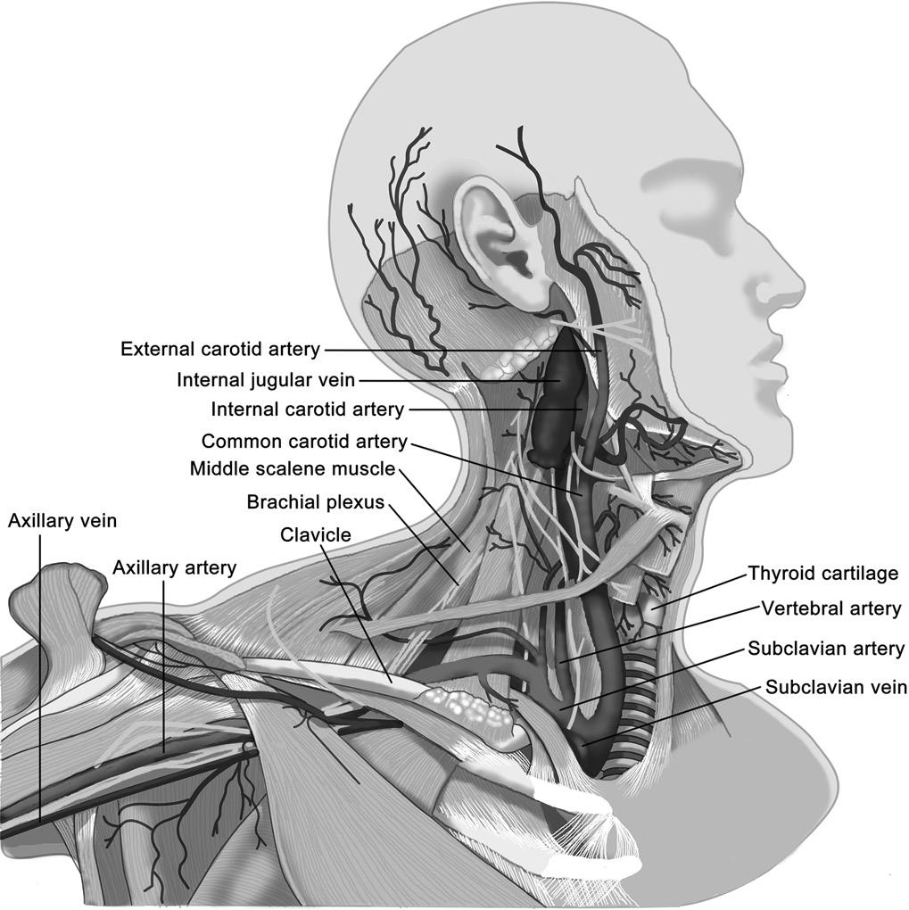
• Accessed by placing a needle inferior to clavicle, 1–2 cm lateral to the midclavicular line, with the tip directed medially and superiorly toward the sternal notch (Figure 1.1)
Vertebral artery
• Branches from the subclavian artery, traveling cephalad to enter the spinal column deep to Chassaignac’s tubercle
• Travels through the transverse foramen of C1–C6 before fusing to form the basilar artery, supplying the posterior Circle of Willis and the spinal cord
• Can be injected directly during an interscalene brachial plexus block
Carotid artery
• Arises from brachiocephalic artery on the right and the aortic arch on the left
• Travels in carotid sheath medial to IJ and anterior to cranial nerve (CN) X
• Bifurcates into internal and external carotid arteries at the level of the C4
• Carotid sinus:
■ Located at bifurcation of carotid artery
■ Compression during carotid endarterectomy can cause a baroreceptor reflex resulting in bradycardia.
Thoracic duct
• Primary endpoint for the lymphatic drainage of the body before joining the venous system
• Arises at the L2 level, courses through the diaphragm posterior to the esophagus, and ascends the thorax just right of the midline between the aorta and azygos vein
• Crosses to the left at T4–T5 and empties into the SCV just lateral to the IJ
• Can be damaged during attempts for central access to the left SCV or IJ, leading to chylothorax
Surface Landmarks
Thyroid cartilage
• In adults, located approximately at the level of C5. Marks the glottic opening, or the start of the larynx
Figure 1.1 Normal anatomical relationships of the major vessels, nerves, bones, and muscles of the neck and axilla
• Motor and sensory innervation to the larynx is derived from CN X (vagus nerve) via the superior, inferior, and the recurrent laryngeal nerves bilaterally
• Musculature of the larynx is innervated entirely by the recurrent laryngeal nerve except for the cricothyroid muscle which is innervated by external branch of superior laryngeal nerve.
Cricothyroid membrane
• Palpable in the anterior neck just inferior to the thyroid cartilage and superior to the cricoid cartilage
• Located approximately at the C6 vertebral level
• Marks the access point for the cricothyroidotomy procedure
Chassaignac’s tubercle
• Another name for the anterior tubercle of the transverse process of C6
• Lies just posterior to the carotid artery, which can be compressed upon this structure to increase vagal tone via carotid massage
• Marks the approximate location of the vertebral artery, which enters deep to this structure into the spinal column after rising from the subclavian artery
• Clinically used to identify the appropriate location to perform nerve blocks of the brachial plexus, cervical plexus, and stellate ganglion
Vertebrae prominens
• Another name for the spinous process of the C7 vertebral body
• This spinous process is the most prominent in the majority of patients (can be C6 or T1 in small subset of patients).
Stellate ganglion
• Named for its “star-like” appearance
• Is the fusion of the inferior cervical and first thoracic sympathetic ganglia
• Located lateral to the vertebral body of C7
• Blockade of this structure is clinically useful for the treatment of sympathetically mediated pain syndromes, such as complex regional pain syndrome (CRPS) or Raynaud’s phenomenon.
• Side effect associated with stellate ganglion blockade is Horner’s syndrome (e.g., ptosis, anhidrosis, miosis), and may frequently occur following many of the cervical and brachial plexus nerve blocks.
Brachial plexus
• Provides cutaneous and motor innervation to the upper extremity
• Lies between the anterior and middle scalene muscles in the neck before running alongside the subclavian and axillary arteries
Radiological Anatomy
See Figure 1.2.
Chest
Surface Landmarks
Trachea
• Begins at C6 and continues inferiorly until it bifurcates at the primary carina
• This bifurcation occurs at the level of the sternal angle, or Angle of Louis, which is the joint between the sternum and manubrium and the connection of the T2 costal cartilages. This structure also marks the approximate level of the T4–T5 intervertebral disk.
Lungs
• Are divided into their lobes by the structures called fissures
• Three lobes on the right and two lobes on the left plus the lingual
• Fissures
• Bilaterally, the oblique fissure divides the superior and inferior lobes on the left and superior and middle lobes on the right.
• The fissures begin posteriorly at the level of T4, traveling caudally and laterally, and then around the torso to terminate anteriorly approximately at the level of the seventh rib on the midclavicular line.
• The right lung is divided a second time by the horizontal fissure, which begins anteriorly approximately at the fourth costal cartilage and traverses laterally to the anterior axillary line, where it intersects with the oblique fissure at the level of the fifth rib. This fissure demarcates the border between the inferior and middle lobes.
Heart
• Point of maximal impulse (PMI)
• Landmark for the apex of the heart located at level of the fifth intercostal space (ICS) 6–10 cm lateral to midline
• Auscultation zones
• Aortic: Second ICS right upper sternal border
• Pulmonary valve: Second ICS left upper sternal border
• Tricuspid valve: Fourth left ICS on the sternal border
• Mitral valve: Fifth left ICS midclavicular line
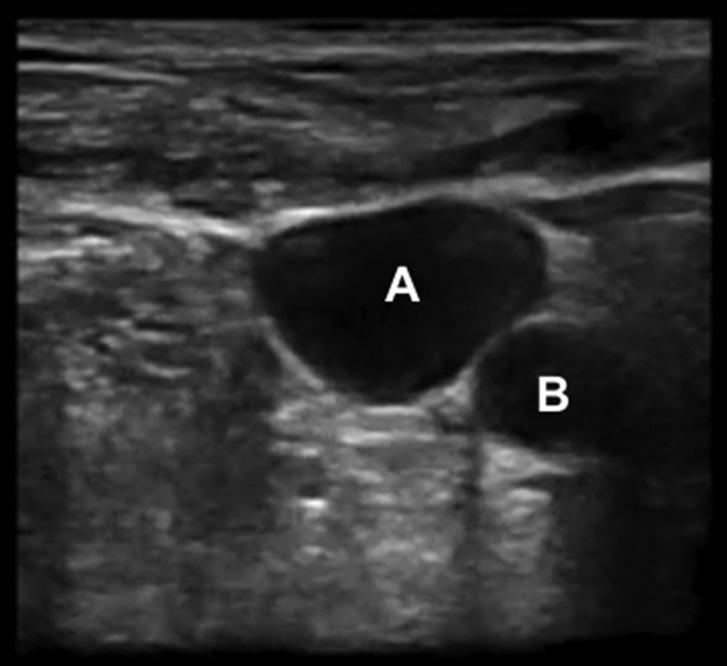
Figure 1.2 Ultrasound image of the lateral neck, displaying (A) the internal jugular vein and (B) the carotid artery
• Coronary arteries
• Left and right main arteries arise from the aorta behind the left and right aortic valve leaflets.
• Left main artery divides into the left anterior descending (LAD) and the circumflex (LCX).
• LAD supplies the anterior wall of the left ventricle (LV) and the anterior two-third of the interventricular septum (IVS).
• LCX supplies the lateral wall of the LV and part of the posterior wall.
• Right coronary artery (RCA)
■ Supplies most of the right side and usually both sinoatrial (SA) and atrioventricular (AV) nodes
• Posterior and anterior walls of the right ventricle (RV) except for the apex (LAD)
• Right atrium including SA node
• Upper half of the atrial septum
• Posterior one-third of IVS
• Inferior wall of LV
• AV node
• Posterior descending artery (PDA) arises from RCA in approximately 80 percent of patients. This is called “right-dominant” circulation.
Radiological Anatomy
See Figures 1.3–1.4.
Upper and Lower Extremities
Upper Extremity Vasculature
Basilic vein
• Travels from the medial posterior forearm at the ulnar head proximally to the anterior elbow, where it lies medial to the tendon of the biceps brachii muscle
• Becomes the axillary vein at the border of the teres major muscle
• Becomes the SCV at the outer border of the first rib
Cephalic vein
• Begins laterally at the wrist within the anatomic snuffbox –a triangle formed by the radial head, the extensor pollicis longus tendon, and the extensor pollicis brevis tendon
• At the elbow, it is most commonly found lateral to the biceps tendon. It then continues proximally in the arm lateral to the biceps brachii muscle before crossing anterior to the deltoid and diving deep to join with the axillary vein under the clavicle.
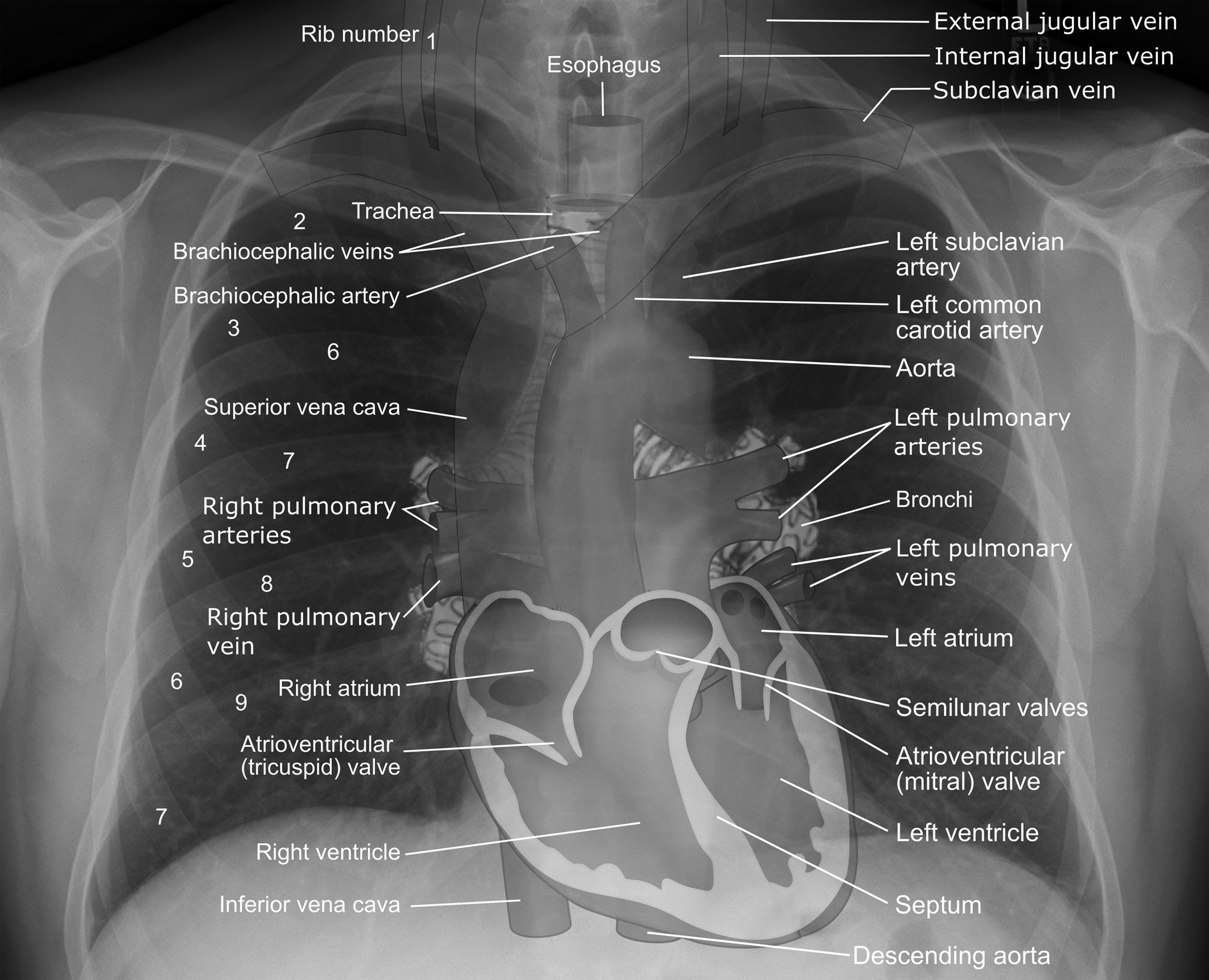
Figure 1.3 Normal radiograph of the chest. Superimposed on this image are outlines of some of the major topographical landmarks of the chest.
Axillary artery
• Direct continuation of the subclavian artery, it begins at the border of the first rib, coursing laterally until the border of the teres muscle where it becomes the brachial artery.
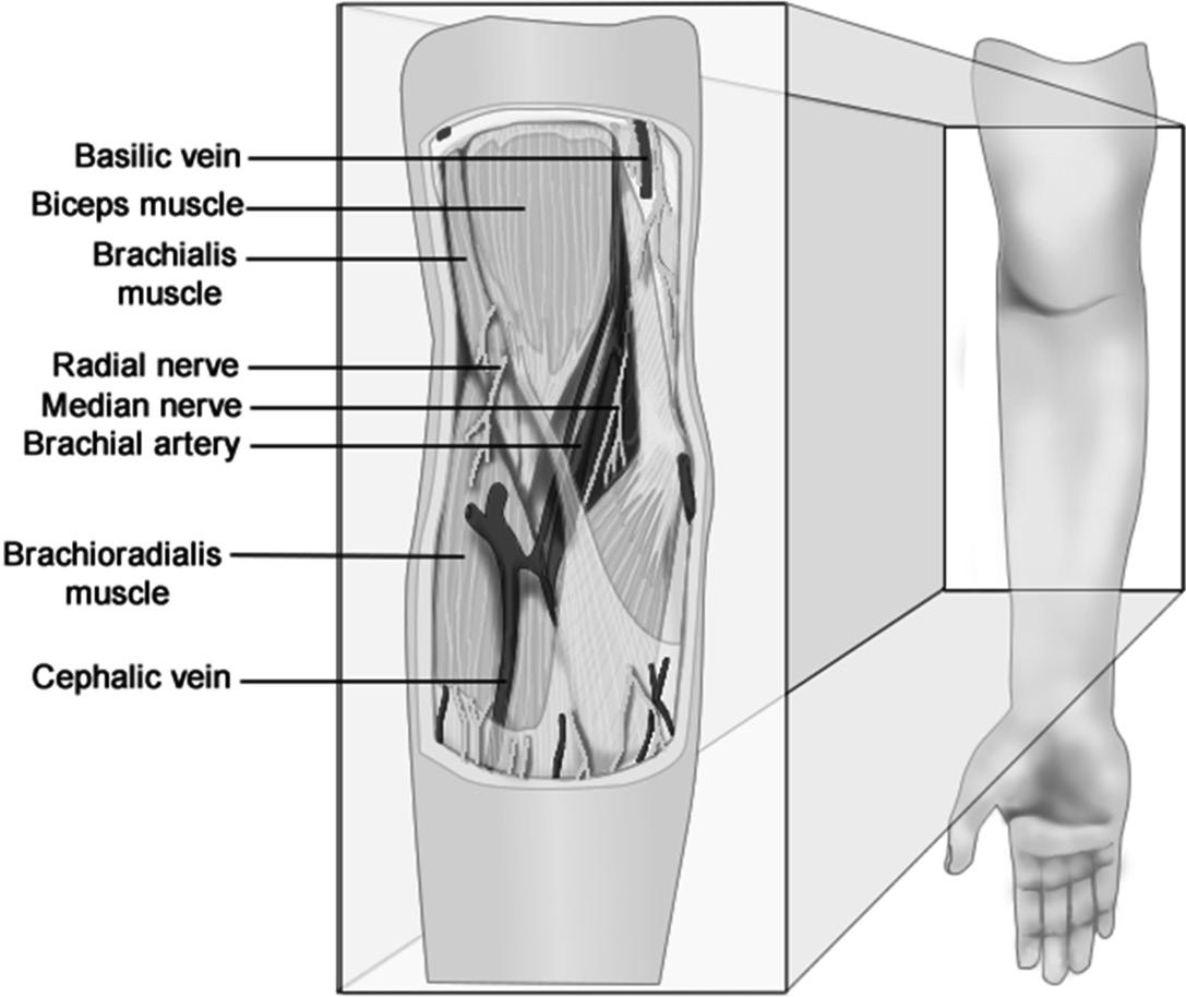
Brachial artery
• The pulsation that is typically felt just medial to the biceps brachii tendon at the cubital fossa
• Subsequently bifurcates into the radial and ulnar arteries (Figure 1.5)
Box 1.1 Upper extremity nerve block landmarks
Interscalene (i.e., brachial plexus: roots)
Supraclavicular (i.e., brachial plexus: trunks/divisions)
Infraclavicular (i.e., brachial plexus: cords)
Axillary (i.e., brachial plexus: branches)
Radial nerve
Ulnar nerve
Median nerve
Between anterior and middle scalene muscles at level of C6
Lateral to the clavicular attachment of the SCM
Three centimeters caudal to the midpoint of a line between the coracoid process and the medial clavicle
At the point of palpation of the axillary artery
Between the brachioradialis and the biceps tendon
Between the medial epicondyle and olecranon
Medial to the brachial artery at the antecubital fossa
Upper Extremity Innervation
Brachial plexus
• Originates from a complex network of nerves formed by ventral rami of C5–T1
• Provides sensory and motor innervation of the upper extremities. Clinically, the anesthesiologist can provide surgical anesthesia to the upper extremity via blockade of the brachial plexus (see Box 1.1)
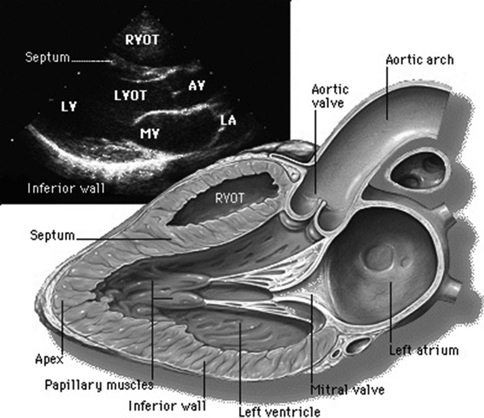
Figure 1.4 Transesophageal echo (TEE) image depicting normal anatomy of the heart
Figure 1.5 Normal anatomical relationships of the major vessels and nerves, bones, and muscles of the antecubital fossa
• ROOTS: After exiting the spinal column, the C5–T1 roots split and recombine to form the superior (C5–C6), middle (C7), and inferior (C8–T1) trunks, which lie between the anterior and middle scalene muscles.
• TRUNKS: Further split into anterior and posterior divisions
• Superior trunk gives rise to the suprascapular nerve which innervates 70 percent of the shoulder joint. Of the brachial plexus blocks, the interscalene block (ISB) is the only one that blocks this nerve. It is also the only block that can be used for shoulder surgery without supplementation.
• Roots/trunks are blocked for the ISB.
• Due to proximity, the phrenic nerve, stellate ganglion, superficial cervical plexus, recurrent laryngeal nerve, and CN XI are frequently blocked with ISB.
• DIVISIONS: Recombine into the lateral, medial, and posterior cords, which are named for their relationship with the subclavian artery
• Level of blockade for supraclavicular block
• CORDS: Split further and recombine to form the terminal branches
• Level of blockade for infraclavicular block
• BRANCHES:
• There are five major terminal branches of the brachial plexus, including:
■ Axillary nerve (C5–C6)
■ Musculocutaneous nerve (C5–C7)
■ Radial nerve (C5–T1)
■ Median nerve (C5–T1)
■ Ulnar nerve (C8–T1)
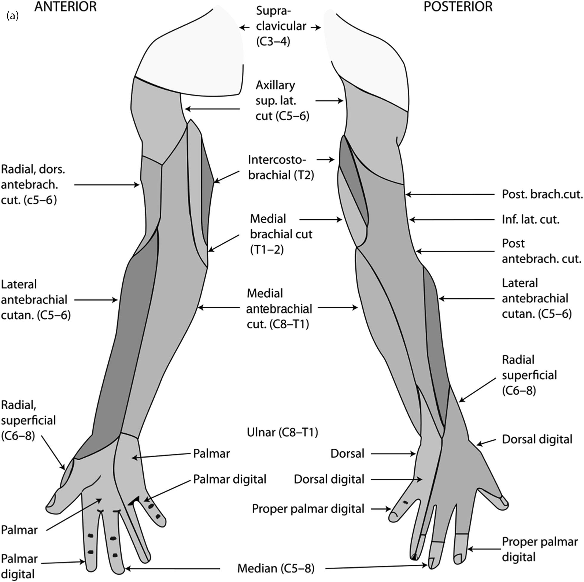
Intercostobrachial nerve
• Skin over axilla and medial arm is the only part of the arm not innervated by the brachial plexus.
• Intercostobrachial nerve which is derived from T2–T3
• If not blocked separately, can contribute to tourniquet pain (Figure 1.7)
Lower Extremity Vasculature
Small saphenous vein
• Begins posterior to the lateral malleolus and extends proximally on the posterior lower leg until the popliteal fossa, where it drains into the popliteal vein
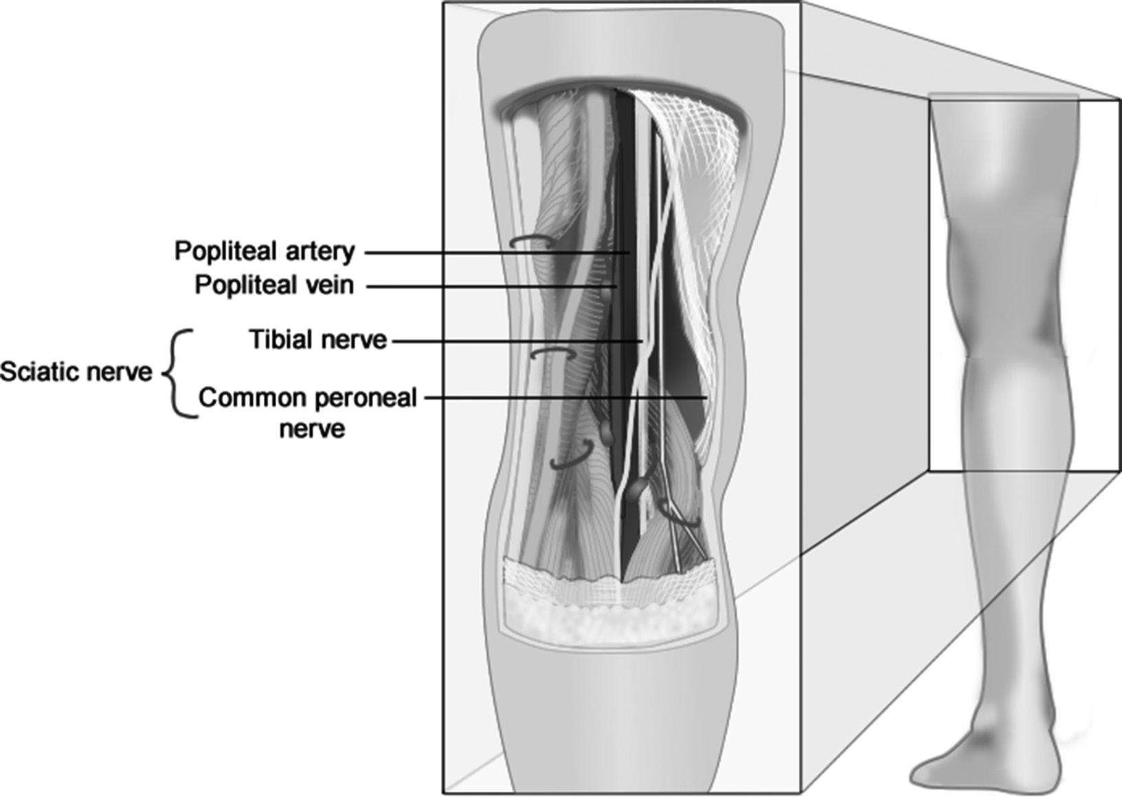
Popliteal vein
• Lies between the popliteal artery and the tibial nerve at the popliteal fossa (Figure 1.6)
• Continues proximally through the adductor magnus muscle, where it becomes the femoral vein
Great saphenous vein
• Longest vein in the body. Typically found superficially at the dorsum of the foot medial to the medial malleolus
• Commonly cannulated in pediatrics for peripheral venous access
• Used as a landmark to block the saphenous nerve at the ankle. It innervates the medial aspect of the foot
• Courses proximally on the medial surface of the leg before entering the fossa ovalis to empty into the femoral vein on the anterior thigh near the inguinal crease
Femoral artery
• Arises as the direct continuation of the external iliac artery
• Lies just lateral to the femoral vein at the inguinal ligament
Figure 1.6 Normal anatomical relationships of the major vessels, nerves, bones, and muscles of the popliteal fossa
• Divides into superficial femoral artery and profunda femoris
• The profunda femoris (deep artery of the thigh) provides vascular supply to the structures of the thigh.
• The superficial femoral artery courses posteriorly and distally until resurfacing at the popliteal fossa as the popliteal artery.
Popliteal
artery
• Divides into two major branches: anterior and posterior tibial arteries
• Anterior tibial artery
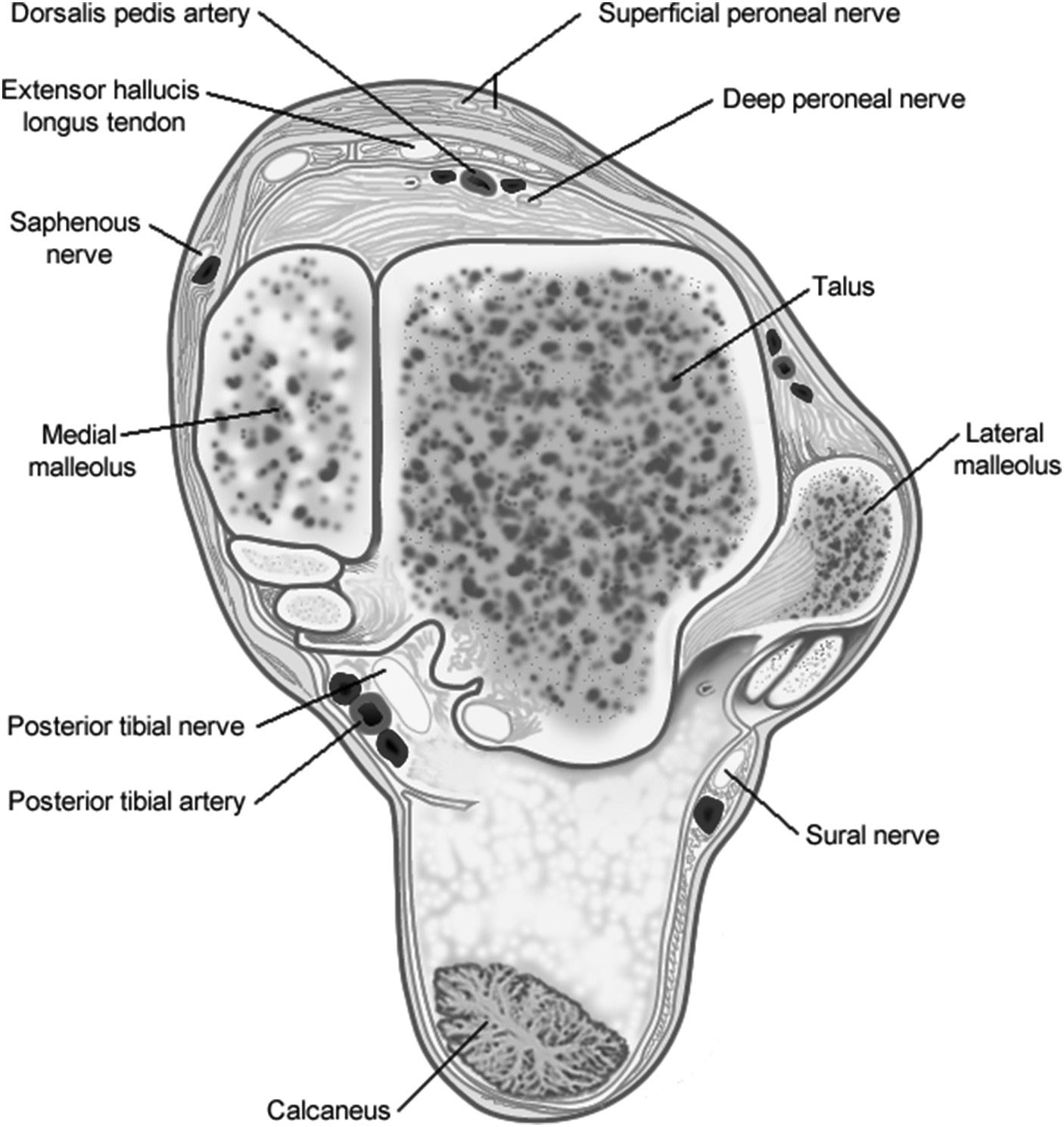
• Terminates as the dorsalis pedis (DP) artery
• DP pulse can be palpated on the dorsal surface of the foot between the extensor hallicus longus and extensor digitorum longus tendons.
• DP pulse is a landmark for deep peroneal nerve blockade which innervates the space between the first and second toes.
• Posterior tibial artery (PT)
• Pulsation can be felt posterior to the medial malleolus at the ankle.
• PT pulse is a landmark for blockade of the posterior tibial nerve which innervates the plantar aspect of the foot (Figure 1.7).
Lower Extremity Innervation
Lumbar plexus
• Originates from a complex network of nerves formed by ventral rami of T12–L4
• Gives rise to femoral, obturator, lateral femoral cutaneous, ilioinguinal, genitofemoral, and iliohypogastric nerves
• Femoral nerve (L2–L4):
• Found deep into the inguinal ligament lateral to the femoral artery
• Provides motor innervation to the muscles for knee extension. Blockade of the femoral nerve results in 80 percent reduction in quadriceps strength
• Sensory innervation anterior and medial thigh via two anterior cutaneous branches
• Lateral femoral cutaneous nerve (LFCN) (L2–L3):
• Provides only cutaneous innervation of the lateral thigh
• Obturator nerve (L2–L4):
• Innervates the adductor muscles
• Sensory innervation varies within the population:
■ One-third posterior knee, one-third medial thigh, one-third no innervation
Sacral plexus (L4–S4)
• Sciatic nerve (L4–S3)
• Front of the piriformis muscle, traveling distally toward the popliteal fossa
• Two major branches
■ Tibial nerve
• Motor function of all the muscles of the posterior compartment of the leg
■ Common peroneal nerve
■ Supplies the muscles of anterior compartment of leg
• Blockade or damage results in foot drop
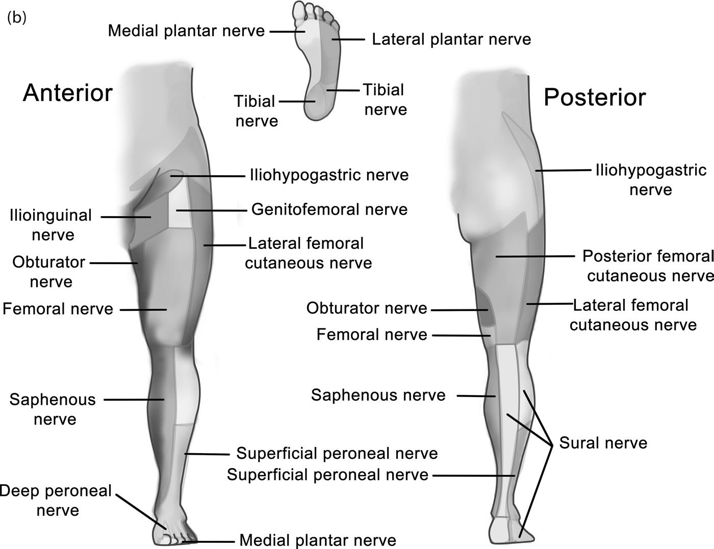
Cutaneous innervation of the distal lower extremity, ankle, and foot is supplied by a combination of five nerves – four derived from the sciatic nerve and one derived from the femoral nerve (Figure 1.8).
• Sciatic branches (all of these can be blocked at once with a popliteal block)
• Tibial nerve: Provides sensory innervation to the heel and plantar surface of the foot
■ Blocked by injection next to PT pulsation posterior to medial malleolus
• Superficial peroneal nerve: Sensory to the dorsum of the foot
■ Blocked by superficial infiltration of local anesthetic between medial and lateral malleoli
• Deep peroneal nerve: Sensory to the web space between the first and second toes
■ Blocked at the intermalleolar axis by injection posterior to the extensor hallicus longus tendon
■ Blocked at the dorsum of the foot by injecting next to DP pulsation
• Sural nerve: Derived from both the tibial and common peroneal nerves. Provides sensory innervation to the posterior lower leg and lateral ankle
■ Blocked by injection of local anesthetic between lateral malleolus and Achilles tendon
• Femoral branch
• Saphenous nerve provides sensory innervation at the medial lower leg and medial ankle and foot.
■ Blocked by injection medial to medial malleolus next to great saphenous vein
Box 1.2 lists some of the normal anatomical relationships and topographic landmarks associated with nerve blocks of the lower extremities.
Radiological Anatomy
See Figures 1.9–1.11.
Spinal Anatomy, Landmarks, and Dermatomes
Surface Landmarks
Box 1.3 describes some of the clinically relevant surface landmarks, and important key sensory and motor areas of innervation.
Spinal Anatomy
Vascular supply
• Anterior two-third of spinal cord receives its blood supply from a single anterior spinal artery, which arises from the vertebral arteries.
• Receives branches from 6–8 radicular arteries, most important of which is the artery of Adamkiewicz, arising most commonly from T9–T12
Figure 1.7 Distribution of the major cutaneous nerve branches of the upper and lower extremities
Box 1.2 Lower extremity nerve block landmarks
Femoral nerve
Lateral femoral cutaneous nerve
Sciatic nerve
Saphenous nerve
Superficial peroneal nerve
Lateral to the pulsation of femoral artery at the inguinal ligament
Medial to anterior superior iliac spine
Four centimeters distal to the midpoint of a line between the greater trochanter and posterior superior iliac spine
Anterior to the medial malleolus near the saphenous vein
Anterior to the lateral malleolus
Deep peroneal nerve Near the pulsation of the dorsalis pedis artery at the ankle
Posterior tibial nerve
Sural nerve
Posterior to the pulsation of the posterior tibial artery
Posterior to the lateral malleolus
• Damage to artery of Adamkiewicz causes anterior spinal cord syndrome
■ Flaccid paralysis of the lower extremities
■ Bowel and bladder dysfunction
■ Proprioception and sensation typically spared
• Occurs most commonly in emergent repair of dissecting or ruptured thoracic aortic aneurysm (40 percent)
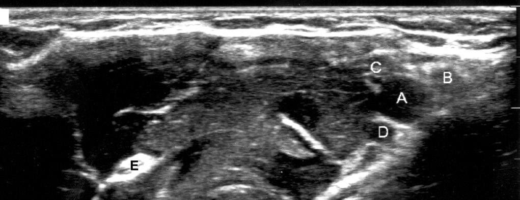
Figure 1.10 Ultrasound image of the brachial plexus at the location for the axillary block. (A) The axillary artery is surrounded by the (B) median, (C) ulnar, and (D) radial nerves. (E) The musculocutaneous nerve is seen laterally within the coracobrachialis muscle.
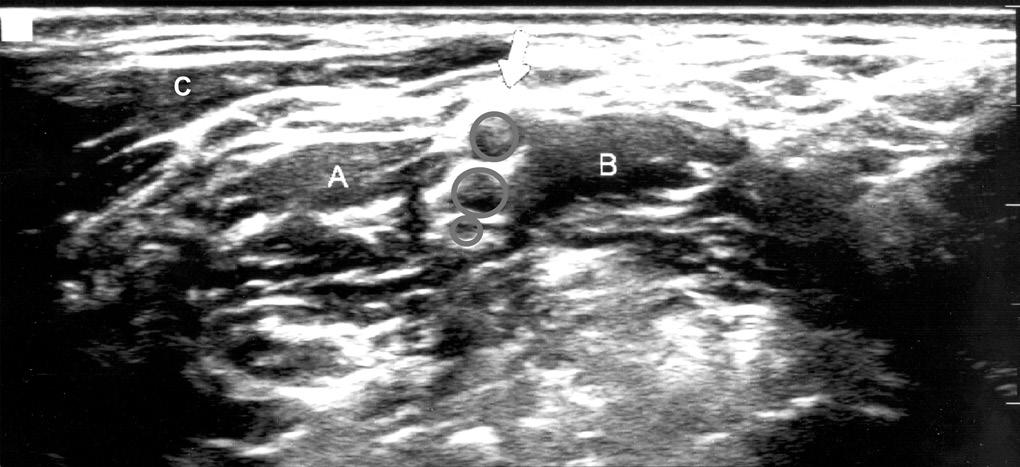
Figure 1.9 Ultrasound image of the brachial plexus at the location for the ISB. At this level, the roots of C5, C6, and C7 appear as hypoechoic circles between (A) the anterior and (B) middle scalene muscles, deep to the (C) SCM muscle.
Figure 1.8 Normal anatomical relationship of the major vessels, nerves, bones, and the ankle