Neuroscience forNeurosurgeons
Editedby
FarhanaAkter
HarvardUniversity
NigelEmptage
UniversityofOxford
FlorianEngert
HarvardUniversity
MitchelS.Berger
UniversityofCalifornia–SanFrancisco
ShaftesburyRoad,CambridgeCB28EA,UnitedKingdom OneLibertyPlaza,20thFloor,NewYork,NY10006,USA
477WilliamstownRoad,PortMelbourne,VIC3207,Australia
314–321,3rdFloor,Plot3,SplendorForum,JasolaDistrictCentre, NewDelhi – 110025,India
103PenangRoad,#05–06/07,VisioncrestCommercial,Singapore238467
CambridgeUniversityPressispartofCambridgeUniversityPress&Assessment, adepartmentoftheUniversityofCambridge.
WesharetheUniversity’smissiontocontributetosocietythroughthepursuitof education,learningandresearchatthehighestinternationallevelsofexcellence.
www.cambridge.org
Informationonthistitle: www.cambridge.org/9781108831468
DOI: 10.1017/9781108917339
©CambridgeUniversityPress&Assessment2024
Thispublicationisincopyright.Subjecttostatutoryexceptionandtotheprovisions ofrelevantcollectivelicensingagreements,noreproductionofanypartmaytake placewithoutthewrittenpermissionofCambridgeUniversityPress&Assessment. Firstpublished2024
PrintedintheUnitedKingdombyCPIGroupLtd,CroydonCR04YY AcataloguerecordforthispublicationisavailablefromtheBritishLibrary. LibraryofCongressCataloging-in-PublicationData
Names:Akter,Farhana(Surgeon),editor.|Emptage,Nigel,editor.|Engert,Florian, editor.|Berger,MitchelS.,editor.
Title:Neuroscienceforneurosurgeons/editedbyFarhanaAkter,NigelEmptage, FlorianEngert,MitchelS.Berger.
Description:Cambridge,UnitedKingdom;NewYork,NY:CambridgeUniversity Press,2023.|Includesbibliographicalreferencesandindex. Identifiers:LCCN2023017267|ISBN9781108831468(hardback)|ISBN 9781108917339(ebook)
Subjects:MESH:NeurosurgicalProcedures|NervousSystemDiseases –physiopathology|NervousSystem – physiology
Classification:LCCRD592.8|NLMWL368|DDC617.4/8092–dc23/eng/20230703 LCrecordavailableat https://lccn.loc.gov/2023017267
ISBN978-1-108-83146-8Hardback
CambridgeUniversityPress&Assessmenthasnoresponsibilityforthepersistence oraccuracyofURLsforexternalorthird-partyinternetwebsitesreferredtointhis publicationanddoesnotguaranteethat anycontentonsuchwebsitesis,orwill remain,accurateorappropriate.
Everyefforthasbeenmadeinpreparingthisbooktoprovideaccurateandup-to-date informationthatisinaccordwithacceptedstandardsandpracticeatthetimeof publication.Althoughcasehistoriesaredrawnfromactualcases,everyefforthasbeen madetodisguisetheidentitiesoftheindividualsinvolved.Nevertheless,theauthors, editors,andpublisherscanmakenowarrantiesthattheinformationcontainedhereinis totallyfreefromerror,notleastbecauseclinicalstandardsareconstantlychangingthrough researchandregulation.Theauthors,editors,andpublishersthereforedisclaimallliability fordirectorconsequentialdamagesresultingfromtheuseofmaterialcontainedinthis book.Readersarestronglyadvisedtopaycarefulattentiontoinformationprovidedbythe manufacturerofanydrugsorequipmentthattheyplantouse.
ListofContributors vii
1 Neuroanatomy 1 FarhanaAkter,CharlesReilly,Christophe Dupre,andShifaHossain
2 CerebralAutoregulation 51 ParisaNikrouz,XingpingQin,FarhanaAkter
3 NeuroimmuneInteractions 59 AllisonChang
4 AnatomyandPhysiologyoftheNeuron 72 OluwatobiAriyoandFarhanaAkter
5 SynapticTransmission 84 KatrinaHon,FarhanaAkter,andNigelEmptage
6 SensoryPathways 99 RachelChau,MeganChau,andFarhanaAkter
7 SomatosensoryandSomaticMotorSystems 121 MariaKaltchenkoandFarhanaAkter
8 NeuronModels 134 KumareshKrishnan
9 AnIntroductiontoArtificialIntelligence andMachineLearning 146 ShunYao,YeWu,andFarhanaAkter
10 ArtificialIntelligenceinNeuroscience 158 WillXiao,MengmiZhang,andGabriel Kreiman
11 ProbabilityandStatistics 167 ZacharyT.Miller
12 Glioma 184 DavidM.AshleyandJustinT.Low
13 BrainMetastases:MoleculestoMedicine 193 EthanS.Srinivasan,VadimTsvankin, EricW.Sankey,MatthewM.Grabowski, PakawatChongsathidkiet, andPeterE.Fecci
14 BenignAdultBrainTumorsandPediatricBrain Tumors 214 ShunYao,UmarRaza,andFarhanaAkter
15 BiomechanicsoftheSpine 234 GaetanoDeBiaseandKingsleyAbode-Iyamah
16 DegenerativeCervicalMyelopathy 239 T.J.Florence,JoelS.Beckett, andLangstonT.Holly
17 Spondylolisthesis 248 YikeJin,AnnLiu,RaviMedikonda, andTimothyF.Witham
18 Radiculopathy 254 YingdaLiandMichaelY.Wang
19 SpinalTumors 267 ZievB.Moses,MatthewTrawczynski, andJohnE.O’Toole
20 AcuteSpinalCordInjuryandSpinal Trauma 278
DominiqueM.O.Higgins,Pavan S.Upadhyayula,MichaelArgenziano, andPaulMcCormick
21 TraumaticBrainInjury 291 KristinA.KeithandJasonH.Huang
22 VascularNeurosurgery 300 KarolP.BudohoskiandAdibA.Abla
23 PediatricVascularMalformations 314 AlaaMontaserandEdwardR.Smith
24 CraniofacialNeurosurgery 326 JohnT.SmetonaandJohnA.Persing
25 Hydrocephalus 335
BenjaminC.Reeves,JasonK.Karimy,Phan Q.Duy,andKristopherT.Kahle
26 PeripheralNerveInjuryResponse Mechanisms 348
AndrewS.JackandLineJacques
27 ClinicalPeripheralNerveInjuryModels 355
AndrewS.Jack,CharlotteJ.Huie,andLine Jacques
28 TheNeuroscienceofFunctional Neurosurgery 369
JosephS.Bell,T.J.Florence,MayaHarary, MaxwellD.Melin,HiroSparks,andNader Pouratian
29 Neuroradiology:FocusedUltrasound inNeurosurgery 382 MassimilianoDelBene,RobertoEleopra, FrancescoPrada,andFrancescoDiMeco
30 MagneticResonanceImaging inNeurosurgery 398 DavidJ.Segar,JasmineA.Thum,Dhiego Bastos,andAlexandraJ.Golby
31 BrainMapping 410 AnthonyT.Lee,CeciliaDalleOre,andShawn L.Hervey-Jumper Index 422
Contributors
AdibA.Abla
UniversityofCaliforniaSanFrancisco,California,USA
KingsleyAbode-Iyamah
MayoClinic,Jacksonville,FL,USA
FarhanaAkter
HarvardUniversity,Cambridge,MA,USA
MichaelArgenziano
ColumbiaUniversityCollegeofPhysiciansandSurgeons, NewYork,NY,USA
OluwatobiAriyo
HarvardUniversity,Cambridge,MA,USA
DavidM.Ashley
DukeUniversitySchoolofMedicine,Durham,NC, USA
JoelS.Beckett
DavidGeffenUCLASchoolofMedicine,CA,USA
JosephS.Bell
UCLADepartmentofNeurosurgery,LosAngeles,CA, USA
DhiegoBastos
BrighamandWomen’sHospital,Boston,MA,USA
KarolP.Budohoski
UniversityofUtah,Utah,USA
AllisonChang
HarvardUniversity,Cambridge,MA,USA
MeganChau
NorthwesternUniversity,Evanston,IL,USA
RachelChau
HarvardUniversity,Cambridge,MA,USA
PakawatChongsathidkiet
DukeUniversityMedicalCenter,Durham,NC,USA
CeciliaDalleOre
UniversityofCaliforniaSanFrancisco,SanFrancisco, CA,USA
GaetanoDeBiase
MayoClinic,Jacksonville,FL,USA
MassimilianoDelBene
FondazioneIRCCSIstitutoNeurologicoCarloBesta,Milan, ItalyandEuropeanInstituteofOncologyIRCCS,Milan, Italy
FrancescoDiMeco
FondazioneIRCCSIstitutoNeurologicoCarloBesta, Milan,Italy,UniversityofMilan,Milan,Italy,andJohns HopkinsMedicalSchool,Baltimore,MD,USA
ChristopheDupre
HarvardUniversity,Cambridge,MA,USA
PhanQ.Duy
YaleSchoolofMedicine,NewHaven,CT,USA
RobertoEleopra
FondazioneIRCCSIstitutoNeurologicoCarloBesta, Milan,Italy
NigelEmptage UniversityofOxford,Oxford,UK
PeterE.Fecci
DukeUniversityMedicalCenter,Durham,NC,USAand DukeUniversityCenterforBrainandSpineMetastasis, Durham,NC,USA
T.J.Florence
DavidGeffenUCLASchoolofMedicine,CA,USA
AlexandraJ.Golby
BrighamandWomen’sHospital,Boston,MA,USA
MatthewM.Grabowski
ClevelandClinicNeurologicalInstitute,Cleveland,OH, USA
MayaHarary
UCLADepartmentofNeurosurgery,LosAngeles,CA,USA
ShawnL.Hervey-Jumper
UniversityofCaliforniaSanFrancisco,SanFrancisco, CA,USA
DominiqueM.O.Higgins
ColumbiaUniversityCollegeofPhysiciansandSurgeons, NewYork,NY,USA
LangstonT.Holly
DavidGeffenUCLASchoolofMedicine,CA,USA
KatrinaHon
HarvardUniversity,Cambridge,MA,USA
ShifaHossain
HarvardUniversity,Cambridge,MA,USA
JasonH.Huang
BaylorScottandWhiteHealth,MedicalCenter,Temple, TX,USAandTexasA&MUniversityHealthScience Center,CollegeofMedicine,Temple,TX,USA
CharlotteJ.Huie
UniversityofCaliforniaSanFrancisco(UCSF),San Francisco,CA,USA
AndrewS.Jack
UniversityofAlberta,Edmonton,AB,Canadaand UniversityofCaliforniaSanFrancisco(UCSF),San Francisco,CA,USA
LineJacques
UniversityofCaliforniaSanFrancisco(UCSF),San Francisco,CA,USA
YikeJin
JohnsHopkinsHospital,Baltimore,MD,USA
KristopherT.Kahle
YaleSchoolofMedicine,NewHaven,CT,USA
MariaKaltchenko
HarvardUniversity,Cambridge,MA,USA
JasonK.Karimy
YaleSchoolofMedicine,NewHaven,CT,USA
KristinA.Keith
BaylorScottandWhiteHealth,MedicalCenter, Temple,TX,USAandTexasA&MUniversityHealth
ScienceCenter,CollegeofMedicine,Temple,TX, USA
GabrielKreiman
HarvardMedicalSchool,Boston,MA,USA
KumareshKrishnan
HarvardUniversity,Cambridge,MA,USA
AnthonyT.Lee
UniversityofCaliforniaSanFrancisco,SanFrancisco, CA,USA
YingdaLi
WestmeadHospital,Sydney,Australia
AnnLiu
JohnsHopkinsHospital,Baltimore,MD,USA
JustinT.Low
DukeUniversitySchoolofMedicine,Durham,NC,USA
PaulMcCormick
ColumbiaUniversityCollegeofPhysiciansandSurgeons, NewYork,NY,USA
RaviMedikonda
JohnsHopkinsHospital,Baltimore,MD,USA
MaxwellD.Melin
UCLA-CaltechMedicalScientistTrainingProgram,Los Angeles,CA,USA
AlaaMontaser
BostonChildren’sHospital,Boston,MA,USA
ZievB.Moses
RushUniversityMedicalCenter,Chicago,IL,USA
ZacharyT.Miller
HarvardUniversity,Cambridge,MA,USA
ParisaNikrouz
MaidstoneandTunbridgeWellsNHSTrust, Kent,UK
JohnE.O’Toole
RushUniversityMedicalCenter,Chicago,IL,USA
JohnA.Persing
YaleSchoolofMedicine,NewHaven,CT,USA
NaderPouratian
UTSouthwesternMedicalCenter,Dallas,TXUSA
FrancescoPrada
FondazioneIRCCSIstitutoNeurologicoCarloBesta, Milan,Italy,UniversityofVirginiaHealthScience Center,Charlottesville,VA,USA,andFocused UltrasoundFoundation,Charlottesville,VA,USA
XingpingQin
HarvardSchoolofPublicHealth,Boston,MA,USA
UmarRaza
NationalUniversityofMedicalSciences(NUMS), Rawalpindi,Pakistan
BenjaminC.Reeves
YaleSchoolofMedicine,NewHaven,CT,USA
CharlesReilly
HarvardUniversity,Cambridge,MA,USA
EricW.Sankey
DukeUniversityMedicalCenter,Durham,NC,USA
DavidJ.Segar
BrighamandWomen’sHospital,Boston,MA,USA
JohnT.Smetona
YaleSchoolofMedicine,NewHaven,CT,USA
EdwardR.Smith
BostonChildren’sHospital,Boston,MA,USA
HiroSparks
UCLADavidGeffenSchoolofMedicine,LosAngeles,CA, USA
EthanS.Srinivasan
DukeUniversityMedicalCenter,Durham,NC,USA
JasmineA.Thum
BrighamandWomen’ sHospital,Boston,MA, USA
MatthewTrawczynski
RushUniversityMedicalCenter,Chicago,IL,USA
VadimTsvankin
ColoradoBrainandSpineInstitute,Denver,CO,USA
VadimTsvankin
ColoradoBrainandSpineInstitute,Denver,CO, USA
PavanS.Upadhyayula
ColumbiaUniversityCollegeofPhysiciansandSurgeons, NewYork,NY,USA
MichaelY.Wang
UniversityofMiamiMillerSchoolofMedicine,Miami,FL, USA
TimothyF.Witham
JohnsHopkinsHospital,Baltimore,MD,USA
YeWu
NanjingUniversityofScienceandTechnology,Nanjing, Jiangsu,China
WillXiao
HarvardMedicalSchool,Boston,MA,USA
ShunYao
TheFirstAffiliatedHospital,SunYat-senUniversity, Guangzhou,Guangdong,China
MengmiZhang
HarvardMedicalSchool,Boston,MA,USA
FarhanaAkter,CharlesReilly,ChristopheDupre,andShifaHossain
1.1AnatomicalPlanesandOrientation oftheBrain
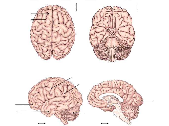
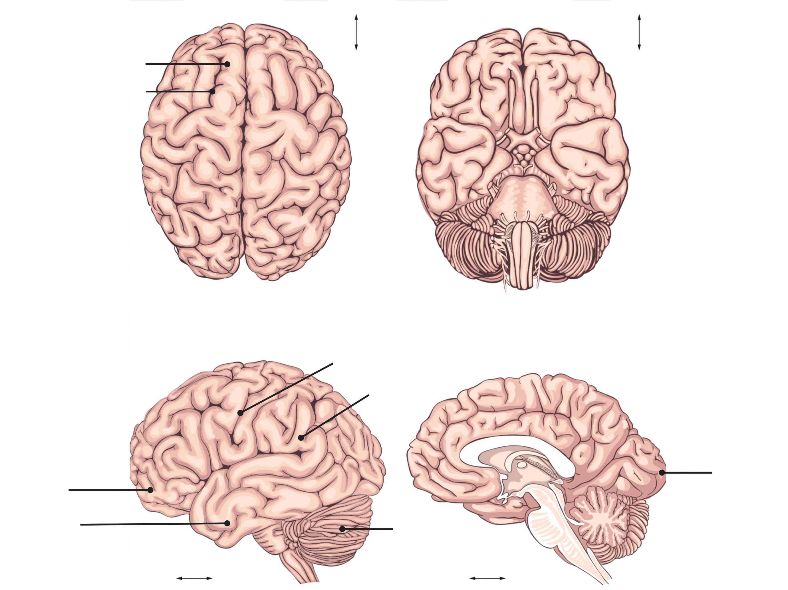
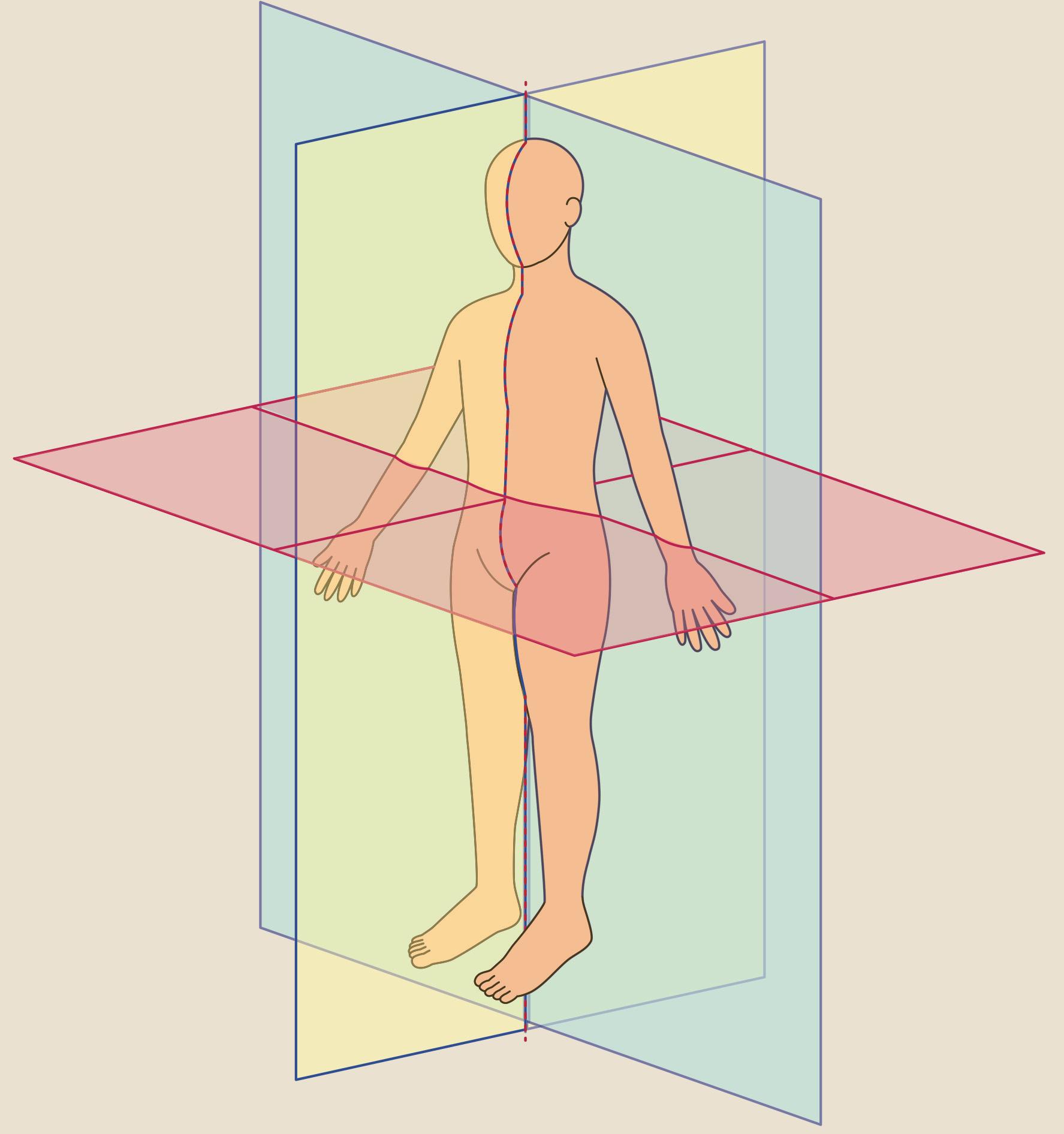
Theneuroaxisofhumansandotherbipedalorthograde animalsisdifferentfromthatofquadrupedanimals. Exampleaxesincludetheanteroposterioraxis,rostrocaudalaxisandthedorsoventralaxis(Figure1.1).Thebrain isusuallyvisualizedinsectionscutthroughthreeorthogonalplanes:sagittal(longitudinal),coronal,andtransverse(axial)planes.
1.2VascularSupplytotheBrain
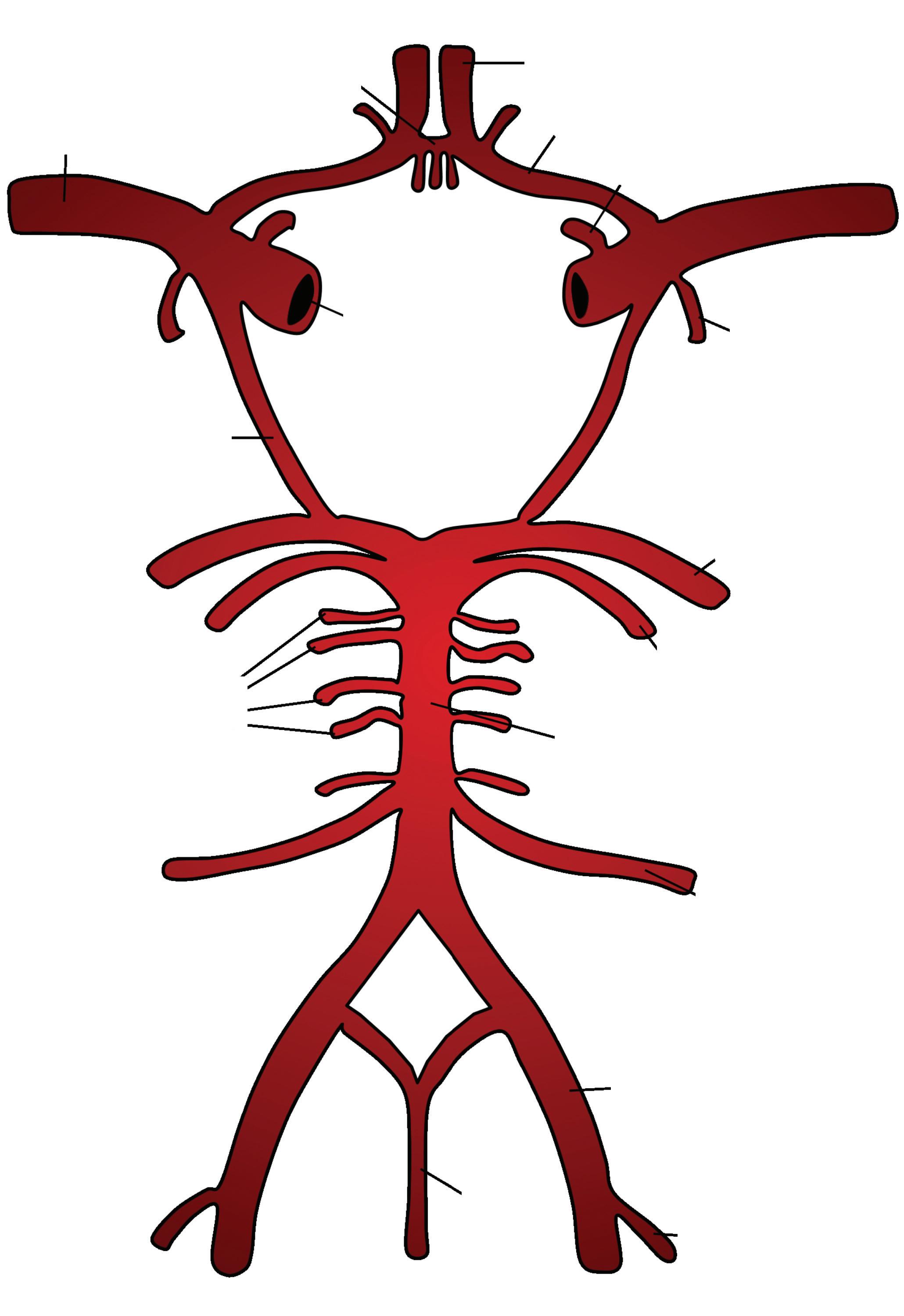
Thebrainrequires15–20%oftherestingcardiacoutput andisexquisitelysensitivetooxygendeprivation.Two mainpairsofarteriessupplybloodtothebrain:the internalcarotidarteriesandthevertebralarteries. Withinthecranialvault,ananastomoticcircle,called theCircleofWillis(Figure1.3),formsfromtheterminal branchesofthesearteries.
Superior frontal gyrus
Superior frontal sulci
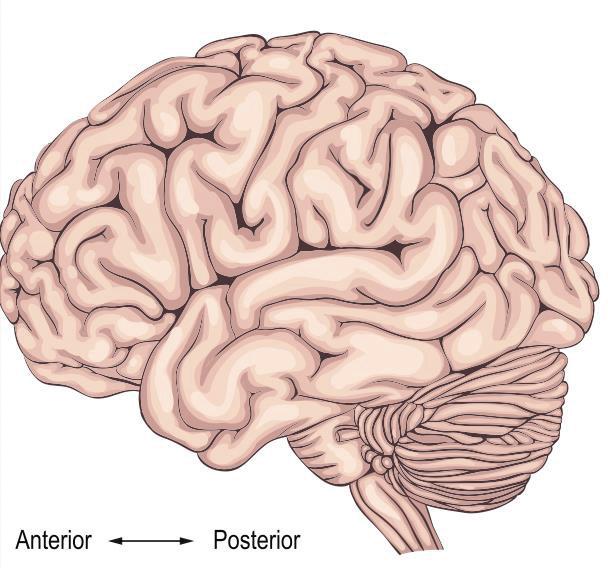
Frontal lobe Temporal lobe Lateral view
FarhanaAkter,CharlesReilly,ChristopheDupre,andShifaHossain
1.1AnatomicalPlanesandOrientation oftheBrain
Theneuroaxisofhumansandotherbipedalorthograde animalsisdifferentfromthatofquadrupedanimals. Exampleaxesincludetheanteroposterioraxis,rostrocaudalaxisandthedorsoventralaxis(Figure1.1).Thebrain isusuallyvisualizedinsectionscutthroughthreeorthogonalplanes:sagittal(longitudinal),coronal,andtransverse(axial)planes.
1.2VascularSupplytotheBrain
Thebrainrequires15–20%oftherestingcardiacoutput andisexquisitelysensitivetooxygendeprivation.Two mainpairsofarteriessupplybloodtothebrain:the internalcarotidarteriesandthevertebralarteries. Withinthecranialvault,ananastomoticcircle,called theCircleofWillis(Figure1.3),formsfromtheterminal branchesofthesearteries.
Superior frontal gyrus
Superior frontal sulci
lobe
Thebrachiocephalictrunkarisesfromtheaortaand bifurcatesintotherightcommoncarotidandrightsubclavianartery.Theleftcommoncarotidarteryandsubclavian arterybranchesoff directlyfromtheaorticarch.The commoncarotidarteriesdivideatthelevelofthethyroid cartilage(C4)intoexternalandinternalcarotidarteries. Thecarotidsinus,adilationatthebaseoftheinternal carotidartery,isabaroreceptor(stretchreceptor)that detectschangesinsystemicbloodpressure.Intimately relatedtoit,isthecarotidbody,madeupofglomustype IchemoreceptorcellsandglomustypeIIsupportingcells. Thechemoreceptorsareprimarilysensorsofpartialpressureofoxygen(PaO2).Thecentralchemoreceptorsinthe medullaoblongataaretheprimarysensorsofthepartial pressureofcarbondioxide(PaCO2)andpH,howeverthe carotidbodiesalsoplayasecondaryrole.
Theexternalcarotidarterydividesintothesuperficial temporalarteryandthemaxillaryarterywithinthe parotidglandandgivesrisetosixbranches(superior thyroidartery,lingualartery,facialartery,ascending pharyngealartery,occipitalartery,andposteriorauricularartery).Themaxillaryarterygivesrisetothemiddle meningealartery,whichpassesthroughtheforamenspinosumtoenterthecranialcavity.
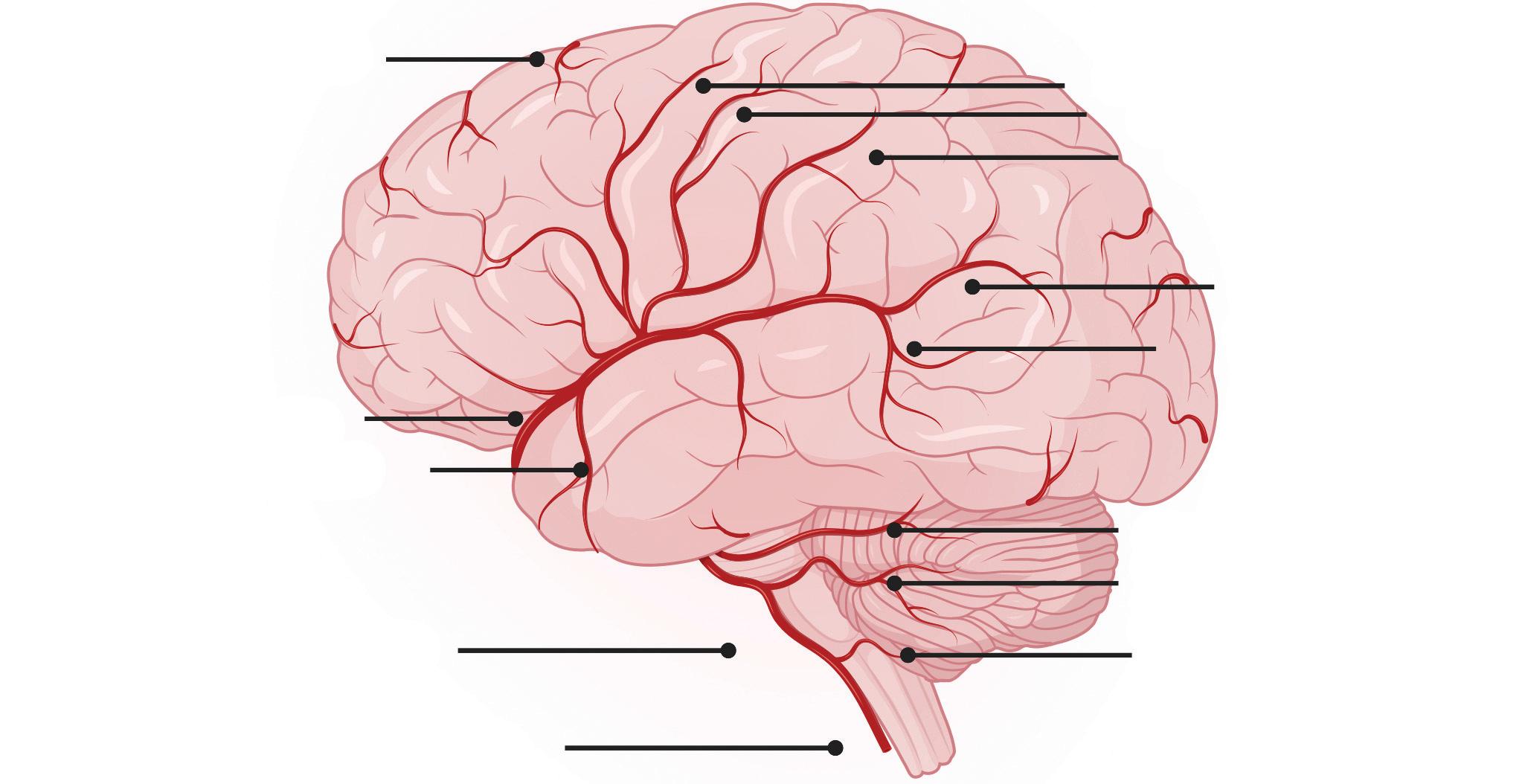
Theinternalcarotidarteryentersthecranialcavityvia thecarotidcanalinthepetrouspartofthetemporalbone, passesthroughthecavernoussinusandpenetratesthe duraintothesubarachnoidspace.Distaltothecavernous sinus,itgivesrisetothefollowingbranches:ophthalmic artery,posteriorcommunicatingartery,anteriorchoroidalartery,andanteriorcerebralartery.Itthencontinues asthemiddlecerebralartery.Theanteriorcerebralartery
suppliesthefrontallobesandmedialaspectsoftheparietalandoccipitallobes(Figure1.4).
Themiddlecerebralarteryisthelargestcerebral arteryandsuppliesthesomatosensoryandmotorcortex, basalganglia,andthecerebralwhitematter.Itcanbe dividedintofourmainsurgicalsegments,denominated M1(sphenoidal/horizontal),M2(insular),M3(opercular),andM4(cortical)segments.M1givesrisetothe lenticulostriateperforatingendarteriessupplyingthe basalgangliaandinternalcapsule.Occlusionofthese vesselscancauselacunarinfarcts,themostcommon typeofischemicstroke.Hypertensivelipohyalinosisof thesevesselscanleadtotheformationofCharcotBouchardaneurysmsandtheseareaprincipalcauseof intracerebralhemorrhage.ThecorticalbranchesofMCA arisefromallofitssegmentsandsupplymostofthe lateralsurfaceofthebrainandincludetheanteriortemporalarteriesfromM1,lateralfrontobasalarteryfrom M2,andparietalbranchesfromM4.Thevertebral arteriesarisefromthesubclavianarteryandpassthough theforamentransversariuminthecervicalvertebrae.The vertebralarteriesenterthecraniumviatheforamenmagnumandmergetoformthebasilarartery.Branchesof thisarteryincludetheanteriorandposteriorinferior cerebellararteriessupplyingthebrainstemandthecerebellumalongsidethesuperiorcerebellararteries.The basilararterybifurcatesintotheposteriorcerebral arteriesandsuppliestheoccipitallobes.
1.2.1CircleofWillis
Ananastomoticcircleisformedaroundtheopticchiasm betweentheanteriorcerebralarteries(viatheanterior
Rostral
Caudal
Dorsal
Ventral
Sagittal
Figure1.2 Planesandorientationsofthe body.
communicatingartery)andtheposteriorcerebralarteries (viatheposteriorcommunicatingarteries).
1.2.2CerebralVenousDrainage
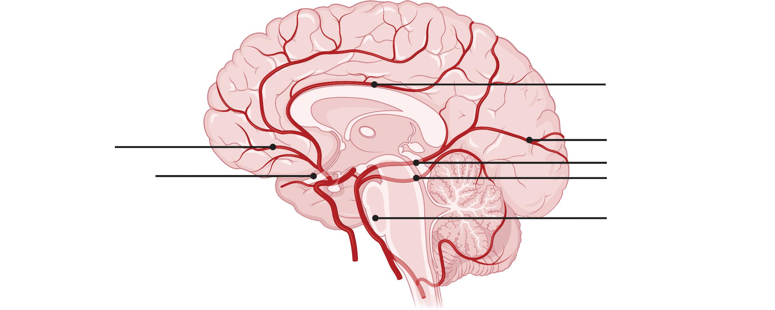
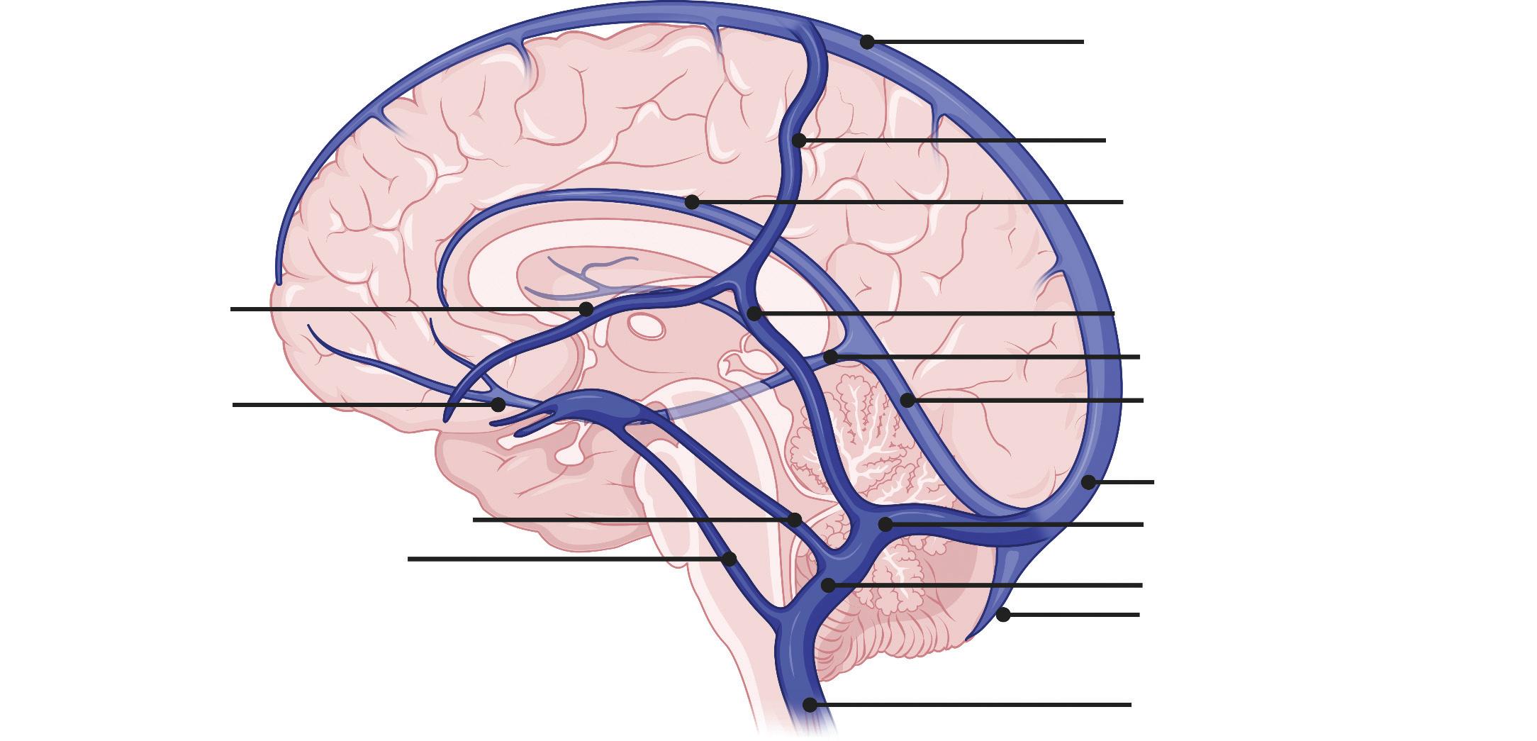
Thesuperficialsystemofveinsdrainingthecerebralcortex includethesuperiorcerebralveins,middlecerebralveins, inferiorcerebralveins,superiorandinferioranastomotic veins.Thesuperficialveinsdrainintothesuperiorand inferiorsagittalsinuses(Figure1.4).
Thedeepveinsincludesubependymalveins,thegreat cerebralvein,andmedullaryveins.Thedeepveinsdrain intothegreatcerebralveinandthentothestraightor transversesinuses.
Theduralvenoussinusesarevalvelessstructures foundbetweentheperiostealandmeningeallayerofthe duramateranddrainintotheinternaljugularvein.The straight,superior,andinferiorsagittalsinusesarefound inthefalxcerebrioftheduramaterandconvergeatthe confluenceofsinuses.Fromherethetransversesinus, locatedbilaterallyinthetentoriumcerebelli,curvesinto thesigmoidsinusandthentotheinternaljugularvein, whichexitsatthejugularforamen.Thegreatcerebral veinandtheinferiorsagittalsinuscontinueasthestraight sinus.
Thecavernoussinusisclinicallyrelevantduetoits vulnerabilityasasiteofinfection.Itcontainstheinternal
Figure1.3 CircleofWillis.
Orbital branches
Anterior cerebral artery
Pericallosal artery
Calcarine artery
Posterior cerebral artery
Superior cerebellar artery
Basilar artery
Anterior cerebral artery
Middle cerebral artery
Anterior temporal artery
Basilar artery
Vertebral artery
(c)
Superficial middle cerebral vein
Anterior cerebral vein
Superior petrosal sinus
Inferior petrosal sinus
Precentral artery
Central artery
Postcentral artery
Angular artery
Temporo-occipital artery
Superior cerebellar artery
Anterior inferior cerebellar artery
Posterior inferior cerebellar artery
Superior sagittal sinus
Superior anastomotic vein
Inferior sagittal sinus
Inferior anastomotic vein
Great cerebral vein
Straight sinus
Confluence of sinuses
Transverse sinus
Sigmoid sinus
Occipital sinus
Internal jugular vein
Figure1.4 Arterialsupplytothebrainandvenousdrainage.
carotidartery,abducensnerve,oculomotornerve,trochlearnerve,andtheophthalmicandmaxillarybranchesof thetrigeminalnerve.Thesinusisconnectedtothevalvelessfacialveinviathesuperiorophthalmicvein,allowing
reverseblood flowtothesinus,andistherefore apotentialrouteofintracranialinfection.Thecavernous sinusalsoreceivesvenousdrainagefromthecentralvein oftheretina,thesphenoparietalsinus,thesuperficial
(a)
(b)
middlecerebralvein,andthepterygoidplexus.These emptyintothesuperiorandinferiorpetrosalsinuses and,ultimately,intotheinternaljugularvein.Theleft andrightcavernoussinusesareconnectedinthemidline bytheanteriorandposteriorintercavernoussinuses.
1.2.3LymphaticsoftheBrain
Ithasbeenalongstandingbeliefthatthevertebrate braindoesnotcontainaclassiclymphaticdrainage system.Instead,itwasthoughtthebrainhas a “glymphaticsystem,” aperivascularpathwaythat allowscerebrospinal fl uid(CSF)torecirculatethrough thebraininterstitiumalongperivascularspacesnearthe cerebralarteries.Thispathwaywasalsothoughttobe responsiblefortheclearance ofinterstitialsolutesalong perivascularchannelsneartheveins,mediatedbyastroglialwaterchannels.However,recent fi ndingshaveprovidedevidenceofafunctionallymphaticsysteminthe brain,whichactstotransportinterstitial fl uid(ISF)and
(a)
solutesfromtheparenchymatothedeepcervicallymph nodes.Thelymphaticsystemalsohelpsmaintainthe waterandionbalanceoftheISFandallowscommunicationwiththeimmunesystem.Thelymphaticvessels areassociatedwiththesuperiorsagittalandtransverse sinusandtheduralmiddlemeningealarteries.Itislikely thatthelymphaticsystemworksinconjunctionwith othere ffl uxpathways,suchasviatheduralarachnoid granulations.
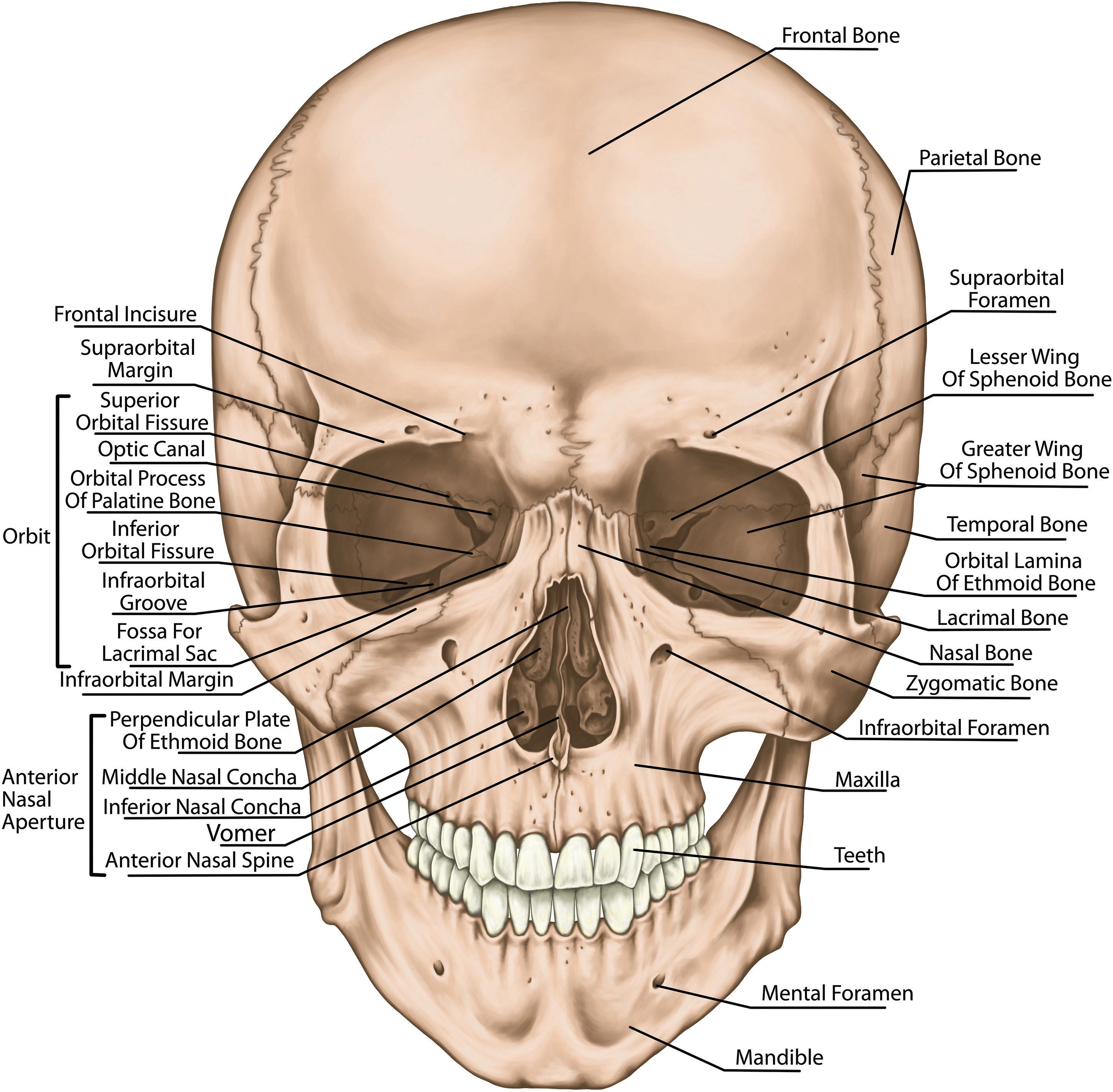
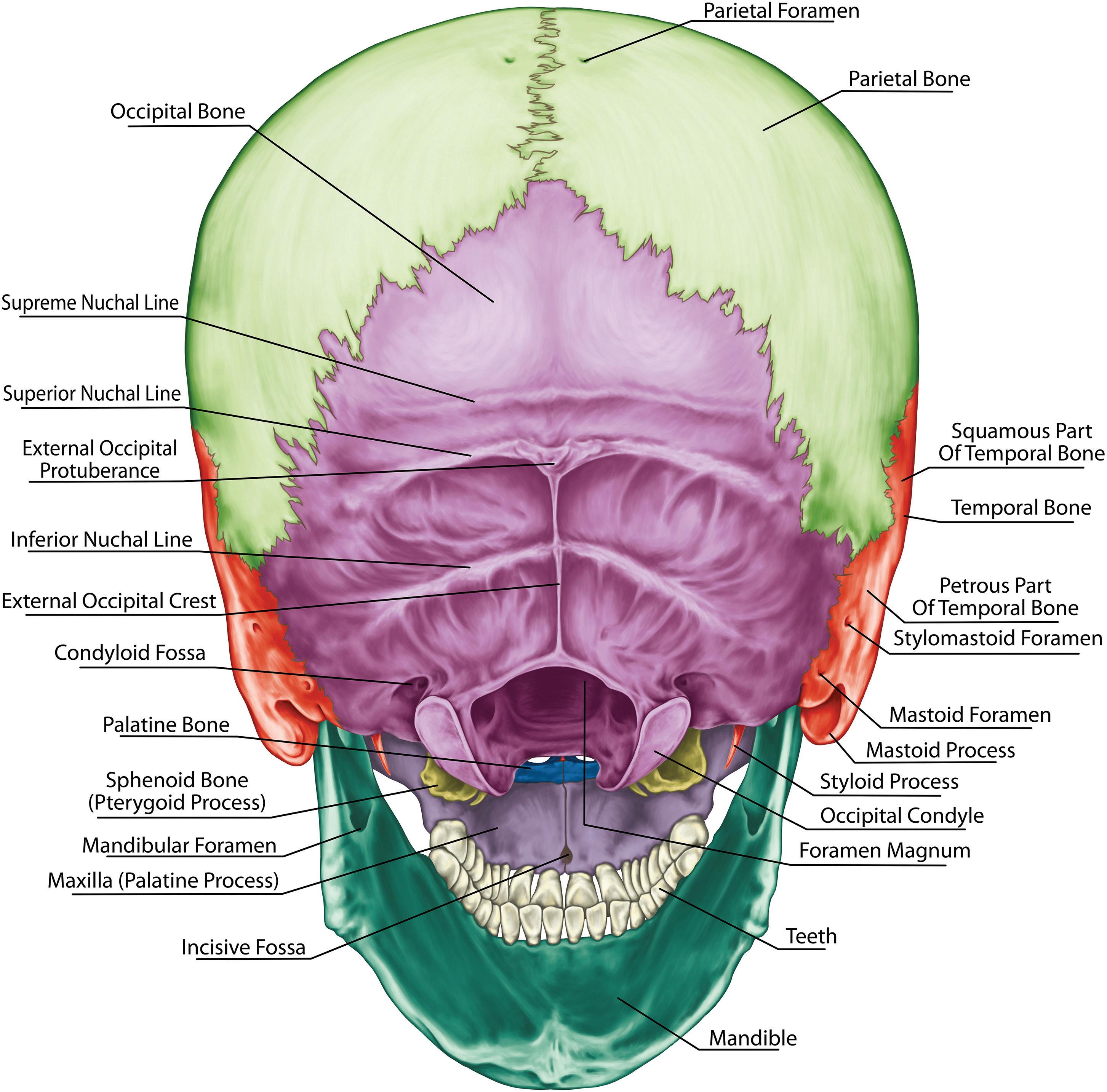
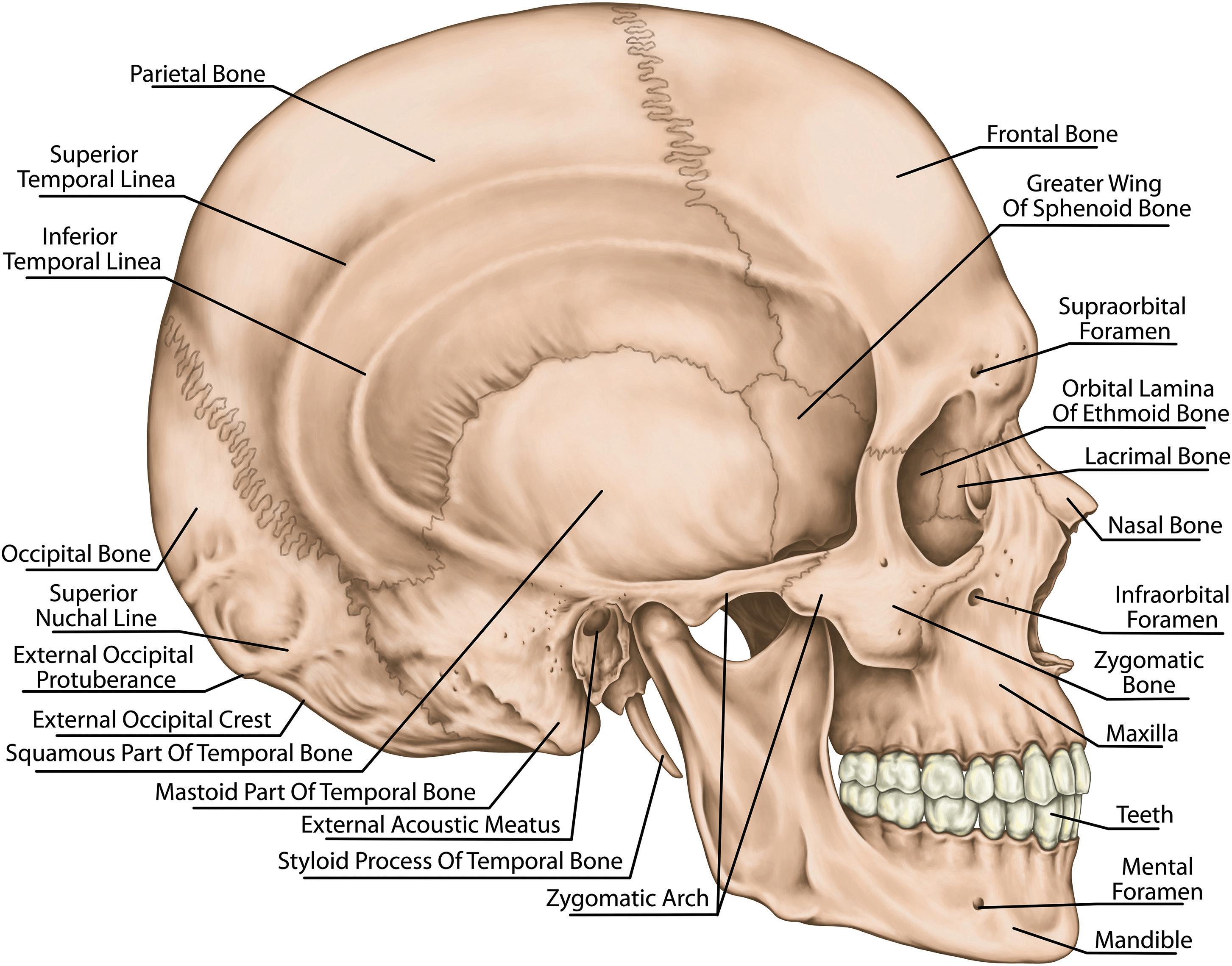
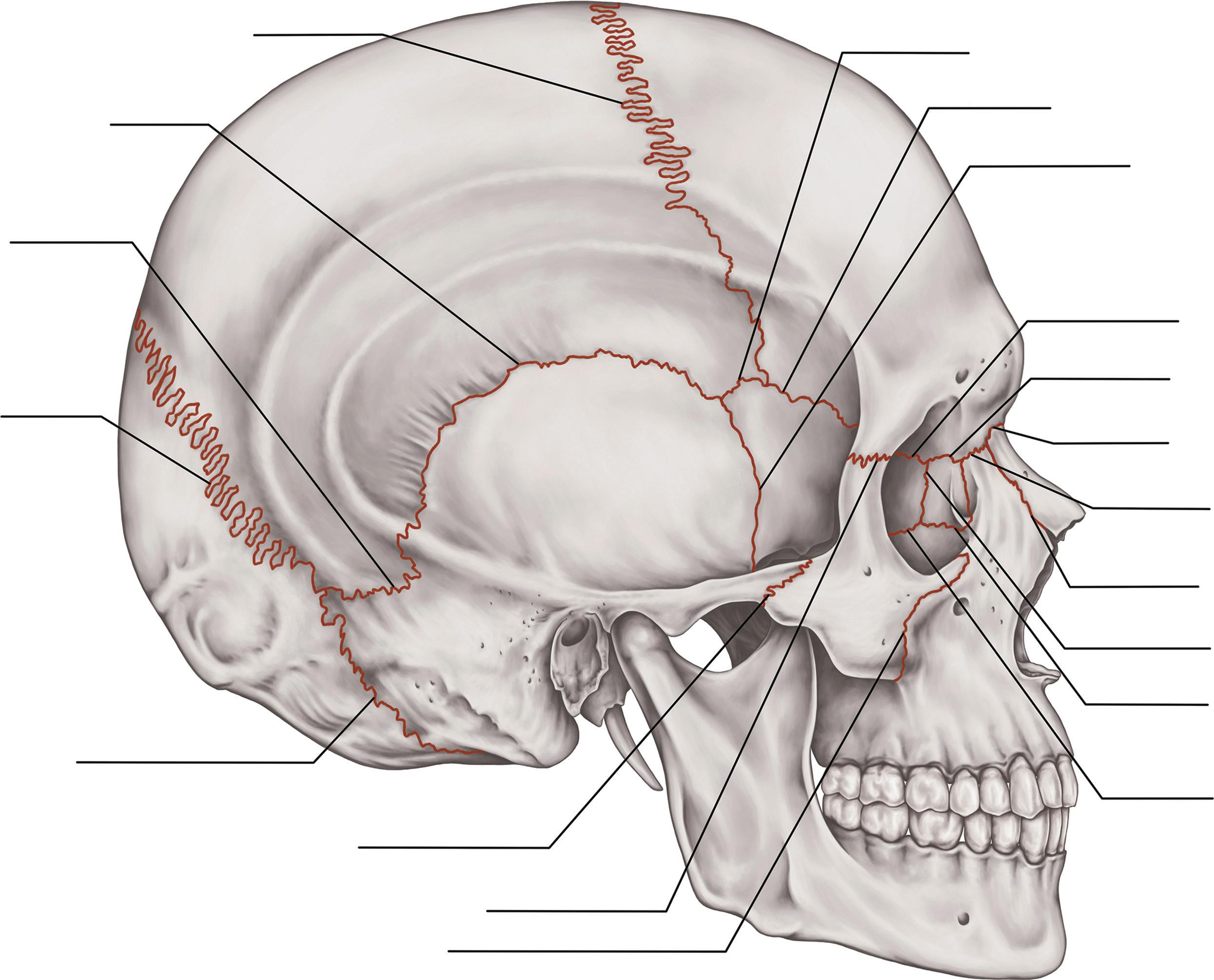
1.3BonesoftheSkull
Theskullisasupportiveprotectionforthebrainandis formedbyintramembranousossificationofcranialand facialbones.Thecalvariumistheroofofthecraniumand iscomprisedofthefrontal,occipital,andparietalbones (Figure1.5).Thebaseofthecraniumiscomprisedofthe ethmoid,occipital,temporal,parietal,frontal,andsphenoidbones.Thejunctionbetweenthelatterfourbonesis knownasthepterionandisofclinicalrelevancebecause
Figure1.5 Theskullandfacial skeleton.
(b) Figure1.5 (cont.)
(c)
Zygomatic Process (Maxilla)
Pterygoid Process (Sphenoid Bone)
Zygomatic Process (Temporal Bone)
Petrous Portion (Temporal Bone)
Mandibular Fossa (Temporal Bone)
Styloid Process (Temporal Bone)
Squamous Portion (Temporal Bone)
Tympanic Portion (Temporal Bone)
Mastoid Process (Temporal Bone)
Occipital Condyle (Occipital Bone)
External Occipital Protuberance (Occipital Bone)
Palatine Bone
Zygomatic Bone
Frontal Bone
Sphenoid Bone
Inferior Nasal Concha Vomer
Temporal Bone
Foramen Magnum
Parietal Bone
Occipital Bone
(e) Palatine Process (Maxilla)
Maxilla
Nasal bone
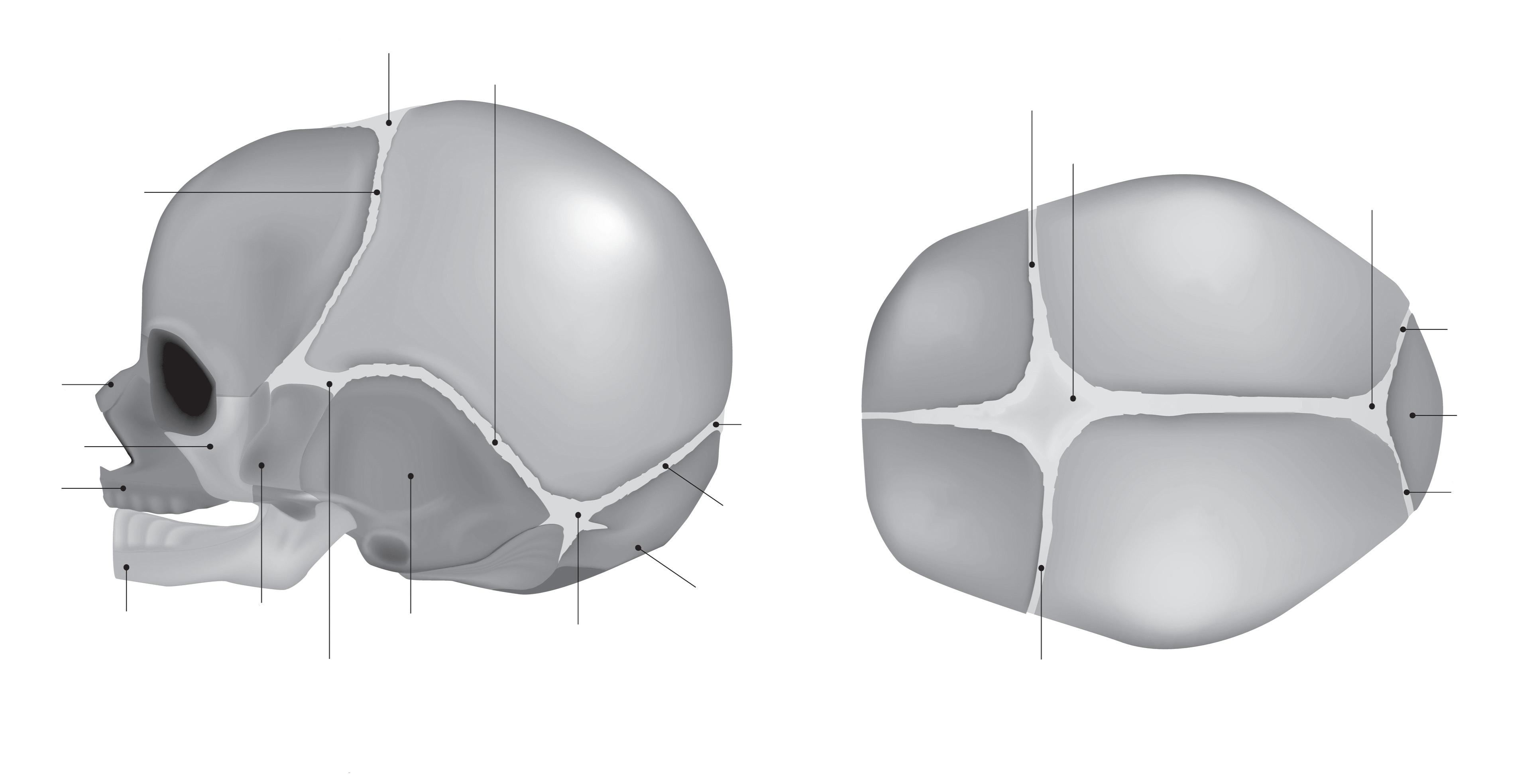
Coronal suture
Anterior fontanelle
Coronal suture Coronal suture
Temporal bone Sphenoidal fontanelle
Lateral view
Posterior fontanelle
Lambdoid suture
Occipital bone
Lambdoid suture
Coronal Suture
Squamosal Suture
Parietomastoid Suture
Lambdoidal Suture
Occipitomastoid Suture
Zygomaticomaxillary Suture Mastoid
Temporozygomatic Suture
Frontozygomatic Suture
Superior view
Sphenoparietal Suture
Sphenofrontal Suture
Sphenosquamosal Suture
Frontoethmoidal Suture
Frontolacrimal Suture
Frontonasal Suture
Frontomaxillary Suture
Nasomaxillary Suture
Ethmoidolacrimal Suture
Maxillolacrimal Suture
Ethmoidomaxillary Suture
itoverliesthemiddlemeningealartery,whichcanrupturefollowingfracturestothisregion,leadingtothe formationofextraduralhematomas.
Cranialfracturesmaybeaccompaniedbyfacialbone fracturesandshouldbesoughtforwhenassessingthe traumapatient.Themostcommonfacialfractures includethoseofthenasalbone,maxilla,mandible,and
zygomaticarch.Otherareaspronetodamagearethe sutures,andtheseincludethecoronal,sagittal,and lambdoidsutures(Figure1.6).Inneonates,incompletelyfusedsuturesgiverisetofontanelles – thefrontal fontanellebetweenthecoronalandsagittalsuturesand theoccipitalfontanellebetweenthesagittalandlambdoidsutures. Anterior fontanelle
Posterior
Zygomatic bone
Figure1.6 Suturesofthebrain.
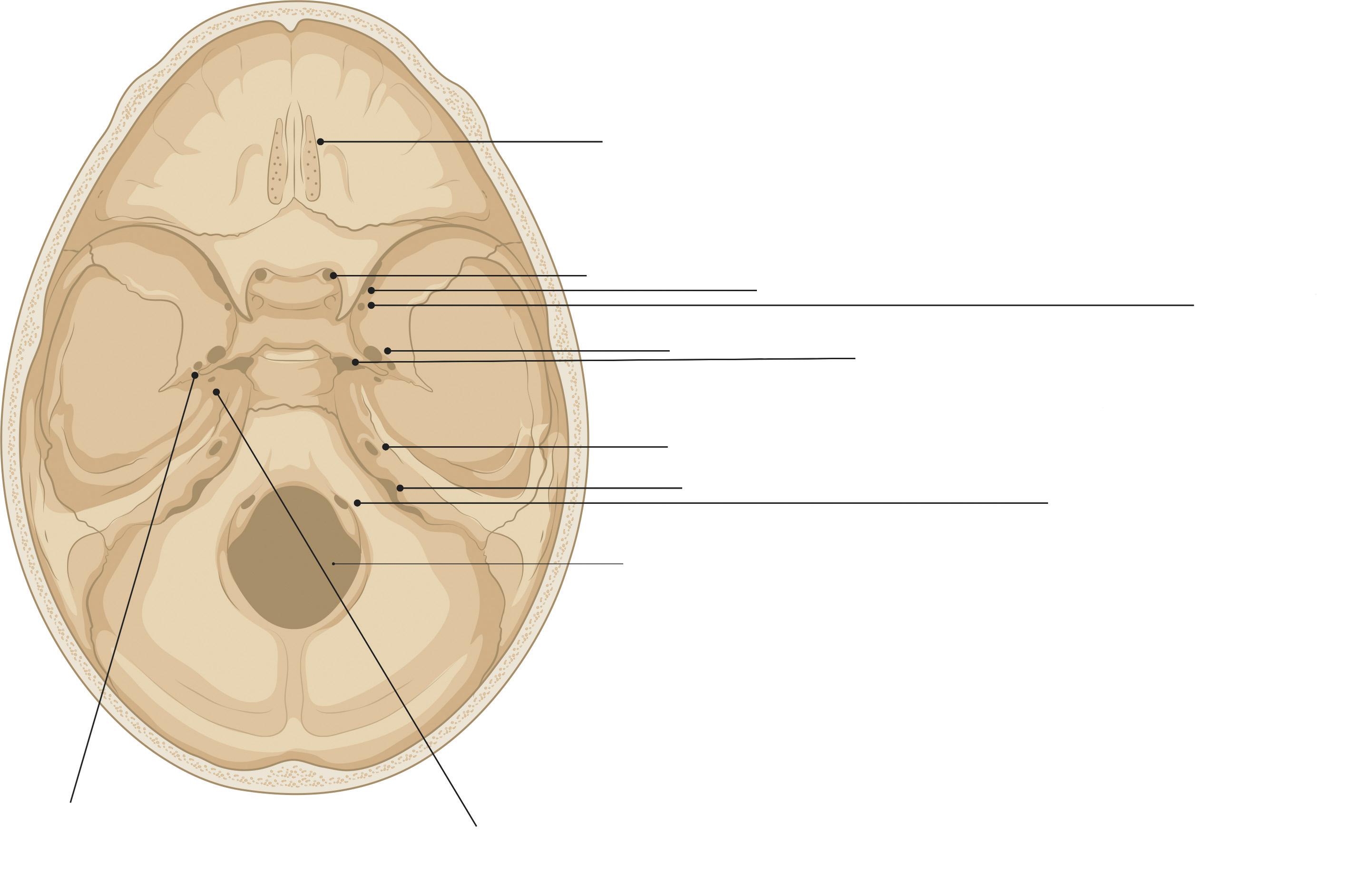
Cribriform
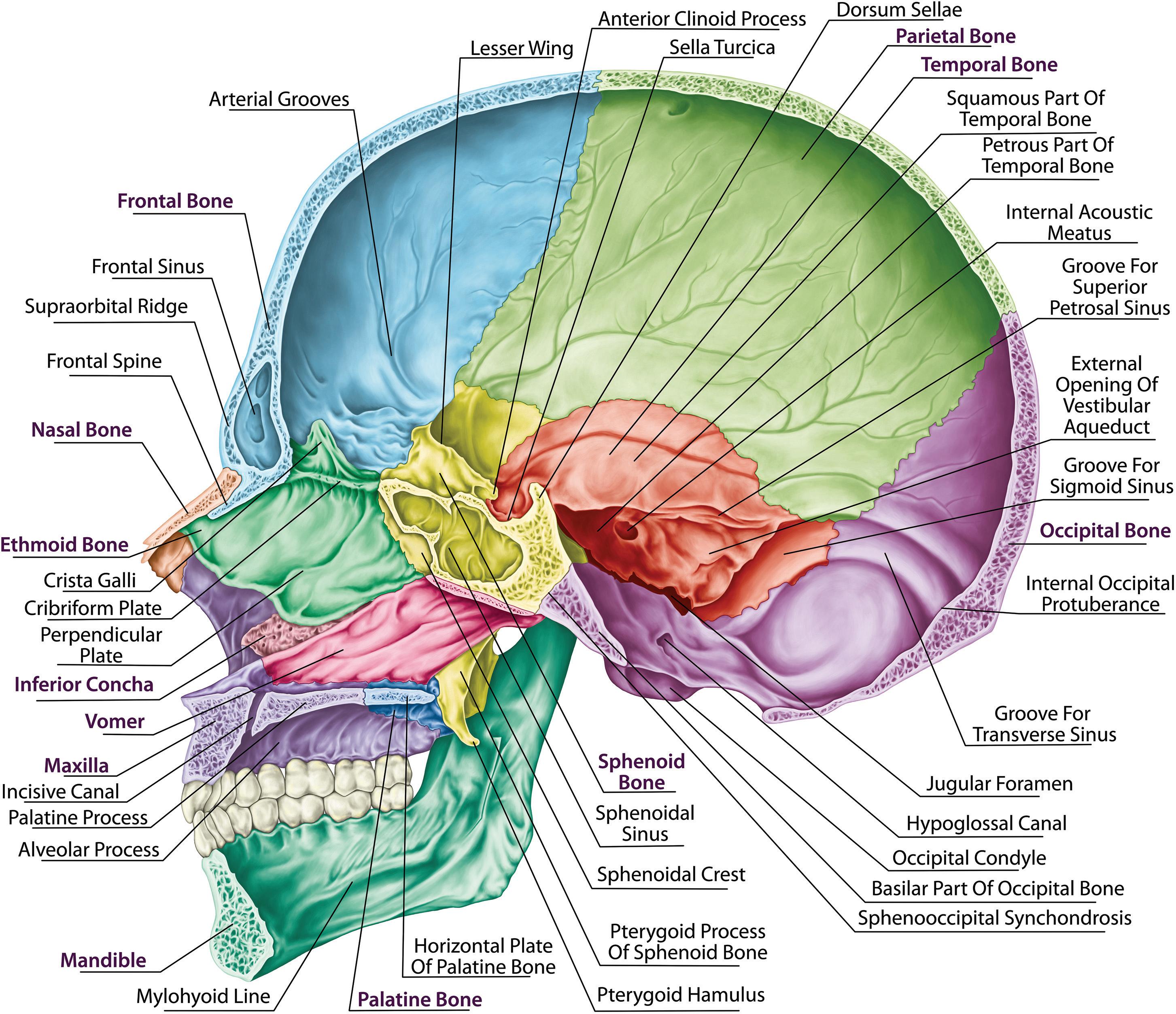
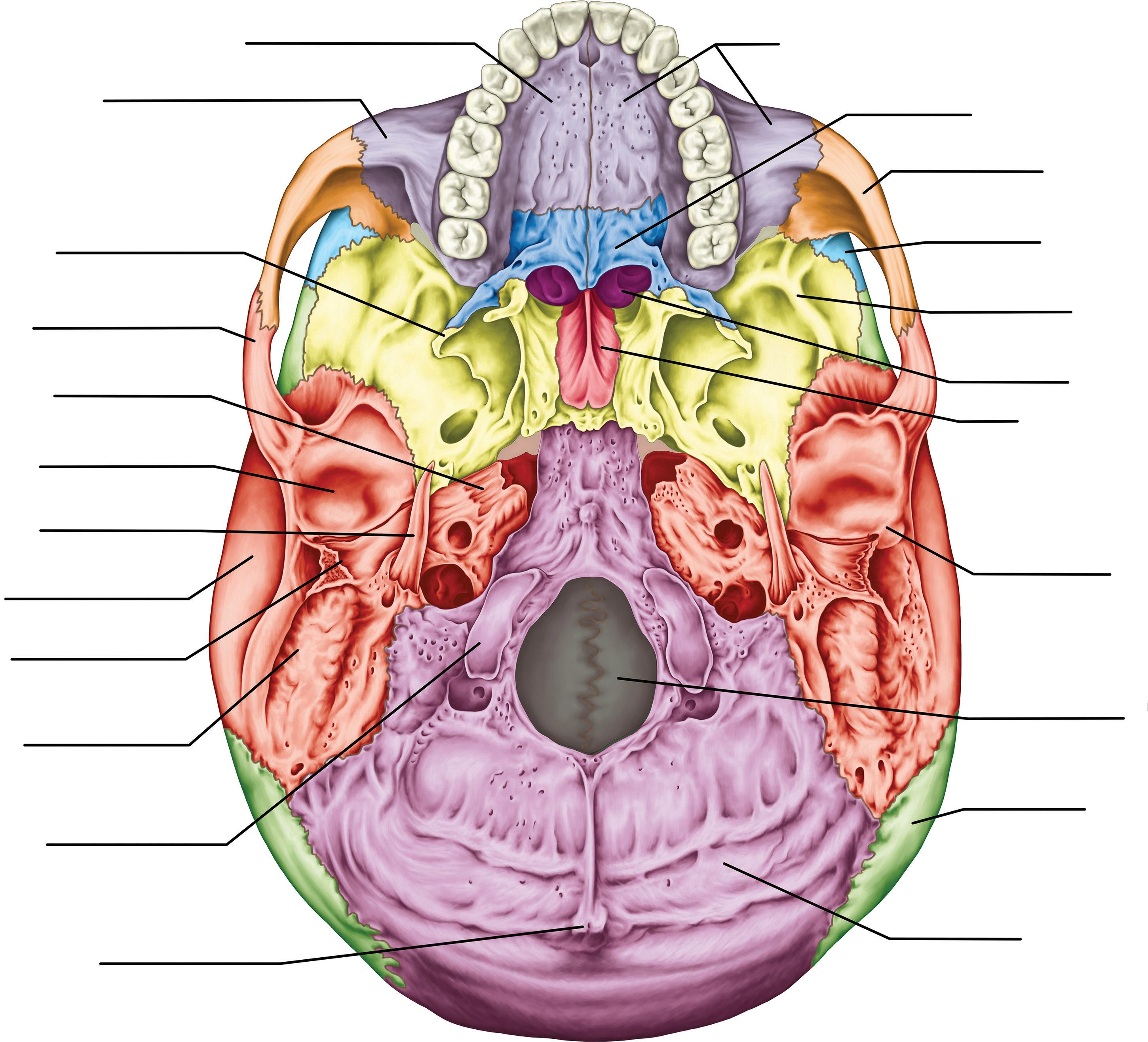
1.4TheCranialFossae
Thecranialcavityisdividedintothreeregionsknownas fossae.Theanteriorcranialfossaoverliesthenasaland orbitalregionsandaccommodatespartsofthefrontal lobe.Itismadeupofthefrontal,ethmoid,andsphenoid bones.Thefrontalcrestonthefrontalboneandthecrista gallioftheethmoidbonearethesitesofattachmentfor apartoftheduramaterthatdividesthecerebralhemispheres,knownasthefalxcerebri.Thecribriformplate supportingtheolfactorybulbislateraltothecristagalli, andcontainsaforamentransmittingtheolfactorynerve andtwoethmoidalforamina(anteriorandposterior, transmittingtheanteriorandposteriorethmoidalvessels andnerves,respectively).Theplateisverythinandcan fracturefollowingfacialtrauma,resultinginCSFrhinorrheaandanosmia.
Theanteriorfossaisseparatedfromthemiddlefossa bythelesserwingofthesphenoidbone.Theanterior clinoidprocessesofthesebonesarethesiteofattachment forthetentoriumcerebelli(duramaterdividingthecerebrumandcerebellum).Themiddlecranialfossaconsists ofthesphenoidandtemporalbones.Withinthecentral
partofthefossaisthesellaturcica,abonyprominence whichsupportsthepituitaryglandwithinthehypophysialfossa.Theposteriorwallofthesellaturcicaisformed bythedorsalsellae,whichseparatethemiddlecranial fossafromtheposteriorcranialfossa.Thelateralpartsof themiddlecranialfossaareformedbythegreaterwings ofthesphenoidboneandthesquamousandpetrousparts ofthetemporalbonesandprovidestructuralsupportto thetemporallobes.Boththesphenoidandtemporal bonescontainnumerousforaminafortransmittingvesselsandnerves.Theposteriorcranialfossaiscomprised oftheoccipitalboneandthetemporalbonesandcontains thebrainstemandcerebellum.Theforaminaoftheskull aremostconsideredinthecontextofthecranialnerves andareshowninFigure1.7.
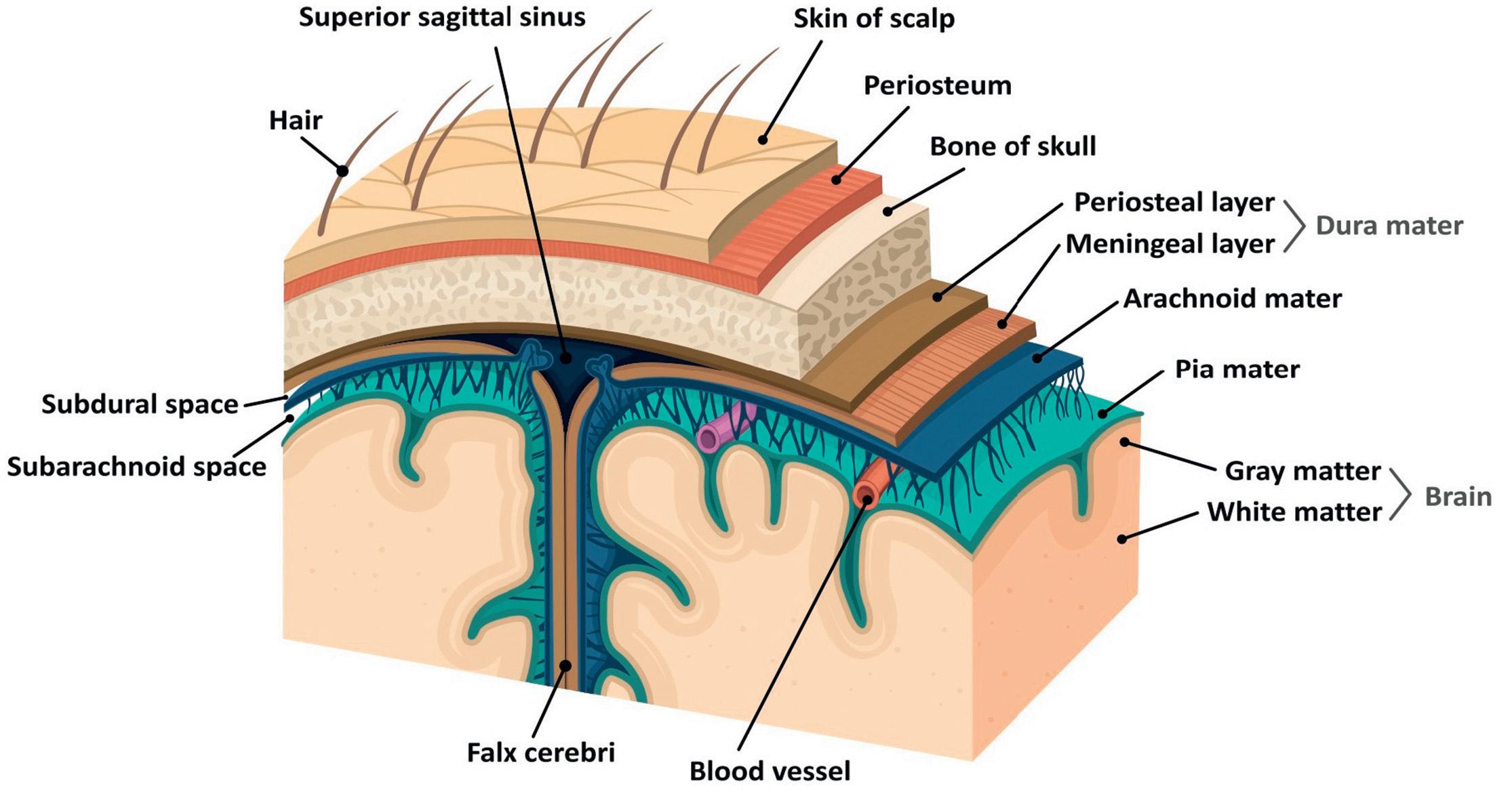
1.5LayersoftheScalp
Thescalpcontainsseverallayers,andtheseinclude skin,denseconnectivetissue,epicranialaponeurosis, looseareolarconnectivetissue,andtheperiosteum (Figure1.8).Thescalpissuppliedbybranchesofthe externalcarotidartery(superficialtemporal,posterior
plate (CNI)
Optic canal (CNII) Superior orbital fissure (CNIII, CNIV, CNV1, CNVI) F. Rotundum (CNV2)
F. lacerum (artery & nerve of pterygoid canal, greater & deep petrosal nerve, meningeal branches of ascending pharyngeal artery, emissary veins)
F. Ovale (CNV3)
Internal acoustic meatus (CNVII, CNVIII) Jugular foramen (CNIX, CNX, CNXI) Hypoglossal canal (CNXII)
Foramen magnum (medulla oblongata, vertebral artery, accessory nerves, spinal arteries)
F. spinosum (middle meningeal artery & vein, meningeal branch of mandibular nerve)
Carotid canal (internal carotid artery, carotid plexus)
Figure1.7 Superiorviewoftheskullbaseshowingtheforamina.
Layersofthescalp.
auricular,andoccipitalarteries)andtheophthalmic artery(supraorbitalandsupratrochleararteries). Venousdrainageincludesasuperficialsystemfollowing thearterialsupply(superficialtemporal,occipital,posteriorauricular,supraorbital,andsupratrochlearveins). Thetemporalregionoftheskullisdrainedbythepterygoidvenousplexus,whichdrainsintothemaxillary vein.Thescalpveinsconnecttothediploicveinsofthe skullviavalvelessemissaryveins,allowingaconnection betweenthescalpandtheduralvenoussinuses.The nervesinnervatingthescalpincludethetrigeminal nervebranches(supratrochlear,supraorbital,zygomaticotemporal,andauriculotemporalnerves).Thecervical nervebranchessupplyingthescalpincludethelesser occipitalnerve,thegreateroccipitalnerve,thegreat auricularnerve,andthethirdoccipitalnerve.
1.6Meninges
Thebrainandspinalcordarecoveredwithmembranous layerscalledthemeninges.Fromoutertoinnertheseare thedura,arachnoid,andpiamater.
1.6.1DuraMater
Thislayerisfoundunderneaththebonesandconsistsof theouterperiosteallayerandtheinnermeningeallayer
andinbetweenhousestheduralvenoussinuses.The meningeallayerfoldsinwardtoformtheduralreflections andformscompartments.Thesecompartmentsarethe falxcerebri,thetentoriumcerebelli,thefalxcerebelli (separatesthecerebellarhemispheres),andthediaphragmasellae(allowsthepassageofthepituitary stalk).Bloodcollectinginbetweentheskullandthe outerperiosteallayerisknownasanextraduralhematomaandusuallyoccursduetodamagetothemiddle meningealartery.Asubduralhematomaoccursdueto damageofthecerebralveinsbetweentheduraandthe arachnoidmater.
1.6.2ArachnoidMater
Thearachnoidmaterconsistsofavascularconnective tissue.Belowitliesthesubarachnoidspacecontaining CSF,whichre-entersthecirculationviatheduralvenous sinusesthroughsmallprojectionscalledarachnoid granulations.
1.6.3PiaMater
Thisisahighlyvascularizedlayerthatadherestothe brainsurfaceandfollowsthecontoursofthebraininto thegyriofthecerebralhemispheresandthefoliaofthe
Figure1.8
cerebellum.Thepiamaterandarachnoidmaterare joinedbyconnectivetissueinthesubarachnoidspace andtogetherareknownasleptomeninges.Thereare compartmentswherethetwoarenotincloseapproximation,resultinginnaturallyenlargedCSF filledpools calledthesubarachnoidcisterns.
1.7OrganizationoftheSympathetic andParasympatheticNervousSystems
Theautonomicnervoussystemiscomposedofthesympatheticnervoussystem(SNS)andtheparasympathetic nervoussystem(PNS)andactstoregulatethebody’ s unconsciousactions.TheSNSstimulatestheso-called “fightor flight” responseandthePNSisinvolvedin “restanddigest” responses(Box1.1).
Box1.1
OrganParasympatheticResponse
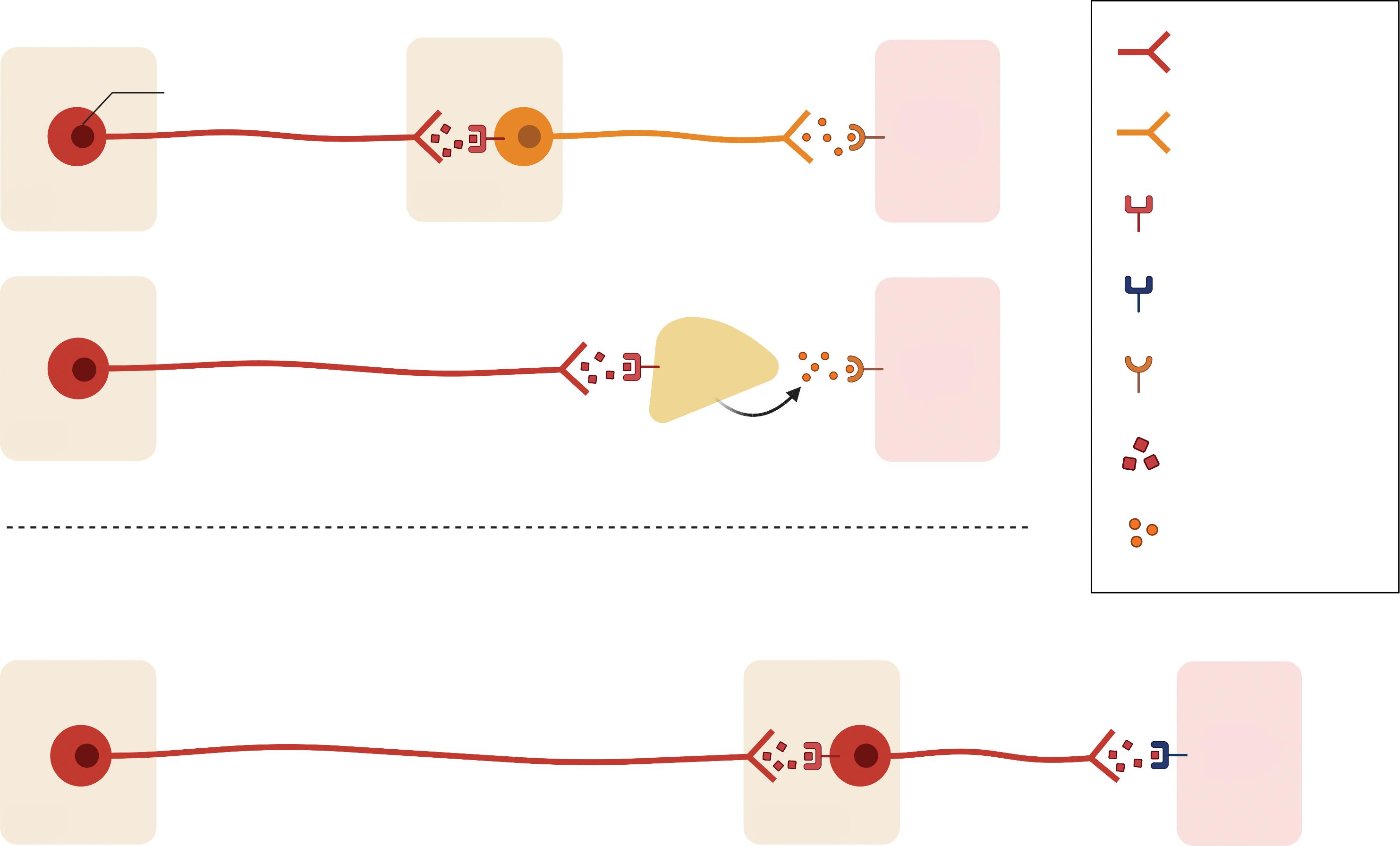
AmajoranatomicalcomponentoftheSNSareapair ofnerve fibersthatspantheskulltococcyx,knownasthe sympatheticchains.Therearetwotypesofneurons involvedinsympatheticsignaltransmissionandthese aretheshortpreganglionicneuronsthatoriginatefrom T1toL2–L3,whichsynapsewithpostganglionicneurons thatextendstotherestofthebody.Atthesynapses, thepreganglionicneuronsreleaseacetylcholine,which activatesthenicotinicacetylcholinereceptorsinthepostganglionicneuronstoreleasenorepinephrine,andthese subsequentlybindtoadrenergicreceptorsinthetarget tissueleadingtosympatheticeffects(Figure1.9).The postganglionicneuronsofsweatglandsreleaseacetylcholinetoactivatemuscarinicreceptors.Thechromaffincells oftheadrenalmedullaactasapostganglionicneuronand releasenorepinephrineandepinephrine.
Iris Miosisandaccommodationduetoconstrictionofthe sphinctermusclesviatheshortciliarynervesoriginating fromtheEdinger–WestphalnucleusofcranialnerveIII
Salivary glands
Lacrimal glands
IncreasedwaterysecretionviacranialnervesIX(parotid gland)andchordatympaniofVII(submandibularand sublingualglands)leadingtoacetylcholinereleaseonto M3muscarinicreceptors
Increasedsecretion(preganglionic fibersreachthe pterygopalatineganglionviathegreaterpetrosalnerve andthenerveofthepterygoidcanaltosynapsewith postganglionic fibers)
Heart Negativechronotopy,inotropyandreducedconduction velocityandcoronaryarteryvasoconstrictionviathevagus nerve
Lung Bronchialmusclecontraction(vagusnerve)
StomachIncreasedperistalsisandmotilityandpyloricsphincter relaxationallowinggastricemptyingviathevagusnerve
GallbladderContraction(vagusnerve)
Internal urethral sphincter
Detrusor muscleof bladder
Relaxation
Contraction(viapelvicsplanchnicnervestoallowbladder emptying)
Penis Erection(pelvicnerve)
Adrenal medulla
SympatheticResponse
Pupildilation(adrenergicinnervationtothedilator pupillaemuscleviathelongciliarynerves,arisingfromthe superiorcervicalganglion)
Reducedsalivasecretion(innervationvia fibersarisingfrom thesuperiorcervicalganglionresultinginnorepinephrine releaseactingonalpha-andbeta-adrenergicreceptors)
Reducedsecretion(innervationvia fibersoriginatinginthe superiorcervicalganglion,whichreachthe pterygopalatineganglionviatheinternalcarotidplexus andthedeeppetrosalnerve)
Positivechronotropy,inotropyandincreasedconduction velocityviathecardiacnervesfromthelowercervicaland upperthoracicganglia
Bronchialmusclerelaxation(thoracicsympatheticganglia)
Reducedgastricmotilityandperistalsisandpyloric sphincterconstrictionpreventinggastricemptyingviathe celiacplexus(T5–T12)
Relaxation(T7–T9throughtheceliacplexus)
Constriction
Relaxation(viasympatheticbranchesfromtheinferior hypogastricplexustoallowbladder filling)
Ejaculation(peristalticcontractionofvasdeferens,seminal vesicles,andprostaticsmoothmusclesviathehypogastric nerveandejaculationviathepudendalnerve)
Norepinephrineandepinephrinesecretion
Parasympathetic nervous system
Sympatheticandparasympathetic fibers.CNS;centralnervoussystem.
Sympatheticnervesariseinthespinalcordinthe intermediolateralnucleusofthelateralgraycolumn. Axonsleavethespinalcordthroughtheanteriorroot andpassnearthesensorygangliontoentertheanterior ramiofspinalnerves.Theaxonsterminateattheparavertebralorprevertebralganglia.Themainprevertebral ganglia(celiac,mesentericandaorticorenalganglia)are locatedanteriortotheaortaandvertebralcolumnand receivepreganglionicaxonsviathesplanchnicnerves. Theparavertebralgangliaarelocatedbilaterallyventrolateraltothevertebralcolumn.Therearethreeparavertebralgangliaandthesearethesuperiorcervical ganglion,middlecervicalganglionandinferiorcervical ganglion.
1.7.1SympatheticGangliaSupplying theHeadandNeck:CervicalGanglia
Therearethreecervicalgangliathatsupplytheheadand neck(thesuperior,middle,andinferiorcervicalganglia). Thesuperiorganglionisfoundposteriortothecarotid arteryandgivesrisetoanumberofpostganglionic nerves:theinternalandexternalcarotidnerves,the
Cholinergic fiber
Adrenergic fiber
Nicotinic cholinergic receptor
Muscarinic cholinergic receptor
Adrenergic cholinergic receptor
Acetylcholine
Norepinephrine/ epinephrine
nervetothepharyngealplexus,thesuperiorcardiac branch,andthegrayramicommunicantes.Themiddle ganglionmaybeabsent,butwhenpresentisfoundanteriortotheinferiorthyroidartery.Itspostganglionic fibers arethegrayramicommunicantes,thethyroidbranches, andthemiddlecardiacbranch.Theinferiorcervical ganglionissituatedanteriorlytotheC7vertebra.Its branchesarethegrayramicommunicantes,branchesto thesubclavianandvertebralarteries,andtheinferior cardiacnerve.Damagetosympathetic fibersenrouteto theheadandneckcanleadtoHorner’ssyndrome,a conditionpresentingwithpartialptosisoftheupper eyelid,miosis(constrictedpupil)andhemi-facialanhidrosis(absenceofsweating).
1.7.2ParasympatheticGangliaSupplying theHeadandNeck
Theparasympathetic fiberssupplyingtheheadandneck arefoundinfourbrainstemnucleiassociatedwithacranial nerve.Theysynapseinaperipheralganglionnearthe targetviscera.Therearefourparasympatheticganglia locatedwithinthehead –ciliary,otic,pterygopalatine,and
Figure1.9
submandibular.Theyreceive fibersfromtheoculomotor, facial,andglossopharyngealnerves(thevagusnerveonly innervatesstructuresinthethoraxandabdomen).
Theciliaryganglionislocatedwithinthebonyorbit. Itspreganglionic fibersarefromtheEdinger–Westphal nucleus,associatedwiththeoculomotornerve.Itspostganglionic fibersleavetheganglionviatheshortciliary nervestoinnervatethesphincterpupillaeandtheciliary muscles.Sympatheticnervesfromtheinternalcarotid plexusandsensory fibersfromthenasocilarynervepass throughtheganglionwithoutsynapsing.
Thepterygopalatineganglionislocatedwithinthe pterygopalatinefossaandissuppliedby fibersfromthe superiorsalivatorynucleus(associatedwiththefacial nerve).Itspostganglionic fibersjoinbranchesofthemaxillarynervetosupplythelacrimalgland,thenasopharynx,andthepalate.
Sympathetic fibersfromtheinternalcarotidplexus andsensorybranchesfromthemaxillarynervepass throughthepterygopalatineganglionwithoutsynapsing.
Thesubmandibularganglionislocatedinferiorlyto thelingualnerveandissuppliedby fibersfromthe superiorsalivatorynucleus.These fibersarecarried withinabranchofthefacialnerve,thechordatympani. Thisnervetravelsalongthelingualbranchofthemandibularnervetoreachtheganglionandleavesthegangliontothesubmandibularandsublingualglands.
Sympathetic fibersfromthefacialarteryplexuspass throughthesubmandibularganglion.Theyarethought toinnervateglandsinthebaseoftheoralcavity.
Theoticganglionislocatedinferiorlytotheforamen ovalewithintheinfratemporalfossa.Itismedialtothe mandibularbranchofthetrigeminalnerve.Theganglionissuppliedby fi bersfromtheinferiorsalivatory nucleus(associatedwiththeglossopharyngealnerve).
Parasympathetic fi berstravelwithinthelesserpetrosal nerve,abranchoftheglossopharyngealnerve,toreach theoticganglion.Theparasympathetic fi berstravel alongtheauriculotemporalnerve(abranchofthemandibulardivisionofthetrigeminalnerve)toprovide secretomotorinnervationtotheparotidgland.
Sympathetic fi bersfromthesuperiorcervicalchain passthroughtheoticganglion,wheretheytravelwith themiddlemeningealarterytoinnervatetheparotid gland.
1.8StructuresoftheBrain
Thenervoussystemformsduringthethirdweekof development.Atthecranialendoftheneuraltube,
threeexpansions(vesicles)develop:theforebrain(prosencephalon),themidbrain(mesencephalon),andthe hindbrain(rhombencephalon).Furtherdivisionseparatestheprosencephalonintothediencephalon(thalamusandhypothalamus)andthetelencephalon (cerebrum).Themesencephalonconsistsofthetectum, cerebralaqueduct,tegmentum,andthecerebralpeduncles.Therhomboencephalonconsistsofthepons,the medulla,andthecerebellum.Thecavitieswithinthe primarybrainvesiclesareprecursorsoftheventricular system.Thecaudalpartsoftheneuraltubeformthe spinalcord.
1.9Forebrain
Thestructuresintheforebrainincludethecerebralcortex andsubcorticalstructuresofthelimbicsystemincludingthe amygdala,hypothalamus,thalamus,hippocampus,basal ganglia,andcingulategyrus.
1.10Cerebrum
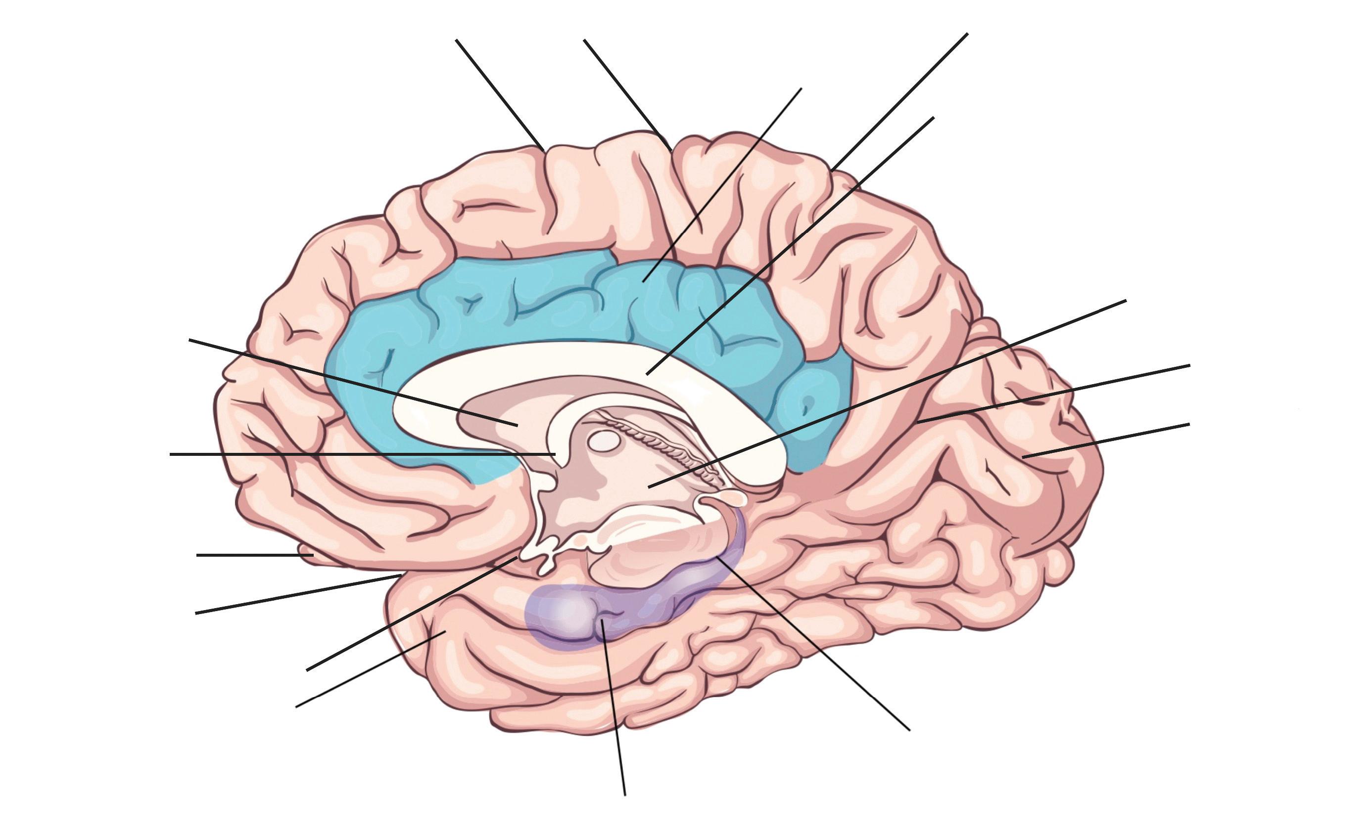
Thelargestpartofthebrainisthecerebrum,containing twohemispheresseparatedbythefalxcerebriofthedura mater.Thevisiblesurfaceofthecerebralhemisphereis afoldedsheetofneuraltissuecalledthecerebralcortex, characterizedbysulci(depressions)andgyri(elevations). Someofthelargerfoldsincludethelateralsulcus(also knownastheSylvian fissure),whichseparatesthetemporallobefromthefrontalandparietallobes,thecentral sulcus(dividingthefrontalandparietallobes),andthe parieto-occipitalsulcus,whichseparatestheoccipitaland parietallobes(Figure1.10).Thetwocerebralhemispheres areconnectedbyawhite-mattertractcalledthecorpus callosum.Thetissueseparatingthetwohemispheresis calledthelongitudinal fissure.Theseptumpellucidum continuesfromthecorpuscallosumtothefornixand separatestheanteriorhornsofthelateralventricles.The calcarine fissureseparatestheoccipitallobeintotheinferiorlingualgyrusandthesuperiorcuneus.
Thecerebrumismadeupofgraymatter(containing cellbodiesanddendrites)andwhitematter(consistingof glialcellsandmyelinatedaxons).Thecerebrumcanbe dividedintofourlobes.Thefrontallobesubservedecision-makingandexecutivecontrol.Theparietallobeis vitalforsensoryperceptionandintegration.Theoccipital lobeisthevisuospatialprocessingareaofthebrainfor color,formandmotion.Thetemporallobecontains corticalareasthatprocessauditorystimuli,encodingof memoryandlanguagecomprehension.
1.10.1Brodmann’sMap
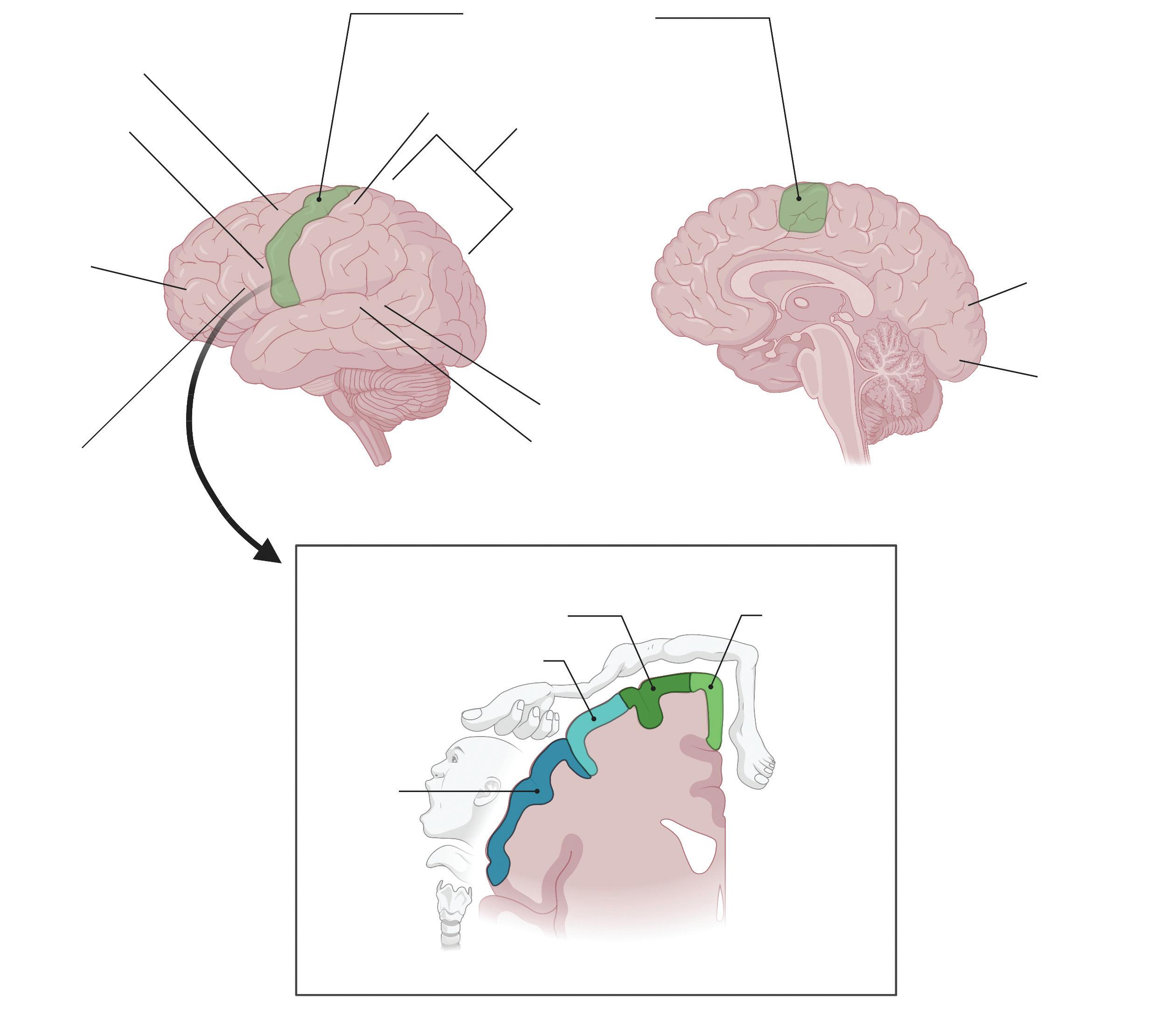
Thecerebralcortexcanalsobesubdividedinto52functionalregions,numberedbytheneuroanatomist KorbinianBrodmannbasedoncytologicalstructure. Theprimarymotorcortex(Brodmannarea4)isanterior tothecentralsulcus(Figure1.10).ItcontainslargeneuronscalledBetzcells,whichsendaxonstothespinalcord andisimportantforplanningandexecutionofmovements.Thetopographicmapofthemotorcortexis arrangedwithan “overrepresentation” ofneurons responsibleforcomplexmotorbehaviors(Figure1.11). Thepremotorcortex(Brodmannarea6)alsoplaysarole inplanningmovement;however,itsfunctionislesswell understood.Thesupplementarymotorarea(Brodmann area6)contributestothecontrolofmovement.The primarysomatosensorycortex(Brodmannareas3,2, and1)isfoundintheparietallobe,posteriortothe centralsulcus.Itprocessesafferentsomatosensoryinput andhelpsintegratesensoryandmotorinformation requiredforskilledmovement.Theprimaryvisualcortex (Brodmannarea17)isattheoccipitalpole.Theextra striatecortexisadjacenttothevisualcortexandprocesses specificfeaturesofvisualinformation.Itcanbeseparated
intotwostreams.Theventralstreamisfromtheprimary visualcortextothetemporallobeandisimportantfor patternandobjectrecognition.Thedorsalstreamfrom thestriatecortexintotheparietallobeisresponsiblefor spatialrecognitionofmotionandlocation.Theprimary auditorycortexisresponsibleforrecognitionofauditory stimuliandisinthelateraltemporallobe.Wernicke’ s area(Brodmannarea22)islocatedinthesuperiortemporalgyrusofthedominantcerebralhemi-sphere.Itis importantforcomprehensionofwrittenandspokenlanguage,andthereforeanydamagetothisarealeadsto fluentbutnonsensicalspeech.Broca’sarea(Brodmann areas44and45)islocatedinthedominantprefrontal cortexandisinvolvedinlanguageprocessingandspeech production.Lesionsinthisregionleadtoexpressive aphasia,wherethepatientretainscomprehensionbut cannotcreate fluentspeech.
1.10.2LayersofTheCerebralCortex
Thecerebralneocortexisarrangedinsixlayers.The outermostlayeristhemolecularlayer(layerI),containing fi bersthatrunparalleltothecorticalsurface withveryfewneurons.LayerIIistheoutergranular
Pre central sulcus
Post central sulcus
Thalamus
Parieto-occipital sulcus
Calcarine fissure
Cut edge of brainstem Hippocampus
Lateral sulcus
Olfactory bulb
Fornix Septum pellucidum
Optic chiasm Temporal lobe (medial surface)
Central sulcus Cingulate gyrus
Corpus callosum
Figure1.10 Structuresofthebrain(medialsurface).
Primary motor cortex
Primary somatosensory cortex
Posterior
Extrastriate cortex
Primary visual striate cortex
layer,containingsmall,roundedneurons.Belowthisis theouterpyramidallayer(layerIII),containingpyramidalneurons.Nextistheinnergranularlayer(layerIV), containingspinystellateandpyramidalcells.Itisa majorinputlayerandreceivesspeci fi csensoryinputs fromthalamocorticala ff erent fi bers.Theprimarysensorycortexhasawell-developedlayerIV,buttheoutputlayersarelesswelldeveloped.LayerIViswell developedintheprimaryvisualcortexandcontains thestriaofGennari,abandofmyelinatedaxonsthat runsparalleltothesurfaceofthecerebralcortex.The innerpyramidallater(layerV)containsmanyoutput neurons.Theprimarymotorcortexhasaveryenlarged layerVcontaininglargeBetzcells,whichsendaxonsto thecontralateralmotornucleiofcranialnervesandto
thelowermotorneuronsintheventralhornofthe spinalcord.Finally,layerVIistheinnermost,multiformlayercontainingmorph ologicallyheterogenous populationofneuronsandsendse ff erent fi berstothe thalamus.
Certainpartsofthecortexarearrangeddifferently: thehippocampushasthreelayersandthecingulategyrus hasfourto fivelayers.
1.10.3VascularSupply
Thebranchesoftheanterior,middle,andposteriorcerebralarteriesareresponsibleforthebloodsupplytothe cerebrum.Thevenousdrainageofthecerebrumisvia cerebralveinsthatemptyintotheduralvenoussinuses (Figure1.4).
Supplementary motor area
Premotor cortex
Prefrontal cortex
Broca's area
Lateral view
Coronal view
Mid-sagittal view
Wernicke's area
Primary auditory cortex
Trunk
Upper extremity
Lower extremity
Face
parietal cortex
Figure1.11 Topographicmapoftheprimarymotorcortex.
1.11Ventricles
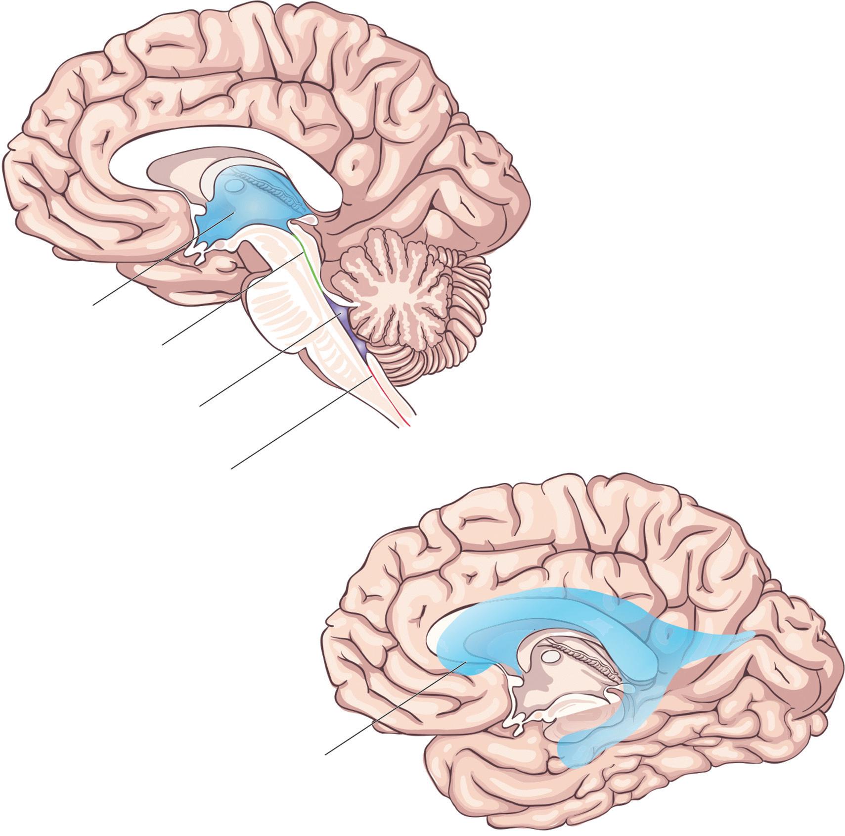
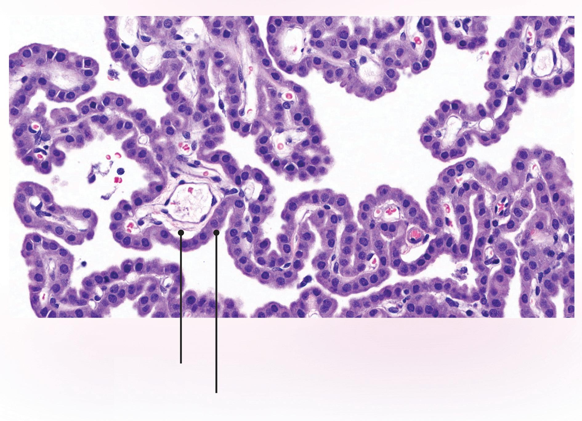
Theventricles(Figure1.12)arelinedbyependymalcells, ciliatedglialcells,whichformthechoroidplexus,astructurewhereCSFisproduced(Figure1.13).Thisprovides hydromechanicalprotectionbyactingasashock absorberandprovidingbuoyancy.
Theventricularsystemiscomposedoffourconnectingcavitiesderivedfromtheneuraltube.Therightand leftventriclesandthethirdventriclearepartofthe forebrain,whilethefourthventricleispartofthe hindbrain.
Duringdevelopment,the fluid-filledcavityoftheprimaryvesiclesbecomethelateralventricles.TheycommunicatewiththethirdventricleviatheforamenofMonro inthediencephalon.Thethirdventriclecommunicates withthefourthventricleinthehindbrainviathecerebral
Capillaries Ependymal cells
Figure1.13 Histologyofthechoroidplexus
Third ventricle
Cerebral aqueduct
Fourth ventricle
Spinal canal
Lateral ventricle (beneath overlying cortex)
Figure1.12 Theventricular system
aqueduct(ofSylvius),whichiscontinuouswiththecentralcanalofthespinalcord.Thefourthventricleisalso connectedtothesubarachnoidspacebyamedianaperture(theforamenofMagendie)andtwolateralapertures (theforaminaofLuschka).
1.12HigherAssociationAreas oftheCortex
Higherassociationareasofthecortexareinvolvedin complexprocessingofvarioussensorymodalities,cognitionandemotion.Theprefrontalandlimbicassociationareasareimportantforregulationofcognition, abstractreasoning,complexemotionsandself-awareness.Thecingulateandparahippocampalgyriare involvedinexpressionofemotionsandformationof memories.Thehippocampalformationisimportant fordeclarativememory.ItcontainstheCornu Ammonis(CA)regions,thesubiculum,andthedentate gyrus,andisfoundinthemedialtemporallobeinthe inferiorhornofthelateralventricle.Theamygdalais asubcorticalnucleusinthemedialtemporallobeandis connectedtotheorbitofrontalcortex,thehypothalamus,andthenucleusaccumbens.
1.13LimbicSystem
Thelimbicsystemisapartofthebraininvolvedin behavioralandemotionalresponses.Thereareseveral importantstructureswithinthelimbicsystemandthese includethebasalganglia,thalamus,hypothalamus,hippocampus,amygdala,andthecingulategyrus.
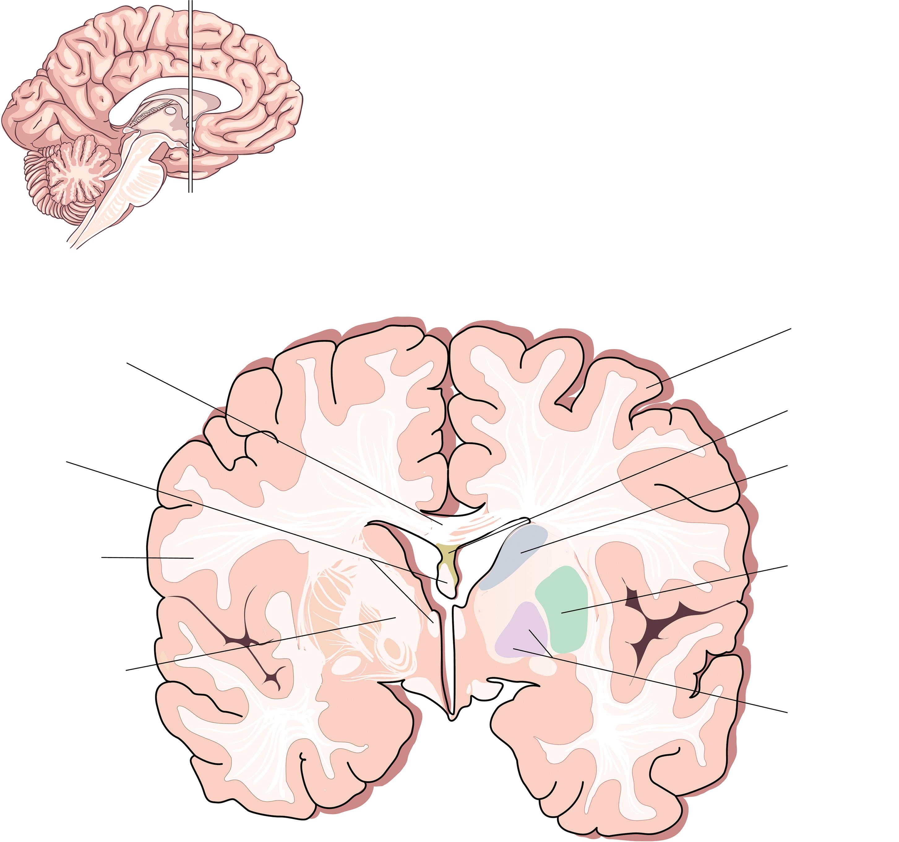
1.13.1BasalGanglia
Thebasalgangliaaresituatedatthebaseoftheforebrain andtopofthemidbrain.Theyareagroupofsubcortical nucleiconnectedtothecerebralcortex,thalamus,andthe brainstem.Thebasalgangliaareresponsibleforcontrol ofvoluntarymotormovements,procedurallearning,cognition,andemotion.Theycon-sistofseveraldistinct structures(Figure1.14)andtheseincludethecaudate nucleusandputamen(togetherknownastheneostriatum)separatedbytheinternalcapsule).Theinternal capsuleisthesiteofthepassageofmany fibersincluding theefferentcorticobulbar fibers,corticospinal fibers, efferentcorticopontine fibers,andafferentthalamocortical fibers.
Thecaudatereceivesinputsfromtheprefrontalcortex andtheputamenreceivesinputsfromthesensorimotor cortex.Theysendoutputstotheexternalandinternal
globuspallidusandthentothethalamusandcerebral cortex,formingasubcorticalloop.Theinternalsegment projectstothemotorareasofthethalamusandthe medialnucleusofthethalamus.Theexternalsegment projectstothesubthalamicnucleus(STN),whichalso projectstotheinternalglobuspallidus.
Theneostriatumalsoprojectstothesubstantianigra parcompacta(SNPC),whichcontainsdopaminergic neuronsandtotheparsreticulata,whichcontains mainlyGABAergicneuronsandisoneoftheoutput nucleiofthebasalgangliatothethalamus(alongside theinternalglobuspallidus)andplaysavitalrolein movementexecution.Theneostriatumissuppliedby smallbranchesofthemiddleandanteriorcerebral arteries.Theneostriatumandinternalcapsulearecommonlya ff ectedinstroke.Damagetotheinternalcapsule leadstocontralateralweakness.Themosta ff ected regionoftheinternalcapsuleisthegenu,wherecorticospinal fi berstothehead,neck,andpartoftheupper limbarelocated.
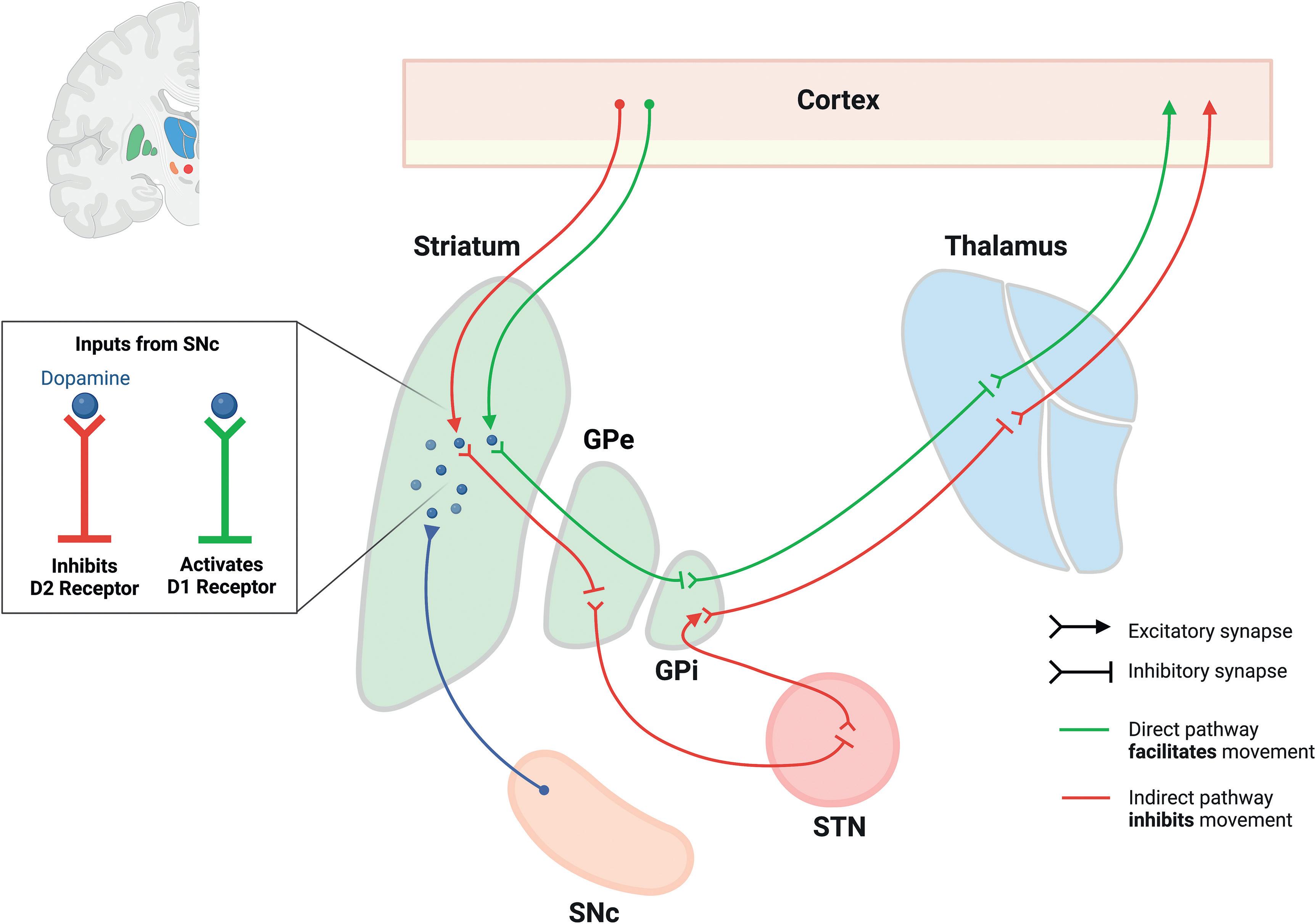
1.13.1.1ConnectionsoftheBasalGanglia
Thestriatumreceivesinputfromthecerebralcortex andtheSNPC(Figure1.15).Thestriatumsendsinhibitoryconnectionstotheinternalandexternalglobuspallidus.TheexternalregionthereforedisinhibitstheSTN, whichthensendsexcitatoryinputtotheinternalglobus pallidus.Thissendsinhibitoryinputtothethalamus, leadingtoinhibitionofinformation flowtothecerebral cortex.
Thebasalgangliaalsoprojecttothemedialdorsal nucleusofthethalamus,whichthenprojectstotheprefrontalassociationcortex.Thisregionisinvolvedin highercorticalandexecutivefunction.
Parkinson’sdiseaseresultsfromdegenerationof dopaminergicneuronsoftheSNPCandexcessiveinhibitionofthethalamus.TheSNPCprojectstobothdirect andindirectpathwaysinthestriatum.Duetothepresenceoftwodifferenttypesofdopaminereceptors,thenet effectistoexcitethedirectpathwayandinhibittheindirectpathway.However,thelossoftheneuronsin Parkinson’sdiseaseupsetsthe finebalanceofthesepathwaysandreducesexcitationofthemotorcortex,resulting inpovertyofmovement.
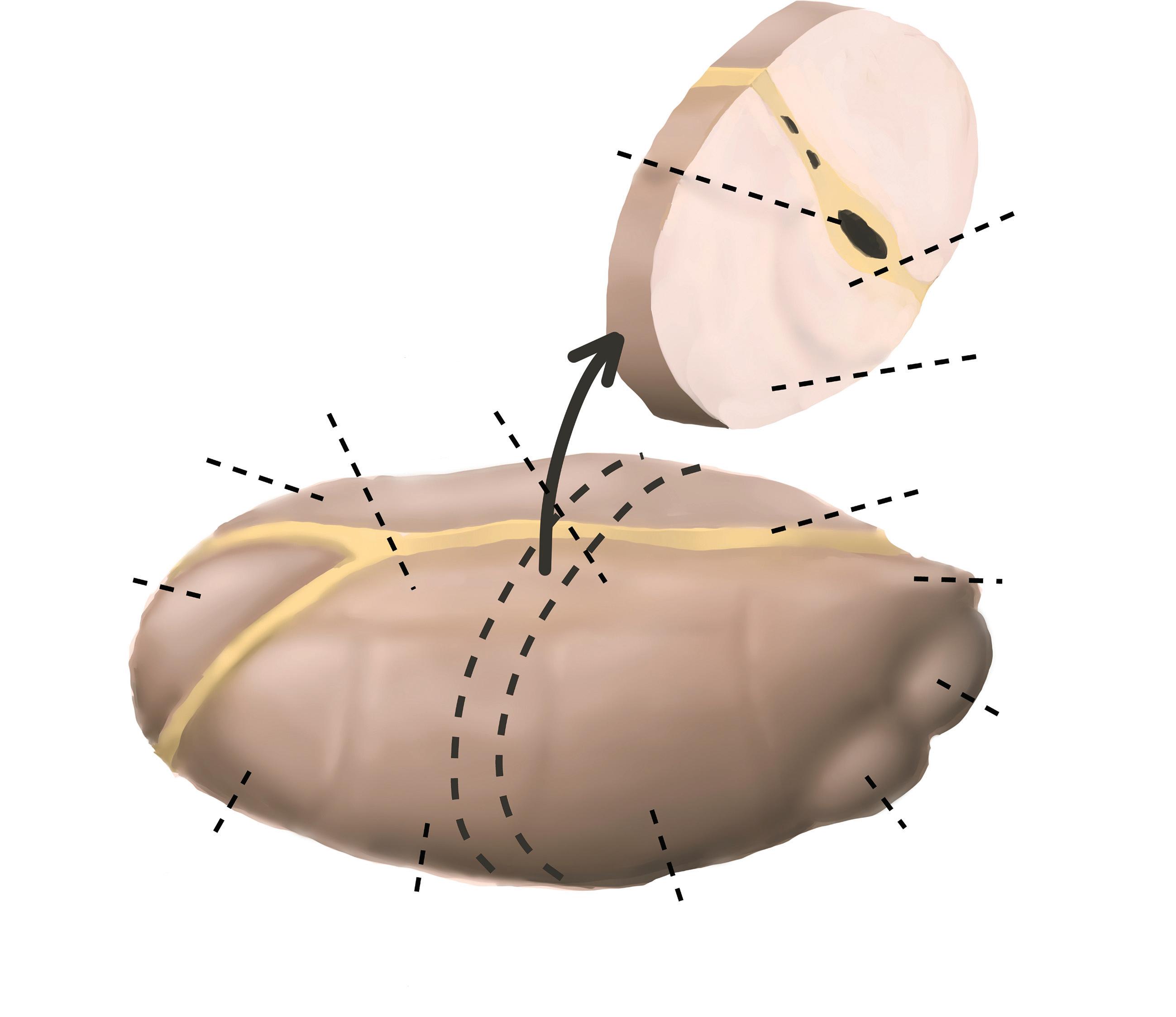
1.13.2Thalamus
Thethalamusislocatedintheforebrainandcontains nucleiwithconnectionstothecerebralcortex,thehippocampus,themammillarybodies,andthefornix.
Cell groups:
Thethalamusisdividedintothreemajornuclear groups(anterior,medial,andventral)(Figure1.16).
Theventralgroupcontainsa scendingsomatosensory relays(ventroposteriornu clei)andrelaysfromthe cerebellumandbasalganglia(ventrolateral).Italso containsthemotorassociat ionareas(ventro-anterior nuclei).Thelateralandmed ialgeniculatenucleiare foundposteriorly.Theanteriorgroupprojectstothe cingulategyrusandreceivesinputfromthemammillarybodiesofthehypothalamus.Themedialnuclei andthepulvinarreceiveinputfromthecerebralcortexandformthecortico-thalamo-corticalrelays, whichprojecttoareasoftheassociationcortex. Thesearetheprefrontalcortexandthetemporal –parietal – occipitalassociationcortex,whichmainly receiveinputfromthemedialnucleiandthepulvinar, respectively.
Thethalamusissuppliedbytheposteriorcerebral arteryandbranchesoftheposteriorcommunicating artery.Thethalamusisinvolvedinlearning,episodic
Figure1.14 Crosssectionofthebasal ganglia.
memory,regulationofsleep,andwakefulness.Lesions intheanteriorthalamuscanleadtoobstructionofthe interventricularforamenofMonroandlesionsinthe posteriomedialthalamuscanobstructthethirdventricle andcerebralaqueduct,leadingtothedevelopmentof hydrocephalus.
1.13.3CingulateGyrus
Thecingulategyrusissituatedabovethecorpuscallosum (Figure1.10).Theanteriorpartrelayssignalsbetweenthe rightandlefthemispheresandisinvolvedinautonomic functionsandcognitiveprocessessuchasrewardbehavior,empathy,andemotion.Theposteriorpartbecomes continuouswiththemostmedialpartofthetemporal lobe,theparahippocampalgyrus.Thecingulategyrusis thoughttobeinvolvedinretrievingepisodicmemory information.Dysfunctionofthisgyrusisfoundinschizophreniaanddepression.Deepbrainstimulationofthe subgenualcortexofthegyrusisusedtotreatintractable
Fiber groups:
Corpus callosum
Cerebral cortex
Septal area
Caudate nucleus
Putamen
Globus pallidus
Fornix
Cortical white matter
Internal capsule
Figure1.15 Basalgangliaconnections.
Lateral dorsal (LD) Medial nuclei
Anterior nuclei
Ventral anterior (VA)
Centromedial nucleus (CM)
Lateral posterior (LP)
Ventral posterior medial (VPM)
Ventral posterior lateral (VPL)
Internal medullary
Lateral geniculate body Medial geniculate body Pulvinar
Figure1.16 Nucleiofthethalamus.
depression.Output fibersaremainlytotheparahippocampalgyrusandarelinkedbythecingulum,awhitemattertract.
1.13.4Hippocampus
Thehippocampusisaseahorse-shapedstructureandthe hippocampalformationreferstothehippocampus proper(CornuAmmonis),thedentategyrus,andthe subiculum.Itisfoundinthetemporallobe,medialto theinferiorhornofthelateralventricle.
Thehippocampushasbeenstudiedextensivelyandis usedasamodelsystemforelectrophysiologicalstudies, particularlyforinvestigatingneuralplasticity.Damageto thehippocampusiscommonlyseeninpatientswith dementia,particularlyAlzheimer’sdisease.
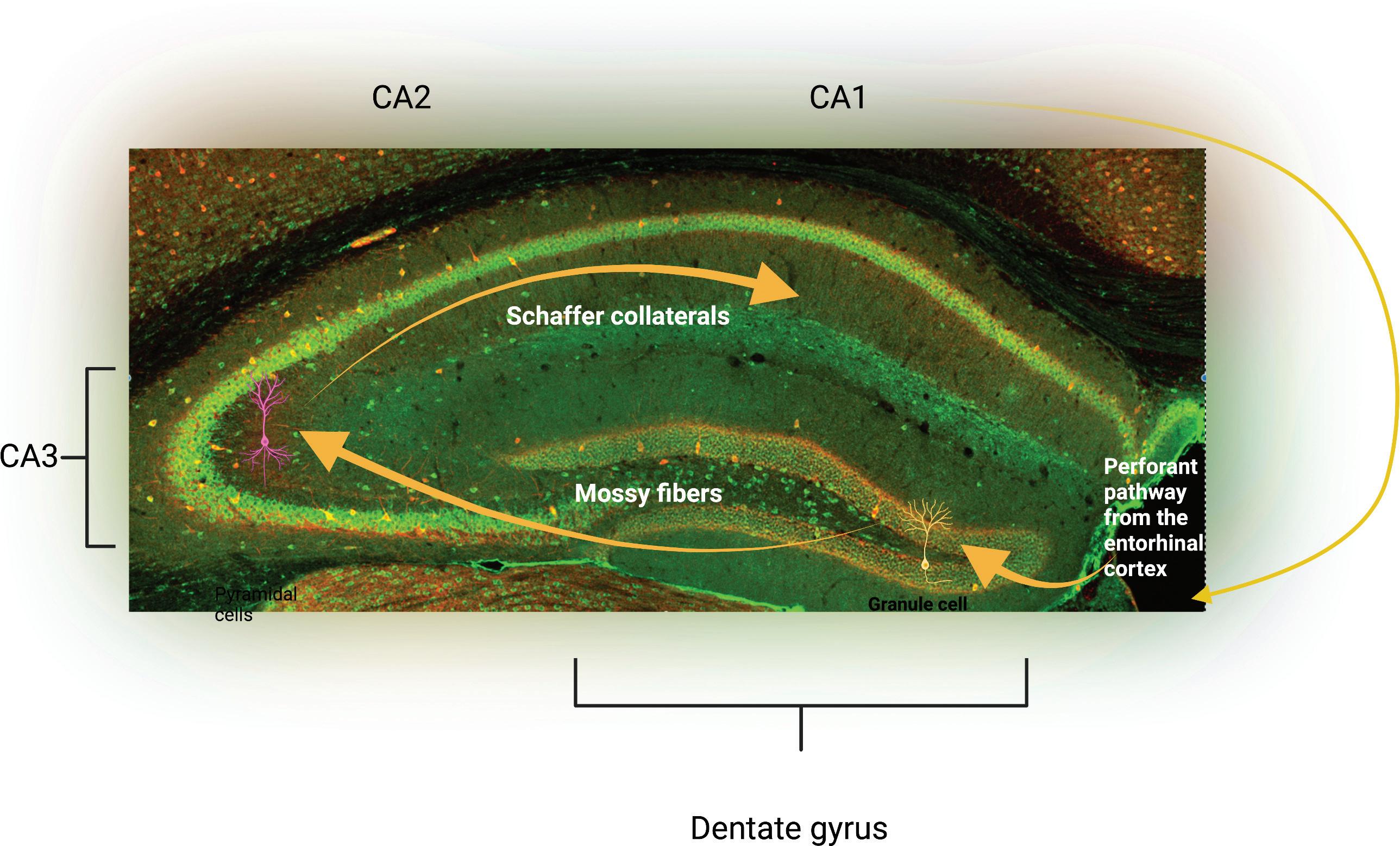
Hippocampaltissueismadeupoflayersandthese include,fromoutertoinner:anexternalplexiformlayer; astratumorienslayercontainingbasaldendritesand basketcells;apyramidalcelllayercontainingtheprimarycellsofthehippocampus;astratumradiatum layer;andthestratumlacunosum-molecularelayers containingtheperforatepathwaymadeupofpyramidal cellapicaldendritesandaff erent fi bersfromtheentorhinalcortex.Theexternalplexiformlayercontainsthe alvearpathway,andthiscontainspyramidalcellaxons throughwhichinformationfromthehippocampusis passedtotheinferiorhornofthelateralventriclebefore reachingtheentorhinalcortex.Inaddition,thehippocampusalsocontainsdistinctregions.Theshapeofthe
Figure1.17 CornuAmmonis axonsofthehippocampus.
hippocampushasbeendescribedassimilartoaseahorse oraram ’shorn(CornuAmmonis).Theabbreviation CAisusedtonamethediff erentregions:CA1,CA2, CA3,andCA4(Figure1.17).
1.13.4.1DentateGyrus
Thedentategyruscontainsgranulecellsandaxonscalled mossy fibers,whichsynapsewiththepyramidalcellsin theCA3 fieldofthehippocampus.Theyalsocontain somepyramidalcellsinthepolymorphiccelllayer.
1.13.4.2HippocampalInputs
Thehippocampusreceivesinformationfromthelateral perforateandmedialperforatepathwaysintheentorhinalcortex(Figure1.18),theprefrontalcortex,theanteriorcingulategyrus,thepre-mammillaryregion,and thereticularformationofthebrainstem.Italsoreceives inputfromthethalamusto fi eldCA1andfromthe serotonin,norepineph-rine,anddopaminesystems. Themedialseptalnucleussendscholinergicand γ-aminobutyricacid(GABA)-ergicinputstothe hippocampus.
Thelargestinputandoutputpathwayofthehippocampusisviathefornix,whichconnectsittoother structuresincludingthemammillarybodiesofthehypothalamus,prefrontalcortexandthelateralseptalarea. Theentorhinalcortex(partoftheparahippocampal gyrus)receivesoutputfromthedeeperlayersofthe hippocampusandgivesinputtothesuperficiallayers.