Immediate download Fundamentals of anatomy & physiology 10th edition frederic h. martini ebooks 2024
Visit to download the full and correct content document: https://ebookmass.com/product/fundamentals-of-anatomy-physiology-10th-edition-fre deric-h-martini/
More products digital (pdf, epub, mobi) instant download maybe you interests ...
Fundamentals of Anatomy and Physiology, 12th Global Edition Frederic Martini
Fundamentals of Anatomy & Physiology, Global Edition 11th Edition, (Ebook PDF)
https://ebookmass.com/product/fundamentals-of-anatomy-physiologyglobal-edition-11th-edition-ebook-pdf/ (eBook PDF) Laboratory Manual for Anatomy & Physiology featuring Martini Art, Main Version 6th Edition
1–1 Levels of Organization 1–10 Diagnostic Imaging Techniques
2–3 Chemical Notation
3–1 Anatomy of a Model Cell 3–7 Protein Synthesis, Processing, and Packaging 3–22 Overview of Membrane Transport 3–23 DNA replication 3–24 Stages of a Cell’s Life Cycle
4–20 Inflammation and Regeneration
5–3 The Epidermis
6–11 Endochondral Ossification 6–16 Types of Fractures and Steps in Repair
7–4 Sectional Anatomy of the Skull
8–10 Sex Differences in the Human Skeleton
9–2 Joint Movement
10–9 Events at the Neuromuscular Junction 10–10 Excitation-Contraction Coupling 10–11 The Contraction Cycle and Cross-Bridge Formation
11–3 Muscle Action
12–9 Resting Membrane Potential 12–14 Generation of an Action Potential 12–15 Propagation of an Action Potential
13–8 Peripheral Distribution of Spinal Nerves 13–14 Spinal Reflexes
14–4 Formation and Circulation of Cerebrospinal Fluid
15–6 Somatic Sensory Pathways
16–2 Overview of the Autonomic Nervous System
17–2 Olfaction and Gustation 17–13 Refractive Problems 17–16 Photoreception
18–2 Structural Classification of Hormones 18–3 G Proteins and Second Messengers 18–18 Diabetes Mellitus 18–20 The General Adaptation Syndrome
19–1 The Composition of Whole Blood 19–8 Hemolytic Disease of the Newborn 20–10 Heart Disease and Heart Attacks 20–14 Cardiac Arrhythmias
21–33 Congenital Heart Problems
22–28 Cytokines of the Immune System
23–15 Respiratory Muscles and Pulmonary Ventilation 23–25 Control of Respiration
24–15 Regulation of Gastric Activity 24–27 Chemical Events of Digestion
25–11 Absorptive and Postabsorptive States
26–16 Summary of Renal Function
27–18 The Diagnosis of Acid-Base Disorders
28–12 Regulation of Male Reproduction 28–24 Regulation of Female Reproduction
29–5 Extraembryonic Membranes and Placenta Formation
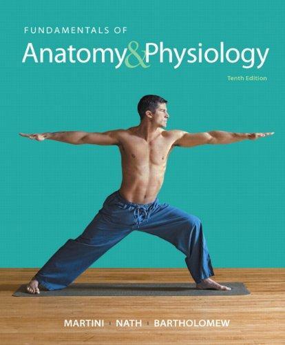
FUNDAMENTALS OF Anatomy Physiology &
Edwin F. Bartholomew, M.S. Tenth
Frederic H. Martini, Ph.D.
University of Hawaii at Manoa
Judi L. Nath, Ph.D. Lourdes University
William C. Ober, M.D. Art Coordinator and Illustrator
Claire E. Ober, R.N. Illustrator
Kathleen Welch, M.D. Clinical Consultant
Clinical Cases by: Ruth Anne O’Keefe
Boston Columbus Indianapolis New York San Francisco Upper Saddle River Amsterdam Cape Town Dubai London Madrid Milan Munich Paris Montreal Toronto Delhi Mexico City São Paulo Sydney Hong Kong Seoul Singapore Taipei Tokyo
Ralph T. Hutchings Biomedical Photographer
Executive Editor: Leslie Berriman
Assistant Editor: Cady Owens
Associate Project Editor: Lisa Damerel
Editorial Assistant: Sharon Kim
Director of Development: Barbara Yien
Development Editor: Anne A. Reid
Managing Editor: Mike Early
Assistant Managing Editor: Nancy Tabor
Project Manager: Caroline Ayres
Director of Digital Product Development: Lauren Fogel
Notice: Our knowledge in clinical sciences is constantly changing. The authors and the publisher of this volume have taken care that the information contained herein is accurate and compatible with the standards generally accepted at the time of the publication. Nevertheless, it is difficult to ensure that all information given is entirely accurate for all circumstances. The authors and the publisher disclaim any liability, loss, or damage incurred as a consequence, directly or indirectly, of the use and application of any of the contents of this volume.
Many of the designations used by manufacturers and sellers to distinguish their products are claimed as trademarks. Where those designations appear in this book, and the publisher was aware of a trademark claim, the designations have been printed in initial caps or all caps.
MasteringA&P®, A&P Flix™, Practice Anatomy Lab™ (PAL™), and Interactive Physiology® are trademarks, in the U.S. and/or other countries, of Pearson Education, Inc. or its affiliates.
Library of Congress Cataloging-in-Publication Data
Martini, Frederic, author.
Fundamentals of anatomy & physiology/Frederic H. Martini, Judi L. Nath, Edwin F. Bartholomew; William C. Ober, art coordinator and illustrator; Claire E. Ober, illustrator; Kathleen Welch, clinical consultant; Ralph T. Hutchings, biomedical photographer. — Tenth edition. p.; cm.
Fundamentals of anatomy and physiology
Includes bibliographical references and index.
ISBN-13: 978-0-321-90907-7
ISBN-10: 0-321-90907-0
I. Nath, Judi Lindsley, author. II. Bartholomew, Edwin F., author. III. Title.
IV. Title: Fundamentals of anatomy and physiology.
Dr. Martini received his Ph.D. from Cornell University in comparative and functional anatomy for work on the pathophysiology of stress. In addition to professional publications that include journal articles and contributed chapters, technical reports, and magazine articles, he is the lead author of ten undergraduate texts on anatomy and physiology or anatomy. Dr. Martini is currently affiliated with the University of Hawaii at Manoa and has a long-standing bond with the Shoals Marine Laboratory, a joint venture between Cornell University and the University of New Hampshire. He has been active in the Human Anatomy and Physiology Society (HAPS) for over 20 years and was a member of the committee that established the course curriculum guidelines for A&P. He is now a President Emeritus of HAPS after serving as President-Elect, President, and Past-President over 2005–2007. Dr. Martini is also a member of the American Physiological Society, the American Association of Anatomists, the Society for Integrative and Comparative Biology, the Australia/New Zealand Association of Clinical Anatomists, the Hawaii Academy of Science, the American Association for the Advancement of Science, and the International Society of Vertebrate Morphologists.
Edwin F. Bartholomew, M.S.
Author
Edwin F. Bartholomew received his undergraduate degree from Bowling Green State University in Ohio and his M.S. from the University of Hawaii. Mr. Bartholomew has taught human anatomy and physiology at both the secondary and undergraduate levels and a wide variety of other science courses (from botany to zoology) at Maui Community College and at historic Lahainaluna High School, the oldest high school west of the Rockies. He is a coauthor of Visual Anatomy & Physiology, Essentials of Anatomy & Physiology, Visual Essentials of Anatomy & Physiology, Structure and Function of the Human Body, and The Human Body in Health and Disease (all published by Pearson). Mr. Bartholomew is a member of the Human Anatomy and Physiology Society (HAPS), the National Association of Biology Teachers, the National Science Teachers Association, the Hawaii Science Teachers Association, and the American Association for the Advancement of Science.
Judi L. Nath, Ph.D.
Author
Dr. Judi Nath is a biology professor at Lourdes University, where she teaches anatomy and physiology, pathophysiology, and medical terminology. She received her Bachelor’s and Master’s degrees from Bowling Green State University and her Ph.D. from the University of Toledo. Dr. Nath is devoted to her students and strives to convey the intricacies of science in captivating ways that are meaningful, interactive, and exciting. She has won the Faculty Excellence Award—an accolade recognizing effectiveteaching,scholarship,andcommunityservice—multiple times. She is active in many professional organizations, notably the Human Anatomy and Physiology Society (HAPS), where she has served several terms on the board of directors. Dr. Nath is a coauthor of Visual Anatomy & Physiology, Visual Essentials of Anatomy & Physiology, and Anatomy & Physiology (all published by Pearson), and she is the sole author of Using Medical Terminology. Her favorite charities are those that have significantly affected her life, including the local Humane Society, the Cystic Fibrosis Foundation, and the ALS Association. On a personal note, Dr. Nath enjoys family life with her husband and their dogs.
William C. Ober, M.D.
Art Coordinator and Illustrator
Dr. Ober received his undergraduate degree from Washington and Lee University and his M.D. from the University of Virginia. He also studied in the Department of Art as Applied to Medicine at Johns Hopkins University. After graduation, Dr. Ober completed a residency in Family Practice and later was on the faculty at the University of Virginia in the Department of Family Medicine and in the Department of Sports Medicine. He also served as Chief of Medicine of Martha Jefferson Hospital in Charlottesville, VA. He is currently a Visiting Professor of Biology at Washington and Lee University, where he has taught several courses and led student trips to the Galapagos Islands. He was on the Core Faculty at Shoals Marine Laboratory for 24 years, where he taught Biological Illustration every summer. Dr. Ober has collaborated with Dr. Martini on all of his textbooks in every edition.
Claire E. Ober, R.N. Illustrator
Claire E. Ober, R.N., B.A., practiced family, pediatric, and obstetric nursing before turning to medical illustration as a full-time career. She returned to school at Mary Baldwin College, where she received her degree with distinction in studio art. Following a five-year apprenticeship, she has worked as Dr. Ober’s partner in Medical & Scientific Illustration since 1986. She was on the Core Faculty at Shoals Marine Laboratory and co-taught the Biological Illustration course with Dr. Ober for 24 years. The textbooks illustrated by Medical & Scientific Illustration have won numerous design and illustration awards.
Kathleen Welch, M.D.
Clinical Consultant
Dr. Welch received her B.A. from the University of Wisconsin–Madison, her M.D.fromtheUniversityof Washington in Seattle, and did her residency in Family Practice at the University of North Carolina in Chapel Hill. Participating in the Seattle WWAMI rural medical education program, she studied in Fairbanks, Anchorage, and Juneau, Alaska, with time in Boise, Idaho, and Anacortes, Washington, as well. For two years, she served as Director of Maternal and Child Health at the LBJ Tropical Medical Center in American Samoa and subsequently was a member of the Department of Family Practice at the Kaiser Permanente Clinic in Lahaina, Hawaii, and on the staff at Maui Memorial Hospital. She has been in private practice since 1987 and is licensed to practice in Hawaii and Washington State. Dr. Welch is a Fellow of the American Academy of Family Practice and a member of the Maui County Medical Society and the Human Anatomy and Physiology Society (HAPS). With Dr. Martini, she has coauthored both a textbook on anatomy and physiology and the A&P Applications Manual. She and Dr. Martini were married in 1979, and they have one son.
Ralph T. Hutchings
Biomedical Photographer
Mr. Hutchings was associated with the Royal College of Surgeons for 20 years. An engineer by training, he has focused for years on photographing the structure of the human body. The result has been a series of color atlases, including the Color Atlas of Human Anatomy, the Color Atlas of Surface Anatomy, and The Human Skeleton (all published by Mosby-Yearbook Publishing). For his anatomical portrayal of the human body, the International Photographers Association has chosen Mr. Hutchings as the best photographer of humans in the twentieth century. He lives in North London, where he tries to balance the demands of his photographic assignments with his hobbies of early motor cars and airplanes.
Ruth Anne O’Keefe, M.D.
Clinical Contributor
Dr. O’Keefe did her undergraduate studies at Marquette University, attended graduate school at the University of Wisconsin, and received her M.D. from George Washington University. She was the first woman to study orthopedics at The Ohio State University during her residency. She did fellowship training in trauma surgery at Loma Linda University in California. In addition to her private orthopedic practice, she has done orthopedic surgery around the world, taking her own surgical teams to places such as the Dominican Republic, Honduras, Peru, New Zealand, and Burkina Faso. She serves on the board of Global Health Partnerships, a group that partners with a clinic serving 35,000 people in remote Kenya. Dr. O’Keefe has always enjoyed teaching and now supervises medical students from the University of New Mexico doing ongoing research in Kenya. She lives in Albuquerque with her Sweet Ed. She is mother of four, grandmother of nine, and foster mother to many.
Preface
The Tenth Edition of Fundamentals of Anatomy & Physiology is a comprehensive textbook that fulfills the needs of today’s students while addressing the concerns of their professors. We focused our attention on the question “How can we make this information meaningful, manageable, and comprehensible?”
During the revision process, we drew upon our content knowledge, research skills, artistic talents, and years of classroom experience to make this edition the best yet.
The broad changes to this edition are presented in the New to the Tenth Edition section below, and the specific changes are presented in the Chapter-by-Chapter Changes in the Tenth Edition section that follows.
New to the Tenth Edition
In addition to the many technical changes in this edition, such as updated statistics and anatomy and physiology descriptions, we have made the following key changes:
NEW 50 Spotlight Figures provide highly visual one- and two-page presentations of tough topics in the book, with a particular focus on physiology. In the Tenth Edition, 18 new Spotlight Figures have been added for a total of 50 across the chapters. There is now at least one Spotlight Figure in every chapter, as well as one Spotlight Figure corresponding to every A&P Flix.
NEW 29 Clinical Cases get students motivated for their future careers. Each chapter opens with a story-based Clinical Case related to the chapter content and ends with a Clinical Case Wrap-Up that incorporates the deeper content knowledge students will have gained from the chapter.
NEW The repetition of the chapter-opening Learning Outcomes below the coordinated section headings within the chapters underscores the connection between the HAPS-based Learning Outcomes and the associated teaching points. Author Judi Nath sat on the Human Anatomy and Physiology Society (HAPS) committee that developed the HAPS Learning Outcomes, recommended to A&P instructors, and the Learning Outcomes in this book are based on them. Additionally, the assessments in MasteringA&P are organized by these Learning Outcomes. As in the previous edition, full-sentence section headings, correlated with the Learning Outcomes, state a core fact or concept to help students readily see and learn the chapter content; and Checkpoints, located at the close of each section, ask students to pause and check their understanding
of facts and concepts. If students cannot answer these questions within a matter of minutes, then they should reread the section before moving on. The Checkpoints reinforce the Learning Outcomes, resulting in a systematic integration of the Learning Outcomes over the course of the chapter. Answers to the Checkpoints are located in the blue Answers tab at the back of the book.
Easier narrative uses simpler, shorter, more active sentences and a reading level that makes reading and studying easier for students.
Improved text-art integration throughout the illustration program enhances the readability of figures. Several tables have been integrated directly into figures to help students make direct connections between tables and art.
Eponyms are now included within the narrative, along with the anatomical terms used in Terminologia Anatomica.
NEW Assignable MasteringA&P activities include the following:
NEW Spotlight Figure Coaching Activities are highly visual, assignable activities designed to bring interactivity to the Spotlight Figures in the book. Multi-part activities include the ranking and sorting types that ask students to manipulate the visuals.
NEW Book-specific Clinical Case Activities stem from the story-based Clinical Cases that appear at the beginning and end of each chapter in the book.
NEW Adaptive Follow-up Assignments allow instructors to easily assign personalized content for each individual student based on strengths and weaknesses identified by his or her performance on MasteringA&P parent assignments.
NEW Dynamic Study Modules help students acquire, retain, and recall information quickly and efficiently. The modules are available as a self-study tool or can be assigned by the instructor. They can be easily accessed with smartphones.
Chapter-by-Chapter Changes in the Tenth Edition
This annotated Table of Contents provides select examples of revision highlights in each chapter of the Tenth Edition. For a more complete list of changes, please contact the publisher.
Chapter 1: An Introduction to Anatomy and Physiology
• New Clinical Case: Using A&P to Save a Life
• New Spotlight Figure 1–10 Diagnostic Imaging Techniques
• New Clinical Note: Autopsies and Cadaver Dissection
• New Clinical Note: Auscultation
• Figure 1–7 Directional References revised
• Figure 1–8 Sectional Planes revised
• Figure 1–9 Relationships among the Subdivisions of the Body Cavities of the Trunk revised
Chapter 2: The Chemical Level of Organization
• New Clinical Case: What Is Wrong with My Baby?
• New Clinical Note: Radiation Sickness
• Clinical Note: Fatty Acids and Health revised
• Section 2-2 includes revised Molecular weight discussion
• Figure 2–4 The Formation of Ionic Bonds revised
• Figure 2–5 Covalent Bonds in Five Common Molecules revised
• Table 2–3 Important Functional Groups of Organic Compounds revised (to clarify structural group and R group)
• Protein Structure subsection includes new discussion of amino acids as zwitterions
• Figure 2–21 Protein Structure revised
Chapter 3: The Cellular Level of Organization
• New Clinical Case: When Your Heart Is in the Wrong Place
• New information added about cholesterol and other lipids
• New overview added about roles of microtubules
• Figure 3–5 The Endoplasmic Reticulum revised
• Clinical Note on DNA Fingerprinting revised
• Figure 3–13 The Process of Translation revised
• Figure 3–14 Diffusion revised
• Figure 3–17 Osmotic Flow across a Plasma Membrane revised
• New Spotlight Figure 3–22 Overview of Membrane Transport incorporates old Figures 3–18, 3–19, and 3–21 and old Table 3–2
• New Spotlight Figure 3–23 DNA Replication incorporates old Figure 3–23
• Spotlight Figure 3–24 Stages in a Cell’s Life Cycle revised
Chapter 4: The Tissue Level of Organization
• New Clinical Case: The Rubber Girl
• Intercellular Connections subsection updated
• Figure 4–2 Cell Junctions revised
• Figure 4–8 The Cells and Fibers of Connective Tissue Proper revised
• Adipose Tissue subsection includes updated discussion of brown fat
• Figure 4–10 Loose Connective Tissues revised
• Spotlight Figure 4–20 Inflammation and Regeneration revised
Chapter 5: The Integumentary System
• New Clinical Case: Skin Cells in Overdrive
• Figure 5–1 The Components of the Integumentary System revised
• New Figure 5–2 The Cutaneous Membrane and Accessory Structures
• New Spotlight Figure 5–3 The Epidermis incorporates old Figures 5–2 and 5–3
• New Figure 5–5 Vitiligo
• New Figure 5–6 Sources of Vitamin D3
• Clinical Note: Decubitus Ulcers revised with new photo
• New Figure 5–8 Reticular Layer of Dermis
• Figure 5–10 Dermal Circulation revised
• Figure 5–12 Hair Follicles and Hairs revised
• New Figure 5–11 Hypodermis
Chapter 6: Osseous Tissue and Bone Structure
• New Clinical Case: A Case of Child Abuse?
• Figure 6–1 A Classification of Bones by Shape revised
• New Figure 6–2 An Introduction to Bone Markings incorporates old Table 6–1
• New Spotlight Figure 6–11 Endochondral Ossification incorporates old Figure 6–10
• New Figure 6–12 Intramembranous Ossification
• Spotlight Figure 6–16 Types of Fractures and Steps in Repair revised
• Clinical Note: Abnormal Bone Development revised
Chapter 7: The Axial Skeleton
• New Clinical Case: Knocked Out
• New Clinical Note: Sinusitis
• Figure 7–2 Cranial and Facial Subdivisions of the Skull revised
• Figure 7–3 The Adult Skull revised to incorporate old Table 7–1
• New Spotlight Figure 7–4 Sectional Anatomy of the Skull incorporates old Figure 7–4 and parts of old Table 7–1
• Figure 7–6 The Frontal Bone revised
• Figure 7–14 The Nasal Complex revised
• Figure 7–22 The Thoracic Cage revised
Chapter 8: The Appendicular Skeleton
• New Clinical Case: The Orthopedic Surgeon’s Nightmare
• New Clinical Note: Hip Fracture
• New Clinical Note: Runner’s Knee
• New Clinical Note: Stress Fractures
• Carpal Bones subsection now lists the 8 carpal bones in two groups of 4 (proximal and distal carpal bones)
• Figure 8–6 Bones of the Right Wrist and Hand revised
• New Spotlight Figure 8–10 Sex Differences in the Human Skeleton incorporates old Figure 8–10, old Table 8–1, and old bulleted list in text
• Clinical Note: Carpal Tunnel Syndrome includes new illustration
• Figure 8–14 Bones of the Ankle and Foot revised
• Clinical Note: Congenital Talipes Equinovarus includes new photo
Chapter 9: Joints
• Chapter title changed from Articulations to Joints
• New Clinical Case: What’s Ailing the Birthday Girl?
• New Clinical Note: Dislocation and Subluxation
• New Clinical Note: Damage to Intervertebral Discs
• Table 9–1 Functional and Structural Classifications of Articulations redesigned
• Spotlight Figure 9–2 Joint Movement incorporates old Figures 9–2 and 9–6 and subsection on Types of Synovial Joints
• Revised discussion of synovial fluid function in shock absorption
• Figure 15–8 Descending (Motor) Tracts in the Spinal Cord reorganized
Chapter 16: The Autonomic Nervous System and Higher-Order Functions
• New Clinical Case: The First Day in Anatomy Lab
• New Spotlight Figure 16–2 Overview of the Autonomic Nervous System incorporates old Figures 16–3 and 16–7
• Figure 16–3 Sites of Ganglia in Sympathetic Pathways revised
• Figure 16–4 The Distribution of Sympathetic Innervation revised
Chapter 17: The Special Senses
• New Clinical Case: A Chance to See
• Figure 17–1 The Olfactory Organs revised
• Spotlight Figure 17–2 Olfaction and Gustation revised
• Figure 17–3 Gustatory Receptors revised
• Figure 17–22 The Middle Ear revised
• Figures 17–23, 17–24, and 17–25 revised to indicate different orientations of maculae in the utricle and saccule
• Figure 17–32 Pathways for Auditory Sensations revised
Chapter 18: The Endocrine System
• New Clinical Case: Stones, Bones, and Groans
• New Spotlight Figure 18–3 G Proteins and Second Messengers incorporates old Figure 18–3
• Figure 18–7 The Hypophyseal Portal System and the Blood Supply to the Pituitary Gland revised
• Figure 18–11 The Thyroid Follicles revised
• New Figure 18–14 The Adrenal Gland incorporates old Figure 18–14 and old Table 18–5
Chapter 19: Blood
• New Clinical Case: A Mysterious Blood Disorder
• Figure 19–3 The Structure of Hemoglobin revised
• Table 19–4 includes revised names for Factors IX and XI and source of Factor X
Chapter 20: The Heart
• New Clinical Case: A Needle to the Chest
• Figure 20–3 The Superficial Anatomy of the Heart revised
• Figure 20–6 The Sectional Anatomy of the Heart revised
• Figure 20–12 Impulse Conduction through the Heart revised
• Figure 20–16 Phases of the Cardiac Cycle revised
• Figure 20–21 Autonomic Innervation of the Heart revised
• Figure 20–24 A Summary of the Factors Affecting Cardiac Output revised
Chapter 21: Blood Vessels and Circulation
• New Clinical Case: Did Ancient Mummies Have Atherosclerosis?
• Figure 21–2 Histological Structures of Blood Vessels revised
• Figure 21–8 Relationships among Vessel Diameter, CrossSectional Area, Blood Pressure, and Blood Velocity within the Systemic Circuit revised
• Figure 21–9 Pressures within the Systemic Circuit revised
• Figure 21–11 Forces Acting across Capillary Walls revised
• Figure 21–20 Arteries of the Chest and Upper Limb revised
• Figure 21–25 Arteries of the Lower Limb revised
• Figure 21–29 Flowcharts of Circulation to the Superior and Inferior Venae Cavae revised
• Figure 21–30 Venous Drainage from the Lower Limb revised
Chapter 22: The Lymphatic System and Immunity
• New Clinical Case: Isn’t There a Vaccine for That?
• Figure 22–6 The Origin and Distribution of Lymphocytes revised
• Figure 22–11 Innate Defenses revised
• Complement System subsection – includes revised number of complement proteins in plasma (from 11 to more than 30)
• Figure 22–18 Antigens and MHC Proteins revised
Chapter 23: The Respiratory System
• New Clinical Case: How Long Should a Cough Last?
• Figure 23–1 The Structure of the Respiratory System reorganized
• Figure 23–3 The Structures of the Upper Respiratory System revised
• Figure 23–5 The Glottis and Surrounding Structures revised
• Figure 23–7 The Gross Anatomy of the Lungs revised
• Figure 23–9 The Bronchi, Lobules, and Alveoli of the Lung revised
• Figure 23–10 Alveolar Organization revised
• Figure 23–13 Mechanisms of Pulmonary Ventilation revised
• New Spotlight Figure 23–15 Respiratory Muscles and Pulmonary Ventilation incorporates old Figure 23–16
• Figure 23–16 Pulmonary Volumes and Capacities revised
• Spotlight Figure 23–25 Control of Respiration revised
Chapter 24: The Digestive System
• New Clinical Case: An Unusual Transplant
• Figure 24–10 The Esophagus revised
• Figure 24–12 The Stomach revised
• Figure 24–16 Segments of the Intestine revised
• Figure 24–21 The Anatomy and Physiology of the Gallbladder and Bile Ducts revised
Chapter 25: Metabolism and Energetics
• New Clinical Case: The Miracle Supplement
• Figure 25–9 Lipid Transport and Utilization revised
• Figure 25–12 MyPlate Plan revised
• Figure 25–14 Mechanisms of Heat Transfer revised
Chapter 26: The Urinary System
• New Clinical Case: A Case of “Hidden” Bleeding
• Revised all relevant figure labels by replacing “Renal lobe” with “Kidney lobe”
• Figure 26–6 The Functional Anatomy of a Representative Nephron and the Collecting System revised
• Spotlight Figure 26–16 Summary of Renal Function revised
Chapter 27: Fluid, Electrolyte, and Acid–Base Balance
• New Clinical Case: When Treatment Makes You Worse
• Figure 27–2 Cations and Anions in Body Fluids revised
• Figure 27–3 Fluid Gains and Losses revised
• Figure 27–11 The Role of Amino Acids in Protein Buffer Systems revised (to emphasize amino acids as zwitterions)
• Figure 27–13 Kidney Tubules and pH Regulation revised
• New Spotlight Figure 27–18 The Diagnosis of Acid–Base Disorders incorporates old Figure 27–18
Chapter 28: The Reproductive System
• New Clinical Case: A Post-Game Mystery
• Figure 28–1 The Male Reproductive System revised
• Figure 28–3 The Male Reproductive System in Anterior View revised and reorganized
• Figure 28–4 The Structure of the Testes revised
• Figure 28–7 Spermatogenesis revised
• Figure 28–13 The Female Reproductive System revised
• Figure 28–15 Oogenesis revised
• Figure 28–18 The Uterus revised
Chapter 29: Development and Inheritance
• New Clinical Case: The Twins That Looked Nothing Alike
• Revised all relevant chapter text by replacing “embryological” with “embryonic” for simplification
• New Spotlight Figure 29–5 Extraembryonic Membranes and Placenta Formation incorporates old Figure 29–5
• Table 29–2 An Overview of Prenatal Development includes revised sizes and weights at different gestational ages
• Figure 29–8 The Second and Third Trimesters revised
• Figure 29–9 Growth of the Uterus and Fetus revised
• Figure 29–13 Growth and Changes in Body Form and Proportion revised
Appendix
• New periodic table
• New codon chart
Acknowledgments
This textbook represents a group effort, and we would like to acknowledge the people who worked together with us to create this Tenth Edition.
Foremost on our thank-you list are the instructors who offered invaluable suggestions throughout the revision process. We thank them for their participation and list their names and affiliations below.
Lisa Conley, Milwaukee Area Technical College
Theresa G. D’Aversa, Iona College
Danielle Desroches, William Paterson University
Debra Galba-Machuca, Portland Community College–Cascade
Lauren Gollahon, Texas Tech University
Gigi Goochey, Hawaii Community College
Mark Haefele, Community College of Denver
Anthony Jones, Tallahassee Community College
William L’ Amoreaux, College of Staten Island
J. Mitchell Lockhart, Valdosta State University
Scott Murdoch, Moraine Valley Community College
Louise Petroka, Gateway Community College
Cynthia Prentice-Craver, Chemeketa Community College
S. Michele Robichaux, Nicholls State University
Susan Rohde, Triton College
Yung Su, Florida State University
Bonnie Taylor, Schoolcraft College
Carol Veil, Anne Arundel Community College
Patricia Visser, Jackson College
Theresia Whelan, State College of Florida – Manatee-Sarasota
Samia Williams, Santa Fe College
The accuracy and currency of the clinical material in this edition and in the A&P Applications Manual in large part reflect the work of Kathleen Welch, M.D. Her professionalism and concern for practicality and common sense make the clinical information especially relevant for today’s students. Additionally,ourcontentexpertontheClinicalCases,RuthAnneO’Keefe, M.D., provided constant, useful feedback on each chapter.
Virtually without exception, reviewers stressed the importance of accurate, integrated, and visually attractive illustrations in helping readers understand essential material. The revision of the art program was directed by Bill Ober, M.D. and Claire E. Ober, R.N. Their suggestions about presentation sequence, topics of clinical importance, and revisions to the proposed art were of incalculable value to us and to the project. The illustration program for this edition was further enhanced by the efforts of several other talented individuals. Jim Gibson
designed most of the new Spotlight Figures in the art program and consulted on the design and layout of the individual figures. His talents have helped produce an illustration program that is dynamic, cohesive, and easy to understand. Anita Impagliazzo helped create the new photo/art combinations that have resulted in clearer presentations and a greater sense of realism in important anatomical figures. We are also grateful to the talented team at Imagineering (imagineeringart.com) for their dedicated and detailed illustrative work on key figures for this edition. The new color micrographs in this edition were provided by Dr. Robert Tallitsch, and his assistance is much appreciated. Many of the striking anatomy photos in the text and in Martini’s Atlas of the Human Body are the work of biomedical photographer Ralph Hutchings; his images played a key role in the illustration program.
We also express our appreciation to the editors and support staff at Pearson Science.
We owe special thanks to Executive Editor, Leslie Berriman, for her creativity and dedication. Her vision helped shape this book in countless ways. Leslie’s enthusiasm for publishing the highest quality material spills over onto the author/ illustrator team. She is our biggest advocate and is always willing to champion our cause—despite the challenges of working with authors. We are appreciative of all her efforts on our behalf.
Assistant Editor, Cady Owens, and Associate Project Editor, Lisa Damerel, are unquestionably the very finest at what they do. While it is expected that editors pay attention to details and keep projects moving forward, Cady and Lisa are true professionals and extremely skilled at not only preparing our material for publication, but making sure it is the best it could possibly be. This past year could not have happened without them.
Annie Reid, our Development Editor, played a vital role in revising the Tenth Edition. Her unfailing attention to readability, consistency, and quality was invaluable to the authors in meeting our goal of delivering complex A&P content in a more student-friendly way.
We are grateful to Mike Rossa for his careful attention to detail and consistency in his copyedit of the text and art.
This book would not exist without the extraordinary dedication of the Production team, including Caroline Ayres, who solved many problems under pressure with unfailing good cheer. Norine Strang skillfully led her excellent team at S4Carlisle to move the book smoothly through composition.
The striking cover and clear, navigable interior design were created by tani hasegawa. Thanks also to Mark Ong, Design Manager, and Marilyn Perry, who devised innovative solutions for several complex design challenges.
Thanks to our photo researcher, Maureen Spuhler, and photo editor, Donna Kalal, for finding, obtaining, and coordinating all the photos in the photo program.
Thanks are also due to Sharon Kim, Editorial Assistant, who served as project editor for the print supplements for instructors and students and coordinated the administrative details of the entire textbook program. Dorothy Cox and Shannon Kong worked tirelessly to shepherd the print and media supplements through production. Thanks also to Stacey Weinberger for handling the physical manufacturing of the book.
We are also grateful to Joe Mochnick, Content Producer, and Liz Winer, Executive Content Producer, for their creative efforts on the media package, most especially MasteringA&P.
We would also like to express our gratitude to the following people at Pearson Science: Paul Corey, President, who continues to support all our texts; Barbara Yien, Director of Development, who kindly kept all phases moving forward under all circumstances; Allison Rona, Senior Marketing Manager; and the dedicated Pearson Science sales representatives for
their continuing support of this project. Special thanks go to Frank Ruggirello, Vice President and Editorial Director, for working closely with Leslie in ensuring we have the resources necessary to publish what students need to succeed. And, a round of applause goes to Derek Perrigo, Senior A&P Specialist, our biggest cheerleader.
To help improve future editions, we encourage you to send any pertinent information, suggestions, or comments about the organization or content of this textbook to us directly, using the e-mail addresses below. We warmly welcome comments and suggestions and will carefully consider them in the preparation of the Eleventh Edition.
Frederic (Ric) H. Martini Haiku, Hawaii martini@pearson.com
Judi L. Nath Sandusky, Ohio nath@pearson.com
Edwin F. Bartholomew Lahaina, Hawaii bartholomew@pearson.com
Preface v
UNIT 1 LEVELS OF ORGANIZATION
1
An Introduction to Anatomy and Physiology
1
An Introduction to Studying the Human Body 2
1-1
1-2
1-3
1-4
1-5
1-6
Anatomy and physiology directly affect your life 2
2 The Chemical Level of Organization
26
An Introduction to the Chemical Level of Organization 27
2-1
Atoms are the basic particles of matter 27
Atomic Structure 27
Elements and Isotopes 28
Atomic Weights 29
Electrons and Energy Levels 30
2-2
1-7
Anatomy is structure, and physiology is function 3
Anatomy and physiology are closely integrated 4
Anatomy 4
Physiology 5
Levels of organization progress from molecules to a complete organism 6
Homeostasis is the state of internal balance 7
Negative feedback opposes variations from normal, whereas positive feedback exaggerates them 10
The Role of Negative Feedback in Homeostasis 10
The Role of Positive Feedback in Homeostasis 12
Systems Integration, Equilibrium, and Homeostasis 13
Anatomical terms describe body regions, anatomical positions and directions, and body sections 14
Superficial Anatomy 14
Sectional Anatomy 16
1-8
Body cavities of the trunk protect internal organs and allow them to change shape 18
The Thoracic Cavity 22
The Abdominopelvic Cavity 22
Chapter Review 23
Spotlights
Levels of Organization 8
Diagnostic Imaging Techniques 20
Clinical Case
Using A&P to Save a Life 2
Clinical Notes
Autopsies and Cadaver Dissection 5
Auscultation 14
2-3
Chemical bonds are forces formed by atom interactions 31
Ionic Bonds 31
Covalent Bonds 34
Hydrogen Bonds 35
States of Matter 35
Decomposition, synthesis, and exchange reactions are important chemical reactions in physiology 36
Basic Energy Concepts 36
Types of Chemical Reactions 37
2-4
2-5
2-6
2-7
2-8
Enzymes catalyze specific biochemical reactions by lowering the energy needed to start them 38
Inorganic compounds lack carbon, and organic compounds contain carbon 39
Physiological systems depend on water 39
The Properties of Aqueous Solutions 40
Colloids and Suspensions 41
Body fluid pH is vital for homeostasis 41
Acids, bases, and salts are inorganic compounds with important physiological roles 42
Salts 43
Buffers and pH Control 43
2-9
2-10
Carbohydrates contain carbon, hydrogen, and oxygen in a 1:2:1 ratio 43
Monosaccharides 44
Disaccharides and Polysaccharides 45
Lipids often contain a carbon-to-hydrogen ratio of 1:2 46
Fatty Acids 46
Eicosanoids 47
Glycerides 48
Steroids 48
Phospholipids and Glycolipids 49
2-11 Proteins contain carbon, hydrogen, oxygen, and nitrogen and are formed from amino acids 51
Protein Structure 51
Protein Shape 52
Enzyme Function 54
Glycoproteins and Proteoglycans 56
2-12 DNA and RNA are nucleic acids 56
Structure of Nucleic Acids 56
RNA and DNA 56
2-13 ATP is a high-energy compound used by cells 58
2-14 Chemicals and their interactions form functional units called cells 59
Chapter Review 60
Spotlights
Chemical Notation 32
Clinical Case
What Is Wrong with My Baby? 27
Clinical Notes
Radiation Sickness 29
Fatty Acids and Health 48
3 The Cellular Level of
Organization 64
An Introduction to Cells 65
3-1 The plasma membrane separates the cell from its surrounding environment and performs various functions 65
Membrane Lipids 68
Membrane Proteins 69
Membrane Carbohydrates 70
3-2 Organelles within the cytoplasm perform particular functions 70
The Cytosol 70
The Organelles 71
3-3 The nucleus contains DNA and enzymes essential for controlling cellular activities 82
Contents of the Nucleus 83
Information Storage in the Nucleus 83
3-4 DNA controls protein synthesis, cell structure, and cell function 84
The Role of Gene Activation in Protein Synthesis 84
The Transcription of mRNA 84
Translation and Protein Synthesis 86
How the Nucleus Controls Cell Structure and Function 87
3-5 Diffusion is a passive transport mechanism that assists membrane passage 87
Diffusion 89
Diffusion across Plasma Membranes 91
3-6
Carrier-mediated and vesicular transport assist membrane passage 94
Carrier-Mediated Transport 94
Vesicular Transport 96
3-7
3-8
3-9
3-10
3-11
The membrane potential results from the unequal distribution of positive and negative charges across the plasma membrane 100
Stages of a cell’s life cycle include interphase, mitosis, and cytokinesis 101
DNA Replication 101
Interphase, Mitosis, and Cytokinesis 101
The Mitotic Rate and Energy Use 103
Several growth factors affect the cell life cycle 103
Tumors and cancers are characterized by abnormal cell growth and division 106
Differentiation is cellular specialization as a result of gene activation or repression 108
Chapter Review 109
Spotlights
Anatomy of a Model Cell 66
Protein Synthesis, Processing, and Packaging 78
Overview of Membrane Transport 98
DNA Replication 102
Stages of a Cell’s Life Cycle 104
Clinical Case
When Your Heart Is in the Wrong Place 65
Clinical Notes
Inheritable Mitochondrial Disorders 80
DNA Fingerprinting 84 Mutations 86
Drugs and the Plasma Membrane 90 Telomerase, Aging, and Cancer 107 Parkinson’s Disease 108
4
The Tissue Level of Organization 113
An Introduction to the Tissue Level of Organization 114
4-1
4-2
4-3
The four tissue types are epithelial, connective, muscle, and neural 114
Epithelial tissue covers body surfaces, lines cavities and tubular structures, and serves essential functions 114
Functions of Epithelial Tissue 115
Specializations of Epithelial Cells 115
Maintaining the Integrity of Epithelia 116
Cell shape and number of layers determine the classification of epithelia 118
Classification of Epithelia 119
Glandular Epithelia 123
4-4
4-5
4-6
Connective tissue provides a protective structural framework for other tissue types 126
Classification of Connective Tissues 126
Connective Tissue Proper 126
Cartilage and bone provide a strong supporting framework 133
Cartilage 133
Bone 136
Tissue membranes are physical barriers of four types:
mucous, serous, cutaneous, and synovial 137
Mucous Membranes 137
Serous Membranes 137
The Cutaneous Membrane 139
Synovial Membranes 139
4-7
Connective tissue creates the internal framework of the body 139
4-8 The three types of muscle tissue are skeletal, cardiac, and smooth 140
Skeletal Muscle Tissue 140
Cardiac Muscle Tissue 142
Smooth Muscle Tissue 142
4-9 Neural tissue responds to stimuli and propagates electrical impulses throughout the body 142
4-10
4-11
The response to tissue injury involves inflammation and regeneration 144
Inflammation 144
Regeneration 144
With advancing age, tissue repair declines and cancer rates increase 144
Aging and Tissue Structure 144
Aging and Cancer Incidence 146
Chapter Review 146
Spotlights
Inflammation and Regeneration 145
Clinical Case
The Rubber Girl 114
Clinical Notes
Exfoliative Cytology 120
Marfan’s Syndrome 129
UNIT 2 SUPPORT AND MOVEMENT
The Integumentary System 150
5-2
5-3
5-4
5-5
Stratum Spinosum 153
Stratum Granulosum 155
Stratum Lucidum 155
Stratum Corneum 155
Factors influencing skin color are epidermal pigmentation and dermal circulation 155
The Role of Epidermal Pigmentation 156
The Role of Dermal Circulation 157
Sunlight causes epidermal cells to convert a steroid into vitamin D3 158
Epidermal growth factor has several effects on the epidermis and epithelia 159
The dermis is the tissue layer that supports the epidermis 160
Dermal Strength and Elasticity 160
Cleavage Lines 161
The Dermal Blood Supply 161
Innervation of the Skin 161
5-6
5-7
5-8
The hypodermis connects the dermis to underlying tissues 162
Hair is composed of keratinized dead cells that have been pushed to the surface 163
Hair Production 165
The Hair Growth Cycle 165
Types of Hairs 165
Hair Color 165
Sebaceous glands and sweat glands are exocrine glands found in the skin 166
Sebaceous Glands 166
Sweat Glands 167
Other Integumentary Glands 168
Control of Glandular Secretions and the Homeostatic Role of the Integument 168
5-9
5-10
5-11
An Introduction to the Integumentary System 151
5-1
The epidermis is composed of layers with various functions 153
Stratum Basale 153
Nails are keratinized epidermal cells that protect the tips of fingers and toes 169
Several phases are involved in repairing the integument following an injury 169
Effects of aging include skin thinning, wrinkling, and reduced melanocyte activity 172
Chapter Review 175
Spotlights
The Epidermis 154
Clinical Case
Skin Cells in Overdrive 151
Clinical Notes
Skin Cancer 158
Decubitus Ulcers 161
Liposuction 163
Burns and Grafts 171
Skin Abnormalities 172
6
Osseous Tissue and Bone Structure
178
An Introduction to the Skeletal System
6-1 The skeletal system has five primary functions 179
6-2 Bones are classified according to shape and structure, and they have a variety of surface markings 180
Bone Shapes 180
Bone Markings 181
Bone Structure 182
6-3 Bone is composed of matrix and several types of cells: osteocytes, osteoblasts, osteogenic cells, and osteoclasts 182
Bone Matrix 183
Bone Cells 183
6-4
6-5
6-6
6-7
Compact bone contains parallel osteons, and spongy bone contains trabeculae 184
Compact Bone Structure 185
Spongy Bone Structure 186
The Periosteum and Endosteum 187
Bones form through ossification and enlarge through appositional growth and remodeling 189
Endochondral Ossification 189
Intramembranous Ossification 192
The Blood and Nerve Supplies to Bone 192
Bone growth and development depend on a balance between bone formation and bone resorption 192
Exercise, hormones, and nutrition affect bone development and the skeletal system 194
The Effects of Exercise on Bone 194
Nutritional and Hormonal Effects on Bone 194
6-8 Calcium plays a critical role in bone physiology 196
The Skeleton as a Calcium Reserve 196
Hormones and Calcium Balance 196
6-9 A fracture is a crack or break in a bone 198
6-10
Osteopenia has a widespread effect on aging skeletal tissue 199
Chapter Review 203
Spotlights
Endochondral Ossification 190
Types of Fractures and Steps in Repair 200
Clinical Case
A Case of Child Abuse? 179
Clinical Notes
Heterotopic Bone Formation 188
Abnormal Bone Development 196
7
The Axial Skeleton 206
An Introduction to the Axial Skeleton 207
7-1
7-2
7-3
7-4
7-5
7-6
The 80 bones of the head and trunk make up the axial skeleton 207
The skull is composed of 8 cranial bones and 14 facial bones 207
Each orbital complex contains an eye, and the nasal complex encloses the nasal cavities 223
The Orbital Complexes 223
The Nasal Complex 223
Fontanelles are non-ossified areas between cranial bones that allow for brain growth in infants and small children 224
The vertebral column has four spinal curves 226
Spinal Curvature 226
Vertebral Anatomy 227
The five vertebral regions are the cervical, thoracic, lumbar, sacral, and coccygeal regions 228
Cervical Vertebrae 229
Thoracic Vertebrae 232
Lumbar Vertebrae 232
The Sacrum 232
The Coccyx 235
7-7
The thoracic cage protects organs in the chest and provides sites for muscle attachment 235
The Ribs 235
The Sternum 236
Chapter Review 238
Spotlights
Sectional Anatomy of the Skull 212
Clinical Case
Knocked Out 207
Clinical Notes
Temporomandibular Joint Syndrome 222
Sinusitis 225
Craniostenosis 226
Kyphosis, Lordosis, and Scoliosis 229
8
The Appendicular Skeleton
241
An Introduction to the Appendicular Skeleton 242
8-1
The pectoral girdles—the clavicles and scapulae— attach the upper limbs to the axial skeleton 242
The Clavicles 244
The Scapulae 244
8-2
The upper limbs are adapted for free movement 245
The Humerus 245
The Ulna 247
The Radius 247
The Carpal Bones 247
The Metacarpal Bones and Phalanges 248
8-3
8-4
8-5
The pelvic girdle—two hip bones—attaches the lower limbs to the axial skeleton 250
The Pelvic Girdle 250
The Pelvis 250
The lower limbs are adapted for movement and support 254
The Femur 254
The Patella 254
The Tibia 256
The Fibula 256
The Tarsal Bones 257
The Metatarsal Bones and Phalanges 258
Sex differences and age account for individual skeletal variation 258
Chapter Review 260
Spotlights
Sex Differences in the Human Skeleton 253
Clinical Case
The Orthopedic Surgeon’s Nightmare 242
Clinical Notes
Carpal Tunnel Syndrome 249
Hip Fracture 254
Runner’s Knee 254
Stress Fractures 258
Congenital Talipes Equinovarus 258
9
Joints 263
An Introduction to Joints 264
9-1 Joints are categorized according to their range of motion or structure 264
9-3 The structure and function of synovial joints enable various skeletal movements 268
9-5
9-6
9-7
9-8
The shoulder is a ball-and-socket joint, and the elbow is a hinge joint 276
The Shoulder Joint 276
The Elbow Joint 278
The hip is a ball-and-socket joint, and the knee is a hinge joint 279
The Hip Joint 279
The Knee Joint 280
With advancing age, arthritis and other degenerative changes impair joint mobility 283
The skeletal system supports and stores energy and minerals for other body systems 284
Chapter Review 286
Spotlights
Joint Movement 270
Clinical Case
What’s Ailing the Birthday Girl? 264
Clinical Notes
Bursitis and Bunions 268
Dislocation and Subluxation 268
Damage to Intervertebral Discs 275
Knee Injuries 283
10
Muscle Tissue 289
An Introduction to Muscle Tissue 290
10-1
10-2
Skeletal muscle performs six major functions
A skeletal muscle contains muscle tissue, connective tissues, blood vessels, and nerves 291
Organization of Connective Tissues 292
Blood Vessels and Nerves 292
10-3
Skeletal muscle fibers have distinctive features 292
The Sarcolemma and Transverse Tubules 292
Myofibrils 293
The Sarcoplasmic Reticulum 294
Sarcomeres 295
Sliding Filaments and Muscle Contraction 298
10-4
The nervous system communicates with skeletal muscles at the neuromuscular junction 299
Electrical Impulses and Excitable
Membranes 302
The Control of Skeletal Muscle Activity 302
Excitation–Contraction Coupling 302
Relaxation 308
Types of Movements at Synovial Joints 269
9-4
Intervertebral discs and ligaments are structural components of intervertebral joints 274
Intervertebral Discs 274
Intervertebral Ligaments 275
Vertebral Movements 275
10-5
Sarcomere shortening and muscle fiber stimulation produce tension 308
Tension Production by Muscle Fibers 308
Tension Production by Skeletal Muscles 312
Motor Units and Tension Production 312
10-6 ATP provides energy for muscle contraction 316
ATP and CP Reserves 316
ATP Generation 317
Energy Use and the Level of Muscular Activity 318
Muscle Fatigue 318
The Recovery Period 318
Hormones and Muscle Metabolism 320
10-7
Muscle performance capabilities depend on muscle fiber type and physical conditioning 320
Types of Skeletal Muscle Fibers 320
Muscle Performance and the Distribution of Muscle Fibers 321
Muscle Hypertrophy and Atrophy 321
Physical Conditioning 322
10-8
10-9
Cardiac muscle tissue differs structurally and functionally from skeletal muscle tissue 324
Structural Characteristics of Cardiac Muscle Tissue 324
Functional Characteristics of Cardiac Muscle Tissue 325
Smooth muscle tissue differs structurally and functionally from skeletal and cardiac muscle tissue 325
Structural Characteristics of Smooth Muscle Tissue 326
Functional Characteristics of Smooth Muscle Tissue 327
Chapter Review 328
Spotlights
Events at the Neuromuscular Junction 300
Excitation–Contraction Coupling 303
The Contraction Cycle and Cross-Bridge Formation 304
Clinical Case
A Real Eye Opener 290
Clinical Notes
Tetanus 306
Rigor Mortis 308
Delayed-Onset Muscle Soreness 323
11
The Muscular System 332
An Introduction to the Muscular System 333
11-1 Fascicle arrangement is correlated with muscle power and range of motion 333
Parallel Muscles 333
Convergent Muscles 333
Pennate Muscles 334
Circular Muscles 335
11-2 The three classes of levers increase muscle efficiency 335
11-3
11-4
Muscle origins are at the fixed end of muscles, and insertions are at the movable end of muscles 336
Origins and Insertions 336
Actions 337
11-5
Descriptive terms are used to name skeletal muscles 339
Location in the Body 339
Origin and Insertion 339
Fascicle Organization 339
Position 339
Structural Characteristics 339
Action 340
Axial and Appendicular Muscles 341
Axial muscles are muscles of the head and neck, vertebral column, trunk, and pelvic floor 341
Muscles of the Head and Neck 341
Muscles of the Vertebral Column 351
Oblique and Rectus Muscles 354
Muscles of the Pelvic Floor 356
11-6
11-7
11-8
Appendicular muscles are muscles of the shoulders, upper limbs, pelvis, and lower limbs 358
Muscles of the Shoulders and Upper Limbs 358
Muscles of the Pelvis and Lower Limbs 369
With advancing age, the size and power of muscle tissue decrease 379
Exercise produces responses in multiple body systems 379
Chapter Review 381
Spotlights
Muscle Action 338
Clinical Case
The Weekend Warrior 333
Clinical Notes
Intramuscular Injections 344
Hernia 378
UNIT 3 CONTROL AND REGULATION
12
Neural Tissue 385
An Introduction to Neural Tissue 386
12-1
The nervous system has anatomical and functional divisions 386
The Anatomical Divisions of the Nervous System 386
The Functional Divisions of the Nervous System 387
12-2
Neurons are nerve cells specialized for intercellular communication 388
The Structure of Neurons 388
The Classification of Neurons 390
12-3
CNS and PNS neuroglia support and protect neurons 392
Neuroglia of the Central Nervous System 392
Neuroglia of the Peripheral Nervous System 397
Neural Responses to Injuries 398
12-4
12-5
12-6
12-7
The membrane potential is the electrical potential of the cell’s interior relative to its surroundings 398
The Membrane Potential 398
Changes in the Membrane Potential 402
Graded Potentials 404
An action potential is an electrical event 406
The All-or-None Principle 406
Generation of Action Potentials 406
Propagation of Action Potentials 407
Axon diameter, in addition to myelin, affects propagation speed 412
At synapses, communication occurs among neurons or between neurons and other cells 413
Synaptic Activity 413
General Properties of Synapses 413
Cholinergic Synapses 414
12-8
12-9
Neurotransmitters and neuromodulators have various functions 416
The Activities of Other Neurotransmitters 416
Neuromodulators 417
How Neurotransmitters and Neuromodulators
Work 420
Individual neurons process information by integrating excitatory and inhibitory stimuli 421
Postsynaptic Potentials 421
Presynaptic Inhibition and Presynaptic Facilitation 423
The Rate of Generation of Action Potentials 423
Chapter Review 425
Spotlights
Resting Membrane Potential 400
Generation of an Action Potential 408
Propagation of an Action Potential 410
Clinical Case
Did Franklin D. Roosevelt Really Have Polio? 386
Clinical Notes
Rabies 390
Tumors 392
Demyelination 395
13
The Spinal Cord, Spinal Nerves, and Spinal Reflexes 429
An Introduction to the Spinal Cord, Spinal Nerves, and Spinal Reflexes 430
13-1 The brain and spinal cord make up the central nervous system (CNS), and the cranial nerves and spinal nerves make up the peripheral nervous system (PNS) 430
13-2
The spinal cord is surrounded by three meninges and carries sensory and motor information 431
Gross Anatomy of the Spinal Cord 431
Spinal Meninges 433
13-3
13-4
Gray matter integrates information and initiates commands, and white matter carries information from place to place 435
Organization of Gray Matter 437
Organization of White Matter 437
Spinal nerves form plexuses that are named according to their level of emergence from the vertebral canal 437
Anatomy of Spinal Nerves 437
Peripheral Distribution of Spinal Nerves 438
Nerve Plexuses 438
13-5
13-6
13-7
13-8
Interneurons are organized into functional groups called neuronal pools 447
Reflexes are rapid, automatic responses to stimuli 449
The Reflex Arc 449
Classification of Reflexes 452
Spinal reflexes vary in complexity 453
Monosynaptic Reflexes 453
Polysynaptic Reflexes 454
The brain can affect spinal cord–based reflexes 455
Voluntary Movements and Reflex Motor Patterns 456
Reinforcement and Inhibition 456
Chapter Review 457
Spotlights
Peripheral Distribution of Spinal Nerves 440
Spinal Reflexes 450
Clinical Case
Prom Night 430
Clinical Notes
Anesthesia 435
Shingles 439
Sensory Innervation in the Hand 444
Sensory Innervation in the Ankle and Foot 447
14
The Brain and Cranial Nerves 461
An Introduction to the Brain and Cranial Nerves 462
14-1
14-2
The brain has several principal structures, each with specific functions 462
Major Brain Regions and Landmarks 462
Embryology of the Brain 464
Ventricles of the Brain 464
The brain is protected and supported by the cranial meninges, cerebrospinal fluid, and the blood–brain barrier 465
The Cranial Meninges 465
Cerebrospinal Fluid 467
The Blood Supply to the Brain 469
14-3 The medulla oblongata is continuous with the spinal cord and contains vital centers 470
14-4 The pons contains nuclei and tracts that carry or relay sensory and motor information 472
14-5 The cerebellum coordinates learned and reflexive patterns of muscular activity at the subconscious level 473
14-6 The midbrain regulates auditory and visual reflexes and controls alertness 475
14-7 The diencephalon integrates sensory information with motor output at the subconscious level 477
The Thalamus 477
The Hypothalamus 478
14-8 The limbic system is a group of tracts and nuclei that function in emotion, motivation, and memory 480
14-9 The cerebrum, the largest region of the brain, contains motor, sensory, and association areas 482
The Cerebral Cortex 482
The White Matter of the Cerebrum 482
The Basal Nuclei 482
Motor and Sensory Areas of the Cortex 486
14-10 Cranial reflexes involve sensory and motor fibers of cranial nerves 503
Chapter Review 504
Spotlights
Formation and Circulation of Cerebrospinal Fluid 468
Clinical Case
The Neuroanatomist’s Stroke 507
Clinical Notes
Epidural and Subdural Hemorrhages 467
Disconnection Syndrome 488
Aphasia and Dyslexia 489
15 Sensory Pathways and the Somatic Nervous System 508
An Introduction to Sensory Pathways and the Somatic Nervous System 509
15-1 Sensory information from all parts of the body is routed to the somatosensory cortex 509
15-2 Sensory receptors connect our internal and external environments with the nervous system 510
The Detection of Stimuli 510
The Interpretation of Sensory Information 511
Adaptation 512
15-3 General sensory receptors are classified by the type of stimulus that excites them 513
Nociceptors 513
Thermoreceptors 514
Mechanoreceptors 514
Chemoreceptors 517
15-4 Separate pathways carry somatic sensory and visceral sensory information 518
Somatic Sensory Pathways 518
Visceral Sensory Pathways 523
15-5 The somatic nervous system is an efferent division that controls skeletal muscles 523
The Corticospinal Pathway 524
The Medial and Lateral Pathways 525
The Basal Nuclei and Cerebellum 526
Levels of Processing and Motor Control 527
Chapter Review 528
Spotlights
Somatic Sensory Pathways 520
Clinical Case
Living with Cerebral Palsy 509
Clinical Notes
Assessment of Tactile Sensitivities 517
Amyotrophic Lateral Sclerosis 526
Cerebral Palsy 527
16 The Autonomic Nervous System and Higher-Order Functions 531
An Introduction to the Autonomic Nervous System and Higher-Order Functions 532
16-1
The autonomic nervous system is involved in the unconscious regulation of visceral functions and has sympathetic and parasympathetic divisions 532
Organization of the ANS 532
Divisions of the ANS 533
16-2 The sympathetic division consists of preganglionic neurons and ganglionic neurons involved in using energy and increasing metabolic rate 536
Organization and Anatomy of the Sympathetic Division 537
Sympathetic Activation 540
16-3 Stimulation of sympathetic neurons leads to the release of various neurotransmitters 540
Sympathetic Stimulation and the Release of NE and E 541
Sympathetic Stimulation and the Release of ACh and NO 541
Summary: The Sympathetic Division 542
16-4 The parasympathetic division consists of preganglionic neurons and ganglionic neurons involved in conserving energy and lowering metabolic rate 542
Organization and Anatomy of the Parasympathetic Division 542
Parasympathetic Activation 542
16-5
Stimulation of parasympathetic neurons leads to the release of the neurotransmitter ACh 544
Neurotransmitter Release 544
Membrane Receptors and Responses 544
Summary: The Parasympathetic Division 544
16-6 The sympathetic and parasympathetic divisions interact, creating dual innervation 545
Anatomy of Dual Innervation 545
Autonomic Tone 546
16-7
16-8
16-9
16-10
Visceral reflexes play a role in the integration and control of autonomic functions 549
Visceral Reflexes 549
Higher Levels of Autonomic Control 550
The Integration of SNS and ANS Activities 551
Higher-order functions include memory and states of consciousness 552
Memory 552
States of Consciousness 554
Neurotransmitters influence brain chemistry and behavior 556
Aging produces various structural and functional changes in the nervous system 557
Chapter Review 559
Spotlight
Overview of the Autonomic Nervous System 534
Clinical Case
The First Day in Anatomy Lab 532
Clinical Notes
Amnesia 553
Categorizing Nervous System Disorders 555
Alzheimer’s Disease 557
17 The Special Senses 563
An Introduction to the Special Senses 564
17-1
17-2
17-3
17-4
17-5
Photoreceptors respond to light and change it into electrical signals essential to visual physiology 581
Visual Physiology 581
The Visual Pathways 587
Equilibrium sensations originate within the internal ear, while hearing involves the detection and interpretation of sound waves 590
Anatomy of the Ear 590
Equilibrium 593
Hearing 596
Chapter Review 604
Spotlights
Olfaction and Gustation 566
Refractive Problems 582
Photoreception 584
Clinical Case
A Chance to See 564
Clinical Notes
Diabetic Retinopathy 574
Detached Retina 574
Glaucoma 577
Motion Sickness 593
18
The Endocrine System 608
An Introduction to the Endocrine System 609
18-1
18-2
Olfaction, the sense of smell, involves olfactory receptors responding to chemical stimuli 564
Olfactory Receptors 564
Olfactory Pathways 565
Olfactory Discrimination 565
Gustation, the sense of taste, involves taste receptors responding to chemical stimuli 568
Taste Receptors 568
Gustatory Pathways 568
Gustatory Discrimination 569
Internal eye structures contribute to vision, while accessory eye structures provide protection 570
Accessory Structures of the Eye 570
The Eye 573
18-3
18-4
18-5
18-6
Homeostasis is preserved through intercellular communication 609
The endocrine system regulates physiological processes through the binding of hormones to receptors 611
Classes of Hormones 611
Secretion and Distribution of Hormones 612
Mechanisms of Hormone Action 614
Control of Endocrine Activity by Endocrine Reflexes 616
The bilobed pituitary gland is an endocrine organ that releases nine peptide hormones 619
The Anterior Lobe of the Pituitary Gland 619
The Posterior Lobe of the Pituitary Gland 623
Summary: The Hormones of the Pituitary Gland 625
The thyroid gland lies inferior to the larynx and requires iodine for hormone synthesis 626
Thyroid Follicles and Thyroid Hormones 626
Functions of Thyroid Hormones 628
The C Cells of the Thyroid Gland and Calcitonin 629
The four parathyroid glands, embedded in the posterior surface of the thyroid gland, secrete parathyroid hormone to elevate blood Ca2 630
The adrenal glands, consisting of a cortex and medulla, cap the kidneys and secrete several hormones 631
The Adrenal Cortex 631
The Adrenal Medulla 634
18-7 The pineal gland, attached to the roof of the third ventricle, secretes melatonin 634
18-8 The pancreas is both an exocrine organ and endocrine gland 635
The Pancreatic Islets 636
Insulin 637
Glucagon 637
18-9 Many organs have secondary endocrine functions 639
The Intestines 639
The Kidneys 639
The Heart 640
The Thymus 640
The Gonads 641
Adipose Tissue 643
18-10 Hormones interact to produce coordinated physiological responses 643
Role of Hormones in Growth 644
Aging and Hormone Production 644
Chapter Review 648
Spotlights
Structural Classification of Hormones 613
G Proteins and Second Messengers 615
Diabetes Mellitus 638
The General Adaptation Syndrome 645
Clinical Case Stones, Bones, and Groans 609
Clinical Notes
Diabetes Insipidus 623
Endocrine Disorders 642
Hormones and Athletic Performance 646
UNIT 4 FLUIDS AND TRANSPORT
19 Blood 652
An Introduction to Blood and the Cardiovascular System 653
19-1 Blood has several important functions and unique physical characteristics 653
19-2 Plasma, the fluid portion of blood, contains significant quantities of plasma proteins 656
Plasma Proteins 656
19-3 Red blood cells, formed by erythropoiesis, contain hemoglobin that can be recycled 657
Abundance of RBCs 657
Structure of RBCs 658
Hemoglobin 659
RBC Formation and Turnover 660
RBC Production 661
19-4 The ABO blood types and Rh system are based on antigen–antibody responses 664
Cross-Reactions in Transfusions 666
Testing for Transfusion Compatibility 666
19-5 The various types of white blood cells contribute to the body’s defenses 667
WBC Circulation and Movement 667
Types of WBCs 670
The Differential Count and Changes in WBC Profiles 671
WBC Production 672
19-6
19-7
Platelets, disc-shaped structures formed from megakaryocytes, function in the clotting process 674
The heart is a four-chambered organ, supplied by the coronary circulation, that pumps oxygen-poor blood to the lungs and oxygen-rich blood to the rest of the body 686
The Pericardium 686
Superficial Anatomy of the Heart 686
The Heart Wall 686
Cardiac Muscle Tissue 689
Internal Anatomy and Organization 689
Connective Tissues and the Cardiac Skeleton 695
The Blood Supply to the Heart 695
20-2 The conducting system distributes electrical impulses through the heart, and an electrocardiogram records the associated electrical events 697
Cardiac Physiology 697
The Conducting System 697
The Electrocardiogram 702
Contractile Cells 703
20-3 Events during a complete heartbeat make up a cardiac cycle 706
Phases of the Cardiac Cycle 707
Pressure and Volume Changes in the Cardiac Cycle 708
Heart Sounds 710
20-4
Cardiodynamics examines the factors that affect cardiac output 711
Overview: Factors Affecting Cardiac Output 711
Factors Affecting the Heart Rate 712
Factors Affecting the Stroke Volume 715
Summary: The Control of Cardiac Output 717
The Heart and the Cardiovascular System 718
Chapter Review 719
Spotlights
Heart Disease and Heart Attacks 698
Cardiac Arrhythmias 704
Clinical Case
A Needle to the Chest 685
Clinical Note
Abnormal Conditions Affecting Cardiac Output 708
21 Blood Vessels and Circulation 723
An Introduction to Blood Vessels and Circulation 724
21-1
Arteries, arterioles, capillaries, venules, and veins differ in size, structure, and functional properties 724
The Structure of Vessel Walls 725
Differences between Arteries and Veins 726
Capillaries 729
Veins 732
The Distribution of Blood 733
21-2 Pressure and resistance determine blood flow and affect rates of capillary exchange 734
Pressure 734
Total Peripheral Resistance 734
An Overview of Cardiovascular Pressures 736
Capillary Pressures and Capillary Exchange 739
21-5
21-6
21-7
The Cardiovascular Response to Hemorrhaging 751
Vascular Supply to Special Regions 752
The pulmonary and systemic circuits of the cardiovascular system exhibit three general functional patterns 753
In the pulmonary circuit, deoxygenated blood enters the lungs in arteries, and oxygenated blood leaves the lungs by veins 754
The systemic circuit carries oxygenated blood from the left ventricle to tissues and organs other than the pulmonary exchange surfaces, and returns deoxygenated blood to the right atrium 755
Systemic Arteries 755
Systemic Veins 763
21-8
21-9
Modifications of fetal and maternal cardiovascular systems promote the exchange of materials, and independence occurs at birth 772
Placental Blood Supply 772
Fetal Circulation in the Heart and Great Vessels 772
Cardiovascular Changes at Birth 773
Aging affects the blood, heart, and blood vessels 775
Chapter Review 777
Spotlight
Congenital Heart Problems 774
Clinical Case
Did Ancient Mummies Have Atherosclerosis? 724
Clinical Notes
Arteriosclerosis 728
Edema 741
22
The Lymphatic System and Immunity
781
An Introduction to the Lymphatic System and Immunity 782
The cardiovascular system adapts to physiological stress and maintains a special vascular supply to the brain, heart, and lungs 749
The Cardiovascular Response to Exercise 749
22-3
Surface barriers and internal defenses make up innate defenses, and lymphocytes provide adaptive defenses 782
Lymphatic vessels, lymphocytes, lymphoid tissues, and lymphoid organs function in body defenses 783
Functions of the Lymphatic System 784
Lymphatic Vessels 784
Lymphocytes 787
Lymphoid Tissues 790
Lymphoid Organs 790
The Lymphatic System and Body Defenses 794
Innate (nonspecific) defenses do not discriminate between potential threats and respond the same regardless of the invader 796
Physical Barriers 796
Phagocytes 796
Immune Surveillance 798
Interferons 799
Complement System 799
Inflammation 801
Fever 802
22-4 Adaptive (specific) defenses respond to individual threats and are either cell-mediated or antibodymediated 802
Forms of Immunity 803
Properties of Adaptive Immunity 804
An Introduction to the Immune Response 804
22-5 T cells play a role in initiating, maintaining, and controlling the immune response 805
Antigen Presentation 805
Antigen Recognition 806
Activation of CD8 T Cells 808
Activation of CD4 T Cells 809
22-6 B cells respond to antigens by producing specific antibodies 810
B Cell Sensitization and Activation 810
Antibody Structure 811
Primary and Secondary Responses to Antigen Exposure 814
Summary of the Immune Response 815
22-7 Immunocompetence enables a normal immune response; abnormal responses result in immune disorders 818
The Development of Immunocompetence 818
Cytokines of the Immune System 818
Immune Disorders 818
Stress and the Immune Response 823
22-8 The immune response diminishes as we age 825
22-9 The nervous and endocrine systems influence the immune response 825
Chapter Review 826
Spotlight
Cytokines of the Immune System 820
Clinical Case
Isn’t There a Vaccine for That? 782
Clinical Notes
Cancer and the Lymphatic System 788
Graft Rejection and Immunosuppression 806
AIDS 819
UNIT 5 ENVIRONMENTAL EXCHANGE
23 The Respiratory System 830
An Introduction to the Respiratory System 831
23-1
The respiratory system, organized into an upper respiratory system and a lower respiratory system, has several basic functions 831
Functions of the Respiratory System 831
Organization of the Respiratory System 832
23-2 Located outside the thoracic cavity, the upper respiratory system consists of the nose, nasal cavity, paranasal sinuses, and pharynx 835
The Nose, Nasal Cavity, and Paranasal Sinuses 835
The Pharynx 837
23-3 Composed of cartilages, ligaments, and muscles, the larynx produces sound 837
Cartilages and Ligaments of the Larynx 837
Sound Production 838
The Laryngeal Musculature 839
23-4 The trachea and primary bronchi convey air to and from the lungs 839
The Trachea 839
The Primary Bronchi 840
23-5
23-6
23-7
Enclosed by pleural cavities, the lungs are paired organs containing alveoli, which permit gaseous exchange 841
Lobes and Surfaces of the Lungs 841
The Bronchi 841
The Bronchioles 841
Alveolar Ducts and Alveoli 843
The Blood Supply to the Lungs 846
The Pleural Cavities and Pleural Membranes 846
External respiration and internal respiration allow gaseous exchange within the body 847
Pulmonary ventilation—the exchange of air between the atmosphere and the lungs—involves pressure changes, muscle movement, and respiratory rates and volumes 848
The Movement of Air 848
Pressure Changes during Inhalation and Exhalation 849
The Mechanics of Breathing 852
Respiratory Rates and Volumes 854
23-8
Gas exchange depends on the partial pressures of gases and the diffusion of molecules 856
The Gas Laws 856
Diffusion and Respiratory Function 858
23-9
Most oxygen is transported bound to hemoglobin; and carbon dioxide is transported in three ways: as carbonic acid, bound to hemoglobin, or dissolved in plasma 860
Oxygen Transport 860
Carbon Dioxide Transport 863
Summary: Gas Transport 864
23-10 Neurons in the medulla oblongata and pons, along with respiratory reflexes, control respiration 864
Local Regulation of Gas Transport and Alveolar Function 865
The Respiratory Centers of the Brain 866
Respiratory Reflexes 870
Voluntary Control of Respiration 872
Changes in the Respiratory System at Birth 872
23-11 Respiratory performance declines with age 873
23-12 The respiratory system provides oxygen to, and eliminates carbon dioxide from, other organ systems 873
Chapter Review 876
Spotlights
Respiratory Muscles and Pulmonary Ventilation 853
Control of Respiration 868
Clinical Case
How Long Should a Cough Last? 831
Clinical Notes
Breakdown of the Respiratory Defense System 834
Pneumothorax 852
Decompression Sickness 858
Blood Gas Analysis 860
Carbon Monoxide Poisoning 863
Emphysema and Lung Cancer 875
24 The Digestive System 880
An Introduction to the Digestive System 881
24-1
24-4
24-5
24-6
The esophagus is a muscular tube that transports solids and liquids from the pharynx to the stomach 895
Histology of the Esophagus 895
Swallowing 896
The stomach is a J-shaped organ that receives the bolus from the esophagus and aids in chemical and mechanical digestion 897
Anatomy of the Stomach 897
Regulation of Gastric Activity 901
Digestion and Absorption in the Stomach 901
The small intestine digests and absorbs nutrients, and associated glandular organs assist with digestive processes 901
The Small Intestine 901
Histology of the Small Intestine 904
Intestinal Secretions 906
Intestinal Movements 907
The Pancreas 907
The Liver 909
The Gallbladder 914
The Coordination of Secretion and Absorption 914
24-7
24-8
The digestive system, consisting of the digestive tract and accessory organs, has overlapping food utilization functions 881
Functions of the Digestive System 882
The Digestive Organs and the Peritoneum 883
Histology of the Digestive Tract 885
The Movement of Digestive Materials 886
Control of Digestive Functions 887
24-2 The oral cavity contains the tongue, salivary glands, and teeth, each with specific functions 889
The Tongue 890
Salivary Glands 890
The Teeth 892
24-3
The pharynx is a passageway between the oral cavity and esophagus 894
24-9
24-10
The large intestine is divided into three parts with regional specialization 916
The Cecum 917
The Colon 917
The Rectum 919
Histology of the Large Intestine 919
Physiology of the Large Intestine 920
Digestion is the chemical alteration of food that allows the absorption and use of nutrients 922
The Processing and Absorbing of Nutrients 922
Carbohydrate Digestion and Absorption 922
Lipid Digestion and Absorption 925
Protein Digestion and Absorption 925
Water Absorption 926
Ion Absorption 926
Many age-related changes affect digestion and absorption 928
The digestive system is extensively integrated with other body systems 928
Chapter Review 930
Spotlights
Regulation of Gastric Activity 902
Chemical Events of Digestion 923
Clinical Case
An Unusual Transplant 881
Clinical Notes
Peritonitis 883
Epithelial Renewal and Repair 886
Mumps 890
Gastritis and Peptic Ulcers 897
Pancreatitis 907
Cirrhosis 909
Colorectal Cancer 917
Dietary Fats and Cholesterol 951
Vitamins 960
Alcohol 962
Inflammatory and Infectious Disorders of the Digestive System 920
25 Metabolism and Energetics
935
An Introduction to Metabolism and Energetics
25-1 Metabolism refers to all the chemical reactions in the body, and energetics refers to the flow and transformation of energy 936
25-2 Carbohydrate metabolism involves glycolysis, ATP production, and gluconeogenesis 939
Glycolysis 939
Mitochondrial ATP Production 939
Energy Yield of Glycolysis and Cellular Respiration 945
Gluconeogenesis 946
25-3 Lipid metabolism involves lipolysis, beta-oxidation, and the transport and distribution of lipids as free fatty acids and lipoproteins 947
Lipid Catabolism 947
Lipid Synthesis 949
Lipid Transport and Distribution 949
25-4 Protein catabolism involves transamination and deamination, whereas protein synthesis involves amination and transamination 952
Amino Acid Catabolism 952
Protein Synthesis 952
25-5 The body experiences two patterns of metabolic activity: the absorptive and postabsorptive states 956
25-6 Adequate nutrition is necessary to prevent deficiency disorders and ensure physiological functioning 957
Food Groups and a Balanced Diet 957
Nitrogen Balance 957
Minerals 959
Vitamins 960
Diet and Disease 961
25-7 Metabolic rate is the average caloric expenditure, and thermoregulation involves balancing heat-producing and heat-losing mechanisms 962
Energy Gains and Losses 962
Thermoregulation 964
Chapter Review 969
Spotlight
Absorptive and Postabsorptive States 954
Clinical Case
The Miracle Supplement 936
Clinical Notes
Carbohydrate Loading 947
Induced Hypothermia 965
Thermoregulatory Disorders 967
26 The Urinary System 972
An Introduction to the Urinary System 973
26-1 The kidneys, ureters, urinary bladder, and urethra make up the urinary system, which has three primary functions 973
26-2
26-3
26-4
Kidneys are highly vascular organs containing functional units called nephrons, which filter, reabsorb, and secrete 974
Sectional Anatomy of the Kidneys 975
Blood Supply and Innervation of the Kidneys 976
The Nephron 978
Different segments of the nephron form urine by filtration, reabsorption, and secretion 983
Basic Processes of Urine Formation 983
Hydrostatic and colloid osmotic pressures influence glomerular filtration pressure, which in turn affects the glomerular filtration rate 987
Filtration Pressures 988
The Glomerular Filtration Rate 989
Control of the GFR 989
26-5
26-6
Countercurrent multiplication, antidiuretic hormone, and aldosterone affect reabsorption and secretion 992
Reabsorption and Secretion at the PCT 992
The Nephron Loop and Countercurrent Multiplication 993
Reabsorption and Secretion at the DCT 996
Reabsorption and Secretion along the Collecting System 997
The Control of Urine Volume and Osmotic Concentration 997
The Function of the Vasa Recta 1001
The Composition of Normal Urine 1001
Urine is transported by the ureters, stored in the bladder, and eliminated through the urethra, aided by the micturition reflex 1004
The Ureters 1005
The Urethra 1006
The Micturition Reflex and Urination 1007
26-7
26-8
Age-related changes affect kidney function and the micturition reflex 1009
The urinary system is one of several body systems involved in waste excretion 1011
Chapter Review 1011
Spotlight
Summary of Renal Function 1002
Clinical Case
A Case of “Hidden” Bleeding 973
Clinical Notes
Analysis of Renal Blood Flow 977
Glomerulonephritis 982
Diuretics 997
Urinary Obstruction 1008
Renal Failure and Kidney Transplant 1009
27 Fluid, Electrolyte,
and Acid–Base Balance 1015
An Introduction to Fluid, Electrolyte, and Acid–Base Balance 1016
27-1 Fluid balance, electrolyte balance, and acid–base balance are interrelated and essential to homeostasis 1016
27-2 The ECF and ICF make up the fluid compartments, which also contain cations and anions 1017
The ECF and the ICF 1017
Basic Concepts in the Regulation of Fluids and Electrolytes 1019
An Overview of the Primary Regulatory Hormones 1020
The Interplay between Fluid Balance and Electrolyte Balance 1021
27-7
Metabolic Acidosis 1041
Metabolic Alkalosis 1043
The Detection of Acidosis and Alkalosis 1043
Aging affects several aspects of fluid, electrolyte, and acid–base balance 1045
Chapter Review 1046
Spotlight
The Diagnosis of Acid–Base Disorders 1044
Clinical Case
When Treatment Makes You Worse 1016
Clinical Notes
Water and Weight Loss 1023
Athletes and Salt Loss 1027
UNIT 6 CONTINUITY OF LIFE
28 The Reproductive System 1050
An Introduction to the Reproductive System 1051
28-1
28-2
27-3
27-4
Hydrostatic and osmotic pressures regulate the movement of water and electrolytes to maintain fluid balance 1021
Fluid Movement within the ECF 1021
Fluid Gains and Losses 1022
Fluid Shifts 1022
Sodium, potassium, calcium, magnesium, phosphate, and chloride balance is essential for maintaining homeostasis 1024
Sodium Balance 1025
Potassium Balance 1027
Balance of Other Electrolytes 1028
27-5 In acid–base balance, regulation of hydrogen ions in body fluids involves buffer systems and respiratory and renal compensatory mechanisms 1030
The Importance of pH Control 1030
Types of Acids in the Body 1030
Mechanisms of pH Control 1032
Maintenance of Acid–Base Balance 1035
27-6
Respiratory acidosis/alkalosis and metabolic acidosis/alkalosis are classes of acid–base balance disturbances 1038
Respiratory Acidosis 1038
Respiratory Alkalosis 1041
28-3
28-4
Basic reproductive system structures are gonads, ducts, accessory glands and organs, and external genitalia 1051
Spermatogenesis occurs in the testes, and hormones from the hypothalamus, anterior lobe of the pituitary gland, and testes control male reproductive functions 1052
The Testes 1052
Spermatogenesis 1056
Mitosis and Meiosis 1056
The Anatomy of a Spermatozoon 1060
The Male Reproductive Tract 1061
The Accessory Glands 1062
Semen 1064
The External Genitalia 1064
Hormones and Male Reproductive Function 1066
Oogenesis occurs in the ovaries, and hormones from the pituitary gland and gonads control female reproductive functions 1068
The Ovaries 1069
The Uterine Tubes 1074
The Uterus 1075
The Vagina 1079
The External Genitalia 1080
The Mammary Glands 1081
Hormones and Female Reproductive Function 1081
The autonomic nervous system influences male and female sexual function 1087
Male Sexual Function 1087
Female Sexual Function 1088
28-5 With age, decreasing levels of reproductive hormones cause functional changes 1088
Menopause 1088
The Male Climacteric 1089
28-6 The reproductive system secretes hormones affecting growth and metabolism of all body systems 1089
Chapter Review 1092
Spotlights
Regulation of Male Reproduction 1067
Regulation of Female Reproduction 1084
Clinical Case
A Post-Game Mystery 1051
Clinical Notes
Dehydroepiandrosterone (DHEA) 1066
Prostatic Hypertrophy and Prostate Cancer 1068
Ovarian Cancer 1074
Cervical Cancer 1075
Breast Cancer 1083
29 Development and Inheritance
1095
An Introduction to Development and Inheritance 1096
29-1 Development, marked by various stages, is a continuous process that occurs from fertilization to maturity 1096
29-2 Fertilization—the fusion of a secondary oocyte and a spermatozoon—forms a zygote 1097
The Oocyte at Ovulation 1097
Oocyte Activation 1099
29-3 Gestation consists of three stages of prenatal development: the first, second, and third trimesters 1099
29-4 Cleavage, implantation, placentation, and embryogenesis are critical events of the first trimester 1100
Cleavage and Blastocyst Formation 1101
Implantation 1101
Placentation 1106
Embryogenesis 1107
29-5 During the second and third trimesters, maternal organ systems support the developing fetus, and
the uterus undergoes structural and functional changes 1112
Pregnancy and Maternal Systems 1112
Structural and Functional Changes in the Uterus 1114
29-6 Labor consists of the dilation, expulsion, and placental stages 1115
Stages of Labor 1115
Premature Labor 1117
Difficult Deliveries 1117
Multiple Births 1117
29-7
29-8
Postnatal stages are the neonatal period, infancy, childhood, adolescence, and maturity, followed by senescence 1118
The Neonatal Period, Infancy, and Childhood 1118
Adolescence and Maturity 1120
Senescence 1122
Genes and chromosomes determine patterns of inheritance 1122
Patterns of Inheritance 1123
Sources of Individual Variation 1126
Sex-Linked Inheritance 1128
The Human Genome Project and Beyond 1129
Chapter Review 1131
Spotlight
Extraembryonic Membranes and Placenta Formation 1104
Clinical Case
The Twins That Looked Nothing Alike 1096
Clinical Notes
Abortion 1114
Chromosomal Abnormalities and Genetic Analysis 1130
Appendix
The Composition of Minor Body Fluids A-1
The Chemistry of Blood, Cerebrospinal Fluid, and Urine A-2
The Periodic Table A-3
Codon Chart A-4
Answers to Checkpoints, End-of-Chapter Questions, and Clinical Case Wrap-Ups AN-1
Glossary G-1
Credits C-1
Index I-1
Learning Outcomes
These Learning Outcomes correspond by number to this chapter’s sections and indicate what you should be able to do after completing the chapter.
1-1 Explain the importance of studying anatomy and physiology.
1-2 Define anatomy and physiology, describe the origins of anatomical and physiological terms, and explain the significance of Terminologia Anatomica (International Anatomical Terminology).
1-3 Explain the relationship between anatomy and physiology, and describe various specialties of each discipline.
1-4 Identify the major levels of organization in organisms, from the simplest to the most complex, and identify major components of each organ system.
1-5 Explain the concept of homeostasis.
1-6 Describe how negative feedback and positive feedback are involved in homeostatic regulation, and explain the significance of homeostasis.
1-7 Use anatomical terms to describe body regions, body sections, and relative positions.
1-8 Identify the major body cavities and their subdivisions, and describe the functions of each.