Foundations oF struCtural Kinesiology 1
Objectives
■ To review the anatomy of the skeletal system
■ To review and understand the terminology used to describe body part locations, reference positions, and anatomical directions
■ To review the planes of motion and their respective axes of rotation in relation to human movement
■ To describe and understand the various types of bones and joints in the human body and their functions, features, and characteristics
■ To describe and demonstrate the joint movements
Kinesiology may be defined as the study of the principles of anatomy (active and passive structures), physiology, and mechanics in relation to human movement. The emphasis of this text is structural kinesiology—the study of muscles, bones, and joints as they are involved in the science of movement. To a much lesser degree, certain physiological and mechanical principles are addressed to enhance the understanding of the structures discussed.
Bones vary in size and shape, which factors into the amount and type of movement that occurs between them at the joints. The types of joint vary in both structure and function. Muscles also vary greatly in size, shape, and structure from one part of the body to another.
Anatomists, athletic trainers, physical therapists, occupational therapists, physicians, nurses, massage therapists, coaches, strength and conditioning specialists, performance enhancement specialists, personal trainers, physical educators, and others in health-related fields should have an adequate knowledge and understanding of all the large muscle groups so they can teach others how to strengthen, improve, and maintain optimal function of the human body. This knowledge forms the basis of exercise programs followed to strengthen and maintain all the muscles. In most cases, exercises that involve the larger primary movers also involve the smaller muscles, however, in certain instances more detailed programs are needed to address certain muscles.
More than 600 muscles are found in the human body. In this book, an emphasis is placed on the larger muscles that are primarily involved in movement of the joints. Details related to many of the small muscles located in the hands, feet, and spinal column are provided to a lesser degree.
Fewer than 100 of the largest and most important muscles, primary movers, are considered in this text. Some small muscles in the human body, such as the multifidus, plantaris, scalenus, and serratus posterior, are omitted because they are exercised with other larger primary movers. In addition, most small muscles of the hands and feet are not given the full attention provided to the larger muscles. Many small muscles of the spinal column, and the facial muscles, are beyond the scope of this text, and are not considered in full detail.
Kinesiology students frequently become so engrossed in learning individual muscles that they lose sight of the total muscular system. They miss
the “big picture”—that muscle groups move joints in given movements necessary for bodily action and skilled performance. Although it is vital to learn the small details of muscle attachments, it is even more critical to be able to apply the information to real-life situations. Once the information can be applied in a useful manner, the specific details are usually much easier to understand and appreciate.
Reference positions
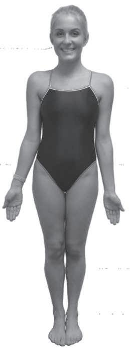
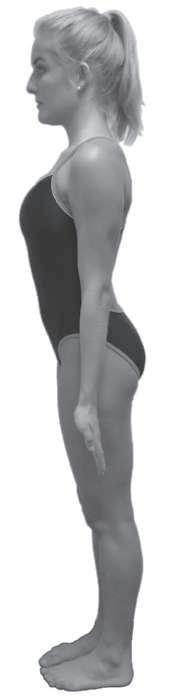
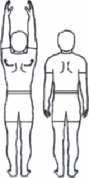
It is crucial for kinesiology students to begin with a reference point in order to better understand the musculoskeletal system, its planes of motion, joint classification, and joint movement terminology. Two reference positions can be used as a basis from which to describe joint movements. The anatomical position is the most widely used and is accurate for all aspects of the body. Fig. 1.1 demonstrates
this reference position, with the subject standing in an upright posture, facing straight ahead, with feet parallel and close and palms facing forward. The fundamental position is essentially the same as the anatomical position, except that the arms are at the sides with the palms facing the body.
Reference lines
To further assist in understanding the location of one body part in relation to another, certain imaginary reference lines may be used. Some examples follow in Fig 1.2.
Mid-axillary line: A line running vertically down the surface of the body passing through the apex of the axilla (armpit)
Mid-sternal line: A line running vertically down the surface of the body passing through the middle of the sternum
FIG. 1.1 • Anatomical position and anatomical directions. Anatomical directions refer to the position of one body part in relation to another.
Courtesy of R.T. Floyd (left, right)
Right mid-clavicular line
Right anterior axillary line
Mid-sternal line
Posterior axillary line
Scapular line
Vertebral line
Anterior axillary line: A line that is parallel to the mid-axillary line and passes through the anterior axillary skinfold
Posterior axillary line: A line that is parallel to the mid-axillary line and passes through the posterior axillary skinfold
Mid-clavicular line: A line running vertically down the surface of the body passing through the midpoint of the clavicle
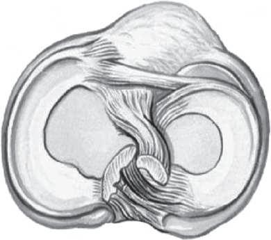
Anterior cruciate ligament
Medial meniscus
Medial tibial plateau
Mid-inguinal point: A point midway between the anterior superior iliac spine and the pubic symphysis
Scapula line: A line running vertically down the posterior surface of the body passing through the inferior angle of the scapula
Vertebral line: A line running vertically down through the spinous processes of the spine
Anatomical directional terminology FIGS. 1.1, 1.3, 1.4
It is important that we all be able to find our way around the human body. To an extent, we can think of this as similar to giving or receiving directions about how to get from one geographic location to another. Just as we use the terms left, right, south, west, northeast, etc. to describe geographic directions, we have terms such as lateral, medial, inferior, anterior, inferomedial, etc. to use for anatomical directions. With geographic directions we may use west to indicate the west end of a street or the western United States. The same is true when we use anatomical directions. We may use superior to indicate the end of a bone in our lower leg closest to the knee, or we may be speaking about the top of the skull. It all depends on the context at the time. Just as we combine south and east to get southeast for the purpose of indicating somewhere
Tibial tuberosity
Lateral meniscus
Lateral tibial plateau
Posterior cruciate ligament
Right knee, superior view with femur and patella removed
1.3 • Anatomical directional terminology.
FIG. 1.2 • Reference lines.
FIG.
Superolateral Superior
Lateral epicondyle
Lateral
Lateral femoral condyle
Lateral tibial condyle
Fibular head
Fibula
Inferolateral
Superomedial
Medial epicondyle
Patella
Medial
Medial femoral condyle
Medial tibial condyle
Tibial tuberosity
Tibia
Inferior
Right knee, anterior view
FIG. 1.4 • Anatomical directional terminology.
in between these directions, we may combine anterior and lateral to get anterolateral for the purpose of describing the general direction or location “in the front and to the outside.” Figs. 1.3 and 1.4 provide further examples.
Anterior: In front or in the front part
Anteroinferior: In front and below
Anterolateral: In front and to the outside
Anteromedial: In front and toward the inner side or midline
Anteroposterior: Relating to both front and rear
Anterosuperior: In front and above
Bilateral: Relating to the right and left sides of the body or of a body structure such as the right and left extremities
Caudal: Below in relation to another structure; inferior
Caudocephalad: Directionally from tail to head in the long axis of the body
Cephalic: Above in relation to another structure; higher, superior
Cephalocaudal: Directionally from head to tail in the long axis of the body
Contralateral: Pertaining or relating to the opposite side
Deep: Beneath or below the surface; used to describe relative depth or location of muscles or tissue
Dexter: Relating to, or situated to the right or on the right side of, something
Distal: Situated away from the center or midline of the body, or away from the point of origin
Inferomedial
Dorsal (dorsum): Relating to the back, being or located near, on, or toward the back, posterior part, or upper surface of; also relating to the top of the foot
Fibular: Relating to the fibular (lateral) side of the knee, leg, ankle, or foot
Inferior (infra): Below in relation to another structure; caudal
Inferolateral: Below and to the outside
Inferomedial: Below and toward the midline or inside
Ipsilateral: On the same side
Lateral: On or to the side; outside, farther from the median or midsagittal plane
Medial: Relating to the middle or center; nearer to the median or midsagittal plane
Median: Relating to, located in, or extending toward the middle; situated in the middle, medial
Palmar: Relating to the palm or volar aspect of the hand
Plantar: Relating to the sole or undersurface of the foot
Posterior: Behind, in back, or in the rear
Posteroinferior: Behind or in back and below
Posterolateral: Behind and to one side, specifically to the outside
Posteromedial: Behind and to the inner side
Posterosuperior: Behind or in back and above
Prone: Face-downward position of the body; lying on the stomach
Proximal: Nearest the trunk or the point of origin
Proximodistal: From the center of the body out toward the distal ends of appendages
Radial: Relating to the radial (lateral) side of the forearm or hand
Scapular plane: In line with the normal resting position of the scapula as it lies on the posterior rib cage; movements in the scapular plane are in line with the scapular, which is at an angle of 30 to 45 degrees from the frontal plane
Sinister: Relating to, or situated to the left or on the left side of, something
Superficial: Near the surface; used to describe relative depth or location of muscles or tissue
Superior (supra): Above in relation to another structure; higher, cephalic
Superolateral: Above and to the outside
Superomedial: Above and toward the midline or inside
Supine: Face-upward position of the body; lying on the back
Tibial: Relating to the tibial (medial) side of the knee, leg, ankle, or foot
Ulnar: Relating to the ulnar (medial) side of the forearm or hand
Ventral: Relating to the belly or abdomen, on or toward the front, anterior part of
Volar: Relating to palm of the hand or sole of the foot
Alignment variation terminology
Anteversion: Abnormal or excessive rotation forward of a structure, such as femoral anteversion
Kyphosis: Increased curving of the spine outward or backward in the sagittal plane
Lordosis: Increased curving of the spine inward or forward in the sagittal plane
Recurvatum: Bending backward, as in knee hyperextension
Retroversion: Abnormal or excessive rotation backward of a structure, such as femoral retroversion
Scoliosis: Lateral curving of the spine
Valgus: Outward angulation of the distal segment of a bone or joint, as in knock-knees
Varus: Inward angulation of the distal segment of a bone or joint, as in bowlegs
Planes of motion
When we study the various joints of the body and analyze their movements, it is helpful to characterize them according to specific planes of motion
(Fig. 1.5). A plane of motion may be defined as an imaginary two-dimensional surface through which a limb or body segment is moved.
There are three specific, or cardinal, planes of motion in which the various joint movements can be classified. The specific planes that divide the body exactly into two halves are often referred to as cardinal planes. The cardinal planes are the sagittal, frontal, and transverse planes. There are an infinite number of planes within each half that are parallel to the cardinal planes. This is best understood in the following examples of movements in the sagittal plane. Sit-ups involve the spine and, as a result, are performed in the cardinal sagittal plane, which is also known as the midsagittal or median plane. Biceps curls and knee extensions are performed in parasagittal planes, which are parallel to the midsagittal plane. Even though these latter examples are not in the cardinal plane, they are thought of as movements in the sagittal plane.
Although each specific joint movement can be classified as being in one of the three planes of motion, our movements are usually not totally in one specific plane but occur as a combination of motions in more than one plane. These movements in the combined planes may be described as occurring in diagonal, or oblique, planes of motion and involve joints that are capable of movement in two or more planes.
Sagittal,
anteroposterior, or AP plane
The sagittal, anteroposterior, or AP plane bisects the body from front to back, dividing it into right and left symmetrical halves. Generally, flexion and extension movements such as biceps curls, knee extensions, and sit-ups occur in this plane.
Frontal, coronal, or lateral plane
The frontal plane, also known as the coronal or lateral plane, bisects the body laterally from side to side, dividing it into front (ventral) and back (dorsal) halves. Abduction and adduction movements such as jumping jacks (shoulder and hip) and spinal lateral flexion occur in this plane.
Transverse, axial, or horizontal plane
The transverse plane, also known as the axial or horizontal plane, divides the body into superior (cephalic) and inferior (caudal) halves. Generally, rotational movements such as forearm pronation and supination and spinal rotation occur in this plane.
Sagittal plane (anteroposterior, AP)
Frontal axis (coronal, lateral, mediolateral)
Medial aspect
Lateral aspect
Sagittal axis (anteroposterior, AP)
Frontal plane (lateral, coronal)
Vertical axis (longitudinal, long)
Transverse plane (axial, horizontal)
FIG. 1.5 • Planes of motion and axes of rotation. A, Sagittal plane with frontal axis; B, Frontal plane with sagittal axis; C, Transverse plane with vertical axis.
Diagonal or oblique plane FIG.
1.6
The diagonal or oblique plane is a combination of more than one plane of motion. In reality, most of our movements in sporting activities fall somewhere between parallel and perpendicular to the previously described planes and occur in a diagonal plane. To further delineate, all movements in diagonal planes occur in a high diagonal plane or one of two low diagonal planes. The high diagonal plane is utilized for overhand movements in the upper extremity, whereas the two low diagonal planes are used to differentiate upper-extremity underhand movements from lower-extremity diagonal movements.
It should be noted that for a joint to move in a diagonal plane, the joint must be capable of movements in at least two planes. In other words, diagonal plane movements involve combining motions in one plane with motions of one or more other planes. Less commonly mentioned are diagonal movements that involve biaxial joints. For example,
combining wrist flexion and ulnar deviation or metacarpophalangeal extension with radial deviation when pointing with the index finger.
Axes of rotation
As movement occurs in a given plane, the joint moves or turns about an axis that has a 90-degree relationship to that plane. The axes are named in relation to their orientation (Fig. 1.5). Table 1.1 lists the planes of motion with their axes of rotation.
Frontal, coronal, lateral, or mediolateral axis
If the sagittal plane runs from anterior to posterior, then its axis must run from side to side. Since this axis has the same directional orientation as the frontal plane of motion, it is named similarly. As the elbow flexes and extends in the sagittal plane during a biceps curl, the forearm is actually rotating about a frontal axis that runs laterally through the elbow joint. The frontal axis may also be referred to as the bilateral axis.
Inferior
Diagonal plane of motion
Diagonal plane of motion
FIG. 1.6 • Diagonal planes and axes of rotation. A, Upper-extremity high diagonal plane movement and axis; B, Upper-extremity low diagonal plane movement and axis; C, Lower-extremity low diagonal plane movement and axis.
TABLE 1.1 • Planes of motion and their axes of rotation
Plane Description of plane Axis of rotation
Sagittal (anteroposterior or AP)
Frontal (coronal or lateral)
Transverse (axial, horizontal)
Divides the body into right and left halves
Divides the body into anterior and posterior halves
Divides the body into superior and inferior halves
Sagittal or anteroposterior axis
Frontal (coronal, lateral, or mediolateral)
Sagittal (anteroposterior or AP)
Vertical (longitudinal or long)
Movement occurring in the frontal plane rotates about a sagittal axis. This sagittal axis has the same directional orientation as the sagittal plane of motion and runs from front to back at a right angle to the frontal plane of motion. As the hip abducts and adducts during jumping jacks, the femur rotates about an axis that runs front to back through the hip joint.
Vertical or longitudinal axis
The vertical axis, also known as the longitudinal or long axis, runs straight down through the top of the head and is at a right angle to the transverse plane of motion. As the head rotates or turns from left to right when indicating disapproval, the skull and cervical vertebrae are rotating around an axis that runs down through the spinal column.
Description of axis
Common movements
Runs medial/lateral Flexion, extension
Runs anterior/ posterior Abduction, adduction
Runs superior/ inferior
Internal rotation, external rotation
Diagonal or oblique axis FIG. 1.6
The diagonal axis, also known as the oblique axis, runs at a right angle to the diagonal plane. As the glenohumeral joint moves from diagonal abduction to diagonal adduction in overhand throwing, its axis runs perpendicular to the plane through the humeral head.
Body regions
As mentioned later under the skeletal system, the body can be divided into axial and appendicular regions. Each of these regions may be further divided into different subregions, such as the cephalic, cervical, trunk, upper limbs, and lower limbs. Within each of these regions are many more subregions and specific regions. Table 1.2 details a breakdown of these regions and their common names, illustrated in Fig. 1.7.
TABLE 1.2 • Body parts and regions
Region name Common name Subregion
Cranial (skull)
Cephalic Head
Cervical Neck
Axial
Trunk
Facial (face)
Thoracic Thorax
Dorsal Back
Abdominal Abdomen
Pelvic Pelvis
Upper limbs Shoulder
Frontal Forehead
Occipital Base of skull
Orbital Eye
Otic Ear
Nasal Nose
Buccal Cheek
Oral Mouth
Mental Chin
Nuchal Posterior neck
Throat Anterior neck
Clavicular Collar bone
Pectoral Chest
Sternal Breastbone
Costal Ribs
Mammary Breast
Scapula Shoulder blade
Vertebral Spinal column
Lumbar Lower back or loin
Celiac Abdomen
Umbilical Navel
Inguinal Groin
Pubic Genital
Coxal Hip
Sacral Between hips
Gluteal Buttock
Perineal Perineum Appendicular
Acromial Point of shoulder
Omus Deltoid
Axillary Armpit
Brachial Arm
Olecranon Point of elbow
Cubital Elbow
Antecubital Front of elbow
Antebrachial Forearm
Lower limbs
Carpal Wrist Manual
Pedal Foot
Palmar Palm
Dorsal Back of hand
Digital Finger
Femoral Thigh
Patella Kneecap
Popliteal Back of knee
Sural Calf
Crural
Leg
Talus Ankle
Calcaneal Heel
Dorsum Top of foot
Tarsal
Plantar
Digital
Instep
Sole
Toe
Nasal (nose)
Otic (ear)
Oral (mouth)
Cervical (neck)
Clavicular (collar bone)
Acromial (point of shoulder)
Axillary (armpit)
Mammary (breast)
Brachial (arm)
Antecubital (front of elbow)
Celiac or abdominal (abdomen)
Antebrachial (forearm)
Carpal (wrist)
Palmar (palm)
Digital (finger)
Femoral (thigh)
Patellar (kneecap)
Anterior crural (leg)
Talus (ankle)
Dorsum of the foot
Cephalic (head)
Frontal (forehead)
Orbital (eye)
Buccal (cheek)
Mental (chin)
Sternal Pectoral region (chest)
Anterior cubital (cubital fossa)
Inguinal (groin)
Posterior thoracic
Coxal (hip)
Genital
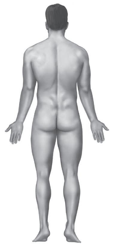
FIG. 1.7 • Body regions. A, Anterior view; B, Posterior view.
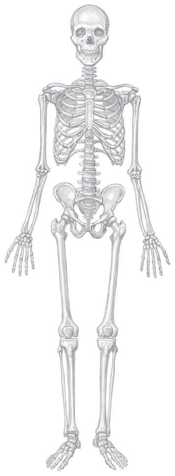
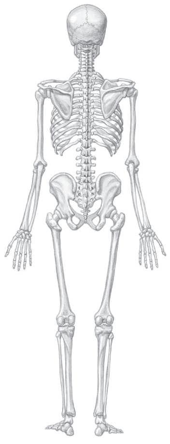
Skeletal systems
Fig. 1.8 shows anterior and posterior views of the skeletal system. Some 206 bones make up the skeletal system, which provides support and protection for other systems of the body and provides for attachments of the muscles to the bones, by which movement is produced. Additional skeletal functions are mineral storage and hemopoiesis, which involves blood cell formation in the red bone marrow. The skeleton may be divided into the appendicular and the axial skeletons. The appendicular skeleton is composed of the appendages, or the upper and lower extremities, and the shoulder and pelvic girdles. The axial skeleton
Cranial (surrounding the brain)
Occipital (base of skull)
Nuchal (posterior neck)
Scapula (shoulder blade)
Shoulder
Vertebral (spinal column)
Brachial (arm)
Abdominal
Olecranon (point of elbow)
Lumbar (lower back or loin)
Dorsum of the hand
Gluteal (buttock)
Sacral
Perineal
Femoral (thigh)
Popliteal fossa (back of knee)
Sural (calf)
Peroneal (fibular)
Plantar (sole)
consists of the skull, vertebral column, ribs, and sternum. Most students who take this course will have had a course in human anatomy, but a brief review is desirable before beginning the study of kinesiology. Later chapters provide additional information and more detailed illustrations of specific bones.
Osteology
The adult skeleton, consisting of approximately 206 bones, may be divided into the axial skeleton and the appendicular skeleton. The axial skeleton contains 80 bones, which include the skull, spinal column, sternum, and ribs. The appendicular skeleton contains
Tarsal (instep)
Digital (toe)
Navel
Throat
Skull
Frontal bone
Zygomatic bone
Manubrium
Coracoid process
Humeral head
Sternum
Rib cage
Ribs (12 pairs)
Medial epicondyle
Radial head
Radial tuberosity
Pelvic girdle
Iliac crest
Ilium
Femoral head
Obturator foramen
Ischium
Pubis
Tibial tuberosity
Medial malleolus
Parietal bone
Temporal bone
Maxilla
Mandible
Spine of scapula Superior angle
Clavicle
Acromion process
Scapula
Greater tubercle
Lesser tubercle
Costal cartilages
Xiphoid process
Humerus
Vertebral column
Ulna
Os coxa
Sacrum
Coccyx
Radius
Carpal bones (8)
Metacarpal bones (5)
Greater trochanter
Lesser trochanter
Ischial tuberosity
Femur
Patella
Fibula head
Tibia
Fibula
Lateral malleolus
Talus
Calcaneus
Tarsal bones (7)
Metatarsal bones (5)
Phalanges (5)
FIG. 1.8 • Skeleton. A, Anterior view; B, Posterior view.
126 bones, which include all the bones of the upper and lower extremities. The pelvis is sometimes classified as being part of the axial skeleton due to its importance in linking the axial skeleton with the lower extremities of the appendicular skeleton. The exact number of bones as well as their specific features occasionally varies from person to person.
Skeletal functions
The skeleton has five major functions:
1. Protection of vital soft tissues such as the heart, lungs, and brain
Occipital bone
Occipital protuberance
Cervical vertebrae (7)
Thoracic vertebrae (12)
Axillary border
Vertebral border
Inferior angle
Lumbar vertebrae (5)
Lateral epicondyle
Olecranon process of ulna
Pelvic girdle
Greater trochanter
Lesser trochanter
Phalanges (5)
Medial femoral condyle
Lateral femoral condyle
2. Support to maintain posture
3. Movement by serving as points of attachment for muscles and acting as levers
4. Storage for minerals such as calcium and phosphorus
5. Hemopoiesis, which is the process of blood formation that occurs in the red bone marrow located in the vertebral bodies, femur, humerus, ribs, and sternum
Types of bones
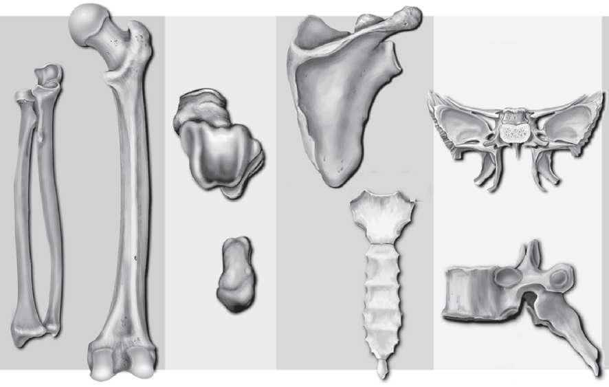
Bones vary greatly in shape and size but can be classified in five major categories (Fig. 1.9).
Long bones: Composed of a long cylindrical shaft with relatively wide, protruding ends; serve as levers. The shaft contains the medullary cavity. Examples include phalanges, metatarsals, metacarpals, tibia, fibula, femur, radius, ulna, and humerus.
Short bones: Small cube-shaped, solid bones that usually have a proportionally large articular surface in order to articulate with more than one bone. Short bones provide some shock absorption and include the carpals and tarsals.
Flat bones: Usually having a curved surface and varying from thick (where tendons attach) to very thin. Flat bones generally provide protection and include the ilium, ribs, sternum, clavicle, and scapula.
Irregular bones: Irregular-shaped bones serve a variety of purposes and include the bones throughout the entire spine and the ischium, pubis, and maxilla.
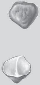
Sesamoid bones: Small bones embedded within the tendon of a musculotendinous unit that provide protection as well as improve the mechanical advantage of musculotendinous units. In addition to the patella, there are small sesamoid bones within the flexor tendons of the great toe and the thumb. Sesamoid
bones are sometimes referred to as accessory bones however, accessory ossicles have no known function and often originate from unfused ossification centers. Both sesamoid and accessory bones occur in varying numbers from one individual to the next. They are most commonly found in smaller joints in the distal extremities of the foot, ankle, and hand but are not always symmetrical in an individual.
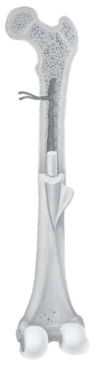
Typical bony features
Long bones possess features that are typical of bones in general, as illustrated in Fig. 1.10. Long bones have a shaft or diaphysis, which is the long cylindrical portion of the bone. The diaphysis wall, formed from hard, dense, compact bone, is the cortex. The outer surface of the diaphysis is covered by a dense, fibrous membrane known as the periosteum. A similar fibrous membrane known as the endosteum covers the inside of the cortex. Between the walls of the diaphysis lies the medullary or marrow cavity, which contains yellow or fatty marrow. At each end of a long bone is the epiphysis, which is usually enlarged and shaped specifically to join with the epiphysis of an adjacent bone at a joint. The epiphysis is formed from spongy or cancellous or trabecular bone. During bony growth the diaphysis and the epiphysis are
Femur
Talus
Scapula
Sternum
Sphenoid bone
Vertebra
Patella posterior view
Great toe sesamoid
Patella anterior view
Capitate (carpal) bone
Radius
Ulna Long Short Flat Irregular Sesamoid
Sesamoid bone
FIG. 1.9 • Classification of bones by shape.
Articular cartilage
Spongy bone
Space occupied by red marrow
Endosteum
Cortex
Medullary cavity
Yellow marrow
Periosteum
Epiphyseal plates
FIG. 1.10 • Major parts of a long bone.
Proximal epiphysis
Diaphysis
Diaphysis
Epiphyseal plate
Epiphysis
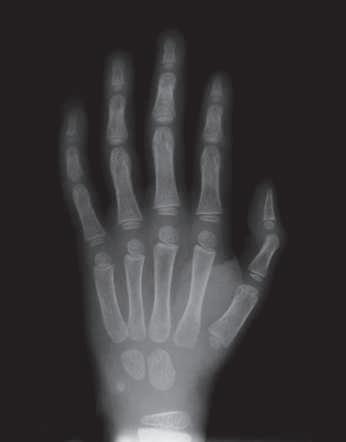
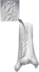
Epiphyseal plates
FIG. 1.11 • The presence of epiphyseal plates, as seen in a radiograph of a child’s hand, indicates that the bones are still growing in length.
TABLE
1.3 • Epiphyseal closure timetables
Approximate age
Distal epiphysis
separated by a thin plate of cartilage known as the epiphyseal plate, commonly referred to as a growth plate (Fig. 1.11). As skeletal maturity is reached, on a timetable that varies from bone to bone as detailed in Table 1.3, the plates are replaced by bone and are closed. To facilitate smooth, easy movement at joints, the epiphysis is covered by articular or hyaline cartilage, which provides a cushioning effect and reduces friction.
Bone development and growth
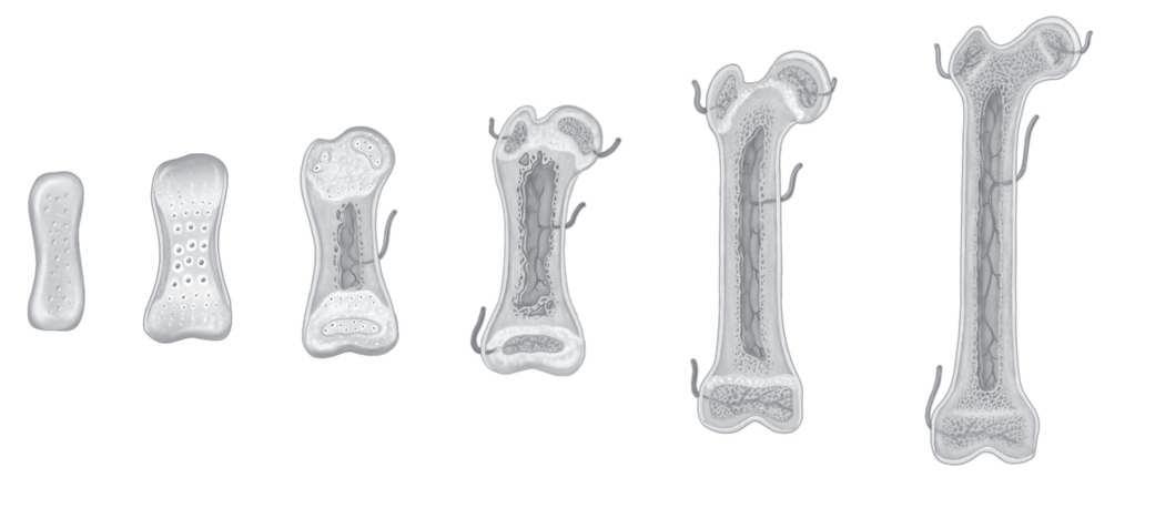
Most of the skeletal bones of concern to us in structural kinesiology are endochondral bones, which develop from hyaline cartilage. As we develop from an embryo, these hyaline cartilage masses grow rapidly into structures shaped similarly to the bones they will eventually become. This growth continues, and the cartilage gradually undergoes
7–8
15–17
18–19
About 20
20–25
25
Bones
Inferior rami of pubis and ischium (almost complete)
Scapula, lateral epicondyle of humerus, olecranon process of ulna
Medial epicondyle of humerus, head and shaft of radius
Humeral head, distal ends of radius and ulna, distal ends of femur and fibula, proximal end of tibia
Acetabulum in pelvis
Vertebrae and sacrum, clavicle, proximal end of fibula, sternum and ribs
Adapted from Goss CM: Gray’s anatomy of the human body, ed 29, Philadelphia, 1973, Lea & Febiger.
significant change to develop into long bone, as detailed in Fig. 1.12.
Bones continue to grow longitudinally as long as the epiphyseal plates are open. These plates begin closing around adolescence and disappear. Most close by age 18, but some may be open until age 25. Growth in diameter continues throughout life. This is done by an internal layer of periosteum building new concentric layers on old layers. Simultaneously, bone around the sides of the medullary
Femur
©Jim
Wehtje/Getty Images
Cartilaginous model
Developing periosteum
Compact bone developing
Calcified cartilage
Primary ossification center
Blood vessel
Secondary ossification center
Remnants of epiphyseal plates
Epiphyseal plates
Medullary cavity
Medullary cavity
Compact bone
Epiphyseal plate
Secondary ossification center
Articular cartilage
Spongy bone
Medullary cavity
Remnant of epiphyseal plate
Spongy bone
Articular cartilage
FIG. 1.12 • Major stages a–f in the development of an endochondral bone (relative bone sizes not to scale).
Epiphyseal growth
Growth in cartilage surrounding epiphysis
Cartilage replaced by bone
Bone remodeled
Growth in length
Cartilage growth in epiphyseal plate
Cartilage replaced by bone
Bone remodeled
Bone resorption
Growth in diameter
Bone addition
Bone resorption
FIG. 1.13 • Remodeling of a long bone. (b) (c)
Growing bone
cavity is resorbed so that the diameter is continually increased. New bone is formed by specialized cells known as osteoblasts, whereas the cells that resorb old bone are osteoclasts. This bone remodeling, as depicted in Fig. 1.13, is necessary for continued bone growth, changes in bone shape, adjustment of bone to stress, and bone repair.
Bone properties
Articular cartilage
Epiphyseal line
Calcium carbonate, calcium phosphate, collagen, and water are the basis of bone composition. About 60% to 70% of bone weight is made up of calcium carbonate and calcium phosphate, with water making up approximately 25% to 30% of bone weight.
Adult bone
Collagen provides some flexibility and strength in resisting tension. Aging causes progressive loss of collagen and increases bone brittleness, resulting in increased likelihood of fractures.
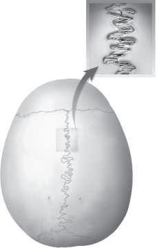
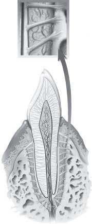
Most outer bone is cortical; cancellous bone is underneath. Cortical bone is harder and more compact, with only about 5% to 30% of its volume being porous, with nonmineralized tissue. In contrast, cancellous bone is spongy, with around 30% to 90% of its volume being porous. Cortical bone is stiffer; it can withstand greater stress, but less strain, than cancellous bone. Due to its sponginess, cancellous bone can undergo greater strain before fracturing.
Bone size and shape are influenced by the direction and magnitude of forces that are habitually applied to them. Bones reshape themselves based on the stresses placed upon them, and their mass increases over time with increased stress.
This concept of bone adaptation to stress is known as Wolff’s law, which essentially states that bone in a healthy individual will adapt to the loads it is placed under. When a particular bone is subjected to increased loading, the bone will remodel itself over time to become stronger to resist that particular type of loading. As a result, the external cortical portion of the bone becomes thicker. The opposite is also true: when the loading on a bone decreases, the bone will become weaker.
Bone markings
Bones have specific markings that exist to enhance their functional relationship with joints, muscles, tendons, nerves, and blood vessels. Many of these markings serve as important bony landmarks in determining muscle location and attachment and joint function. Essentially, all bone markings may be divided into
1. Processes (including elevations and projections), which either form joints or serve as a point of attachment for muscles, tendons, or ligaments, and
2. Cavities (depressions), which include openings and grooves that contain tendons, vessels, nerves, and spaces for other structures.
Detailed descriptions and examples of many bony markings are provided in Table 1.4.
Types of joints
The articulation of two or more bones allows various types of movement. The extent and type of
movement determine the name applied to the joint. Bone structure limits the kind and amount of movement in each joint. Some joints or arthroses have no movement, others are only very slightly movable, and others are freely movable with a variety of movement ranges. The type and range of movements are similar in all humans; but the freedom, range, and vigor of movements are limited by the configuration of the bones where they fit together, and by ligaments and muscles.
Articulations may be classified according to the structure or function. Classification by structure places joints into one of three categories: fibrous, cartilaginous, or synovial. Functional classification also results in three categories: synarthrosis (synarthrodial), amphiarthrosis (amphiarthrodial), and diarthrosis (diarthrodial). There are subcategories in each classification. Due to the strong relationship between structure and function, there is significant overlap between the classification systems. That is, there is more similarity than difference between the two members in each of the following pairs: fibrous and synarthrodial joints, cartilaginous and amphiarthrodial joints, and synovial and diarthrodial joints. However, not all joints fit neatly into both systems. Table 1.5 provides a detailed listing of all joint types according to both classification systems. Since this text is concerned primarily with movement, the more functional system (synarthrodial, amphiarthrodial, and diarthrodial joints) will be used throughout, following a brief explanation of structural classification.
Fibrous joints are joined together by connective tissue fibers and are generally immovable. Subcategories are suture and gomphosis, which are immovable, and syndesmosis, which allows a slight amount of movement. Cartilaginous joints are joined together by hyaline cartilage or fibrocartilage, which allows very slight movement. Subcategories include synchondrosis and symphysis. Synovial joints are freely movable and generally are diarthrodial. Their structure and subcategories are discussed in detail under diarthrodial joints.
The articulations are grouped into three classes based primarily on the amount of movement possible, with consideration given to their structure.
Synarthrodial (immovable) joints FIG. 1.14
Structurally, these articulations are divided into two types:
Suture
Found in the sutures of the cranial bones. The sutures of the skull are truly immovable beyond infancy.
Processes that form joints
Marking
Condyle
Facet
Processes to which muscles, tendons, or ligaments attach
Large, rounded projection that usually articulates with another bone
Small, flat or nearly flat surface
Head Prominent, rounded projection of the proximal end of a bone, usually articulating
Angle Bend or protruding angular projection
Border or margin Edge or boundary line of a bone
Crest Prominent, narrow, ridgelike projection
Epicondyle
Projection located above a condyle
Line Ridge of bone less prominent than a crest
Process Any prominent projection
Ramus
Part of an irregularly shaped bone that is thicker than a process and forms an angle with the main body
Spine (spinous process) Sharp, slender projection
Suture Line of union between bones
Trochanter Very large projection
Tubercle Small, rounded projection
Tuberosity
Facet
Cavities (depressions)
Large, rounded or roughened projection
Flattened or shallow articulating surface
Foramen Rounded hole or opening in bone
Fossa Hollow, depressed, or flattened surface
Fovea Very small pit or depression
Meatus
Tubelike passage within a bone
Notch Depression in the margin of a bone
Sinus Cavity or hollow space within a bone
Sulcus (groove)
Furrow or groovelike depression on a bone
Medial or lateral condyle of femur 266
Articular facet of vertebra
331
Head of femur, head of humerus 220, 222, 224, 117
Superior and inferior angle of scapula 92, 94
Lateral and medial border of scapula 92, 94
Iliac crest of pelvis 222
Medial or lateral epicondyle of humerus
150
Linea aspera of femur 222
Acromion process of scapula, olecranon process of humerus 92, 94, 117, 118 150
Superior and inferior ramus of pubis 221
Spinous process of vertebra, spine of scapula 330, 331, 94
Sagittal suture between parietal bones of skull 16
Greater or lesser trochanter of femur 220, 222
Greater and lesser tubercles of humerus 117
Radial tuberosity, tibial tuberosity 150, 266
Intervertebral facets in cervical, thoracic, and lumbar spine 331
Obturator foramen in pelvis 220, 221
Supraspinatus fossa, iliac fossa 94, 220
Fovea capitis of femur 224
External auditory meatus of temporal bone 346
Trochlear and radial notch of the ulna 150
Frontal sinus
Intertubercular (bicipital) groove of humerus 117
TABLE 1.5 • Joint classification by structure and function
Structural classification
Fibrous Cartilaginous
Synarthrodial Gomphosis Suture
Amphiarthrodial Syndesmosis
Functional classification
Diarthrodial
Symphysis Synchondrosis
Gomphosis
Synovial
Arthrodial
Condyloidal
Enarthrodial Ginglymus
Sellar
Trochoidal
Found in the sockets of the teeth. The socket of a tooth is often referred to as a gomphosis (type of joint in which a conical peg fits into a socket). Normally, there should be essentially no movement of the teeth in the mandible or maxilla.
Amphiarthrodial (slightly movable) joints FIG. 1.15
Structurally, these articulations are divided into three types:
Syndesmosis
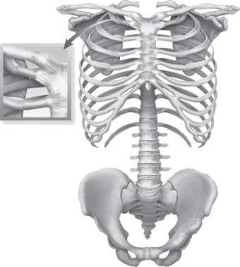
Type of joint held together by strong ligamentous structures that allow minimal movement between the bones. Examples are the coracoclavicular joint and the inferior tibiofibular joint.
Symphysis
Type of joint separated by a fibrocartilage pad that allows very slight movement between the bones. Examples are the symphysis pubis and the intervertebral disks.
Intervertebral disk (fibrocartilage)
Interpubic disk (fibrocartilage)
FIG. 1.15 • Amphiarthrodial joints. A, Syndesmosis joint; B, Symphysis joint;
FIG. 1.14 • Synarthrodial joints.
Fibrous connective tissue
Suture
Gomphosis
Fibrous connective tissue
Body of vertebra
Clavicle
Rib
Costal cartilage
Sternum
Synchondrosis
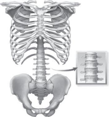
Type of joint separated by hyaline cartilage that allows very slight movement between the bones. Examples are the costochondral joints of the ribs with the sternum.
Diarthrodial
(freely
movable) joints FIG. 1.16
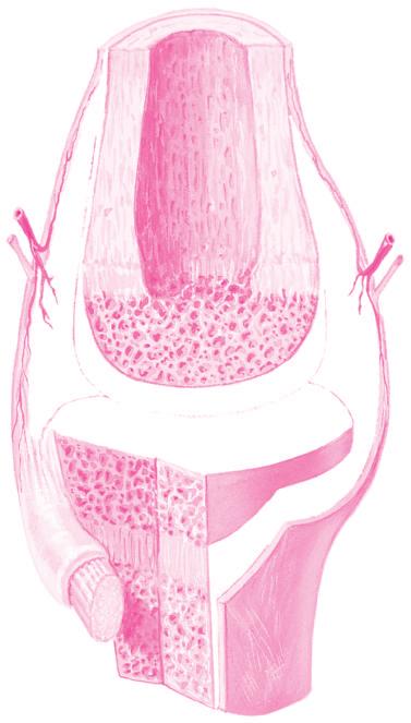
Diarthrodial joints, also known as synovial joints, are freely movable. A sleevelike covering of ligamentous tissue known as the joint capsule surrounds the bony ends forming the joints. This ligamentous capsule is lined with a thin vascular synovial capsule that secretes synovial fluid to lubricate the area inside the joint capsule, known as the joint cavity. In certain areas the capsule is thickened to form tough, nonelastic ligaments that provide additional support against abnormal movement or joint opening. These ligaments vary in location, size, and strength depending upon the particular joint. Ligaments, in connecting bones to bones, provide static stability to joints.
In many cases, additional ligaments, not continuous with the joint capsule, provide further
support. In some cases, these additional ligaments may be contained entirely within the joint capsule; or intraarticularly, such as the anterior cruciate ligament in the knee; or extraarticularly, such as the fibular collateral ligament of the knee, which is outside the joint capsule.
The articular surfaces on the ends of the bones inside the joint cavity are covered with layers of articular or hyaline cartilage that helps protect the ends of the bones from wear and damage. This cartilage is quite resilient because it is slightly compressible and elastic, which enables it to absorb compressive and shear forces. The articular surface, thanks in part to lubrication from synovial fluid, has a very low amount of friction and is very durable. When the joint surfaces are unloaded or distracted, this articular cartilage slowly absorbs a slight amount of the joint synovial fluid, only to slowly secrete it during subsequent weight bearing and compression. Articular cartilage has a very limited blood supply and as a result depends on joint movement to provide its nutrition through this synovial flow. Therefore, maintaining and utilizing
FIG. 1.16 • Structure of a diarthrodial synovial joint.
Bursa
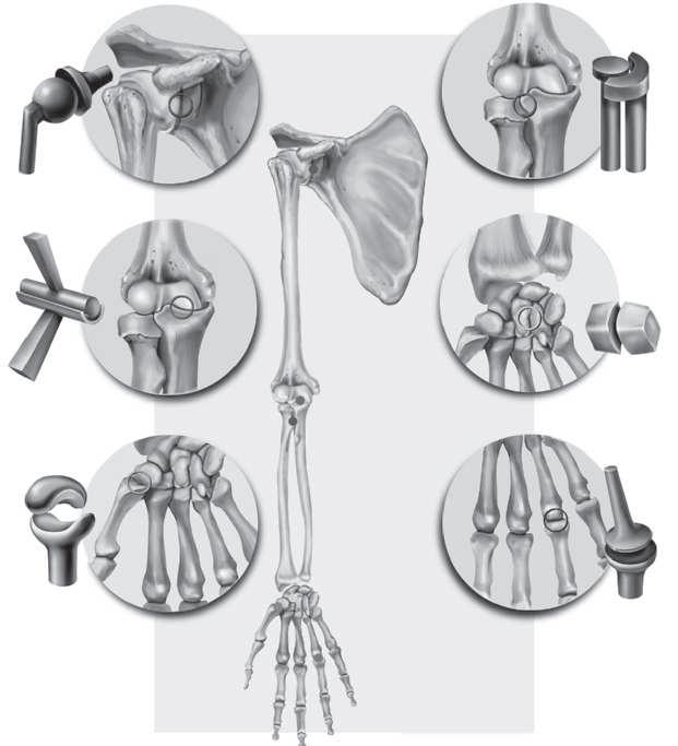
a joint through its normal range of motion are important to sustaining joint health and function. Additionally, some diarthrodial joints have a fibrocartilage disk between their articular surfaces to provide additional shock absorption and load distribution, and to further enhance joint stability. Examples are the knee’s medial and lateral menisci and the acetabular and glenoid labrum of the hip and shoulder joints, respectively. Structurally, this type of articulation can be divided into six groups, as shown in Fig. 1.17.
Diarthrodial joints have motion possible in one or more planes. Those joints having possible motion in one plane are said to have one degree of freedom of motion, whereas joints having motion in two and three planes are described as having two and three degrees of freedom of motion, respectively. Refer to Table 1.6 for a comparison of diarthrodial joint features by subcategory.
Arthrodial (gliding, plane) joint
This joint type is characterized by two flat, or plane, bony surfaces that butt against each other. This type of joint permits limited gliding movement.
Examples are the carpal bones of the wrist and the tarsometatarsal joints of the foot.
Condyloidal (ellipsoid, ovoid, biaxial ball-andsocket) joint
This is a type of joint in which the bones permit movement in two planes without rotation. Examples are the wrist (radiocarpal joint) between the radius and the proximal row of the carpal bones or the second, third, fourth, and fifth metacarpophalangeal joints.
Enarthrodial (spheroidal, multiaxial ball-andsocket) joint
This type of joint is most like a true ball-and-socket in that it permits movement in all planes. Examples are the shoulder (glenohumeral) and hip (acetabularfemoral) joints.
Ginglymus (hinge) joint
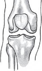
This is a type of joint that permits a wide range of movement in only one plane. Examples are the elbow (humeroulnar), ankle (talocrural), and knee (tibiofemoral) joints.
FIG. 1.17 • Types of diarthrodial or synovial joints.
Ball-and-socket joint (glenohumeral)
Hinge joint (humeroulnar)
Gliding joint (intercarpal)
Pivot joint (radioulnar)
Saddle joint (carpometacarpal)
Ellipsoid joint
Enarthrodial
Ginglymus
Arthrodial
Trochoidal
Sellar
Condyloidal (metacarpophalangeal)
Head of humerus
Scapula
Radius Ulna
Carpal bone
Metacarpal bone
Phalanx
Metacarpal bone
Humerus
Ulna
Carpal bones
TABLE 1.6 • Diarthrodial joint classification
Classification name Number of axes Degrees of freedom Typical movements
Ginglymus (hinge)
Trochoidal (pivot, screw)
Condyloidal (ellipsoid, ball-and-socket, ovoid)
Arthrodial (gliding, plane)
Enarthrodial (ball-and-socket, spheroidal)
Sellar (saddle)
Uniaxial One
Flexion, extension
Internal rotation, external rotation
Biaxial Two
Multiaxial Three
Flexion, extension, abduction, adduction
Flexion, extension, abduction, adduction, internal rotation, external rotation
Sellar (saddle) joint
This type of reciprocal reception is found in the thumb at the carpometacarpal joint and permits ball-and-socket movement, with the exception of slight rotation. Some anatomists also classify the sternoclavicular joint as a sellar joint.
Trochoidal (pivot, screw) joint
This is a type of joint with a rotational movement around a long axis. An example is the rotation of the radius on the ulna at the proximal and distal radioulnar joints.
Stability
and mobility of diarthrodial joints
Generally, the more mobile a joint is, the less stable it is, and vice versa. This is true when comparing the same joints between individuals, but also when comparing one joint versus another in the same individual. Both heredity and developmental factors (Wolff’s law for bone and Davis’s law for soft tissue) contribute to these variances. In a manner similar to the adaption of bone to loading, as previously discussed in Wolff’s law, soft tissue also adapts to stress or the lack thereof. This corollary to Wolff’s law is known as Davis’s law, which essentially states that ligaments, muscle,
Joint examples
Elbow joint (humeroulnar)
Ankle joint (talocrural)
Proximal and distal radioulnar joint
Plane for examples Axis for examples
Sagittal Frontal
Atlantoaxial joint Transverse Vertical
Wrist (radiocarpal)
2nd–5th
metacarpophalangeal joints
Transverse tarsal joint
Vertebral facets in spine
Sagittal Frontal Frontal Sagittal
Intercarpal joints in wrist Variable Frontal Variable Variable Sagittal Variable
Glenohumeral joint
Hip joint (acetabularfemoral)
1st carpometacarpal joint
Sagittal Frontal Transverse Frontal Sagittal Vertical
Sagittal Frontal Transverse Frontal Sagittal Vertical
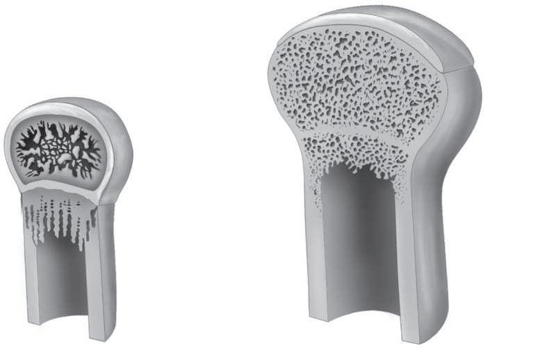
and other soft tissue when placed under appropriate tension will adapt over time by lengthening, and conversely, when maintained in a loose or shortened state over a period of time will gradually shorten. Five major factors affect the total stability, and consequently the mobility, of a joint (see Fig. 1.18).
• Bones—Although bones are usually very similar in bilateral comparisons within an individual, the actual anatomical configuration at the joint surfaces in terms of depth and shallowness may vary significantly between individuals.
• Cartilage—The structures of both hyaline cartilage and specialized cartilaginous structures, such as the knee menisci, glenoid labrum, and acetabular labrum, further assist in joint congruency and stability. As with bones, these structures normally are the same in bilateral comparisons within an individual, but may vary between individuals in size and configuration.
• Ligaments and connective tissue—Ligaments and connective tissue provide static stability to joints. As with bones and cartilage, variances exist between individuals in the degree of restrictiveness of ligamentous tissue. An
• Bony architecture
• Cartilaginous structure
• Ligamentous and connective tissue laxity
• Muscle strength, endurance, and flexibility
• Proprioception and motor control
Diarthrodial joint mobility
individual’s amount of hypo- or hyperlaxity is primarily due to the proportional amount of elastin versus collagen within the joint structures. Simply put, individuals with proportionally higher elastin-to-collagen ratios are hyperlax, or “loose-jointed,” whereas individuals with proportionally lower ratios are tighter.
• Muscles—Muscles provide dynamic stability to joints when actively contracting. Without active tension via contraction, muscles provide minimal static stability. Consequently, strength and endurance are significant factors in stabilizing joints, whereas muscle flexibility may affect the total range of joint motion possible.
• Proprioception and motor control—Proprioception is the subconscious mechanism by which the body is able to regulate posture and movements by responding to stimuli originating in the proprioceptors embedded in joints, tendons, muscles, and the inner ear. Motor control is the process by which bodily actions and movements are organized and executed. To determine the appropriate amount of muscular forces and joint activations needed, sensory information from the environment and the body must be integrated and then coordinated in a cooperative manner between the central nervous system and the musculoskeletal system. Muscle strength and endurance are not very useful in providing joint stability unless they can be activated precisely when needed.
The integrity of any of these structures may be affected by acute or chronic injury. These structures adapt over time both positively and negatively to the specific biomechanical demands placed upon them. When any of the above factors are compromised, additional demands are placed on the
remaining structures to provide stability, which in turn may compromise their integrity, resulting in abnormal mobility. This abnormal mobility, whether hypermobility or hypomobility, may lead to further pathological conditions such as tendinitis, bursitis, arthritis, internal derangement, and joint subluxations.
Movements in joints
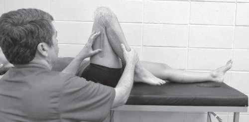
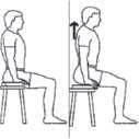
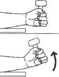
In many joints, several different movements are possible. Some joints permit only flexion and extension; others permit a wide range of movements, depending largely on the joint structure. We refer to the area through which a joint may normally be freely and painlessly moved as the range of motion (ROM). The specific amount of movement possible in a joint or range of motion may be measured by using an instrument known as a goniometer to compare the change in joint angles. The goniometer has a moving arm, a stationary arm, and an axis or fulcrum. Measuring the available range of motion in a joint or the angles created by the bones of a joint is known as goniometry
The goniometer axis, or hinge point, is placed even with the axis of rotation at the joint line. The stationary arm is held in place either along or parallel to the long axis of the more stationary bone (usually the more proximal bone), and the moving arm is placed either along or parallel to the long axis of the bone that moves the most (usually the more distal bone). The joint angle can then be read from the goniometer, as shown in Fig. 1.19. As an example, we could measure the angle between the femur and the trunk in the anatomical position (which would usually be zero), and
FIG. 1.18 • Factors affecting diarthrodial joint stability.
then ask the person to flex the hip as far as possible. If we measured the angle again at full hip flexion, we would find a goniometer reading of around 130 degrees.
Depending on the size of the joint and its movement potential, different goniometers may be more or less appropriate. Fig. 1.20 depicts a variety of goniometers that may be utilized to determine the range of motion for a particular joint. Inclinometers may also be used to measure range of motion, particularly in the spine.
Please note that the normal range of motion for a particular joint varies to some degree from person to person. Appendix 1 provides the average normal ranges of motion for all joints.
When using movement terminology, it is important to understand that the terms are used to describe the actual change in position of the bones

relative to each other. That is, the angles between the bones change, whereas the movement occurs between the articular surfaces of the joint. We may say, in describing knee movement, “flex the leg at the knee”; this movement results in the leg moving closer to the thigh. Some describe this as leg flexion occurring at the knee joint and may say “flex the leg,” meaning flex the knee. Additionally, movement terms are utilized to describe movement occurring throughout the full range of motion or through a very small range. Using the knee flexion example again, we may flex the knee through the full range by beginning in full knee extension (zero degrees of knee flexion) and flexing it fully, so that the heel comes in contact with the buttocks; this would be approximately 140 degrees of flexion. We may also begin with the knee in 90 degrees of flexion and then flex it 30 degrees more; this movement results in a knee flexion angle of 120 degrees, even though the knee flexed only 30 degrees. In both examples, the knee is in different degrees of flexion. We may also begin with the knee in 90 degrees of flexion and extend it 40 degrees, which would result in a flexion angle of 50 degrees. Even though we extended the knee, it is still flexed, only less so than before.
In this example, we more commonly move the distal extremity in relation to the proximal extremity, which is usually more stationary. However, there are examples in every joint where the distal segment may be more stationary and we move the proximal segment in relation to it. An example is the knee in doing a squat from the standing position. As the squat occurs, the thigh moves toward the stabler leg, still resulting in knee flexion that could be stated as flexing the thigh at the knee.
Some movement terms may be used to describe motion at several joints throughout the body, whereas other terms are relatively specific to a joint or group of joints (Fig. 1.21). Rather than list the terms alphabetically, we have chosen to group them according to the body area and pair them with opposite terms where applicable. Additionally, the prefixes hyper- and hypo- may be combined with these terms to emphasize motion beyond and below normal, respectively. Of these combined terms, hyperextension is the most commonly used.
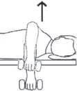
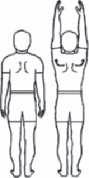
Terms describing general movements
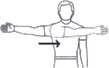
Abduction: Lateral movement away from the midline of the trunk in the frontal plane. An example is
FIG. 1.19 • Goniometric measurement of knee joint flexion.
FIG. 1.20 • Various goniometers used for measuring joint range of motion.
Courtesy of R.T. Floyd
Courtesy of R.T. Floyd
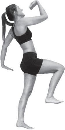
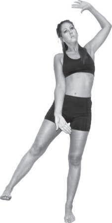
FIG. 1.21 • Joint movements. A, Examples of sagittal plane movements: extension of left toes, ankle (plantar flexion), knee, hip, shoulder, elbow, wrist, fingers, lumbar and cervical spine; flexion of right toes, ankle (dorsiflexion), knee, hip, shoulder, elbow, wrist, and fingers. B, Examples of frontal plane movements: abduction of left transverse tarsal/ subtalar joints (eversion), shoulder, wrist, fingers, and shoulder girdle (upward rotation), lumbar (lateral flexion to right) and cervical spine (lateral flexion to left), and right hip; adduction of right transverse tarsal/subtalar joints (inversion), shoulder, wrist, fingers, and shoulder girdle (downward rotation). C, Examples of transverse plane movements: internal rotation of right shoulder, right radioulnar joints (pronation); external rotation of right knee, right hip, left shoulder, left radioulnar joints (supination); and lumbar (left rotation) and cervical spine (left rotation).
raising the arms or thighs to the side away from the anatomical position.
Adduction: Movement medially toward and/or across the midline of the trunk in the frontal plane. An example is lowering the arm to the side or the thigh back to the anatomical position.
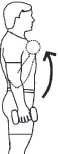
Flexion: Bending movement that results in a decrease of the angle in a joint by bringing bones together, usually in the sagittal plane. An example is the elbow joint when the hand is drawn to the shoulder.
Extension: Straightening movement that results in an increase of the angle in a joint by moving bones apart, usually in the sagittal plane. Using the elbow, an example is when the hand moves away from the shoulder.
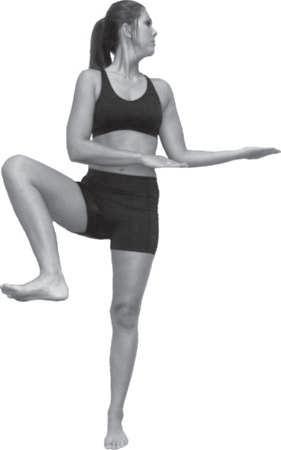
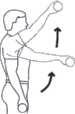
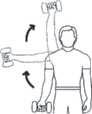
Circumduction: Circular movement of a limb that delineates an arc or describes a cone. It is a combination of flexion, extension, abduction, and adduction. Sometimes referred to as circumflexion. An example is when the shoulder joint or the hip joint moves in a circular fashion around a fixed point, either clockwise or counterclockwise.
Diagonal abduction: Movement by a limb through a diagonal plane away from the midline of the body, such as in the hip or glenohumeral joint.
Diagonal adduction: Movement by a limb through a diagonal plane toward and across the midline of the body, such as in the hip or glenohumeral joint.
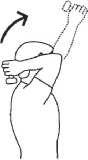
External rotation: Rotary movement around the longitudinal axis of a bone away from the midline of the body. Occurs in the transverse plane and is also known as rotation laterally, outward rotation, and lateral rotation.
Internal rotation: Rotary movement around the longitudinal axis of a bone toward the midline of the body. Occurs in the transverse plane and is also known as rotation medially, inward rotation, and medial rotation.
Terms describing ankle and foot movements
Eversion: Turning the subtalar and traverse tarsal joints of the foot outward or laterally in the frontal plane; abduction, characterized by the soles of the feet being turned outward. An example is standing with the weight on the inner edge of the foot.
Courtesy of R.T. Floyd (all)
Inversion: Turning the subtalar and traverse tarsal joints medially into adduction in the frontal plane; characterized by the soles of the feet being turned inward. An example is standing with the weight on the outer edge of the foot.
Dorsal flexion (dorsiflexion): Flexion movement of the ankle that results in the top of the foot moving toward the anterior tibia in the sagittal plane.
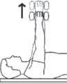
Plantar flexion: Extension movement of the ankle that results in the foot and/or toes moving away from the body in the sagittal plane.
Pronation: A position of the foot and ankle resulting from a combination of ankle dorsiflexion, subtalar eversion, and forefoot abduction (toe-out).
Supination: A position of the foot and ankle resulting from a combination of ankle plantar flexion, subtalar inversion, and forefoot adduction (toe-in).
Terms describing radioulnar joint movements
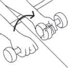
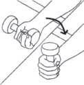
Pronation: Internally rotating the radius in the transverse plane so that it lies diagonally across the ulna, resulting in the palm-down position of the forearm.
Supination: Externally rotating the radius in the transverse plane so that it lies parallel to the ulna, resulting in the palm-up position of the forearm.
Terms describing shoulder girdle (scapulothoracic) movements
Depression: Inferior movement of the shoulder girdle in the frontal plane. An example is returning to the normal position from a shoulder shrug.
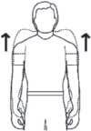
Elevation: Superior movement of the shoulder girdle in the frontal plane. An example is shrugging the shoulders.
Protraction (abduction): Forward movement of the shoulder girdle in the horizontal plane away from the spine. Abduction of the scapula.
Retraction (adduction): Backward movement of the shoulder girdle in the horizontal plane toward the spine. Adduction of the scapula.
Rotation downward: Rotary movement of the scapula in the frontal plane with the inferior angle of the scapula moving medially and downward. Occurs primarily in the return from upward rotation. The inferior angle may actually move upward slightly as the scapula continues in extreme downward rotation.
Rotation upward: Rotary movement of the scapula in the frontal plane with the inferior angle of the scapula moving laterally and upward.
Terms describing shoulder joint (glenohumeral) movements
Horizontal abduction: Movement of the humerus or femur in the horizontal plane away from the midline of the body. Also known as horizontal extension or transverse abduction.
Horizontal adduction: Movement of the humerus or femur in the horizontal plane toward the midline of the body. Also known as horizontal flexion or transverse adduction.
Scaption: Movement of the humerus away from the body in the scapular plane. Glenohumeral abduction in a plane 30 to 45 degrees between the sagittal and frontal planes.
Terms describing spine movements
Lateral flexion (side bending): Movement of the head and/or trunk in the frontal plane laterally away from the midline. Abduction of the spine.
Reduction: Return of the spinal column in the frontal plane to the anatomic position from lateral flexion. Adduction of the spine.
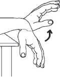
Terms describing wrist and hand movements
Dorsal flexion (dorsiflexion): Extension movement of the wrist in the sagittal plane with the dorsal or posterior side of the hand moving toward the posterior side of the forearm.
Palmar flexion: Flexion movement of the wrist in the sagittal plane with the volar or anterior side of the hand moving toward the anterior side of the forearm.
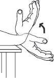
Radial flexion (radial deviation): Abduction movement at the wrist in the frontal plane of the thumb side of the hand toward the lateral forearm.
Ulnar flexion (ulnar deviation): Adduction movement at the wrist in the frontal plane of the little finger side of the hand toward the medial forearm.
Opposition of the thumb: Diagonal movement of the thumb across the palmar surface of the hand to make contact with the fingers.
Reposition of the thumb: Diagonal movement of the thumb as it returns to the anatomical position from opposition with the hand and/or fingers.
These movements are considered in detail in the chapters that follow as they apply to the individual joints.
Combinations of movements can occur. Flexion or extension can occur with abduction, adduction, or rotation.
Movement icons (pedagogical feature)
Throughout this text a series of movement icons will be utilized to represent different joint movements. These icons will be displayed in the page margins to indicate the joint actions of the muscles
displayed on that page. As further explained in Chapter 2, the actions displayed represent the movements that occur when the muscle contracts concentrically. Table 1.7 provides a complete list of the icons. Refer to them as needed when reading Chapters 4–7 and 9–12.
1.7 • Movement icons representing joint actions
Shoulder girdle
Scapula elevation Scapula depression Scapula abduction Scapula adduction
Glenohumeral
Scapula upward rotation
Scapula downward rotation
Shoulder flexion Shoulder extension Shoulder abduction Shoulder adduction Shoulder external rotation Shoulder internal rotation Shoulder horizontal abduction Shoulder horizontal adduction Elbow Radioulnar joints
TABLE