PDF Pearson foundation series biology class 8 eight trishna knowledge systems download
Pearson Foundation Series Biology Class 8 eight Trishna Knowledge Systems
Visit to download the full and correct content document: https://ebookmass.com/product/pearson-foundation-series-biology-class-8-eight-trish na-knowledge-systems/
More products digital (pdf, epub, mobi) instant download maybe you interests ...
Pearson Foundation Series Biology class 10 Ten Trishna Knowledge Systems
> > Provides learner-friendly content, application-analyse based questions and hints and solutions for strong foundation
Uses a graded approach to generate, build and retain interest in concepts and knowledge simulation
About Pearson
Pearson is the world’s learning company, with presence across 70 countries worldwide. Our unique insights and world-class expertise comes from a long history of working closely with renowned teachers, authors and thought leaders, as a result of which, we have emerged as the preferred choice for millions of teachers and learners across the world.
We believe learning opens up opportunities, creates fulfilling careers and hence better lives. We hence collaborate with the best of minds to deliver you class-leading products, spread across the Higher Education and K12 spectrum. Superior learning experience and improved outcomes are at the heart of everything we do. This product is the result of one such effort.
Your feedback plays a critical role in the evolution of our products and you can contact us - reachus@pearson.com. We look forward to it.
Supplement Reading Opener: Olivier Le Queinec. Shutterstock
Icons of Practice Questions: graphixmania. Shutterstock
Icons of Hints and Explanation: graphixmania. Shutterstock
Senior Editor—Acquisitions: Nandini Basu
Senior Editor—Production: Vipin Kumar
The aim of this publication is to supply information taken from sources believed to be valid and reliable. This is not an attempt to render any type of professional advice or analysis, nor is it to be treated as such. While much care has been taken to ensure the veracity and currency of the information presented within, neither the publisher nor its authors bear any responsibility for any damage arising from inadvertent omissions, negligence or inaccuracies (typographical or factual) that may have found their way into this book.
This book is sold subject to the condition that it shall not, by way of trade or otherwise, be lent, resold, hired out, or otherwise circulated without the publisher’s prior written consent in any form of binding or cover other than that in which it is published and without a similar condition including this condition being imposed on the subsequent purchaser and without limiting the rights under copyright reserved above, no part of this publication may be reproduced, stored in or introduced into a retrieval system, or transmitted in any form or by any means (electronic, mechanical, photocopying, recording or otherwise), without the prior written permission of both the copyright owner and the publisher of this book.
No part of this eBook may be used or reproduced in any manner whatsoever without the publisher's prior written consent.
This eBook may or may not include all assets that were part of the print version. The publisher reserves the right to remove any material in this eBook at any time.
ISBN 978-93-530-6206-4
eISBN
First Impression
Published by Pearson India Education Services Pvt. Ltd, CIN: U72200TN2005PTC057128
Head Office: 15th Floor, Tower-B, World Trade Tower, Plot No. 1, Block-C, Sector-16, Noida 201 301, Uttar Pradesh, India.
Registered Office: 4th Floor, Software Block, Elnet Software City, TS-140, Block 2 & 9, Rajiv Gandhi Salai, Taramani, Chennai 600 113, Tamil Nadu, India. Fax: 080-30461003, Phone: 080-30461060
Pearson Foundation Series has evolved into a trusted resource for students who aspire to be a part of the elite undergraduate institutions of India. This new Biology series is an addition to the existing Foundation series particularly targeted for Medical and other related examinations. Each title in this series providing authentic and class-tested content for effective preparation— strong foundation, and better scoring.
The structure of the content is designed in such a manner that it motivates students to go beyond the usual school curriculum, and acts as a source of higher learning to strengthen the fundamental concepts of Biology.
The core objective of the series is to be a one-stop solution for faster and effective preparation for various competitive examinations. Irrespective of the field of study that the student may choose to take up later, it is important to understand that Mathematics and Science form the basis for most modern-day activities. Hence, utmost effort has been made to develop student interest in these basic blocks through real-life examples, critical thinking skills, and asking questions based on application-analyze from the key concepts. Ultimately, the aim is to ingrain the art of problem-solving in the mind of the reader.
To ensure high level of accuracy and practicality, this series has been authored by a team of highly qualified teachers with a rich experience, and are actively involved in grooming young minds. That said, we believe that there is always scope for doing things better and hence invite you to provide us with your feedback and suggestions on how this series can be improved further.
Cell—The basic Unit of life
Chapter Insights
Chapter Cell—The basic Unit of life 1
ReMeMbeR
Info Box!
(a green
is taken as a food supplement.
Before beginning this chapter, you should be able to:
ReMeMbeR
• Recall the basic features of a cell
Before beginning this chapter, you should be able to:
• Remember the meaning and organization of tissue
keY IDeas
• Recall the basic features of a cell
• Remember the meaning and organization of tissue
Remember section will help them to memories and review the previous learning on a particular topics
Nervous Tissue
After completing this chapter, you should be able to:
Key points will help the students to identify the essential points in a chapter
keY IDeas
• Understand the role of cell in maintaining life
After completing this chapter, you should be able to:
• Identify the different cell organelles and their functions
• Understand the role of cell in maintaining life
• Differentiate between mitosis and meiosis
• Identify the different cell organelles and their functions
Tissues and Body Movements 2.11
• Differentiate between mitosis and meiosis
F I g. 5.3 Vinegar, produced by the action of bacteria, is used for pickling microorganisms in medicinal Industry
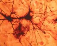
Cells of nervous tissue are called neurons. Neurons transmit information through electrical and chemical signals. Brain, spinal cord and nerves are made up of neurons. Neurons are protected by another type of cell called neuroglial cell. Neurons consist of three major parts:
1. Cell body: Contains nucleus and cytoplasm.
2. Axon: Longest part and carries impulses away from the cell body.
3. Dendrites: Short and branched parts arising from the cell body. They carry impulses towards cell body.
Microorganisms are used to make antibiotics. These antibiotics are used to kill or stop the growth of other disease-causing microorganisms. A simple example is penicillin. Penicillin can be obtained from the fungus Penicillium These antibiotics can be used to cure many diseases that are caused by microorganisms, (except viral diseases). Microorganisms can also be used in the production of vaccine. A vaccine is a biological preparation that consists of either dead or weakened microbes. When a vaccine is introduced into the body, the body produces antibodies against the vaccine, which remains in the body and protects the body from attack by the same microbe in the future. The common examples are rabies and tuberculosis vaccines.
INTRODUCTION
Info boxes are some add-on information on related topics
Info Box!
There are more bacterial cells in human body than human cells!
Concepts are explained in a well structured and lucid manner
and some are non-living. However, both living and non-living things are composed of cer tain basic chemical elements, such as Carbon, Hydrogen, Oxygen and Nitrogen. These chemical elements are found in an organized manner in the for m of cells in the living beings, which distinguishes them from non-living Cells are the basic str uctural and functional units of life A cell is the smallest individual unit of matter capable of perfor ming all essential life processes by itself
1. Define tissue with one example.
Groups of cells either similar or dissimilar and which perform a specific function are called tissue. for example, muscular tissue in humans aids in movement and locomotion.
Info Box!
2. Differentiate between apical meristem and lateral meristem Meristems are the plant tissues that can divide throughout their life. apical meristem lateral meristem
F I g. 5.4 Medical vials containing vaccines
Antibodies are the substances produced by the body to fight against foreign particles called antigens.
Seen at the growing tips of roots and shoots Seen along the sides of the stem and the root Increases the length of root and stem Increases the girth of plant body Involved in primary growth of plants Involved in secondary growth of plants
Sir Alexander Fleming was the person who introduced to the world, the greatest discovery of that time, the antibiotic ‘Penicillin’. It saved the lives of thousands of soldiers during the second world war. He was awarded the Nobel Prize in Physiology or Medicine along with Florey and Chain in the year 1945. Florey and Chain developed processes to produce penicillin in sufficient quantites to become widely available.
Quick Recap section will help to review all important concepts, discussed in that particular chapter
Chlorella
alga)
Each section contains detailed diagrams, images, real life microscopic views for better understanding and conceptual clarity.
Chapter 1 1.22
TEST YOUR CONCEPTS
Directions for questions 1 to 20: Fill in the blanks in each question.
1. The first living cell was discovered by ________
2. ________ is the basic structural and functional unit of life.
3. Take the odd one out.
Amoeba, Bacteria, Plant, Honeybee.
4. The concept ‘Omnis cellula e cellula’ was put forward by ________.
5. The term ‘protoplasm’ was coined by ________.
6. The smallest cell is ________.
7. The largest cell is ________.
F I g. 4.25 Longitudinal section of a flower showing all parts
Stamen or Androecium
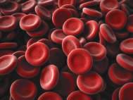
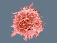
23. Which of the following can change its shape? (i) Amoeba (ii) Neuron (iii) WBC (iv) RBC (a) (i) only (b) (i) and (ii) (c) (i) and (iii) (d) (i), (ii), (iv)
8. Lactobacillus is ________ shaped.
9. Mycoplasma lacks ________.
Supplement Reading S.10
2 2.18
10. Phosphate granules are examples for ________.
tEst YoUR ConCEpts
1. Albert Einstein
‘Test Your Concepts’ at the end of the chapter for classroom preparations
11. The inner foldings of the mitochondrial inner membrane are called ________.
12. Chlorophylls are present in ________.
2. Robert Hooke
3. Hypothesis
4. Observation
Stamen is the male reproductive organ of a flower and male gamete is produced inside the stamen. Stamen has two parts: Anther and filament
24. Nucleoid is the region where ________ is/are located
(a) Genetic material (b) Proteins (c) Pigments (d) Nutrients
25. Bacterial cell wall is made up of (a) Cellulose (b) Peptidoglycan (c) Hemicellulose (d) Pectin
26. Match the following
MASTERINg THE CONCEPTS
A. Inclusion bodies (i) Eukaryotic
B. Mesosome (ii) Protein manufacture
Knowledge and Understanding
13. ________ is known as the energy currency of the cell.
14. Detoxification of poisons takes place in ________.
5. Social science
15. Thylakoids of different grana are connected by ________.
6. (d)
7. (d)
8. (d)
Different levels of questions have been included in the Test Your Concept as well as on Mastering the Concepts which will help students to develop the problemsolving skill
1. Anther: It is the structure bearing pollen grains located at the top of the filament of stamen.
2. Filament: It is a long stalk like structure that attaches the stamen with flower parts, such as petal.
C. Ribosome (iii) Cell wall formation
D. Golgi complex (iv) Nutrient reservoir
A B C D
1. What are stomata and what are their significance in a plant?
10. (d)
(a) (iii) (ii) (i) (iv)
10. Differentiate between mitosis and meiosis.
Structure of stamen
F I g. 4.26 Parts of stamen
11. Differentiate between bone and cartilage.
11. scientist Contribution
(b) (iv) (iii) (i) (ii) (c) (iii) (i) (iv) (ii) (d) (iv) (iii) (ii) (i)
2. Make a comparison between the three types of simple permanent tissue based on their structures.
Thomas Alva Edison Motion picture camera
12. How epithelial tissues have been classified according to their shape?
Dimitri Ivanovich
3. Differentiate between xylem and phloem.
27. Select the wrong statement.
Mendeleev Periodic table and periodic law
16. Specialized peroxisomes in plants are called ________.
4. Write the significance of collenchyma in plants?
Srinivasa Ramanujan Infinite series
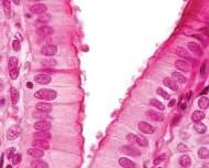
13. What is ciliated epithelium, and why are they important in the respiratory tract?
(a) Plant cells are surrounded by a living, rigid outer layer called cell wall.
Albert Einstein E = mc2
5. How permanent tissue is formed from meristematic tissue?
17. Thin thread-like structure present inside the nucleus is called ________.
9. (b)
Mastering the concepts are further divided as per Knowledge/ Understanding, and Application/Analyze
14. Make a comparison between tendon and ligament.
(b) Plant cell wall is made up of cellulose. (c) Bacterial cell wall is made up of peptidoglycan.
Sir Isaac Newton Universal gravitation
Charles Darwin Theories of evolution
15. Which type of tissue is blood and what is its function?
18. The membrane surrounding vacuole is called ________.
19. In plant, reserve food is stored in the form of ________.
mastERing tHE ConCEpts
20. Plant cell wall is made up of ________
Knowledge and Understanding
6. What is a permanent tissue and how it is classified?
James Watson DNA structure
(d) Both prokaryotic and eukaryotic plasma membranes are composed of lipids and proteins.
7. What is phloem tissue, and explain its various elements with their function.
28. Adjacent plant cells are joined together by (a) Middle lamella (b) Plasmodesmata
8. Give a note on cuticle.
Directions for questions 21 to 40: For each of the following questions, four choices have been provided. Select the correct alternatives.
16. Give a note on the structure of neuron.
17. Define pseudopodia.
18. Give a note on axial skeleton.
(c) Mesosomes (d) Plasma membrane
9. Give a brief note on xylem tissue.
29. Which of the following is the correct statement?
19. What are the different types of joints and give an example for each?
21. Site for protein manufacture is (a) Cell wall (b) Cytoplasm (c) Ribosome (d) Inclusion bodies
1. Science plays a major role in everyday life. It has made our communication easy, even if we are at far away places. It has also made our transportation easy and comfortable. Through science, it is now possible to treat millions of diseases, thereby increasing the lifespan of people.
to peers may lead to new questions every time which may in turn lead to another investigation.
(a) Endoplasmic reticulum is the site of protein manufacture in bacteria.
Application and Analysis
3. Vikram Sarabhai: Father of Indian space programme.
(b) The membrane-bound inclusion bodies help in the storage of nutrients in prokaryotes.
1. You might have seen that even after grazing by herbivores, grasses regenerate from the remaining parts. Which tissue is responsible for this and where it is located?
2. Communicating the results obtained after data analysis to let others know about it is the last step in a scientific method. The results can be presented as written or oral reports. Communicating the results
Application and Analysis
Srinivasa Ramanujan: Indian mathematician who made major contributions to number theory, infinite series, etc.
2. Further growth of an onion root stops after the removal of tip portion. Why?
3. Identify the parts.
1. Scientists follow a specific pathway for their innovations, which is known as a scientific method. It involves a series of techniques such as:
• Observation
• Asking questions
CV Raman: Indian physicist who carried out major work in the field of light scattering
~ Hypothesis - I - pea seeds do not require water for germination ~ Hypothesis - II - pea seeds require air for germination ~ Hypothesis III - pea seeds require adequate
Hints and Explanation for key questions along with highlights on the common mistakes that students usually make in the examination
4. It is possible to move the elbow freely. Which types of joints are present here that help in this movement?
Stament
Anther
Filament
Ovule
Receptacle
Stigma Style
Petal
Sepal
Pedicel
Anthers
Filament
Stamen
Pollen sacs
Series Chapter Flow
This page is intentionally left blank.
Chapter Cell—The basic Unit of life 1
ReMeMbeR
Before beginning this chapter, you should be able to:
• Recall the cellular organization in humans
• Remember important cell organelles and their functions
keY IDeas
After completing this chapter, you should be able to:
• Understand the role of cell in maintaining life
• Identify the different cell organelles and their functions
• Differentiate between mitosis and meiosis
There are more bacterial cells in human body than human cells!
INTRODUCTION
If we look around, we see many different objects, some of them are living and some are non-living. However, both living beings and non-living things are composed of certain basic chemical elements, such as Carbon, Hydrogen, Oxygen and Nitrogen. These chemical elements are found in an organized manner in the form of cells in the living beings, which distinguishes them from non-living. Cells are the basic structural and functional units of life. A cell is the smallest individual unit of matter capable of performing all essential life processes by itself.
Cell—The basIC UNIT Of lIfe
The term ‘cell’ is derived from Latin word cellula , meaning ‘little room’. The term cell was coined by Robert Hooke in 1665. A cell is the smallest building block of life. It is defined as the basic structural and functional unit of a living organism. A cell is capable of performing all vital functions to sustain life.
Discovery of Cell
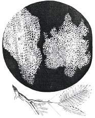
In 1665, Robert Hooke observed the thin slices of cork under a microscope. He noticed several small compartment-like structures that resembled the structure of a honeycomb.
fI g. 1.1 Microscope used by Robert Hooke to make his observations of cells
fI g. 1.2 Robert Hooke’s observations of cellular structure of cork
Unicellular and Multicellular Organisms
Cell is the basic structural and functional unit of life. All living organisms are made up of cells. In one case, a single cell may constitute an organism (unicellular) and in other case, an organism may consist of many cells (multicellular). According to this, organisms can be divided into two types:
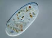
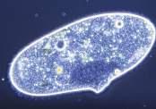
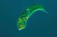
1. Unicellular organisms: Organisms with only one cell, for example, Amoeba, Chlamydomonas, Paramoecium, bacteria, etc.
iInfo Box!
In 1674, Anton Van Leeuwenhoek observed free living cells in pond water.
2. Multicellular organisms: Organisms with many cells, for example, plants and animals, are called multicellular organisms.
Cell Theory
Cell theory was proposed by Matthias Jakob Schleiden (1838) and Theodor Schwann (1839) and was later modified by Rudolf Virchow. The two main postulates are:
1. All organisms are made up of cells.
2. Cell is the basic structural and functional unit of life.
Info Box!
The number and type of cells in a given tissue is called cellularity.
Size and mass of a typical cell are 10 micrometre and 1 nanogram.
An important addition to the Cell Theory was made by Rudolph Virchow in 1868. He stated that new cells arise from pre-existing cells (in Latin: omnis cellula e cellula).
fI g. 1.3 Some common unicellular organisms
Table 1.1 Some famous scientists and their contribution to the field of cell biology
scientist
Robert Hooke
Anton Van Leeuwenhoek
Robert Brown
Purkinje
Schleiden, Schwann, Virchow
Camillo Golgi
Longest cell: Neuron (in humans) i
Info Box!
Smallest cell: Mycoplasma
Largest cell: Ostrich’s egg
size of Cell
Contribution
Discovered cell
Observed first living cell
Discovered cell nucleus
Coined the term ‘protoplasm’
Cell theory
First described Golgi apparatus
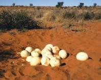
Most cells are minute and are visible only under the microscope. They are only a few micrometres in diameter. The cells range in size from micrometers to as long as 1 m. There are some cells which are big enough to be seen with naked eye. For example, egg of an ostrich is about 15-cm long and 13-cm wide.
fI g. 1.4 (Left) Microscopic view of cells that are not visible to naked eye (Right) Ostrich eggs in in nest, Kalahari desert (Africa)
Naked eye means an unaided vision, without a telescope, microscope or other optical device i
Info Box!
shape of Cell
The shape of the cells varies according to their function. They may be disclike, polygonal, columnar, cuboid or thread-like. Nerve cells have an elongated structure and this helps them in conducting impulses quickly. Similarly, there are cells that have a flexible shape, for example, Amoeba and some blood cells. The flexibility in Amoeba’s shape helps it to perform various activities, such as movement, feeding, etc. The change in shape of Amoeba is due to the formation of pseudopodia that are projections on its body.
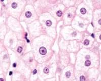
fI g. 1.5 Different types of cells showing different shapes
Types of Cells
The cells can be divided into two types: prokaryotic and eukaryotic, according to the differences in their cellular organization. The cells which posses a true nucleus along with membrane-bound organelles are called eukaryotic cells, example plant cell and animal cell. The cells which do not possess a well-defined nucleus as well as membrane-bound organelles are called prokaryotic cells, example bacterial cell.
Prokaryotic Cells
Prokaryotic cells are primitive and lack a true nucleus. Their genetic material is not surrounded by a nuclear membrane and is confined to a particular region known as nucleoid.
Red blood cells (round and biconcave)
Columnar epithelial cells (long and narrow)
White blood cells (amoeboid)
Nerve cells (branched and long)
Ribosomes Cytoplasm
fI g. 1.6 A prokaryotic cell
Prokaryotic cells are generally smaller than eukaryotic cells. There are four basic shapes for prokaryotes (bacteria) listed as follows:
fI g. 1.7 Illustration of types of bacterial shapes
Though their shapes and sizes vary, all cells possess a common structure. All prokaryotic cells have major components that are discussed below.
Cell Wall
It forms the outermost layer of the cell. It provides integrity to the cell. It is made up of peptidoglycan, which in turn is made up of carbohydrates. It is absent in Mycoplasma
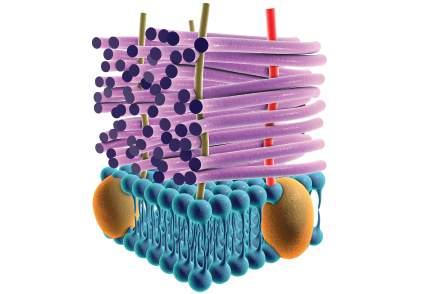
It lies just inside the cell wall. It isolates the cell’s content from the outside environment and forms a barrier to substances entering in and out of the cell. Membranes are semipermeable, meaning they allow only certain substances to pass through it while preventing others. It is made up of a phospholipid bilayer.
Cytoplasm
Membrane protein
Info Box!
Bacteria are divided into Gram-positive and Gramnegative depending upon Gram staining method developed by Christian Gram
All of the fluid substance present inside the cell constitutes its cytoplasm. It comprises all kinds of cell organelles present in a cell, with the exception of no nucleus in a prokaryotic cell.
Mesosomes
These are formed by the plasma membrane extension into the cell. They help in processes, such as cell wall formation, respiration, etc.
Ribosomes
These are the sites of protein synthesis. Ribosomes are found to be associated with plasma membrane. They are not bound by any membrane system.
Peptidoglycan
Cytoplasmic Inclusions
Eukaryotic Cells
Chloroplast
Cytoplasm
Mitochondrion
Cell Membrane
Cell Wall
Inclusion bodies lie free in the cytoplasm. They help in storage of reserve materials, for example, phosphate granules.
All eukaryotic cells possess a true nucleus, that is, their genetic material is surrounded by a nuclear envelope. All protists, fungi, plants and animals are eukaryotes. They possess membrane bound organelles, such as endoplasmic reticulum, mitochondria, Golgi bodies, etc.
Nuclear envolope
Nucleolus
Nucleus
All eukaryotic cells typically have:
Endoplasmic reticulum
Golgi apparatus
Cell wall
Plant cells are surrounded by a non-living, rigid, permeable outer layer known as a cell wall. It provides structural integrity and protection to the cell. It is made up of cellulose. Cell walls of adjacent cells are joined together by a jelly-like substance called middle lamella.
mRNA
Large subunit
Small subunit
fI g. 1.9 Diagram showing ribosome structure
Vacuole
fI g. 1.10 A typical plant (eukaryotic) cell
Cell Membrane
It is made up of proteins and lipids. Cell membrane is semi-permeable which means that it allows entry and exit of only some materials.
Extracellular fluid
Transmembrane glycoprotein
Glycolipid
Cholesterol
Carbohydrates
Pore
Peripheral protein
Transmembrane protein
Cytoplasm
Channel protein
g. 1.11 Plasma membrane structure
Protoplasm
The living material comprising cytoplasm, nucleus and other organelles is called protoplasm.
Cytoplasm
Fluid content inside the cell membrane. Contains many membrane-bound organelles. These are discussed in detail in the following section.
Cell wall
Plasma membrane
Cytoplasm
Mesosomes
Prokaryotic
Cell
Eukaryotic
Ribosomes
Inclusion bodies
Plant cell
Animal cell
Nucleus
Plasma membrane
Cytoplasm
Nucleus Cell wall
Plasma membrane
Cytoplasm
fI g. 1.12 Cellular organization in prokaryotes and eukaryotes
Table
1.2
Differences between prokaryotic and eukaryotic cell
Prokaryotic cell
Size generally small (1–10 mm)
True nucleus is absent
Contains single chromosome
Membrane-bound cell organelles absent, such as endoplasmic reticulum, Golgi complex, etc.
Cell Organelles
eukaryotic cell
Size generally large (5–100 mm)
Nucleus is present
Contains more than one chromosome
Membrane-bound cell organelles present
An organelle is a unit inside the cell that performs a special function. The various functions performed by a cell are actually divided among the organelles, that is, there is a division of labour inside the cell in eukaryotic cells. This division of labour is absent in prokaryotic cells. For example, energy production is performed by mitochondria, photosynthesis by plastids, etc. The various cytoplasmic organelles are given below:
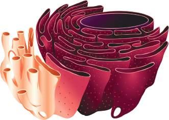
Endoplasmic Reticulum
It is a large network or reticulum of membrane-bound tiny tubular structures that start at the nuclear membrane and extend into the cytoplasm. They are mainly involved with the synthesis, folding, modification and transport of proteins.
Endoplasmic reticulum is of two types:
1. Rough Endoplasmic Reticulum (RER): which possesses ribosomes on its surface and helps in protein synthesis.
2. Smooth Endoplasmic Reticulum (SER): which lacks ribosomes on its surface. They are a major site for lipid synthesis and also involved in the detoxification of poisons.
Nucleus Rough
Ribosomes
Ribosomes
It consists of dense and spherical particles which are present in both prokaryotic and eukaryotic cells. These are the sites of protein manufacture. They are found either free in cytoplasm or attached with RER. These are not bound by any membrane and are complex of ribonucleic acid (RNA) and proteins.
Golgi Apparatus
It was first described by Camillo Golgi in 1898. It consists of a system of many, flat disc-shaped sacs called cisternae, which are arranged parallel to each other. Its functions include packaging, storing and dispatching of various products received from endoplasmic reticulum. It remains in close association with endoplasmic reticulum. It is involved in synthesis of complex sugars and lysosome.
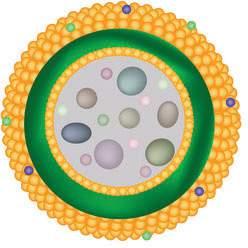
Lysosomes
Mitochondria
Glycosylated mambrane transport proteins
Lysosome structure
These are composed of tiny spherical structures surrounded by a single membrane. They contain many digestive enzymes that are manufactured in RER. Lysosomes are known as waste disposal system of the cell as they help in cleaning the cell by removing the worn out or damaged cellular organelles. They are also called suicide bags of cells as they can digest their own cell, if anything happens to the cell structure or if the cell gets damaged.
Hydrolytic enzyme mixture
They are referred to as the powerhouses of the cell. They are the main sites for energy production. They produce energy in the form of adenosine triphosphate (ATP). ATP is known as energy currency of the cell. Number of mitochondria per cell vary according to the physiological activity of the cell.
Structure of a typical mitochondrion is characterized by the following features.
• It is surrounded by a double membrane covering.
• Its outer membrane is porous and inner membrane is deeply folded.
• The foldings of inner membrane are called cristae which increase the surface area.
• The compartment enclosed by inner membrane is called matrix.
• Mitochondrion possesses its own DNA (single circular DNA) and ribosomes.
Lipid layer
Membrane
fI g. 1.15
fI g. 1.14 Golgi Apparatus
DNA Ribosomes Matrix Outer Membrane Inner Membrane
F0, F1 Complexes
Info Box!
Green chilly is converted into red chilly due to conversion of chloroplast into chromoplast. i
Plastids
Intermembrane Space
fI g. 1.16 Structure of mitochondria
Plastids are present in most plant cells and are absent in animal cells. Like mitochondria, plastids contain their own DNA and ribosomes. Plastids are of three types as detailed below:
Plastids
Chromoplasts
fI g. 1.17 Division of plastids on the basis of presence or absence of pigments
1. Chromoplasts: Coloured plastids in which chlorophyll is absent and contain yellow or orange pigment. They are found in fruits, flowers, etc.
2. Chloroplasts: Consist of a green pigment called chlorophyll and it mainly functions in photosynthesis. Chloroplast is also known as kitchen of the cell.
3. Leucoplasts: They are white or colourless and mainly function in storage. Depending upon the storing material, leucoplasts are of three types; Aleuroplast (store protein), elaioplast (stores oil), amyloplast (stores carbohydrate).
Structure of Chloroplasts
It is surrounded by a double membrane covering. It consists of many membrane-bound flat discs called thylakoids which has chlorophyll. Thylakoids are arranged in stacks called grana. The matrix found inside the chloroplast is called stroma. Thylakoids of different grana are connected by stromal lamellae
Cristae Junction
Vacuoles
Microbodies
These are membrane bound spaces seen in the cytoplasm of mainly plant and fungal cells. These are the storage sacs that store proteins, amino acids, water and other nutrients. They can occupy up to 90% of the cell volume in plant cells. In animal cells, vacuoles are either very small or absent. It is surrounded by a single membrane called tonoplast. The substance present inside the vacuole is called cell sap. It provides turgidity and rigidity to the plant cell.
Microbodies are a kind of organelles present in plants, animals and protozoa. They include peroxisomes, glyoxysomes, glycosomes and hydrogenosomes.
1. Peroxisomes: Present in cytoplasm and mainly function in the detoxification of toxic substances.
2. Glyoxysomes: Specialized peroxisomes in plants.
3. Glycosomes: Specialized peroxisomes present in protists.
4. Hydrogenosomes: Found in anaerobic eukaryotes.
Centrosomes
They are only found in animal cells. They are not bounded by a membrane and consist of two centrioles. Centrosomes help in cell division.
Nucleus
It was first described by Robert Brown in the year 1831. It is a large, spherical, centrally placed organelle found in eukaryotic cells which is the central controlling unit of all the activities of a cell.
Structure of nucleus
It is surrounded by a double membrane called nuclear envelope or nuclear membrane. Nuclear envelope contains many pores called nuclear pores. Nuclear pores allow transport of materials across nucleus that is, from nucleus to cytoplasm and from cytoplasm to nucleus. Liquid substance present inside the nucleus is called nucleoplasm. It contains nucleolus and chromatin material.