Chapter 1
Respiratory emergencies
Jennifer Stirling, Clare Sutton and Georgina Pickering
Charles Sturt University, Bathurst, NSW, Australia
CHAPTER CONTENTS
Level 1: Asthma
Level 1: Chronic obstructive pulmonary disease (COPD)
Level 2: Pulmonary embolism (PE)
Level 2: Life‐threatening asthma
Level 3: Respiratory sepsis
Level 3: Smoke inhalation
LEVEL 1 CASE STUDY Asthma
Information typeData
Time of origin17:08
Time of dispatch17:10
On‐scene time17:20
Day of the weekFriday
Nearest hospital30 minutes
Nearest backup15 minutes
Patient detailsName: Betsy Booper DOB:10/09/2002
CASE
You have been called to an outdoor running track for an 18‐year‐old female with shortness of breath. The caller states she has taken her inhaler to no effect.
Pre‐arrival information
The patient is conscious and breathing. You can access the area via the back gate of the sports field and drive right up to the patient, who is sat down on the track.
Windscreen report
The location appears safe. Approx. 10 people around the patient. Environment – warm summer evening and good light.
Entering the location
The sports coach greets you as you get out of the ambulance and informs you that the patient suffers with exercise‐induced asthma, but this is worse than normal and her inhaler has been ineffective.
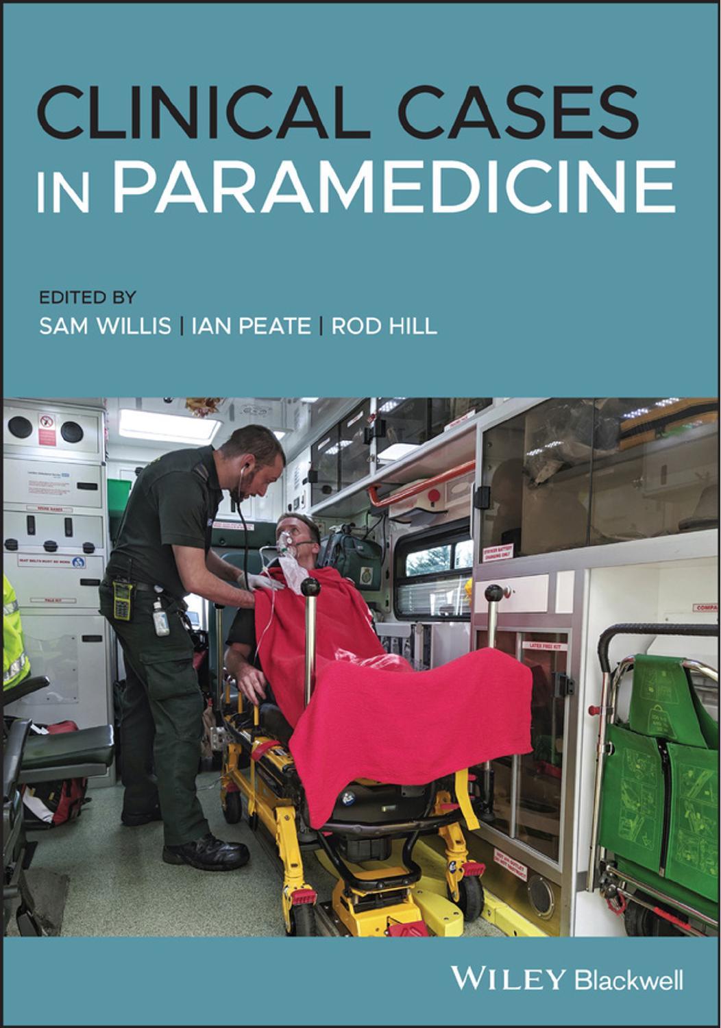
Clinical Cases in Paramedicine, First Edition. Edited by Sam Willis, Ian Peate, and Rod Hill. © 2021 John Wiley & Sons Ltd. Published 2021 by John Wiley & Sons Ltd.
On arrival with the patient
The patient is sat on a bench on the side of the track. She is leaning forward, resting her elbows on her thighs (tripodding). She says hello as you introduce yourself to her.
Patient assessment triangle
General appearance
Alert. Speaking in short sentences. She looks panicked.
Circulation to the skin
Flushed cheeks.
Work of breathing
Breathing appears rapid and shallow. An audible wheeze is noted.
SYSTEMATIC APPROACH
Danger None at this time.
Response
Alert on the AVPU scale.
Airway
Clear.
Breathing
RR: 28. Regular and shallow. No accessory muscle use. Expiratory wheeze on auscultation.
Circulation
HR: 100. Regular and strong. Capillary refill time <2 seconds. Flushed cheeks and peripherally warm.
Disability
Moving all four limbs.
Pupils equal and reactive to light (PEARL).
Exposure
Bystanders have left. Next of kin are now on scene.
Temperature: warm summer evening – approx. 20 °C.
Vital signs
RR: 28 bpm
HR: 100 bpm
BP: 125/74 mmHg
SpO2: 93%
Blood glucose: 5.2 mmol/L
Temperature: 36.9 °C
PEF: 300 L/min
GCS: 15/15
4 Lead ECG: sinus tachycardia
TASK
Look through the information provided in this case study and highlight all of the information that might concern you as a paramedic.
Aside from auscultation, which you have already done, what examination techniques should you incorporate into this patient assessment?
• Inspection – observe the chest for an abnormalities such as wounds, scars, bruising, asymmetry and recession.
• Palpation – feel for any asymmetry, vocal fremitus and tenderness.
• Percussion – hyper‐ or hypo‐resonance.
What adventitious (added) sounds might indicate asthma and why?
Expiratory wheeze. This sound is made when air has a restricted path through the bronchi, due to inflammation and muscle spasm in the airways.
What medicine (pharmacology) is likely to relieve the patient’s symptoms and why?
Nebulised salbutamol – it is a Beta2, adrenergic agonist that relaxes smooth muscle in the bronchi.
Case Progression
You treat the patient with 5 mg of nebulised salbutamol and 6 L of oxygen. The nebuliser finishes and you remove the mask.
Patient assessment triangle
General appearance
The patient is now speaking in full sentences.
Circulation to the skin
Flushed.
Work of breathing
Normal effort of breathing.
SYSTEMATIC APPROACH
Danger
None at this time.
Response Alert.
Airway
Clear.
Breathing
RR:16. Regular. Normal depth. No accessory muscle use. No wheeze or adventitious sounds.
Circulation
HR: 105. Regular and strong. Capillary refill time <2 seconds. Flushed cheeks and peripherally warm.
Disability No change.
Exposure
No change.
Vital signs
RR: 16 bpm
HR: 105 bpm
BP: 128/78 mmHg
SpO2: 97%
Blood glucose: not repeated
Temperature: not repeated
PEF: 380 L/min
GCS: 15/15
4 lead ECG: sinus tachycardia
What kinds of questions would you ask this patient specifically related to asthma as part of the history‐taking process?
See Table 1.1.
Table 1.1 History‐taking questions
Asthma history
Does this feel like your normal asthma?
Is this the worst it’s ever been?
What time did this episode start today?
Do you take your asthma medication regularly?
What were you doing when it started today?
What usually triggers your symptoms?
When was the last time your visited your GP and/or went to hospital with these symptoms?
Have you ever been intubated or been in ICU with these symptoms?
Medication history
What asthma medications do you take?
How frequently do you have to take your medication?
Do you usually have to take your inhaler while exercising?
When was the last time you had a medication review with your GP?
Have you had any recent changes in medication?
Do you take any other medications?
Have you had any coaching on the best way to take your inhaler?
F/SH (family and social history)
Does anyone else in your family experience asthma?
Do you smoke? If so, how frequently?
Do you drink or take any drugs recreationally?
Who do you live with?
What do you do for work?
Do you exercise regularly?
Are you under any particular stress at the moment?
Past medical history (PMH)
Do you have any other medical problems?
Do you have any allergies?
Have you had a cough or cold recently?
The patient is 160 cm tall, what should her predicted peak expiratory flow reading (PEFR) be? Her first reading was 300 – what percentage is that from predicted?
(Hint: you will be required to look this up using the Australian National Asthma Council chart found here: http://www.peakflow.com/pefr_normal_values.pdf or by doing an internet search.)
• 400 L/min.
• 75%.
LEVEL 1 CASE STUDY
Chronic obstructive pulmonary disease (COPD)
Information typeData
Time of origin07:09
Time of dispatch07:12
On‐scene time07:30
Day of the weekWednesday
Nearest hospital15 minutes
Nearest backup40 minutes
Patient detailsName: Dave Beater
DOB: 21/09/1954
CASE
You have been called to a residential address for a 66‐year‐old male with difficulty in breathing. The caller states he has been breathless all night and has had a cough recently. He has seen his GP who prescribed antibiotics and steroids but he feels his breathing has got worse overnight.
Pre‐arrival information
The patient is conscious and breathing and is in a first‐floor flat/unit.
Windscreen report
The location appears safe. Greeted at the main door by the patient’s wife.
Entering the location
Wife escorts you up in the lift to the patient’s flat.
On arrival with the patient
Patient is sat in the tripod position and appears distressed. He makes eye contact when you arrive, but does not speak as is so short of breath. He has a productive cough that results in a string of green‐looking sputum that he manages to capture in his handkerchief to show you.
Patient assessment triangle
General appearance
Alert, and makes eye contact, but is acutely distressed. Can only speak in single words and is reluctant to talk. In tripod position, coughing.
Circulation to the skin
Pink face, breathing through pursed lips.
Work of breathing
Increased work of breathing – rapid and shallow breaths with accessory muscle use.
SYSTEMATIC APPROACH
Danger
None at this time.
Response Alert.
Airway Clear.
Breathing
RR: 36. Rapid and shallow, with accessory muscle use. Widespread bilateral wheeze noted on auscultation.
Circulation
HR: 110. Radial palpable – irregular. Capillary refill time 2 seconds.
Disability
Pupils equal and reactive to light (PEARL).
Exposure
The patient is in his own home.
Vital signs
RR: 36 bpm
HR: 110 bpm
BP: 150/90 mmHg
SpO2: 86%
Blood glucose: 4.5 mmol/L
Temperature: 37.8 °C
PEF: unable to record
GCS: 15/15
4 Lead ECG: atrial fibrillation
Allergies: nil
TASK
Look through the information provided in this case study and highlight all of the information that might concern you as a paramedic.
What is COPD?
COPD is a progressive disease and is characterized by air flow obstruction that is not fully reversible. The airway obstruction results from damage to alveoli, alveolar ducts and bronchioles due to chronic inflammation.
List the features of an acute exacerbation of COPD.
• Increased dyspnoea.
• Increased sputum production.
• Increased cough.
• Upper airway symptoms, such as a cold and sore throat.
• Increased wheeze.
• Reduced exercise tolerance.
• Fluid retention.
• Increased fatigue.
• Acute confusion.
• Worsening of previously stable condition.
Case Progression
After administration of 5 mg salbutamol via nebuliser, the patient’s condition improves slightly and he hands you a medical card that his ‘breathing doctor’ gave to him. The card states the patient is at risk of retaining CO2 and should only be administered with 28% oxygen to achieve saturations between 88 and 92%.
Patient assessment triangle
General appearance Alert and more interactive.
Circulation to the skin Pink.
Work of breathing
Increased work of breathing – breathing rapid, but not as shallow as before.
SYSTEMATIC APPROACH
Danger None at this time.
Response Alert.
Airway Clear.
Breathing
RR: 30. Audible wheeze on auscultation.
Circulation
HR: 120. Palpable radial. Capillary refill time 2 seconds.
Disability
Moving all four limbs.
Exposure
Normal temperature in the ambulance.
Vital signs
RR: 30 bpm
HR: 120 bpm
BP: 148/78 mmHg
SpO2: 90%
Blood glucose: not repeated
Temperature: not repeated
GCS: 15/15
4 lead ECG: atrial fibrillation
Allergies: nil
When the nebuliser has finished, you notice that the patient’s SpO2 is dropping so you decide to keep the patient on oxygen. What percentage of oxygen would you administer to this patient and why?
28% oxygen through a nasal cannula. The patient is at risk of developing hypercapnia respiratory failure, so it is important the oxygen is titrated to maintain saturations between 88 and 92%. Research suggests that over‐oxygenation increases the mortality and morbidity of COPD patients and that titration of oxygen administration can reduce mortality.
What is meant by the term hypercapnia?
• ‘A condition of abnormally elevated carbon dioxide (CO2) levels in the blood, caused by hypoventilation, lung disease, or diminished consciousness’ (NAEMT, 2015, p. 92).
• ‘Alveolar hypoventilation with increased alveolar carbon dioxide limits the amount of oxygen available for diffusion into the blood, leading to secondary hypoxemia’ (McCance et al., 2010, p. 1269).
LEVEL 2 CASE STUDY
Pulmonary embolism (PE)
Information typeData
Time of origin17:55
Time of dispatch18:01
On‐scene time18:10
Day of the weekFriday
Nearest hospital30 minutes
Nearest backup15 minutes
Patient detailsName: Jasmine Wallis
DOB: 27/12/2000
CASE
You have been called to a car park for a 20‐year‐old female who is complaining of feeling dizzy and faint.
Pre‐arrival information
She is conscious and breathing.
Windscreen report
The car park is behind a row of shops and is poorly lit. The patient is hard to spot at first, as she is sitting on the metal fire escape steps with her head in her hands at the back of a building. She is alone. The car park is full, which prevents you parking near to the patient.
Entering the location
You park your ambulance as near as possible and cross the car park to get to your patient.
On arrival with the patient
The patient is able to raise her head and make eye contact.
Patient assessment triangle
General appearance
The patient looks at you when you speak and is able to speak in full sentences.
Circulation to the skin
Mildly pale.
Work of breathing
Increased. The patients looks mildly short of breath.
SYSTEMATIC APPROACH
Danger
None at this time.
Response Alert.
Airway Clear.
Breathing
RR: 26. Mildly increased effort, no accessory muscle use. Auscultation – clear.
Circulation
HR: 120. Tachycardic, weak and regular pulse. Capillary refill time >2 seconds.
Disability
Pupils equal and reactive to light (PEARL).
Exposure
The patient is sitting on metal fire escape stairs, in a dark, cold car park in an undesirable part of town.
Vital signs
RR: 26 bpm
HR: 120 bpm
BP: 90/60 mmHg
SpO2: 90%
Blood glucose: 4.4 mmol/L
Temperature: 36.5 °C
ECG: sinus tachycardia
Allergies: nil
TASK
Look through the information provided in this case study and highlight all of the information that might concern you as a paramedic.
List your differential diagnoses for this patient.
• Musculoskeletal pain.
• Pericarditis.
• Hyperventilation.
• Chest infection.
• Syncope.
• Pneumothorax.
List as many predisposing factors associated with PE as you can. Which could assist you with working through your differential diagnosis and history taking?
See Table 1.2.
Table 1.2 Pulmonary embolism predisposing factors
Surgery, especially recent
Abdominal
Pelvic
Hip or knee
Post‐operative intensive care
Obstetrics
Pregnancy
Cardiac
Recent acute myocardial infarction
Limb problems
Recent lower limb fractures
Varicose veins
Lower limb problems secondary to stroke or spinal cord injury
Malignancy
Abdominal and /or pelvic, in particular advanced metastatic disease
Concurrent chemotherapy
Other
Risk increases with age
>60 years of age
Previous proven deep vein thrombosis (DVT)/PE
Immobility
Thrombotic disorder
Neurological disease with extremity paresis
Thrombophilia
Hormone replacement therapy and oral contraception
Prolonged bed rest >3 days
Other recent trauma
Source: JRCALC (2019), p. 367.
What validated assessment tool could assist you with assessing the probability of PE in this patient?
See Table 1.3.
Table 1.3 Wells’ criteria for PE
CriteriaScore
Clinical signs and symptoms of DVT (leg swelling and pain with palpation of the deep veins)3
An alternative diagnosis is PE is less likely3
Pulse rate >100 bpm1.5
Immobilisation or surgery in the previous 4 weeks1.5
Previous DVT/PE1.5
Haemoptysis1
Malignancy (treatment ongoing or within the last 6 months or palliative)1
Clinical probability
High>6 points
Moderate2–6 points
Low<2 points
Source: JRCALC (2019), p. 368.
Note: When using the Wells’ criteria, a low probability does not rule out PE.
Case Progression
You decide to move your patient to the back of the ambulance to continue the examination in a warm and private environment. On standing, the patient complains of feeling dizzy and faint and is unable to walk even a couple of steps. You instruct your crewmate to fetch the carry chair as you can’t get the stretcher close enough to the patient.
Patient assessment triangle
General appearance
Patient feels better when lying flat.
Circulation to the skin
Normal.
Work of breathing
Increased. Patient complains of not being able to ‘catch her breath’.
SYSTEMATIC APPROACH
Danger
None at this time.
Response
Alert.
Airway
Clear.
Breathing
RR: 30.
Circulation
HR: 128. Weak radial.
Disability
Moving all four limbs.
Exposure
Normal temperature in the ambulance.
Vital signs
RR: 30 bpm
HR: 128 bpm
BP: 88/60 mmHg
SpO2: unable to obtain
Blood glucose: not repeated
Temperature: not repeated
GCS: 15/15
12 lead ECG: sinus tachycardia with right bundle branch block (RBBB)
What is the most common
ECG finding in PE? What other ECG changes are associated with PE?
The most common ECG finding in PE is sinus tachycardia. PE can cause any of the following ECG changes:
• T‐wave inversion.
• New‐onset atrial fibrillation.
• Right bundle branch block.
• Right axis deviation.
• S1Q3T3 (this is a specific pattern that is seen rarely in PE):
◦ S waves in lead I.
◦ Q waves in lead III.
◦ T‐wave inversion in lead III.
Explain why females taking the oral contraceptive pill are at greater risk of developing a PE.
Virchow’s triad explains the three broad categories that play a part in thrombus formation:
1. Hypercoagulability.
2. Hemodynamic changes (stasis, turbulence).
3. Endothelial injury/dysfunction.
Taking contraceptive drugs that contain oestrogen can actually change the constitution of the blood, increasing plasma and other clotting factors. This causes the woman to be in a hypercoagulative state, increasing the risk of developing DVT/PE.
LEVEL 2 CASE STUDY
Life‐threatening asthma
Information typeData
Time of origin07:13
Time of dispatch07:15
On‐scene time07:26
Day of the weekMonday
Nearest hospital20 minutes
Nearest backup10 minutes
Patient detailsName: Billy Bob DOB: 01/06/1995
CASE
You have been called to a residential address for a 25‐year‐old male with difficulty in breathing. Caller states he has been breathless all night and has had a cough recently.
Pre‐arrival information
The patient is conscious and breathing and is located in a third‐floor flat/unit – there is no lift.
Windscreen report
The location appears safe and you are greeted at the communal entrance by the patient’s partner.
Entering the location
The partner appears agitated and hurries you up the stairs, stating that the patient was having his breakfast and his breathlessness got a lot worse.
On arrival with the patient
The patient is sat leaning forward and appears panicked. He does not say hello when you introduce yourself and states repeatedly that he cannot breathe, in short sharp breaths.
Patient assessment triangle
General appearance
Alert, but does not acknowledge your presence. Acutely distressed. Unable to speak in full sentences, leaning forward with clear dyspnoea.
Circulation to the skin
Pale and peripherally cyanosed.
Work of breathing
He has increased breathing effort and only giving 1 word answers.
SYSTEMATIC APPROACH
Danger
None at this time.
Response Alert.
Airway Clear.
Breathing
RR: 32. Rapid and shallow. No accessory muscle use. Minimal air movement bilaterally on auscultation.
Circulation
HR: 130. Radial weak and barely palpable, regular. Capillary refill time 3 seconds. Nail beds appear bluish.
Disability
Pupils equal and reactive to light (PEARL) – 5 mm.
Exposure
The chest is exposed to conduct an assessment. The patient is in a private residence and the unit has a warm temperature.
Vital signs
RR: 32 bpm
HR: 130 bpm
BP: 100/54 mmHg
SpO2: 87%
Blood glucose: 4.3 mmol/L
Temperature: 37.2 °C
Peak expiratory flow reading (PEFR): unable to record GCS: E4, Verbal – not complying with your questioning, only stating he cannot breathe, M6 4 Lead ECG: sinus tachycardia, regular
Allergies: nil
TASK
Look through the information provided in this case study and highlight all of the information that might concern you as a paramedic.
Using the latest guidelines from the Australia and New Zealand Thoracic Society (ANZTS), the British Thoracic Society (BTS) or a source that draws on these resources, compare and contrast the differences between life‐threatening asthma and anaphylaxis, and explain why this is more likely to be asthma than any other differential diagnosis.
Similarities: asthma and anaphylaxis both present with respiratory distress and a wheeze. Both are due to an inflammatory response. And both may appear flushed – from exertion in asthma, and in anaphylaxis the skin’s reaction to the allergen.
Differences: in anaphylaxis the whole airway can be affected, producing particular symptoms not associated with asthma, such as voice changes, stridor, inspiratory wheeze and tongue and lip swelling. Also asthma is predominantly a respiratory problem, whereas anaphylaxis can present with gastrointestinal problems and hypotension, which can lead to distributive shock.
Although this did occur after eating, the patient seems to be presenting with symptoms limited purely to the respiratory system. There are no dermatological, gastrointestinal or cardiovascular changes that would indicate anaphylaxis.
Is this patient suffering from moderate, severe or life‐threatening asthma, and why?
Life‐threatening asthma. See Table 1.4.
Table 1.4 Comparison of asthma severity
Near‐fatal asthma
Life‐threatening asthma
Acute severe asthma
Moderate acute asthma
Raised PaCO2 and/or requiring mechanical ventilation with raised inflation pressures
In a patient with severe asthma any one of:
PEF <33% best or predicted
SpO2 <92%
PaO2 <8 Kpa
‘Normal’ PaCO2 (4.6–6.0 Kpa)
Altered conscious level
Exhaustion
Arrhythmia
Hypotension
Cyanosis
Silent chest
Poor respiratory effort
Any one of:
PEF 33–50% best or predicted
Respiratory rate ≥25/min
Heart rate ≥110/min
Inability to complete sentences in one breath
Increasing symptoms
PEF >50–75% best or predicted
No features of acute severe asthma
Source: British Thoracic Society (2019).
List your treatment, route and dosages.
• Adrenaline – 500 μg IM.
• Salbutamol – 5 mg nebulised.
• Ipatropium bromide – 500 μg nebulised.
• Oxygen – 6/8 L.
• Hydrocortisone – 100 mg IV (IM possible if unable to gain IV access).
Case Progression
You treat this patient rapidly with 500 μg of intramuscular (IM) adrenaline while your crewmate administers 5 mg of salbutamol and ipratropium bromide via a nebulizer, on 6 L of oxygen. After nebuliser therapy and 1 dose of IM Adrenaline, you rapidly extricate your patient to the ambulance. You deliver a pre-alert to the nearest emergency department.
Patient assessment triangle
General appearance
Alert and now looking at you and nodding or shaking his head in response to your questions.
Circulation to the skin
Pale.
Work of breathing
Increased work of breathing – breathing still rapid, but less shallow.
SYSTEMATIC APPROACH
Danger
None at this time.
Response Alert.
Airway
Clear and peripherally cyanosed.
Breathing
RR:28. Audible wheeze on auscultation.
Circulation
HR: 128. Palpable radial. Capillary refill time 2 seconds. Nail beds appear bluish.
Disability
Moving all four limbs.
Exposure
Normal temperature in the ambulance.
Vital signs
RR: 28 bpm
HR: 128 bpm
BP: 110/78 mmHg
SpO2: 91%
Blood glucose: not repeated
Temperature: not repeated
GCS: 15/15
4 lead ECG: sinus tachycardia
This type of incident may lead to high levels of stress during the time you are with the patient. Name at least four short‐term effects of stress.
• Increased heart rate
• Increased blood pressure
• Pupil dilation
• Sweating
• Increased blood sugar levels
• Inhibitions of digestive secretions
• Peripheral vasoconstriction
• Bronchodilation
Source: ANZ (2015).
It is important to recognise symptoms of long‐term (chronic) stress in yourself or others. Name at least two long‐term effects of stress.
• Behaviour changes:
◦ Difficulty sleeping.
◦ Altered eating habits.
◦ Smoking/drinking more.
◦ Avoiding friends and family.
◦ Sexual problems.
• Physical responses:
◦ Tiredness.
◦ Indigestion and nausea.
◦ Headaches.
◦ Aching muscles.
◦ Palpitations.
• Mental responses:
◦ Increased indecision.
◦ Difficulty concentrating.
◦ Poor memory.
◦ Feeling inadequate.
◦ Low self‐esteem.
• Emotional responses:
◦ Mood swings, becoming irritable or angry.
◦ Increased anxiety.
◦ Feeling numb.
◦ Hypersensitivity.
◦ Feeling drained and listless.
Source: Ambulance care practice (2019).
LEVEL 3 CASE STUDY Respiratory sepsis
Information typeData
Time of origin09:15
Time of dispatch09:30
On‐scene time09:43
Day of the weekSunday
Nearest hospital20 minutes
Nearest backup40 minutes
Patient detailsName: Nicholas Beaumont DOB: 01/01/1947
CASE
You have been called to a residential address for a 73‐year‐old male complaining of weakness and shortness of breath.
Pre‐arrival information
Patient is conscious and breathing. Upstairs in bed.
Windscreen report
The scene is safe. You are met at the door by the patient’s wife.
Entering the location
The wife tells you her husband has had a productive cough for 3 days and is now unable to get out of bed.
On arrival with the patient
The patient is lying in bed and appears lethargic.
Patient assessment triangle
General appearance
Alert but lethargic.
Circulation to the skin
Flushed, warm to touch and clammy.
Work of breathing
Increased work of breathing.
SYSTEMATIC APPROACH
Danger None at this time.
Response
Alert on the AVPU scale.
Airway Clear.
Breathing
RR: 34. Rapid. Mild accessory muscle use. Right basal crackles on auscultation.
Circulation
HR: 130. Radial palpable but weak – regular. Capillary refill time 3 seconds.
Disability
Pupils equal and reactive to light (PEARL).
Exposure
The patient is in his own bed and the ambient temperature is warm.
Vital signs
RR: 34 bpm
HR: 130 bpm
BP: 108/54 mmHg
SpO2: 87%
Blood glucose: 8.3 mmol/L
Temperature: 38.4 °C
GCS: 15/15
4 lead ECG: sinus tachycardia
Allergies: nil
TASK
Look through the information provided in this case study and highlight all of the information that might concern you as a paramedic.
Case Progression
You administer high‐flow oxygen titrated to maintain an SpO2 of 94–98%. Fluid is not indicated at this time and your local guidelines do not allow for the administration of prehospital antibiotics. You commence rapid transport to the Emergency Department with a sepsis pre‐alert.
Patient assessment triangle General appearance Alert.
Circulation to the skin Flushed.
Work of breathing
Increased work of breathing.
SYSTEMATIC APPROACH
Danger None at this time.
Response Alert.
Airway Clear.
Breathing
RR: 30. Right basal crackles.
Circulation
HR: 126. Weak radial. Capillary refill time 3 seconds.
Disability
Moving all four limbs.
Exposure
Normal temperature in the ambulance. Patient covered with sheet not blanket to assist with ambient cooling.
Vital signs
RR: 30 bpm
HR: 126 bpm
BP: 100/58 mmHg
SpO2: 90%
Blood glucose: not repeated
Temperature: 38.3 °C
GCS: 15/15
4 lead ECG: sinus tachycardia
What is sepsis?
‘Sepsis is characterised by a life‐threatening organ dysfunction due to a dysregulated host response to infection’ (UKST, 2019, p. 14).
Outline the pathophysiology of sepsis.
Sepsis is when the body’s natural inflammatory immune response to a localised infection becomes systemic, setting off a chain of physiological responses that quickly become life‐threatening. It is an exaggerated response involving both the complement system (immune response) and the coagulation cascades.
The body tries to keep up with the increased demand for oxygen by raising the respiratory rate (RR) to increase the level of oxygen in the blood and to oxygenate the extra blood flow through the lungs. The heart rate (HR) and stroke volume (SV) are raised, leading to an increased cardiac output (CO). Vasodilation occurs, allowing the blood vessels to transport a greater blood volume, which eventually leads to reduced preload and reduced SV. The HR increases further to compensate, resulting in tachycardia. Some patients may be on medications that mask tachycardia (e.g. betablockers).
Profound vasodilation leads to a ‘relative loss’ in circulating volume and the increased permeability of the blood vessels following the release of histamine results in an ‘absolute loss’ as fluid escapes into the extravascular space. 25% of patients with septic shock present with a normal BP (cryptic shock or occult hypoperfusion) and others may present with relative hypotension (systolic BP >40 mmHg lower than normal systolic BP).
In the early stages patients may be warm and flushed as vasodilation leads to an increased blood volume in the peripheries. Heat generated soon becomes lost through the skin, reducing the temperature. In the later stages, the patient begins to peripherally shut down as the body attempts to redirect the blood to its core organs, which results in a further cooling of the skin. Hypothermia/cold sepsis occurs in 10–20% of patients and is more common in elderly patients. The mortality rate for these patients is double that of those with pyrexia.
Which groups are most at risk of developing sepsis?
• Elderly patients (>75 years or frail).
• Young patients (under 1 year).
• Immunocompromised patients whose immune system is impaired by medication or illness (e.g. chemotherapy patients) or where immune function is impaired due to medical conditions (diabetes and sickle cell) or medications (immunosuppressants or steroids).
• Post‐surgery (within the last 6 weeks).
• Open wounds.
• Patients with indwelling medical devices (catheters or cannulas).
• Intravenous drug users.
• Pregnant women with recent history of miscarriage or termination and post‐delivery.
What prompts or tools are used to determine when to screen for sepsis?
Guidelines used to recommend use of the modified systemic inflammatory response syndrome (SIRS) criteria, whereby patients presenting with two of more SIRS criteria with a confirmed or suspected infection were deemed to require further investigation to confirm or exclude a diagnosis of sepsis. This screening tool captured those patients presenting with ‘uncomplicated’ sepsis who were otherwise well and were at low risk for clinical deterioration. The definition of sepsis has now been updated so only those with a degree of organ dysfunction or clinical compromise are included. The SIRS criteria are no longer used as a screening tool.
The red flag system was developed to be used in conjunction with the SIRS criteria as a guide to which patients needed early intervention. This was to ensure responsible antibiotic stewardship due to the sensitivity of the SIRS criteria. The red flag system is quick to apply and is used by over 90% of UK hospitals.
The revised version of the National Early Warning Score (NEWS2) track and trigger system has been shown to be the most effective screening tool for predicting adverse outcomes for patients presenting with sepsis. This has
now been incorporated into many systems, where screening is recommended for those with a NEWS2 of greater than 5 with identified risk factors or clinician concerns.
Which components of the Sepsis Six apply to the prehospital environment?
• Oxygen: titrate to maintain SpO2 at 94–98%.
• Fluids: bolus of 500 mL over 15 minutes if indicated (systolic BP <90 mmHg).
• Antibiotics: benzylpenicillin for meningococcal septicaemia. Refer to local guidelines regarding the use of broad‐spectrum antibiotics. Not routinely recommended.
• Lactate: measure lactate if indicated by local guidelines. Not routinely recommended.
LEVEL 3 CASE STUDY
Smoke inhalation
Information typeData
Time of origin02:24
Time of dispatch02:25
On‐scene time02:30
Day of the weekFriday
Nearest hospital15 minutes
Nearest backup10 minutes
Patient detailsName: Sam Bryant
DOB: 09/09/1990
CASE
You have been called to a fire at a residential address for a 30‐year‐old male with smoke inhalation.
Pre‐arrival information
The patient is conscious and breathing and has extricated himself from the fire. He is at the neighbour’s house when you arrive.
Windscreen report
Fire and police units are on scene. The incident has been contained.
Entering the location
The patient is sat on the couch at a neighbour’s house.
On arrival with the patient
The patient is talking to a police officer and appears distressed.
Patient assessment triangle
General appearance
He is alert and has soot around his mouth and nose. He is coughing quite badly.
Circulation to the skin
Normal skin colour.
Work of breathing
Increased work of breathing.
SYSTEMATIC APPROACH
Danger
None at this time – the hazard has been contained.
Response
Alert on the AVPU scale.
Airway
Clear. Soot is noted in the mouth and nose. Singed nasal hairs and hoarse voice.
Breathing
RR: 28. No accessory muscle use. Equal air entry in both lungs, no adventitious (added) sounds on auscultation.
Circulation
HR: 106. The radial pulse is palpable – regular. Capillary refill time 1 second.
Disability
Pupils equal and reactive to light (PEARL), 4 mm.
Exposure
The chest is exposed in a private dwelling to undertake a physical exam – the ambient temperature is warm.
Vital signs
RR: 28 bpm
HR: 106 bpm
BP: 125/82 mmHg
SpO2: 97%
Blood glucose: 5.1 mmol/L
Temperature: 36.6 °C
GCS: 15/15
4 lead ECG: sinus tachycardia
TASK
Look through the information provided in this case study and highlight all of the information that might concern you as a paramedic.
Case Progression
En route to hospital, the patient starts to become lethargic and complains of a headache and dizziness. On checking the patients carbon monoxide (CO) level, you notice it is much higher than expected. You administer high‐flow oxygen through a non‐rebreathe mask using 15 L of oxygen and transport the patient to the nearest Emergency Department with a pre‐alert call due to the smoke inhalation and potential for CO poisoning.
Patient assessment triangle
General appearance
Alert but no longer meeting gaze.
Circulation to the skin
Normal.
Work of breathing
Increased work of breathing – but improved since treatment provided.
SYSTEMATIC APPROACH
Danger
None at this time.