No part of this publication may be reproduced, stored in a retrieval system or transmitted in any form or by any means, electronic, mechanical, photocopying, recording, scanning or otherwise, except as permitted under Sections 107 or 108 of the 1976 United States Copyright Act, without either the prior written permission of the Publisher or authorization through payment of the appropriate per-copy fee to the Copyright Clearance Center, 222 Rosewood Drive, Danvers, MA 01923, (978) 750-8400, fax (978) 646-8600. Requests to the Publisher for permission should be addressed to the Permissions Department, John Wiley Sons, Inc., 111 River Street, Hoboken, NJ 07030-5774, (201) 748-6011, fax (201) 748-6008.
Evaluation copies are provided to qualified academics and professionals for review purposes only, for use in their courses during the next academic year. These copies are licensed and may not be sold or transferred to a third party. Upon completion of the review period, please return the evaluation copy to Wiley. Return instructions and a free of charge return shipping label are available at www.wiley.com/go/returnlabel. Outside of the United States, please contact your local representative.
ISBN 9781119712855
Library of Congress Cataloging-in-Publication Data
Names: Pratt, Charlotte W., author. | Cornely, Kathleen, author. Title: Essential biochemistry / Charlotte W. Pratt, Seattle Pacific University, Kathleen Cornely, Providence College. Description: Fifth edition. | Hoboken : Wiley, 2021. | Includes index. Identifiers: LCCN 2020055734 (print) | LCCN 2020055735 (ebook) | ISBN 9781119713203 (paperback) | ISBN 9781119718987 (adobe pdf) | ISBN 9781119712855 (epub)
LC record available at https://lccn.loc.gov/2020055734
LC ebook record available at https://lccn.loc.gov/2020055735
CHARLOTTE PRATT received a B.S. in biology from the University of Notre Dame and a Ph.D. in biochemistry from Duke University. She is a protein chemist who has conducted research in blood coagulation and inflammation at the University of North Carolina at Chapel Hill. She is currently Associate Professor in the Biology Department at Seattle Pacific University. Her interests include molecular evolution, enzyme action, and the relationship between metabolic processes and disease. She has written numerous research and review articles, has worked as a textbook editor, and is a co-author, with Donald Voet and Judith G. Voet, of Fundamentals of Biochemistry, published by John Wiley & Sons, Inc.
KATHLEEN CORNELY holds a B.S. in chemistry from Bowling Green (Ohio) State University, an M.S. in biochemistry from Indiana University, and a Ph.D. in nutritional biochemistry from Cornell University. She is currently the Robert H. Walsh ’39 Endowed Professor in Chemistry and Biochemistry at Providence College, where she has focused on expanding the use of case studies and guided inquiry across a broad spectrum of classes. Her interest in active pedagogy has led to her involvement in national programs including Project Kaleidoscope, the POGIL Project, and the Howard Hughes Medical Institute SEA PHAGES program, which has also fueled her current experimental research in phage genomics. She has been a member of the editorial board of Biochemistry and Molecular Biology Education and has served for several years as coordinator of the undergraduate poster competition at the annual meeting of the American Society for Biochemistry and Molecular Biology.
Part 1
Foundations
1 The Chemical Basis of Life 1
1.1 What Is Biochemistry? 1
1.2 Biological Molecules 3
Cells contain four major types of biomolecules 3
There are three major kinds of biological polymers 6
Box 1.A Units Used in Biochemistry 7
1.3 Energy and Metabolism 10
Enthalpy and entropy are components of free energy 11
∆G is less than zero for a spontaneous process 12
Life is thermodynamically possible 12
1.4 The Origin of Cells 14
Prebiotic evolution led to cells 15
Box 1.B How Does Evolution Work? 17
Eukaryotes are more complex than prokaryotes 17
The human body includes microorganisms 19
2 Aqueous Chemistry
27
2.1 Water Molecules and Hydrogen Bonds 27
Hydrogen bonds are one type of electrostatic force 29
Water dissolves many compounds 31
Box 2.A Why Do Some Drugs Contain Fluorine? 31
2.2 The Hydrophobic Effect 33
Amphiphilic molecules experience both hydrophilic interactions and the hydrophobic effect 35
The hydrophobic core of a lipid bilayer is a barrier to diffusion 35
Box 2.B Sweat, Exercise, and Sports Drinks 36
2.3 Acid–Base Chemistry 37
[H+] and [OH–] are inversely related 38
The pH of a solution can be altered 39
Box 2.C Atmospheric CO2 and Ocean Acidification 39
A pK value describes an acid’s tendency to ionize 40
The pH of a solution of acid is related to the pK 41
2.4 Tools and Techniques: Buffers 44
2.5 Clinical Connection: Acid–Base Balance in Humans 46
Part 2 Molecular Structure and Function
3 Nucleic Acid Structure and Function 57
3.1
Nucleotides 57
Nucleic acids are polymers of nucleotides 58
Some nucleotides have other functions 60
3.2 Nucleic Acid Structure 61
DNA is a double helix 62
RNA is single-stranded 64
Nucleic acids can be denatured and renatured 64
3.3 The Central Dogma 67
Box 3.A Replication, Mitosis, Meiosis, and Mendel’s
Laws 67
DNA must be decoded 70
A mutated gene can cause disease 71
Genes can be altered 72
Box 3.B Genetically Modified Organisms 73
3.4
Genomics 74
The exact number of human genes is not known 75
Genome size varies 75
Genomics has practical applications 77
Box 3.C Viruses 78
4 Protein Structure 86
4.1 Amino Acids, the Building Blocks of Proteins 86
The 20 amino acids have different chemical properties 88
Box 4.A Does Chirality Matter? 89
Box 4.B Monosodium Glutamate 91
Peptide bonds link amino acids in proteins 91
The amino acid sequence is the first level of protein structure 94
4.2
Secondary Structure: The Conformation of the Peptide Group 95
The α helix exhibits a twisted backbone conformation 96
The β sheet contains multiple polypeptide strands 96
Proteins also contain irregular secondary structure 98
4.3 Tertiary Structure and Protein Stability 99
Proteins can be described in different ways 99
Globular proteins have a hydrophobic core 100
Protein structures are stabilized mainly by the hydrophobic effect 101
Box 4.C Thioester Bonds as Spring-Loaded Traps 103
Protein folding is a dynamic process 103
Box 4.D Baking and Gluten Denaturation 104
Disorder is a feature of many proteins 105
Protein functions may depend on disordered regions 106
4.4 Quaternary Structure 107
4.5 Clinical Connection: Protein Misfolding and Disease 109
4.6 Tools and Techniques: Analyzing Protein Structure 111
Chromatography takes advantage of a polypeptide’s unique properties 111
Mass spectrometry reveals amino acid sequences 114
Box 4.E Mass Spectrometry Applications 116
Protein structures are determined by NMR spectroscopy, X-ray crystallography, and cryo-electron microscopy 116
5 Protein Function 125
5.1 Myoglobin and Hemoglobin: Oxygen-Binding Proteins 126
Oxygen binding to myoglobin depends on the oxygen concentration 127
Myoglobin and hemoglobin are related by evolution 128
Oxygen binds cooperatively to hemoglobin 129 A conformational shift explains hemoglobin’s cooperative behavior 130
Box 5.A Carbon Monoxide Poisoning 130 H+ ions and bisphosphoglycerate regulate oxygen binding to hemoglobin in vivo 132
5.2 Clinical Connection: Hemoglobin Variants 134
5.3 Structural Proteins 136
Actin filaments are most abundant 137
Actin filaments continuously extend and retract 138
Tubulin forms hollow microtubules 139
Keratin is an intermediate filament 142
Collagen is a triple helix 144
Box 5.B Vitamin C Deficiency Causes Scurvy 144
Collagen molecules are covalently cross-linked 145
Box 5.C Bone and Collagen Defects 147
5.4 Motor Proteins 148
Myosin has two heads and a long tail 148
Myosin operates through a lever mechanism 150
Kinesin is a microtubule-associated motor protein 151
Box 5.D Myosin Mutations and Deafness 151
Kinesin is a processive motor 152
5.5 Antibodies 154
Immunoglobulin G includes two antigen-binding sites 154
B lymphocytes produce diverse antibodies 156
Researchers take advantage of antibodies’ affinity and specificity 157
6 How Enzymes Work 167
6.1 What Is an Enzyme? 167
Enzymes are usually named after the reaction they catalyze 170
6.2 Chemical Catalytic Mechanisms 171
A catalyst provides a reaction pathway with a lower activation energy barrier 173
Enzymes use chemical catalytic mechanisms 173
Box 6.A Depicting Reaction Mechanisms 175
The catalytic triad of chymotrypsin promotes peptide bond hydrolysis 177
6.3 Unique Properties of Enzyme Catalysts 180
Enzymes stabilize the transition state 180
Efficient catalysis depends on proximity and orientation effects 181
The active-site microenvironment promotes catalysis 182
6.4 Chymotrypsin in Context 183
Not all serine proteases are related by evolution 183
Enzymes with similar mechanisms exhibit different substrate specificity 184
Chymotrypsin is activated by proteolysis 185
Protease inhibitors limit protease activity 186
6.5 Clinical Connection: Blood Coagulation 187
7 Enzyme Kinetics and Inhibition
7.1 Introduction to Enzyme Kinetics 198
198
7.2 Derivation and Meaning of the Michaelis–Menten Equation 201
Rate equations describe chemical processes 201
The Michaelis–Menten equation is a rate equation for an enzyme-catalyzed reaction 202
KM is the substrate concentration at which velocity is half-maximal 204
The catalytic constant describes how quickly an enzyme can act 204
kcat/KM indicates catalytic efficiency 205
KM and Vmax are experimentally determined 205
Not all enzymes fit the simple Michaelis–Menten model 207
7.3 Enzyme Inhibition 209
Some inhibitors act irreversibly 209
Competitive inhibition is the most common form of reversible enzyme inhibition 210
Transition state analogs inhibit enzymes 212
Other types of inhibitors affect Vmax 213
Box 7.A Inhibitors of HIV Protease 214
Allosteric enzyme regulation includes inhibition and activation 216
Several factors may influence enzyme activity 219
7.4 Clinical Connection: Drug Development 219
8 Lipids and Membranes 234
8.1 Lipids 234
Fatty acids contain long hydrocarbon chains 235
Box 8.A Omega-3 Fatty Acids 236
Some lipids contain polar head groups 237
Lipids perform a variety of physiological functions 239
Box 8.B The Lipid Vitamins A, D, E, and K 240
8.2 The Lipid Bilayer 241
The bilayer is a fluid structure 242
Natural bilayers are asymmetric 243
8.3 Membrane Proteins 244
Integral membrane proteins span the bilayer 245
An α helix can cross the bilayer 245
A transmembrane β sheet forms a barrel 246
Lipid-linked proteins are anchored in the membrane 246
8.4 The Fluid Mosaic Model 248
Membrane proteins have a fixed orientation 249
Lipid asymmetry is maintained by enzymes 250
9 Membrane Transport 258
9.1 The Thermodynamics of Membrane Transport 258
Ion movements alter membrane potential 259
Membrane proteins mediate transmembrane ion movement 260
9.2 Passive Transport 263
Porins are β barrel proteins 263
Ion channels are highly selective 264
Gated channels undergo conformational changes 265
Box 9.A Pores Can Kill 265
Aquaporins are water-specific pores 266
Some transport proteins alternate between conformations 268
9.3 Active Transport 269
The Na,K-ATPase changes conformation as it pumps ions across the membrane 269
ABC transporters mediate drug resistance 271
Secondary active transport exploits existing gradients 271
9.4 Membrane Fusion 272
SNAREs link vesicle and plasma membranes 273
Box 9.B Antidepressants Block Serotonin Transport 275
Endocytosis is the reverse of exocytosis 276
Autophagosomes enclose cell materials for degradation 277
Box 9.C Exosomes 278
10 Signaling 287
10.1
General Features of Signaling Pathways 287
A ligand binds to a receptor with a characteristic affinity 288
Most signaling occurs through two types of receptors 289
The effects of signaling are limited 290
10.2 G Protein Signaling Pathways 291
G protein–coupled receptors include seven transmembrane helices 292
The receptor activates a G protein 293
The second messenger cyclic AMP activates protein kinase A 294
Arrestin competes with G proteins 296
Signaling pathways must be switched off 296
The phosphoinositide signaling pathway generates two second messengers 297
Many sensory receptors are GPCRs 298
Box 10.A Opioids 299
10.3 Receptor Tyrosine Kinases 300
The insulin receptor dimer changes conformation 300
The receptor undergoes autophosphorylation 302
Box 10.B Cell Signaling and Cancer 303
10.4 Lipid Hormone Signaling 303
Eicosanoids are short-range signals 305
Box 10.C Inhibitors of Cyclooxygenase 306
11 Carbohydrates 315
11.1 Monosaccharides 315
Most carbohydrates are chiral compounds 316
Cyclization generates α and β anomers 317
Monosaccharides can be derivatized in many different ways 318
Box 11.A The Maillard Reaction 319
11.2 Polysaccharides 320
Lactose and sucrose are the most common disaccharides 321
Starch and glycogen are fuel-storage molecules 321
Cellulose and chitin provide structural support 322
Box 11.B Cellulosic Biofuel 323
Bacterial polysaccharides form a biofilm 324
11.3 Glycoproteins 325
Oligosaccharides are N-linked or O-linked 325
Oligosaccharide groups are biological markers 326
Box 11.C The ABO Blood Group System 327
Proteoglycans contain long glycosaminoglycan chains 327
Bacterial cell walls are made of peptidoglycan 328
Part 3 Metabolism
12 Metabolism and Bioenergetics 337
12.1 Food and Fuel 337
Cells take up the products of digestion 338
Monomers are stored as polymers 339
Fuels are mobilized as needed 340
12.2 Metabolic Pathways 343
Some major metabolic pathways share a few common intermediates 343
Many metabolic pathways include oxidation–reduction reactions 344
Metabolic pathways are complex 346
Human metabolism depends on vitamins 347
Box 12.A The Transcriptome, the Proteome, and the Metabolome 348
Box 12.B Iron Metabolism 351
12.3 Free Energy Changes in Metabolic Reactions 352
The free energy change depends on reactant concentrations 352
Unfavorable reactions are coupled to favorable reactions 354
Energy can take different forms 356
Regulation occurs at the steps with the largest free energy changes 357
13 Glucose Metabolism 366
13.1 Glycolysis 366
Energy is invested at the start of glycolysis 367
ATP is generated near the end of glycolysis 373
Box 13.A Catabolism of Other Sugars 378
Some cells convert pyruvate to lactate or ethanol 379
Box 13.B Alcohol Metabolism 380
Pyruvate is the precursor of other molecules 381
13.2 Gluconeogenesis 383
Four gluconeogenic enzymes plus some glycolytic enzymes convert pyruvate to glucose 383
Gluconeogenesis is regulated at the fructose bisphosphatase step 385
The signal recognition particle targets some proteins for membrane translocation 685
Many proteins undergo covalent modification 687
GLOSSARY G-1
ODD-NUMBERED SOLUTIONS S-1
INDEX I-1
Preface
Our goal in writing Essential Biochemistry has been to provide students with a succinct guide to modern biochemistry that includes plenty of chemistry, biological context, and problemsolving opportunities. We want our book to be readable and practical, presenting the basic concepts of molecular structure and function, metabolism, and molecular biology along with some insights into the biochemistry of health and disease.
This fifth edition of Essential Biochemistry is our most ambitious revision—almost every page has at least minor changes. We looked for better ways to focus on the chemistry behind the biology, and we added some new topics to help students make connections between biochemistry and other subjects. We worked hard to integrate the updates so that students would find this book—like the previous editions—clear, concise, and manageable.
We kept the organization mostly the same, and the chapters include all the helpful pedagogical features you’re used to. And because we believe that students learn by doing, at the end of each chapter you’ll find even more problems for students to test their understanding. Of the 1960 problems— averaging about 90 per chapter—20% are new to this edition.
Among the major differences in the new edition is a streamlined Chapter 3, now called Nucleic Acid Structure and Function, to provide a solid foundation for understanding protein structure and function. New material also helps students relate biochemistry to previously encountered biological concepts such as chromosome division and the laws of inheritance. Material related to manipulating DNA has been moved to Chapter 20 (DNA Replication and Repair), which provides the appropriate context for appreciating techniques for copying, pasting, and sequencing DNA.
In this edition, Chapter 4 includes an updated section on protein folding and stability, with information about metamorphic proteins and intrinsically disordered proteins. We added a new section on antibody structure and function to Chapter 5 to present key features of immunoglobulins and some current applications. We used a new approach to define uncompetitive and noncompetitive enzyme inhibition, which should help students better visualize these situations (Chapter 7). Additional diagrams now present all the intermediates of the pentose phosphate pathway (Chapter 13) and the Calvin cycle (Chapter 16).
Other notable changes are 10 new boxes covering topics such as virus structures and viral replication, thioesters, opioids, the Maillard reaction, iron metabolism, and the nuclear pore complex. Significant updates in the text itself address practical application of genomics (Chapter 3), autophagy (Chapter 9), the structures and mechanisms of sensory and other signaling proteins (Chapter 10), supermolecular complexes in respiration and photosynthesis (Chapters 15 and 16), the regulation of metabolism by sterols and oxygen (Chapters 17 and 19), and next-generation DNA sequencing (Chapter 20).
In addition to many small adjustments to improve clarity and readability, we have refreshed some diagrams and have moved information from figure captions to the text so that students won’t overlook key details. Finally, the list of selected readings for each chapter has been revised with the goal of providing current and accessible reviews that students can follow. We hope you’ll find the fifth edition a valuable resource for your students. As always, we welcome your feedback and suggestions,
Charlotte Pratt Kathleen Cornely
Pedagogical Elements
We hope that students will not simply memorize what they read but will use the textbook as a guide and learn to think and explore on their own. To that end, we have built into the chapters a variety of features to facilitate learning outside the lecture hall.
• Do You Remember review questions at the start of each chapter help students tie new topics to previously encountered material.
• Learning Objectives for each section are based on verbs, giving students an indication of what they need to be able to do, not just know.
• Before Going On study hints at the end of each section suggest activities that support learning.
• Questions following selected tables and figures prompt students to inspect information more closely and make comparisons.
• Key sentences summarizing main points are printed in italics to help students focus and review.
• Metabolism overview figures introduced in Chapter 12 and revisited in subsequent chapters help students place individual metabolic pathways into a broader context.
• Chapter Summaries, organized by section headings, highlight the most important concepts in each section.
• Key terms are in boldface and are listed at the end of the chapter, with complete definitions in the Glossary
• Tools and Techniques sections appear at the end of Chapters 2, 4, and 20 to showcase practical aspects of biochemistry and provide an overview of experimental techniques that students will encounter in their reading or laboratory experience.
• Clinical Connection sections in Chapters 2, 4, 5, 6, 7, 13, 19, and 20 provide more in-depth exploration of the biochemical basis of diseases.
• Selected Readings are listed at the end of each chapter, with short descriptions, for students to consult as sources of additional information and context.
• Problems for each chapter—20% more than in the last edition—are grouped by section and are offered in pairs, with the answers to odd-numbered problems provided in an appendix.
Organization
We have chosen to focus on aspects of biochemistry that tend to receive little coverage in other courses or present a challenge to many students. Thus, in this textbook, we devote proportionately more space to topics such as acid–base chemistry, enzyme mechanisms, enzyme kinetics, oxidation–reduction reactions, oxidative phosphorylation, photosynthesis, and the enzymology of DNA replication, transcription, and translation. At the same time, we appreciate that students can become overwhelmed with information. To counteract this tendency, we have intentionally left out some details, particularly in the chapters on metabolic pathways, in order to emphasize some general themes, such as the stepwise nature of pathways, their evolution, and their regulation.
The 22 chapters of Essential Biochemistry are relatively short, so that students can spend less time reading and more time extending their learning through active problem-solving. Most of the problems require some analysis rather than simple recall of facts. Many problems based on research data provide students a glimpse of the “real world” of science and medicine.
Although each chapter of Essential Biochemistry, Fifth Edition is designed to be self-contained so that it can be covered at any point in the syllabus, the 22 chapters are organized into four parts that span the major themes of biochemistry, including some chemistry background, structure–function relationships, the transformation of matter and energy, and how genetic information is stored and made accessible.
Part 1 of the textbook includes an introductory chapter and a chapter on water. Students with extensive exposure to chemistry can use this material for review. For students with little previous experience, these two chapters provide the chemistry background they will need to appreciate the molecular structures and metabolic reactions they will encounter later.
Part 2 begins with a chapter on the genetic basis of macromolecular structure and function (Chapter 3, Nucleic Acid Structure and Function). This is followed by chapters on protein structure (Chapter 4) and protein function (Chapter 5), with coverage of myoglobin and hemoglobin, cytoskeletal and motor proteins, and antibodies. An explanation of how enzymes work (Chapter 6) precedes a discussion of enzyme kinetics (Chapter 7), an arrangement that allows students to grasp the importance of enzymes and to focus on the chemistry
of enzyme-catalyzed reactions before delving into the more quantitative aspects of enzyme kinetics. A chapter on lipid chemistry (Chapter 8, Lipids and Membranes) is followed by two chapters that discuss critical biological functions of membranes (Chapter 9, Membrane Transport, and Chapter 10, Signaling). The section ends with a chapter on carbohydrate chemistry (Chapter 11), completing the survey of molecular structure and function.
Part 3 begins with an introduction to metabolism that provides an overview of fuel acquisition, storage, and mobilization as well as the thermodynamics of metabolic reactions (Chapter 12). This is followed, in traditional fashion, by chapters on glucose and glycogen metabolism (Chapter 13); the citric acid cycle (Chapter 14); electron transport and oxidative phosphorylation (Chapter 15); the light and dark reactions of photosynthesis (Chapter 16); lipid catabolism and biosynthesis (Chapter 17); and pathways involving nitrogen-containing compounds, including the synthesis and degradation of amino acids, the synthesis and degradation of nucleotides, and the nitrogen cycle (Chapter 18). The final chapter of Part 2 explores the integration of mammalian metabolism, with extensive discussions of hormonal control of metabolic pathways, disorders of fuel metabolism, and cancer (Chapter 19).
Part 4, the management of genetic information, includes three chapters, covering DNA replication and repair (Chapter 20), transcription (Chapter 21), and protein synthesis (Chapter 22). Because these topics are typically also covered in other courses, Chapters 20–22 emphasize the relevant biochemical details, such as topoisomerase action, nucleosome structure, mechanisms of polymerases and other enzymes, structures of accessory proteins, proofreading strategies, and chaperone-assisted protein folding.
The WileyPLUS Advantage
WileyPLUS is a research-based online environment for effective teaching and learning. The customization features, quality question banks, interactive eTextbook, and analytical tools allow you to quickly create a customized course that tracks student learning trends. Your students can stay engaged and on track with the use of intuitive tools like the syncing calendar and the student mobile app. All Wiley educational products and services are born accessible, designed for users of all abilities.
Guided Tours cover the major topics of the course. These multi-part tutorials explain biochemistry in time and space. Interactive questions at the end of each tour reinforce learning. Assignable End-of-Chapter Questions, over 20 per chapter, can be assigned to students through WileyPLUS. Twenty-four Sample Calculation Videos walk students through each step of the sample calculations.
Brief Bioinformatics Exercises crafted by Rakesh Mogul at California State Polytechnic University, Pomona, provide detailed instructions for novices to access and use bioinformatics databases and software tools. Each of the 57 exercises includes multiple-choice questions to help students gauge their success in learning from these resources.
Do You Remember Practice Quizzes help students prepare for new material by reinforcing relevant topics from previous chapters.
Concept Check Questions for each section allow students to test their knowledge.
Discussion Questions are thought-provoking questions that serve as a point of departure for student discussion and engagement with content.
Twenty-three Animated Process Diagrams bring multi-step figures to life.
Bioinformatics Projects, written by Paul Craig at Rochester Institute of Technology, provide guidance for 12 extended explorations of online databases, with questions, many openended, for students to learn on their own.
Acknowledgments
Unless otherwise noted, molecular graphics images were created using data from the Protein Data Bank (www.rcsb.org) with the PyMol Molecular Graphics System Version 2.1, Schrödinger LLC, originally developed by Warren DeLano, or with the Swiss-Pdb Viewer [Guex, N., Peitsch, M.C., SWISS-MODEL and the Swiss-Pdb Viewer: an environment for comparative protein modeling, Electrophoresis 18, 2714–2723 (1997).
We would like to thank everyone who helped develop Essential Biochemistry, Fifth Edition, including Associate Publisher Sladjana Bruno, Senior Editor Jennifer Yee, Senior Course Production Operations Specialist Patricia Gutierrez, Course Content Developers Corrina Santos and Andrew Moore, Senior Designer Thomas Nery, Editorial Assistant Samantha Hart, and the Lumina Datamatics team.
We also thank all the reviewers who provided essential feedback on manuscript and media, corrected errors, and made valuable suggestions for improvements that have been so important in the writing and development of Essential Biochemistry, Fifth Edition
Fifth Edition Reviewers:
Arkansas
Cindy L. White, Harding University
California
M. Nidanie Henderson-Stull, Soka University of America
Rakesh Mogul, California State Polytechnic University, Pomona
Florida
Vijaya Narayanan, Florida International University
Massachusetts
Emily Westover, Brandeis University
Michigan
Allison C. Lamanna, University of Michigan
Additional Instructor Resources in WileyPLUS
• PowerPoint Art Slides.
• Exercise Questions with immediate descriptive feedback by Rachel Milner, University of Alberta; Adrienne Wright, University of Alberta; and Mary Peek, Georgia Institute of Technology.
• Test Bank Questions by Scott Lefler, Arizona State University and Anne Grippo, Arkansas State University.
• Practice and Pre-Lecture Questions by Steven Vik, Southern Methodist University, and Mary Peek, Georgia Institute of Technology.
• PowerPoint Lecture Slides by Mary Peek, Georgia Institute of Technology.
• Personal Response System (“Clicker”) Questions by Gail Grabner, University of Texas at Austin, and Mary Peek, Georgia Institute of Technology.
Nebraska
Jing Zhang, University of Nebraska, Lincoln
North Dakota
Dennis R. Viernes, University of Mary
New York
Youngjoo Kim, SUNY College at Old Westbury
Texas
Autumn L. Sutherlin, Abilene Christian University
Virginia
Michael Klemba, Virginia Tech
Canada
Isabelle Barrette-Ng, University of Windsor
Sian T. Patterson, University of Toronto
Previous Edition Reviewers:
Arkansas
Anne Grippo, Arkansas State University
Arizona
Allan Bieber, Arizona State University
Matthew Gage, Northern Arizona University
Scott Lefler, Arizona State University, Tempe
Allan Scruggs, Arizona State University, Tempe
Richard Posner, Northern Arizona State University
California
Elaine Carter, Los Angeles City College
Daniel Edwards, California State University, Chico
Gregg Jongeward, University of the Pacific
Pavan Kadandale, University of California, Irvine
Paul Larsen, University of California, Riverside
Rakesh Mogul, California State Polytechnic University, Pomona
Brian Sato, University of California, Irvine
Colorado
Paul Azari, Colorado State University
Andrew Bonham, Metropolitan State University of Denver
Johannes Rudolph, University of Colorado
Connecticut
Matthew Fisher, Saint Vincent’s College
Florida
David Brown, Florida Gulf Coast University
Georgia
Chavonda Mills, Georgia College
Mary E. Peek, Georgia Tech University
Rich Singiser, Clayton State University
Hawaii
Jon-Paul Bingham, University of Hawaii-Manoa, College of Tropical Agriculture and Human Resources
Illinois
Lisa Wen, Western Illinois University
Gary Roby, College of DuPage
Jon Friesen, Illinois State University
Constance Jeffrey, University of Illinois, Chicago
Stanley Lo, Northwestern University
Kristi McQuade, Bradley University
Indiana
Brenda Blacklock, Indiana University-Purdue University Indianapolis
Todd Hrubey, Butler University
Christine Hrycyna, Purdue University
Mohammad Qasim, Indiana University-Purdue University
Iowa
Don Heck, Iowa State University
Kansas
Peter Gegenheimer, The University of Kansas
Ramaswamy Krishnamoorthi, Kansas State University
Louisiana
James Moroney, Louisiana State University
Jeffrey Temple, Southeastern Louisiana University
Maine
Robert Gundersen, University of Maine, Orono
Massachusetts
Jeffry Nichols, Worcester State University
Michigan
Marilee Benore, University of Michigan
Kim Colvert, Ferris State University
Kathleen Foley, Michigan State University
Deborah Heyl-Clegg, Eastern Michigan University
Melvin Schindler, Michigan State University
Jon Stoltzfus, Michigan State University
Mark Thomson, Ferris State University
Minnesota
Sandra Olmsted, Augsburg College
Tammy Stobb, St. Cloud State University
Mississippi
Jeffrey Evans, University of Southern Mississippi
James R. Heitz, Mississippi State University
Arthur Chu, Delta State University
Missouri
Karen Bame, University of Missouri, Kansas City
Nuran Ercal, Missouri University of Science & Technology
Nebraska
Jodi Kreiling, University of Nebraska, Omaha
Madhavan Soundararajan, University of Nebraska
Russell Rasmussen, Wayne State College
New Jersey
Yufeng Wei, Seton Hall University
Bryan Spiegelberg, Rider University
New Mexico
Beulah Woodfin, University of New Mexico
New York
Wendy Pogozelski, SUNY Geneseo
Susan Rotenberg, Queens College of CUNY
Sergio Abreu, Fordham University
Ohio
Edward Merino, University of Cincinnati
Heeyoung Tai, Miami University
Lai-Chu Wu, The Ohio State University
Oklahoma
Charles Crittell, East Central University
Oregon
Jeannine Chan, Pacific University
Steven Sylvester, Oregon State University
Pennsylvania
Mahrukh Azam, West Chester University of Pennsylvania
Jeffrey Brodsky, University of Pittsburgh
David Edwards, University of Pittsburgh School of Pharmacy
Robin Ertl, Marywood University
Amy Hark, Muhlenberg College
Justin Huffman, Pennsylvania State University, Altoona
Michael Sypes, Pennsylvania State University
Sandra Turchi-Dooley, Millersville University
Laura Zapanta, University of Pittsburgh
Rhode Island
Lenore Martin, University of Rhode Island
Erica Oduaran, Roger Williams University
South Carolina
Carolyn S. Brown, Clemson University
Weiguo Cao, Clemson University
Ramin Radfar, Wofford College
Paul Richardson, Coastal Carolina University
Kerry Smith, Clemson University
Tennessee
Meagan Mann, Austin Peay State University
Texas
Johannes Bauer, Southern Methodist University
David W. Eldridge, Baylor University
Edward Funkhouser, Texas A&M University
Gail Grabner, University of Texas, Austin
Barrie Kitto, University of Texas at Austin
Marcos Oliveira, Feik School of Pharmacy, University of the Incarnate
Word
Richard Sheardy, Texas Woman’s University
Linette Watkins, Southwest Texas State University
Utah
Craig Thulin, Utah Valley University
Wisconsin
Sandy Grunwald, University of Wisconsin La Crosse
Canada
Isabelle Barrette-Ng, University of Windsor
The Chemical Basis of Life
This first chapter offers a preview of the study of biochemistry, broken down into three sections that reflect how topics in this book are organized. First come brief descriptions of the four major types of small biological molecules and their polymeric forms. Next is a summary of the thermodynamics that apply to metabolic reactions. Finally, there is a discussion of the origin of selfreplicating lifeforms and their evolution into modern cells. These short discussions introduce some of the key players and major themes of biochemistry and provide a foundation for the topics that will be encountered in subsequent chapters.
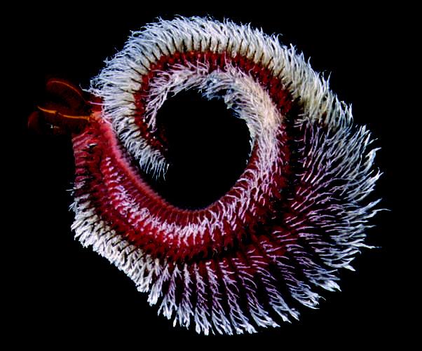
The chemical reactions of living systems take place across a wide range of conditions. Although many microbial species can tolerate extreme heat, multicellular organisms require much more temperate habitats. One exception is Alvinella pompejana, the Pompeii worm, which lives near deepsea hydrothermal vents and thrives at 42°C (107°F). Hairlike colonies of symbiotic bacteria may help insulate its body.
1.1 What Is Biochemistry?
LEARNING OBJECTIVE
Recognize the main themes of biochemistry.
Biochemistry is the scientific discipline that seeks to explain life at the molecular level. It uses the tools and terminology of chemistry to describe the various attributes of living organisms. Biochemistry offers answers to such fundamental questions as “What are we made of?” and “How do we work?” Biochemistry is also a practical science: It generates powerful techniques that underlie advances in other fields, such as genetics, cell biology, and immunology; it offers insights into the treatment of diseases such as cancer and diabetes; and it improves the efficiency of industries such as wastewater treatment, food production, and drug manufacturing.
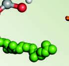
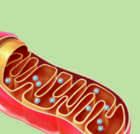
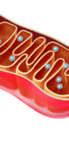
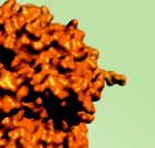
Some aspects of biochemistry can be approached by studying individual molecules isolated from cells. A thorough understanding of each molecule’s physical structure and chemical reactivity helps lead to an understanding of how molecules cooperate and combine to form larger functional units and, ultimately, the intact organism (Fig. 1.1). However, just as a clock completely disassembled no longer resembles a clock, information about a multitude of biological molecules does not necessarily reveal how an organism lives. Biochemists therefore investigate how organisms behave under different conditions or when a particular molecule is modified or absent. In addition, they collect vast amounts of information about molecular structures and functions—information that is stored and analyzed by computer, a field of study known as bioinformatics A biochemist’s laboratory is as likely to hold racks of test tubes as flasks of bacteria or computers.
Chapters 3 through 22 of this book are divided into three groups that roughly correspond to three major themes of biochemistry:
DSP/deepseaphotography
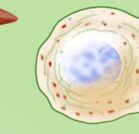
FIGURE 1.1 Levels of organization in a living organism. Biochemistry focuses on the structures and functions of molecules. Interactions between molecules give rise to higherorder structures (for example, organelles), which may themselves be components of larger entities, leading ultimately to the entire organism.
1. Living organisms are made of macromolecules. Some molecules are responsible for the physical shapes of cells. Others carry out various activities in the cell. (For convenience, we often use cell interchangeably with organism since the simplest living entity is a single cell.) In all cases, the structure of a molecule is intimately linked to its function. Understanding a molecule’s structural characteristics is therefore an important key to understanding its functional significance.
2. Organisms acquire, transform, store, and use energy. The ability of a cell to carry out metabolic reactions—to synthesize its constituents and to move, grow, and reproduce— requires the input of energy. A cell must extract this energy from the environment and spend it or store it in a manageable form.
3. Biological information is transmitted from generation to generation. Modern human beings look much like they did 100,000 years ago. Certain bacteria have persisted for millions, if not billions, of years. In all organisms, the genetic information that specifies a cell’s structural composition and functional capacity must be safely maintained and transmitted each time the cell divides.
Several other themes run throughout biochemistry and we will highlight these where appropriate.
4. Cells maintain a state of homeostasis. Even within its own lifetime, a cell may dramatically alter its shape or metabolic activities, but it does so within certain limits. In order to remain in a steady, nonequilibrium state—homeostasis—the cell must recognize changing internal and external conditions and regulate its activities.
5. Organisms evolve. Over long periods of time, the genetic composition of a population of organisms changes. Examining the molecular makeup of living organisms allows biochemists to identify the genetic features that distinguish groups of organisms and to trace their evolutionary history.
6. Diseases can be explained at the biochemical level. Identifying the molecular defects that underlie human diseases, or investigating the pathways that allow one organism to infect another, is the first step in diagnosing, treating, preventing, or curing a host of ailments.
Ubiquinone
Biological Molecules
LEARNING OBJECTIVES
Identify the major classes of biological molecules.
• List the elements found in biological molecules.
• Draw and name the common functional groups in biological molecules.
• Draw and name the common linkages in biological molecules.
• Distinguish the main structural features of amino acids, carbohydrates, nucleotides, and lipids.
• Identify the monomers and linkages in polypeptides, polysaccharides, and nucleic acids.
• Summarize the biological functions of the major classes of biological molecules.
Even the simplest organisms contain a staggering number of different molecules, yet this number represents only an infinitesimal portion of all the molecules that are chemically possible. For one thing, only a small subset of the known elements are found in living systems (Fig. 1.2). The most abundant of these are C, N, O, and H, followed by Ca, P, K, S, Cl, Na, and Mg. Certain trace elements are also present in very small quantities.
Virtually all the molecules in a living organism contain carbon, so biochemistry can be considered to be a branch of organic chemistry. In addition, nearly all biological molecules are constructed from H, N, O, P, and S. Most of these molecules belong to one of a few structural classes, which are described below.
Similarly, the chemical reactivity of biomolecules is limited relative to the reactivity of all chemical compounds. A few of the functional groups and intramolecular linkages that are common in biochemistry are listed in Table 1.1. Familiarity with these functional groups is essential for understanding the behavior of the different types of biological molecules we will encounter throughout this book.
Cells contain four major types of biomolecules
Most of the cell’s small molecules can be divided into four classes. Although each class contains many members, they are united under a single structural or functional definition. Identifying a particular molecule’s class may help predict its chemical properties and possibly its role in the cell.
FIGURE 1.2 Elements found in biological systems. The most abundant elements are most darkly shaded; trace elements are most lightly shaded. Not every organism contains every trace element. Biological molecules primarily contain H, C, N, O, P, and S.
TABLE 1.1
Common Functional Groups and Linkages in Biochemistry
Amine b
Ether
Ketone iol
Aldehyde
Carboxylic acid b (Carboxylate)
Ester
C O OR
Amide
Imine b
or R NHR ioester
Phosphoric acid ester b, c
Diphosphoric acid ester b, d
or (amino group)
OH (hydroxyl group) (ether linkage)
(carbonyl group), (acyl group)
(carbonyl group), (acyl group)
(carboxyl group) or
(carboxylate group)
O (ester linkage)
C S OR (thioester linkage)
O
NHR
NH or
SH (sulfhydryl group) RNH3 or R2NH2 or R3NH or R NH2
(amido group)
or or or
(phosphoester linkage)
(imino group) (phosphoanhydride linkage)
(phosphoryl group)
group, pyrophosphoryl group)
a R represents any carboncontaining group. In a molecule with more than one R group, the groups may be the same or different.
b Under physiological conditions, these groups are ionized and hence bear a positive or negative charge.
c When R = H, the molecule is inorganic phosphate (abbreviated Pi ), usually H2PO 4 or HPO 4 2− .
d When R = H, the molecule is pyrophosphate (abbreviated PPi ).
Question Cover the structure column and draw the structure for each compound listed on the left. Do the same for each functional group.
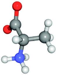
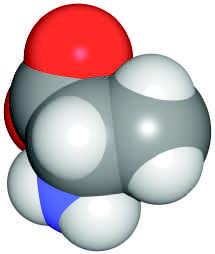
A structural formula includes all the atoms and the major bonds. Some bonds, such as the C O and N H bonds, are implied. The central carbon has tetrahedral geometry. The horizontal bonds extend slightly above the plane of the page and the vertical bonds extend slightly behind it.
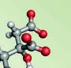
A ball-and-stick representation more accurately reveals the arrangement of atoms in space, although it does not show their relative sizes or electrical charges. The atoms are color-coded by convention: C gray, N blue, O red, and H white. b.
The space-filling model best represents the actual shape of the molecule but may obscure some of its atoms and linkages. Each atom is shown as a sphere whose radius (the van der Waals radius) corresponds to the distance of closest approach by another atom. c.
FIGURE 1.3 Representations of alanine. a. Structural formula, b. ballandstick model, and c. spacefilling model.
1. Amino Acids Among the simplest compounds are the amino acids, so named because they contain an amino group ( NH2) and a carboxylic acid group ( COOH). Under physiological conditions, these groups are actually ionized to NH 3 + and COO . The common amino acid alanine—like other small molecules—can be depicted in different ways, for example, by a structural formula, a ballandstick model, or a spacefilling model (Fig. 1.3). Other amino acids resemble alanine in basic structure, but instead of a methyl group ( CH3), they have another group—called a side chain or R group—that may also contain N, O, or S; for example,
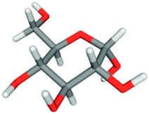
2. Carbohydrates Simple carbohydrates (also called monosaccharides or just sugars) have the formula (CH2O)n, where n is ≥ 3. Glucose, a monosaccharide with six carbon atoms, has the formula C6H12O6. It is sometimes convenient to draw it as a ladderlike chain (left); however, glucose forms a cyclic structure in solution (right):
In the representation of the cyclic structure, the darker bonds project in front of the page and the lighter bonds project behind it. In many monosaccharides, one or more hydroxyl groups are replaced by other groups, but the ring structure and multiple OH groups of these molecules allow them to be easily recognized as carbohydrates.
Asparagine
3. Nucleotides A five carbon sugar, a nitrogen containing ring, and one or more phosphate groups are the components of nucleotides. For example, adenosine triphosphate (ATP) contains the nitrogenous group adenine linked to the monosaccharide ribose, to which a triphosphate group is also attached:
triphosphate (ATP)
The most common nucleotides are mono, di, and triphosphates containing the nitrogenous ring compounds (or “bases”) adenine, cytosine, guanine, thymine, or uracil (abbreviated A, C, G, T, and U).
4. Lipids The fourth major group of biomolecules consists of the lipids These compounds cannot be described by a single structural formula since they are a diverse collection of molecules. However, they all tend to be poorly soluble in water because the bulk of their structure is hydrocarbonlike. For example, palmitic acid consists of a highly insoluble chain of 15 carbons attached to a carboxylic acid group, which is ionized under physiological conditions. The anionic lipid is therefore called palmitate. Palmitate
Cholesterol, although it differs significantly in structure from palmitate, is also poorly soluble in water because of its hydrocarbonlike composition.
Cells also contain a few other small molecules that cannot be easily classified into the groups above or that are constructed from molecules belonging to more than one group.
There are three major kinds of biological polymers
In addition to small molecules consisting of relatively few atoms, organisms contain macromolecules that may consist of thousands of atoms. Such huge molecules are not synthesized in one piece but are built from smaller units. This is a universal feature of nature: A few kinds
Box 1.A Units Used in Biochemistry
Biochemists follow certain conventions when quantifying objects on a molecular scale. For example, the mass of a molecule can be expressed in atomic mass units; however, the masses of biological molecules—especially very large ones—are typically given without units. Here it is understood that the mass is expressed relative to onetwelfth the mass of an atom of the common carbon isotope 12C (12.011 atomic mass units). Occasionally, units of daltons (D) are used (1 dalton = 1 atomic mass unit), often with the prefix kilo, k (kD). This is useful for macromolecules such as proteins, many of which have masses in the range from 20,000 (20 kD) to over 1,000,000 (1000 kD).
The standard metric prefixes are also necessary for expressing the minute concentrations of biomolecules in living cells. Concentrations are usually given as moles per liter (mol · L−1 or M), with the appropriate prefix, such as m, µ, or n:
mega (M)
(k)
(m)
micro ( µ) 10−6
(n)
(f)
For example, the concentration of the sugar glucose in human blood is about 5 mM, but many intracellular molecules are present at concentrations of µM or less.
Distances are customarily expressed in angstroms, Å (1 Å = 10−10 m) or in nanometers, nm (1 nm = 10−9 m). For example, the distance between the centers of carbon atoms in a C C bond is about 1.5 Å and the diameter of a DNA molecule is about 20 Å.
Question The diameter of a typical spherical bacterial cell is about 1 µm. What is the cell’s volume?
of building blocks can be combined in different ways to produce a wide variety of larger structures. This is advantageous for a cell, which can get by with a limited array of raw materials. In addition, the very act of chemically linking individual units (monomers) into longer strings (polymers) is a way of encoding information (the sequence of the monomeric units) in a stable form. Biochemists use certain units of measure to describe both large and small molecules (Box 1.A).
Amino acids, monosaccharides, and nucleotides each form polymeric structures with widely varying properties. In most cases, the individual monomers become covalently linked in headtotail fashion:
The linkage between monomeric units is characteristic of each type of polymer. The monomers are called residues after they have been incorporated into the polymer. Strictly speaking, lipids do not form polymers, although they do tend to aggregate to form larger structures such as cell membranes. Most of the mass of a cell consists of polymers, with proteins accounting for the greatest share (Fig. 1.4).
and lipids account for about 75% of the dry mass of a typical mammalian cell.
Monomers
Polymer
Residue
Proteins
Carbohydrates
FIGURE 1.4 Mass of a mammalian cell. Proteins
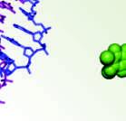
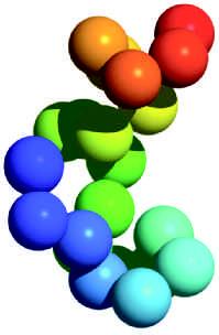
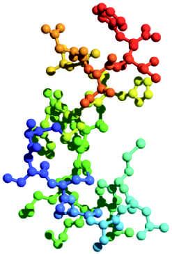
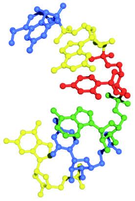
FIGURE 1.5 Structure of human endothelin. The 21 amino acid residues of this polypeptide, shaded from blue to red, form a compact structure. In a, each amino acid residue is represented by a sphere. The ballandstick model b shows all the atoms except hydrogen.
1. proteins Polymers of amino acids are called polypeptides or proteins. Twenty different amino acids serve as building blocks for proteins, which may contain many hundreds of amino acid residues. The amino acid residues are linked to each other by amide bonds called peptide bonds. A peptide bond (arrow) links the two residues in a dipeptide (the side chains of the amino acids are represented by R1 and R2).
Because the side chains of the 20 amino acids have different sizes, shapes, and chemical properties, the exact conformation (three dimensional shape) of the polypeptide chain depends on its amino acid composition and sequence. For example, the small polypeptide endothelin, with 21 residues, assumes a compact shape in which the polymer bends and folds to accommodate the functional groups of its amino acid residues (Fig. 1.5).
The 20 different amino acids can be combined in almost any order and in almost any proportion to produce myriad polypeptides, all of which have unique threedimensional shapes. This property makes proteins as a class the most structurally variable and therefore the most functionally versatile of all the biopolymers. Accordingly, proteins perform a wide variety of tasks in the cell, such as mediating chemical reactions and providing structural support
2.
Nucleic Acids
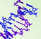
Polymers of nucleotides are termed polynucleotides or nucleic acids, better known as DNA and RNA. Unlike polypeptides, with 20 different amino acids available for polymerization, each nucleic acid is made from just four different nucleotides. For example, the residues in RNA contain the bases adenine, cytosine, guanine, and uracil, whereas the residues in DNA contain adenine, cytosine, guanine, and thymine. Polymerization involves the phosphate and sugar groups of the nucleotides, which become linked by phosphodiester bonds.
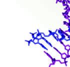
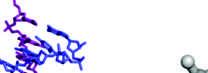
In part because nucleotides are much less variable in structure and chemistry than amino acids, nucleic acids tend to have more regular structures than proteins. This is in keeping with their primary role as carriers of genetic information, which is contained in their sequence of
nucleotide residues rather than in their three-dimensional shape (Fig. 1.6). Nevertheless, many nucleic acids do bend and fold into compact globular shapes, as proteins do.
3. polysaccharides
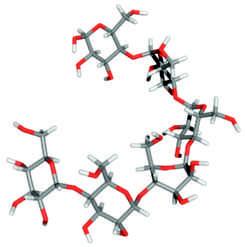
Polysaccharides usually contain only one or a few different types of monosaccharide residues, so even though a cell may synthesize dozens of different kinds of monosaccharides, most of its polysaccharides are homogeneous polymers. This tends to limit their potential for carrying genetic information in the sequence of their residues (as nucleic acids do) or for adopting a large variety of shapes and mediating chemical reactions (as proteins do). On the other hand, polysaccharides perform essential cell functions by serving as fuel-storage molecules and by providing structural support. For example, plants link the monosaccharide glucose, which is a fuel for virtually all cells, into the polysaccharide starch for longterm storage. The glucose residues are linked by glycosidic bonds (the bond is shown in red in this disaccharide):
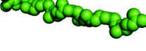
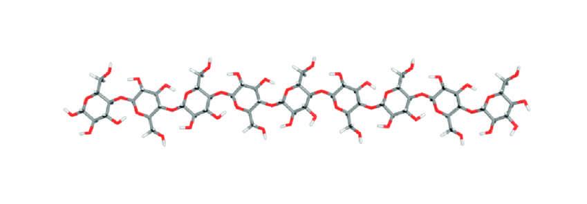
Glucose monomers are also the building blocks for cellulose, the extended polymer that helps make plant cell walls rigid (Fig. 1.7). The starch and cellulose polymers differ in the arrangement of the glycosidic bonds between glucose residues.
The brief descriptions of biological polymers given above are generalizations, meant to convey some appreciation for the possible structures and functions of these macromolecules. Exceptions to the generalizations abound. For example, some small polysaccharides encode information that allows cells bearing the molecules on their surfaces to recognize each other. Likewise, some nucleic acids perform structural roles, for example, by serving as scaffolding in ribosomes, the small machines where protein synthesis takes place. Under certain conditions, proteins are called on as fuelstorage molecules. A summary of the major and minor functions of proteins, polysaccharides, and nucleic acids is presented in Table 1.2.
FIGURE 1.6 Structure of a nucleic acid. a. Sequence of nucleotide residues, using oneletter abbreviations. b. Ballandstick model of the polynucleotide, showing all atoms except hydrogen (this structure is a sixresidue segment of RNA).
FIGURE 1.7 Glucose and its polymers. Both starch and cellulose are polysaccharides containing glucose residues. They differ in the type of chemical linkage between the monosaccharide units. Starch molecules have a loose helical conformation, whereas cellulose molecules are extended and relatively stiff.
Cellulose
Glucose
Starch
TABLE 1.2 Functions of Biopolymers
Biopolymer
✔ major function
✓ minor function
Before Going On
• List the six most abundant elements in biological molecules.
• Name the common functional groups and linkages shown in Table 1.1.
• Give the structural or functional definitions for amino acids, monosaccharides, nucleotides, and lipids.
• Describe the advantage of building a polymer from monomers.
• Give the structural definitions and major functions of proteins, polysaccharides, and nucleic acids.
• Name the linkage in each type of polymer.
• List the major functions of proteins, polysaccharides, and nucleic acids.
1.3
Energy and Metabolism
LEARNING OBJECTIVES
Explain how enthalpy, entropy, and free energy apply to biological systems.
• Define enthalpy, entropy, and free energy.
• Write the equation that links changes in enthalpy, entropy, and free energy.
• Relate changes in enthalpy and entropy to the spontaneity of a process.
• Describe the energy flow that makes living systems thermodynamically possible.
Assembling small molecules into polymeric macromolecules requires energy. And unless the monomeric units are readily available, a cell must synthesize the monomers, which also requires energy. In fact, cells require energy for all the functions of living, growing, and reproducing.
It is useful to describe the energy in biological systems using the terminology of thermodynamics (the study of heat and power). An organism, like any chemical system, is subject to the laws of thermodynamics. According to the first law of thermodynamics, energy cannot be created or destroyed. However, it can be transformed. For example, the energy of a river flowing over a dam can be harnessed as electricity, which can then be used to produce heat or perform mechanical work. Cells can be considered to be very small machines that use chemical energy to drive metabolic reactions, which may also produce heat or carry out mechanical work.