Veterinary Ophthalmology
Volume I and Volume II
Editor Kirk N. Gelatt
Associate Editors
Gil Ben-Shlomo
Brian C. Gilger
Diane V.H. Hendrix
Thomas J. Kern
Caryn E. Plummer
Sixth Edition
This edition first published 2021 © 2021 by John Wiley & Sons, Inc.
Fifth Edition © 2013 John Wiley & Sons, Inc. Fourth Edition © 2007 Blackwell Publishing. Third Edition © 1999 Lippincott Williams & Wilkins. Second Edition © 1991 Lea & Febiger. First Edition © 1981 Lea & Febiger.
Blackwell Publishing was acquired by John Wiley & Sons in February 2007. Blackwell’s publishing program has been merged with Wiley’s global Scientific, Technical and Medical business to form Wiley‐Blackwell.
The right of Kirk N. Gelatt to be identified as the author of the editorial material in this work has been asserted in accordance with law.
Registered Office
John Wiley & Sons, Inc., 111 River Street, Hoboken, NJ 07030, USA
Editorial Office
111 River Street, Hoboken, NJ 07030, USA
For details of our global editorial offices, customer services, and more information about Wiley products visit us at www.wiley.com.
Wiley also publishes its books in a variety of electronic formats and by print‐on‐demand. Some content that appears in standard print versions of this book may not be available in other formats.
Limit of Liability/Disclaimer of Warranty
The contents of this work are intended to further general scientific research, understanding, and discussion only and are not intended and should not be relied upon as recommending or promoting scientific method, diagnosis, or treatment by physicians for any particular patient. In view of ongoing research, equipment modifications, changes in governmental regulations, and the constant flow of information relating to the use of medicines, equipment, and devices, the reader is urged to review and evaluate the information provided in the package insert or instructions for each medicine, equipment, or device for, among other things, any changes in the instructions or indication of usage and for added warnings and precautions. While the publisher and authors have used their best efforts in preparing this work, they make no representations or warranties with respect to the accuracy or completeness of the contents of this work and specifically disclaim all warranties, including without limitation any implied warranties of merchantability or fitness for a particular purpose. No warranty may be created or extended by sales representatives, written sales materials or promotional statements for this work. The fact that an organization, website, or product is referred to in this work as a citation and/or potential source of further information does not mean that the publisher and authors endorse the information or services the organization, website, or product may provide or recommendations it may make. This work is sold with the understanding that the publisher is not engaged in rendering professional services. The advice and strategies contained herein may not be suitable for your situation. You should consult with a specialist where appropriate. Further, readers should be aware that websites listed in this work may have changed or disappeared between when this work was written and when it is read. Neither the publisher nor authors shall be liable for any loss of profit or any other commercial damages, including but not limited to special, incidental, consequential, or other damages.
Library of Congress Cataloging‐in‐Publication Data
Names: Gelatt, Kirk N., editor.
Title: Veterinary ophthalmology / editor, Kirk N. Gelatt ; associate editors, Gil Ben‐Shlomo, Brian C. Gilger, Diane V.H. Hendrix, Thomas J. Kern, Caryn E. Plummer.
Other titles: Textbook of veterinary ophthalmology
Description: Sixth edition. | Hoboken, NJ : Wiley‐Blackwell, 2020. | Includes bibliographical references and index.
Identifiers: LCCN 2020010772 (print) | LCCN 2020010773 (ebook) | ISBN 9781119441830 (hardback) | ISBN 9781119441823 (adobe pdf) | ISBN 9781119441816 (epub)
Subjects: MESH: Eye Diseases–veterinary | Diagnostic Techniques, Ophthalmological–veterinary | Ophthalmologic Surgical Procedures–veterinary
Classification: LCC SF891 (print) | LCC SF891 (ebook) | NLM SF 891 | DDC 636.089/77–dc23
LC record available at https://lccn.loc.gov/2020010772
LC ebook record available at https://lccn.loc.gov/2020010773
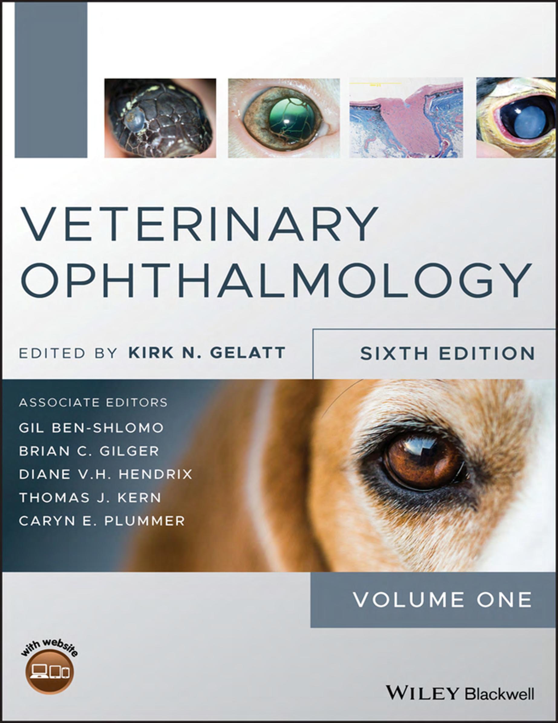
Cover image: Eye of a black horse © happylights / Shutterstock, Closeup of cat face. Fauna background © darkbird77 / Getty Images, Inquisitive Beagle Hound © bpretorius / Getty Images, Inset images courtesy of Kirk N. Gelatt. Cover design by Wiley
Set in 9.5/12pt STIX TwoText by SPi Global, Pondicherry, India
10 9 8 7 6 5 4 3 2 1
This book is dedicated to the memory of Dr. Gil Ben-Shlomo, an exceptional scholar, teacher, father and friend. The veterinary ophthalmology community has lost a gentle doctor and a gentleman.
Contributors xi
Preface xvii
About the Companion Website xix
Volume 1
Section I Basic Vision Sciences 1
Edited by Diane V.H. Hendrix
1 Ocular Embryology and Congenital Malformations 3
Cynthia S. Cook
2 Ophthalmic Anatomy 41
Jessica M. Meekins, Amy J. Rankin, and Don A. Samuelson
3 Physiology of the Eye 124
Diane V.H. Hendrix, Sara M. Thomasy, and Glenwood G. Gum
4 Optics and Physiology of Vision 168
Ron Ofri and Björn Ekesten
5 Fundamentals of Animal Vision 225
Björn Ekesten and Ron Ofri
Section II Foundations of Clinical Ophthalmology 261
Edited by Diane V.H. Hendrix, Gil Ben-Shlomo, and Brian C. Gilger
6 Ocular Immunology 263
Robert English and Brian C. Gilger
7 Clinical Microbiology and Parasitology 293
David Gould, Emma Dewhurst, and Kostas Papasouliotis
8 Clinical Pharmacology and Therapeutics
Part 1 Ocular Drug Delivery 349
Alain Regnier
Part 2 Antibacterial Agents, Antifungal Agents, and Antiviral Agents 385
Alison Clode and Erin M. Scott
Part 3 Anti-Inflammatory and Immunosuppressant Drugs 417
Amy J. Rankin
Part 4 Mydriatics/Cycloplegics, Anesthetics, and Tear Substitutes and Stimulators 435
Ian P. Herring
Part 5 Medical Therapy for Glaucoma 451
Caryn E. Plummer
9 Veterinary Ophthalmic Pathology 479
Bruce H. Grahn and Robert L. Peiffer
10 Ophthalmic Examination and Diagnostics
Part 1 The Eye Examination and Diagnostic Procedures 564
Heidi J. Featherstone and Christine L. Heinrich
Part 2 Ocular Imaging 662
David Donaldson and Claudia Hartley
Part 3 Diagnostic Ophthalmic Ultrasound 733
Ellison Bentley, Stefano Pizzirani, and Kenneth R. Waller, III
Part 4 Clinical Electrodiagnostic Evaluation of the Visual System 757
Gil Ben-Shlomo
11 Ophthalmic Genetics and DNA Testing 778
Simon M. Petersen-Jones
12 Fundamentals of Ophthalmic Microsurgery 787
David A. Wilkie
13 Digital Ophthalmic Photography 815
Richard J. McMullen, Jr., Nicholas J. Millichamp, and Christopher G. Pirie
Section IIIA Canine Ophthalmology 877
Edited by Gil Ben-Shlomo, Brian C. Gilger, Kirk N. Gelatt, and Caryn E. Plummer
14 Diseases and Surgery of the Canine Orbit 879
Simon A. Pot, Katrin Voelter, and Patrick R. Kircher
15 Diseases and Surgery of the Canine Eyelid 923
Frans C. Stades and Alexandra van der Woerdt
16 Diseases and Surgery of the Canine Nasolacrimal System 988
Lynne S. Sandmeyer and Bruce H. Grahn
17
Diseases and Surgery of the Canine Lacrimal Secretory System 1008
Elizabeth A. Giuliano
18 Diseases and Surgery of the Canine Conjunctiva and Nictitating Membrane 1045
Claudia Hartley and Diane V.H. Hendrix
19 Diseases and Surgery of the Canine Cornea and Sclera 1082
R. David Whitley and Ralph E. Hamor
20 The Canine Glaucomas 1173
Caryn E. Plummer, András M. Komáromy, and Kirk N. Gelatt
Index i1
Volume 2
Section IIIB Canine Ophthalmology 1257
Edited by Gil Ben-Shlomo, Brian C. Gilger, Kirk N. Gelatt, Caryn E. Plummer, and Thomas J. Kern
21 Diseases and Surgery of the Canine Anterior Uvea 1259
Diane V.H. Hendrix
22 Diseases of the Lens and Cataract Formation 1317
Marta Leiva and Teresa Peña
23 Surgery of the Lens 1371
Tammy Miller Michau
24 Diseases and Surgery of the Canine Vitreous 1459
Michael H. Boevé and Frans C. Stades
25 Diseases of the Canine Ocular Fundus 1477
Simon M. Petersen-Jones and Freya Mowat
26 Surgery of the Canine Posterior Segment 1575
Allison R. Hoffman, Joseph C. Wolfer, Samuel J. Vainisi, and András M. Komáromy
27 Diseases of the Canine Optic Nerve 1622
Gillian J. McLellan
Section IV Special Ophthalmology 1663
Edited by Caryn E. Plummer and Thomas J. Kern
28 Feline Ophthalmology 1665
Mary Belle Glaze, David J. Maggs, and Caryn E. Plummer
29 Equine Ophthalmology 1841
Caryn E. Plummer
30 Food and Fiber Animal Ophthalmology 1983
Bianca C. Martins
31 Avian Ophthalmology 2055
Lucien V. Vallone and Thomas J. Kern
32 Ophthalmology of New World Camelids 2085
Juliet R. Gionfriddo and Ralph E. Hamor
33 Laboratory Animal Ophthalmology 2109
Seth Eaton
34 Small Mammal Ophthalmology 2179
David L. Williams
35 Exotic Animal Ophthalmology 2200
Thomas J. Kern
36 Neuro-Ophthalmology 2237
Aubrey A. Webb and Cheryl L. Cullen
37 Ocular Manifestations of Systemic Disease
Part 1 The Dog 2329
Aubrey A. Webb and Cheryl L. Cullen
Part 2 The Cat 2421
Aubrey A. Webb and Cheryl L. Cullen
Part 3 The Horse 2495
Aubrey A. Webb and Cheryl L. Cullen
Part 4 Food Animals 2535
Aubrey A. Webb and Cheryl L. Cullen
Index i1
Contributors
Gil Ben-Shlomo, DVM, PhD
Diplomate ACVO, Diplomate ECVO
Associate Professor of Ophthalmology
Departments of Clinical Sciences
College of Veterinary Medicine
Cornell University Ithaca, NY, USA
Ellison Bentley, DVM
Diplomate ACVO
Clinical Professor, Comparative Ophthalmology
Department of Surgical Sciences
School of Veterinary Medicine
University of Wisconsin‐Madison Madison, WI, USA
Michael H. Boevé, DVM, PhD
Diplomate ECVO
Staff Ophthalmologist
Department of Clinical Sciences of Companion Animals
Faculty of Veterinary Medicine Utrecht University, The Netherlands
Alison Clode, DVM
Diplomate ACVO Port City Veterinary Referral Hospital Portsmouth, NH, USA
Cynthia S. Cook, DVM, PhD
Diplomate ACVO
Veterinary Vision San Carlos and San Francisco, CA, USA
Cheryl L. Cullen, DVM
Diplomate ACVO
CullenWebb Animal Eye Specialists Riverview, NB, Canada
Emma Dewhurst, MA, VetMB, MRCVS
Diplomate ECVCP, FRCPath
IDEXX Laboratories Wetherby, West Yorkshire, UK
David Donaldson, BVSc(Hons), MRCVS, MANZCVS
Diplomate ECVO
European Specialist in Veterinary Ophthalmology
Senior Clinician in Ophthalmology
Langford Vets
University of Bristol Veterinary School Langford, Bristol, UK
Seth Eaton, VMD
Diplomate ACVO
Clinical Assistant Professor, Comparative Ophthalmology
Department of Surgical Sciences
School of Veterinary Medicine
University of Wisconsin – Madison Madison, WI, USA
Björn Ekesten, DVM, PhD
Diplomate ECVO
Professor of Ophthalmology
Department of Clinical Sciences
Faculty of Veterinary Medicine
Swedish University of Agricultural Science Uppsala, Sweden
Robert English, DVM, PhD
Diplomate ACVO
Animal Eye Care of Cary Cary, NC, USA
Heidi J. Featherstone, BVetMed, DVOphthal, MRCVS
Diplomate ECVO, Diplomate RCVS
Head of Ophthalmology
The Ralph Veterinary Referral Centre; Honorary Associate Professor
Nottingham Veterinary School Marlow, Buckinghamshire, UK
Kirk N. Gelatt, VMD
Diplomate ACVO
Emeritus Distinguished Professor of Comparative Ophthalmology
Department of Small and Large Animal Clinical Sciences College of Veterinary Medicine
University of Florida Gainesville, FL, USA
Brian C. Gilger, DVM, MS
Diplomate ACVO, Diplomate ACT
Professor of Ophthalmology
Department of Clinical Sciences College of Veterinary Medicine
North Carolina State University Raleigh, NC, USA
Juliet R. Gionfriddo, DVM
Diplomate ACVO
Professor Emeritus of Ophthalmology
Department of Clinical Sciences
College of Veterinary Medicine
Colorado State University Fort Collins, CO, USA
Elizabeth A. Giuliano, DVM, MS
Diplomate ACVO
Associate Professor of Ophthalmology
Department of Veterinary Medicine and Surgery
College of Veterinary Medicine
University of Missouri Columbia, MO, USA
Mary Belle Glaze, DVM
Diplomate ACVO
Gulf Coast Animal Eye Clinic Houston, TX, USA
David Gould, BSc (Hons), BVM&S, PhD, DVOphthal, FRCVS
Diplomate ECVO
RCVS and European Specialist in Veterinary Ophthalmology
Head of Ophthalmology
Davies Veterinary Specialists
Manor Farm Business Park
Higham Gobion, Hertfordshire, UK
Bruce H. Grahn, DVM
Diplomate ACVO
Professor Emeritus of Veterinary Ophthalmology
Western College of Veterinary Medicine and Prairie Ocular Pathology of Prairie Diagnostic Laboratories
University of Saskatchewan Saskatoon, SK, Canada
Glenwood G. Gum, MS, PhD
Director of Ophthalmology
Absorption Systems
San Diego, CA, USA
Ralph E. Hamor, DVM, MS
Diplomate ACVO
Clinical Professor of Comparative Ophthalmology
Departments of Small and Large Animal Clinical Sciences
College of Veterinary Medicine
University of Florida Gainesville, FL, USA
Claudia Hartley, BVSc, CertVOphthal, MRCVS
Diplomate ECVO
European Specialist in Veterinary Ophthalmology
Head of Ophthalmology
Langford Vets
University of Bristol Veterinary School
Langford, Bristol, UK
Christine L. Heinrich, DVOphthal, MRCVS
Diplomate ECVO
RCVS and European Specialist in Veterinary Ophthalmology
Eye Veterinary Clinic Leominster, Herefordshire, UK
Diane V.H. Hendrix, DVM
Diplomate ACVO
Professor of Ophthalmology
Department of Small Animal Clinical Sciences
College of Veterinary Medicine
University of Tennessee Knoxville, TN, USA
Ian P. Herring, DVM, MS
Diplomate ACVO
Associate Professor of Ophthalmology
Department of Small Animal Clinical Sciences
Virginia‐Maryland College of Veterinary Medicine
Virginia Tech
Blacksburg, VA, USA
Allison R. Hoffman, DVM
Diplomate ACVO Eye Care for Animals Pasadena, CA, USA
Thomas J. Kern, DVM
Diplomate ACVO
Associate Professor of Ophthalmology Department of Clinical Sciences College of Veterinary Medicine
Cornell University Ithaca, NY, USA
Patrick R. Kircher, Dr.Med.Vet., PhD
Diplomate ECVDI, Exec. MBA, UZH
Professor of Veterinary Diagnostic Imaging Clinic for Diagnostic Imaging
Department of Clinical Diagnostics and Services
Vetsuisse Faculty, University of Zurich Zurich, Switzerland
András M. Komáromy, DrMedVet, PhD
Diplomate ACVO, Diplomate ECVO Professor of Ophthalmology
Department of Small Animal Clinical Science College of Veterinary Medicine
Michigan State University East Lansing, MI, USA
Marta Leiva, DVM, PhD
Diplomate ECVO
Professor Associate Hospital Clinic Veterinari
Fundació Universitat Autònoma de Barcelona; Departament de Medicina i Cirurgia Animal Facultat de Veterinària
Universitat Autònoma de Barcelona Bellaterra, Barcelona, Spain
David J. Maggs, BVSc
Diplomate ACVO
Professor of Ophthalmology
Department of Surgical and Radiological Sciences
School of Veterinary Medicine
University of California – Davis Davis, CA, USA
Bianca C. Martins, DVM, MSc, PhD
Diplomate ACVO
Clinical Associate Professor of Ophthalmology
Department of Surgical and Radiological Sciences
School of Veterinary Medicine
University of California - Davis Davis, CA, USA
Gillian J. McLellan, BVMS, PhD, DVOphthal, MRCVS
Diplomate ECVO, Diplomate ACVO
Associate Professor of Comparative Ophthalmology
Department of Surgical Sciences
School of Veterinary Medicine and Department of Ophthalmology and Visual Sciences
School of Medicine and Public Health
University of Wisconsin‐Madison Madison, WI, USA
Richard J. McMullen, Jr., Dr.Med.Vet, CertEqOphth (Germany)
Diplomate ACVO, Diplomate ECVO
Associate Professor of Equine Ophthalmology
Department of Clinical Sciences
Auburn University
JT Vaughan Large Animal Teaching Hospital Auburn, AL, USA
Jessica M. Meekins, DVM, MS
Diplomate ACVO
Associate Professor of Ophthalmology
Department of Clinical Sciences
Veterinary Health Center College of Veterinary Medicine
Kansas State University Manhattan, KS, USA
Tammy Miller Michau, DVM, MS, MSpVM
Diplomate ACVO
Vice President, Medical Affairs Operations
Mars Veterinary Health Vancouver, WA, USA
Nicholas J. Millichamp, BVetMed, PhD, DVOphthal, MRCVS
Diplomate ACVO, Diplomate ECVO
Eye Care for Animals‐Houston Houston, TX, USA
Freya Mowat, BVSc, PhD
Diplomate ECVO, Diplomate ACVO
Assistant Professor of Ophthalmology
Department of Surgical Sciences
School of Veterinary Medicine
University of Wisconsin‐Madison Madison, WI, USA
Ron Ofri, DVM, PhD
Diplomate ECVO
Professor, Veterinary Ophthalmology
Koret School of Veterinary Medicine
Hebrew University of Jersualem Rehovot, Israel
Kostas Papasouliotis, DVM, PhD, MRCVS
Diplomate RCPath (Vet.Clin.Path.), Diplomate ECVCP
EBVS® European Specialist in Veterinary Clinical Pathology
Senior Clinical Pathologist
IDEXX Laboratories
Wetherby, West Yorkshire, UK
Robert L. Peiffer, Jr., DVM, PhD
Diplomate ACVO
Professor Emeritus of Ophthalmology and Pathology
School of Medicine
University of North Carolina Chapel Hill, NC, USA
Teresa Peña, DVM, PhD
Diplomate ECVO
Professor
Hospital Clinic Veterinari
Fundació Universitat Autònoma de Barcelona; Departament de Medicina i Cirurgia Animal
Facultat de Veterinària
Universitat Autònoma de Barcelona
Bellaterra, Barcelona, Spain
Simon M. Petersen-Jones, DVetMed, PhD, DVOphthal, MRCVS
Diplomate ECVO
Myers‐Dunlap Endowed Chair in Canine Health
Professor of Comparative Ophthalmology
Department of Small Animal Clinical Sciences College of Veterinary Medicine
Michigan State University East Lansing, MI, USA
Christopher G. Pirie, DVM
Diplomate ACVO
Associate Professor of Ophthalmology
Department of Small Animal Clinical Sciences
College of Veterinary Medicine
Michigan State University East Lansing, MI, USA
Stefano Pizzirani, DVM, PhD
Diplomate ACVO, Diplomate ECVS‐inactive
Associate Professor of Ophthalmology
Department of Clinical Science
Tufts Cummings School of Veterinary Medicine
North Grafton, MA, USA
Caryn E. Plummer, DVM
Diplomate ACVO
Professor of Comparative Ophthalmology
Departments of Small and Large Animal Clinical Sciences College of Veterinary Medicine
University of Florida Gainesville, FL, USA
Simon A. Pot, DVM
Diplomate ACVO, Diplomate ECVO
Ophthalmology Section
Equine Department
Vetsuisse Faculty, University of Zurich Zurich, Switzerland
Amy J. Rankin, DVM, MS
Diplomate ACVO
Associate Professor of Ophthalmology
Department of Clinical Sciences
Veterinary Health Center College of Veterinary Medicine
Kansas State University Manhattan, KS, USA
Alain Regnier, Dr.Med.Vet, PhD
Emeritus Professor of Ophthalmology Department of Clinical Sciences School of Veterinary Medicine Toulouse, France
Don A. Samuelson, PhD, MS
Professor of Comparative Ophthalmology (Retired) Department of Small Animal Clinical Sciences College of Veterinary Medicine University of Florida Gainesville, FL, USA
Lynne S. Sandmeyer, DVM, DVSc
Diplomate ACVO
Associate Professor of Ophthalmology (Retired) Department of Small Animal Clinical Sciences
Western College of Veterinary Medicine University of Saskatchewan Saskatoon, SK, Canada
Erin M. Scott, VMD
Diplomate ACVO
Assistant Professor of Ophthalmology
Department of Small Animal Clinical Sciences College of Veterinary Medicine and Biomedical Sciences
Texas A&M University College Station, TX, USA
Frans C. Stades, DVM, PhD
Diplomate ECVO
Emeritus Professor of Veterinary Ophthalmology
Department of Clinical Sciences of Companion Animals
Faculty of Veterinary Medicine Utrecht University Utrecht, The Netherlands
Sara M. Thomasy, DVM, PhD
Diplomate ACVO
Professor of Comparative Ophthalmology
Department of Surgical and Radiological Sciences
School of Veterinary Medicine; Department of Ophthalmology & Vision Science School of Medicine
University of California Davis, CA, USA
Samuel J. Vainisi, DVM
Diplomate ACVO
Animal Eye Clinic Denmark, WI, USA
Lucien V. Vallone, DVM
Diplomate ACVO
Assistant Clinical Professor of Comparative Ophthalmology
Department of Small Animal Clinical Sciences College of Veterinary Medicine
Texas A&M University College Station, TX, USA
Alexandra van der Woerdt, DVM, MS
Diplomate ACVO, Diplomate ECVO
The Animal Medical Center New York, NY, USA
Katrin Voelter, DVM, Dr.Med.Vet., PhD
Diplomate ECVO
Equine Department
Vetsuisse Faculty, University of Zurich Zurich, Switzerland
Kenneth R. Waller III, DVM, MS
Diplomate ACVR
Clinical Associate Professor of Diagnostic Imaging Department of Surgical Sciences School of Veterinary Medicine University of Wisconsin-Madison Madison, WI, USA
Aubrey A. Webb, DVM, PhD CullenWebb Animal Eye Specialists Riverview, NB, Canada
R. David Whitley, DVM, MS
Diplomate ACVO Gulf Coast Veterinary Specialists Houston, TX; Departments of Small and Large Animal Clinical Sciences College of Veterinary Medicine University of Florida Gainesville, FL, USA
David A. Wilkie, DVM, MS
Diplomate ACVO
Professor of Comparative Ophthalmology Department of Veterinary Clinical Sciences College of Veterinary Medicine The Ohio State University Columbus, OH, USA
David L. Williams, MA, VetMB, PhD, CertVOphthal, MRCVS
Senior Lecturer, Veterinary Ophthalmology Department of Clinical Veterinary Medicine University of Cambridge Cambridge, UK
Joseph C. Wolfer, DVM
Diplomate ACVO
Toronto Animal Eye Clinic Toronto, ON, Canada
Preface
In 1965 when I entered veterinary ophthalmology, it became very quickly apparent that there was a very limited information base or knowledge in veterinary ophthalmology. If this new clinical discipline was to grow and develop into a respected clinical specialty, we would need to develop our own scientific base, and compete with the other emerging clinical specialties in veterinary medicine. And we have! With our limited English‐language books of Veterinary Ophthalmology by R.H. Smythe (1956), W.G. Magrane’s first edition of Canine Ophthalmology (1965; Lea and Febiger), and Diseases of the Canine Eye by F.G. Startup (1969; Williams and Wilkins), and the chapter in Advances in Veterinary Science called ‘Examination of the Eye’ and ‘Eye Operations in Animals’ by Otto Überreiter (1959; Academic Press), we needed to “roll up our sleeves” and get to work big time!
We have available now (2021) a large number of veterinary ophthalmology books concentrating on the dog, cat, exotic animals, horses, ophthalmic pathology, and ophthalmic surgery. Two veterinary ophthalmology journals have proven invaluable to our success as a discipline. The first journal, Veterinary and Comparative Ophthalmology, was published by Fidia Research Foundation and Veterinary Practice Publishing (1991–1998), and our second journal was Veterinary Ophthalmology (published by Blackwell and Wiley‐Blackwell, 1998 to present); they greatly assisted our development and proved critical for the distribution of new scientific information. In fact, the current journal provides more than 90% of animal ophthalmic literature annually worldwide.
In the late 1950s and extending into the 1970s, professional groups of budding veterinary ophthalmologists organized scientific societies to gather and exchange their knowledge and clinical experiences, which rapidly evolved to Colleges of Veterinary Ophthalmologists whose primary missions were to train new veterinary ophthalmologists (termed residents), and foster (and fund) research to “grow” the clinical discipline long term and worldwide. Nowadays these significant changes have greatly enriched veterinary ophthalmology, and markedly improved the quality of our ophthalmic animal patients.
The advances in this text, Veterinary Ophthalmology, have paralleled and documented the changes in veterinary ophthalmology, and has become our symbol of where we are today. In 1981, the first edition was released, consisting of 21 chapters (788 pages) by 22 authors, and was well received. As a result, subsequent editions followed: second edition (1991; 765 pages and 19 authors), and then in 1999 our last single‐volume release (1544 pages, 37 chapters, and 44 authors). The third edition was markedly expanded and had color illustrations throughout the text.
The last two editions were two‐volume sets: for 2007, volume one 535 pages, 9 chapters, and 45 authors, and for the second larger volume 1672 pages, 20 chapters, and 36 authors; and in 2012 for volume one 789 pages, 12 chapters, and 26 authors, and for the second volume 1479 pages, 22 chapters, and 39 authors. All editions were well referenced; in fact, a great value of this text is that it documents the advances in veterinary ophthalmology during the past half of the twentieth century, and the first two decades of the twenty‐first century!
The sixth edition again consists of two volumes, 37 chapters, and 64 contributors. Like the last two editions, the first volume contains the basic science and foundations of clinical ophthalmology chapters and the first part of the third section on canine ophthalmology. Basic vision science courses in veterinary medical colleges are often an afterthought, and our veterinary ophthalmic basic sciences are frequently documented by veterinary ophthalmologists (rather than anatomists, physiologists, pharmacologists, etc.).
The first volume of the basic sciences and foundations of veterinary ophthalmology is designed to provide the base of those subjects that underpin the clinical sciences. They include embryology, anatomy, ophthalmology physiology, optics and physiology of vision, and fundamentals of vision in animals. In the foundations of clinical ophthalmology section, the chapters include immunity, microbiology, clinical pharmacology and therapeutics, ophthalmic pathology, ophthalmic examination and diagnostics, ophthalmic genetics and DNA testing, fundamentals of microsurgery, and photography. The third section starts with the chapters for
the first part of the canine ophthalmology including orbit, eyelids, nasolacrimal system, lacrimal secretory system, conjunctiva and nictitating membrane, cornea and sclera, and glaucoma.
The second volume focuses on clinical ophthalmology in the different species, and starts with the second part of canine ophthalmology (chapters- anterior uvea, lens and cataract formation, surgery of the lens, vitreous, ocular fundus, surgery of the posterior segment, and optic nerve), and continues with feline, equine, food and fiber‐producing animals, avian, New World camelids, laboratory animals, pocket pet animals, and exotics, and concludes with comparative neuro‐ophthalmology, ophthalmic manifestations of systemic diseases, and the index. The sixth edition more or less has devoted space relative to the amount of time based on different animal species encountered in veterinary ophthalmology practice.
Now, in 2021, the sixth edition of Veterinary Ophthalmology continues to document this discipline’s advances. The magnitude of this edition has now required five associate editors, who devoted their time and expertise to make it happen. Like for me, I’m certain it was a learning experience! They are Drs. Brian C. Gilger, Diane V.H. Hendrix, Thomas J. Kern, Caryn E. Plummer, and Gil Ben‐Shlomo. Each editor chose their authors and respective chapters, based on their
expertise and preferences. A book like this is a huge undertaking, and all of us have devoted hundreds of hours to make it a successful product for the profession. Our 64 authors contributed hundreds of hours to this edition, taking time away from family and practice, and we thank them.
When all the chapters had been submitted and production had started, the COVID‐19 pandemic spread across the world like a massive hurricane. Terms like “face masks,” “social distancing,” “isolation,” and “quarantine or shelter at home” became common terms, and our daily personal and professional routines were markedly disrupted. But progress in the production of the sixth edition continued uninterrupted.
We thank Erica Judisch, Executive Editor, Veterinary Medicine and Dentistry, and Purvi Patel, Project Editor, of Wiley‐Blackwell for their expertise and assistance in making the sixth edition of Veterinary Ophthalmology a reality. Our copyeditors, Jane Grisdale and Sally Osborn, and project manager Mirjana Misina were superb. And lastly, we thank and appreciate the continued support and encouragement of our spouses and family members who bear with us as we struggle to meet our time schedules and other life priorities.
Distinguished Kirk N. Gelatt, VMD, Diplomate, Professor of Comparative American College of Veterinary Ophthalmology, Emeritus Ophthalmologists
Basic Vision Sciences
Ocular Embryology and Congenital Malformations
Cynthia S. Cook
An understanding of normal and abnormal ocular development is essential to the broader subjects of anatomy, physiology, and pathology. Embryology provides both insight into the development of structures such as the cornea, iridocorneal angle, and retina and their normal and pathologic functions, as well as a means of understanding how congenital malformations occur.
Investigations of ocular development have often used rodents as animal models. Comparison with studies of humans and other animals demonstrates that the sequence of developmental events is very similar across species (Cook, 1995; Cook & Sulik, 1986; Hilfer, 1983; O’Rahilly, 1983). Factors that must be considered when making interspecies comparisons include duration of gestation, differences in anatomic end point (e.g., presence of a tapetum, macula, or Schlemm’s canal), and when eyelid fusion breaks (during the sixth month of gestation in the human versus 2 weeks postnatal in the dog; Table 1.1).
This chapter describes normal events and abnormalities in this developmental sequence that can lead to malformations. Bearing in mind the species differences alluded to earlier, the mouse is a valuable model in the study of normal and abnormal ocular morphogenesis. In particular, studying the effects of acute exposure to teratogens during development has provided valuable information about the specific timing of events that lead to malformations.
Gastrulation and Neurulation
Cellular mitosis following fertilization results in transformation of the single‐cell zygote into a cluster of 12–16 cells. With continued cellular proliferation, this morula becomes a blastocyst, containing a fluid‐filled cavity. The cells of the blastocyst will form both the embryo proper and the extraembryonic tissues (i.e., amnion and chorion). At this early stage, the embryo is a bilaminar disc, consisting of hypoblast
and epiblast. This embryonic tissue divides the blastocyst space into the amniotic cavity (adjacent to epiblast) and the yolk sac (adjacent to hypoblast; Fig. 1.1).
Gastrulation (formation of the mesodermal germ layer) begins during day 10 of gestation in the dog (day 7 in the mouse; days 15–20 in the human). The primitive streak forms as a longitudinal groove within the epiblast (i.e., future ectoderm). Epiblast cells migrate toward the primitive streak, where they invaginate to form the mesoderm. This forms the three classic germ layers: ectoderm, mesoderm, and endoderm. Gastrulation proceeds in a cranial‐to‐caudal progression; simultaneously, the cranial surface ectoderm proliferates, forming bilateral elevations called the neural folds (i.e., the future brain). The columnar surface ectoderm in this area now becomes known as the neural ectoderm (Fig. 1.2).
As the neural folds elevate and approach each other, a specialized population of mesenchymal cells, the neural crest, emigrates from the neural ectoderm at its junction with the surface ectoderm (Fig. 1.3). Migration and differentiation of the neural crest cells are influenced by the hyaluronic acid‐rich extracellular matrix. This acellular matrix is secreted by the surface epithelium as well as by the crest cells, and it forms a space through which the crest cells migrate. Fibronectin secreted by the noncrest cells forms the limits of this mesenchymal migration (LeDouarin & Teillet, 1974). Interactions between the migrating neural crest and the associated mesoderm appear to be essential for normal crest differentiation (LeDouarin & Teillet, 1974; Noden, 1993). The neural crest cells migrate peripherally beneath the surface ectoderm to spread throughout the embryo, populating the region around the optic vesicle and ultimately giving rise to nearly all the connective tissue structures of the eye (Table 1.2; Hilfer & Randolph, 1993; Johnston et al., 1979; Noden, 1993). The patterns of neural crest emergence and migration correlate with the segmental disposition of the developing brain.
Veterinary Ophthalmology: Volume I, Sixth Edition. Edited by Kirk N. Gelatt, Gil Ben-Shlomo, Brian C. Gilger, Diane V.H. Hendrix, Thomas J. Kern, and Caryn E. Plummer.
Veterinary Vision, San Carlos and San Francisco, CA, USA
Table 1.1 Sequence of ocular development (Cook, 1995; O’Rahilly, 1983).
Human (Approximate Postfertilization Age)
Month Week Day
Dog (Day Postfertilization)
Mouse (Day Postfertilization)Postnatal (P)Developmental Events
13228 13
4249 15
Optic sulci present in forebrain
Optic sulci convert into optic vesicles 10 17
Optic vesicle contacts surface ectoderm
Lens placode begins to thicken 26
252810.5
3211 19
3311.5 25
Optic vesicle surrounded by neural crest mesenchyme
Optic vesicle begins to invaginate, forming optic cup
Lens pit forms as lens placode invaginates
Retinal primordium thickens, marginal zone present
Optic vesicle invaginated to form optic cup
Optic fissure delineated
Retinal primordium consists of external limiting membrane, proliferative zone, primitive zone, marginal zone, and internal limiting membrane
Oculomotor nerve present
Pigment in outer layer of optic cup
Hyaloid artery enters through the optic cup
Lens vesicle separated from surface ectoderm
Retina: inner marginal and outer nuclear zones 11.5 29
63712
7
Basement membrane of surface ectoderm intact
Primary lens fibers form
Trochlear and abducens nerves appear
Lid folds present
Edges of optic fissure in contact 12 30
Tunica vasculosa lentis present
Lens vesicle cavity obliterated
Ciliary ganglion present 4112 32
Posterior retina consists of nerve fiber layer, inner neuroblastic layer, transient fiber layer of Chievitz, proliferative zone, outer neuroblastic layer, and external limiting membrane 17 32
Eyelids fuse (dog)
Secondary lens fibers present 4814 32
851
Anterior chamber beginning to form 12.5 40
Corneal endothelium differentiated
Optic nerve fibers reach the brain
Optic stalk cavity is obliterated
Lens sutures appear
Acellular corneal stroma present 54 30–35
95717 40
Scleral condensation present
First indication of ciliary processes and iris
Extraocular muscles visible
Table 1.1 (Continued)
Human (Approximate Postfertilization Age)
Eyelids fuse (occurs earlier in the dog)
45 Pigment visible in iris stroma
Ciliary processes touch lens equator
Rudimentary rods and cones appear
45–1 P
312
4 51
56
56
Hyaloid artery begins to atrophy to the disc
Branches of the central retinal artery form
Pupillary sphincter differentiates
Retinal vessels present
Ciliary muscle appears
Eye axis forward (human)
Tapetum present (dog)
2–14 P Tunica vasculosa lentis atrophies
5 40
Short eyelashes appear
Layers of the choroid are complete with pigmentation
6 Eyelids begin to open, light perception
1 P
7 1–14 P
1–16 P
Pupillary dilator muscle present
Pupillary membrane atrophies
Rod and cone inner and outer segments present in posterior retina
10–13 PPars plana distinct
9 16–40 PRetinal layers developed
14 P Regression of pupillary membrane, tunica vasculosa lentis, and hyaloid artery nearly complete
Lacrimal duct canalized
Data from Aguirre et al. (1972), Akiya et al. (1986), Cook (1995), and van der Linde‐Sipman et al. (2003).
It is important to note that mesenchyme is a general term for any embryonic connective tissue. Mesenchymal cells generally appear stellate and are actively migrating populations surrounded by extensive extracellular space. In contrast, the term mesoderm refers specifically to the middle embryonic germ layer. In other parts of the body (e.g., the axial skeletal system), mesenchyme develops primarily from mesoderm, with a lesser contribution from the neural crest. In the craniofacial region, however, mesoderm plays a relatively small role in the development of connective tissue structures. In the eye, mesoderm probably gives rise only to the striated myocytes of the extraocular muscles and vascular endothelium. Most of the craniofacial mesenchymal tissue comes from neural crest cells (Johnston et al., 1979).
The neural tube closes initially in the craniocervical region with closure proceeding cranially and caudally. Once closure is complete, the exterior of the embryo is fully covered by
surface ectoderm, and the neural tube is lined by neural ectoderm. Neural segmentation then occurs to form the specific parts of the brain: forebrain (i.e., prosencephalon), midbrain (i.e., mesencephalon), and hindbrain (i.e., rhombencephalon; see Fig. 1.3 and Fig. 1.4). The optic vesicles develop from neural ectoderm within the forebrain, with the ocular connective tissue derivatives originating from the midbrain neural crest.
Formation of the Optic Vesicle and Optic Cup
The optic sulci are visible as paired evaginations of the forebrain neural ectoderm on day 13 of gestation in the dog (see Fig. 1.3, Fig. 1.4, Fig. 1.5, Fig. 1.6, and Fig. 1.7). The transformation from optic sulcus to optic vesicle occurs
Maternal sinusoid
Endometrial stroma
Extra-embryonic somatopleuric mesoderm
Amnioblast cavity
Epiblast Hypoblast
Endoderm
Bilaminar embryo
Extra-embryonic coelom
Exocoelomic membrane
Figure 1.1 A blastocyst that has penetrated the maternal endometrium. An embryoblast has formed and consists of two cell layers: the epiblast above, and the hypoblast below. (Reprinted with permission from Cook, C., Sulik, K.K., & Wright, K.W. (2003) Embryology. In: Pediatric Ophthalmology and Strabismus (eds. Wright, K.W. & Spiegel, P.H.), pp. 3–38. New York: Springer.)
Amniotic cavity
Primitive node
Primitive streak
Primitive streak Epiblast Amnion
Figure 1.2 A. Dorsal view of an embryo in the gastrulation stage with the amnion removed. B. Cross-section through the primitive streak, representing invagination of epiblast cells between the epiblast and hypoblast layers. Note that the epiblast cells filling the middle area form the mesodermal layer. C. Cross-section through the neural plate. Note that the ectoderm in the area of the neural groove (shaded cells) has differentiated into neural ectoderm, whereas the ectoderm on each side of the neural groove is surface ectoderm (clear water cells). (Reprinted with permission from Cook, C., Sulik, K.K., & Wright, K.W. (2003) Embryology. In: Pediatric Ophthalmology and Strabismus (eds. Wright, K.W. & Spiegel, P.H.), pp. 3–38. New York: Springer.)
Neural crest cells
Pericardial bulge
Optic sulci
Brain regions:
Forebrain Midbrain
Hindbrain
Table 1.2 Embryonic origins of ocular tissues (Johnston et al., 1979; Noden, 1993; Yamashita & Sohal, 1987).
Neural Ectoderm Neural Crest
Neural retina Stroma of iris, ciliary body, choroid, and sclera
Retinal pigment epithelium Ciliary muscles
Posterior iris epitheliumCorneal stroma and endothelium
Pupillary sphincter and dilator muscle (except in avian species)
Bilayered ciliary epithelium
Figure 1.3 Dorsal view showing partial fusion of the neural folds to form the neural tube. Brain vesicles have divided into three regions: forebrain, midbrain, and hindbrain. The neural tube, groove, and facing surfaces of the large neural folds are lined with neural ectoderm (shaded cells), whereas surface ectoderm covers the rest of the embryo. Neural crest cells are found at the junction of the neural ectoderm and surface ectoderm. Neural crest cells migrate beneath the surface ectoderm, spreading throughout the embryo and specifically to the area of the optic sulci. Somites have formed along the lateral aspect of the closed cephalic neural tube. On the inside of both forebrain vesicles is the optic sulci. (Reprinted with permission from Cook, C., Sulik, K.K., & Wright, K.W. (2003) Embryology. In: Pediatric Ophthalmology and Strabismus (eds. Wright, K.W. & Spiegel, P.H.), pp. 3–38. New York: Springer.)
concurrent with the closure of the neural tube (day 15 in the dog). Intracellular filaments and microtubules within the cytoskeleton alter cell shape and allow for cell movement. In addition to the mechanical influences of the cytoskeleton and the extracellular matrix, localized proliferation and cell growth contribute to expansion of the optic vesicle (Fig. 1.5; Hilfer & Randolph, 1993; Hilfer et al., 1981).
The optic vesicle enlarges and, covered by its own basal lamina, approaches the basal lamina underlying the surface ectoderm (Fig. 1.5). The optic vesicle appears to play a significant role in the induction and size determination of the palpebral fissure and of the orbital and periocular structures (Jones et al., 1980). An external bulge indicating the presence of the enlarging optic vesicle can be seen at approximately day 17 in the dog.
Perivascular connective tissue and smooth muscle cells
Striated muscles of iris (avian species only)
Meninges of optic nerve
Orbital cartilage and bone
Connective tissue of the extrinsic ocular muscles
Endothelium of trabecular meshwork
Surface Ectoderm Mesoderm
Lens Extraocular myoblasts
Corneal and conjunctival epithelium
Lacrimal gland
Vascular endothelium
Schlemm’s canal (human)
Posterior sclera (?)
Data from Ashton (1966), Cook et al. (1991a), and Cook and Sulik (1986, 1988).
Anterior neuropore
Optic sulci
Pericardial bulge
Cut edge of amnion
Forebrain
Future lens placode
Midbrain
1st and 2nd pharyngeal pouches
Hindbrain
Somite
sac
Figure 1.4 Development of the optic sulci, which are the first sign of eye development. Optic sulci on the inside of the forebrain vesicles consisting of neural ectoderm (shaded cells). The optic sulci evaginate toward the surface ectoderm as the forebrain vesicles simultaneously rotate inward to fuse. (Reprinted with permission from Cook, C., Sulik, K.K., & Wright, K.W. (2003) Embryology. In: Pediatric Ophthalmology and Strabismus (eds. Wright, K.W. & Spiegel, P.H.), pp. 3–38. New York: Springer.)
Yolk