Sustainable Uses of Byproducts from Silk Processing
Narendra Reddy and Pornanong Aramwit
Authors
Professor Narendra Reddy Center for Incubation, Innovation, Research, and Consultancy
Jyothy Institute of Technology
Thataguni Post 560109 Bengaluru India
Professor Pornanong Aramwit Department of Pharmacy Practice Faculty of Pharmaceutical Sciences and Center of Excellence in Bioactive Resources for Innovative Clinical Applications
Chulalongkorn University, Bangkok Thailand 10330
The Academy of Science
The Royal Society of Thailand Dusit, Bangkok Thailand 10330
Cover Cover Image: © Baona/iStock/Getty Images Plus/ Getty Images
All books published by Wiley-VCH are carefully produced. Nevertheless, authors, editors, and publisher do not warrant the information contained in these books, including this book, to be free of errors. Readers are advised to keep in mind that statements, data, illustrations, procedural details or other items may inadvertently be inaccurate.
Library of Congress Card No.: applied for British Library Cataloguing-in-Publication Data
A catalogue record for this book is available from the British Library.
Bibliographic information published by the Deutsche Nationalbibliothek
The Deutsche Nationalbibliothek lists this publication in the Deutsche Nationalbibliografie; detailed bibliographic data are available on the Internet at <http://dnb.d-nb.de>.
© 2021 WILEY-VCH GmbH, Boschstraße 12, 69469 Weinheim, Germany
All rights reserved (including those of translation into other languages). No part of this book may be reproduced in any form – by photoprinting, microfilm, or any other means – nor transmitted or translated into a machine language without written permission from the publishers. Registered names, trademarks, etc. used in this book, even when not specifically marked as such, are not to be considered unprotected by law.
Print ISBN: 978-3-527-34786-5
ePDF ISBN: 978-3-527-82875-3
ePub ISBN: 978-3-527-82874-6
oBook ISBN: 978-3-527-82876-0
Typesetting SPi Global, Chennai, India
Printing and Binding
Printed on acid-free paper
Contents
Preface xi
1 Sericin: Structure and Properties 1
1.1 Type of Silk Sericin 1
1.2 Localization of Silk Sericin 1
1.3 Molecular Mass of Sericin 2
1.3.1 Middle Silk Gland Sericin 2
1.3.2 Mulberry Cocoon sericin 2
1.3.3 Non‐mulberry Cocoon and Peduncle Sericin 5
1.4 Layers of Sericin 6
1.5 Sericin Amino Acid Components 6
1.5.1 Silk Gland of Mulberry Sericin 6
1.5.2 Sericin from Mulberry Cocoons 8
1.5.3 Sericin from Non‐mulberry Cocoons 12
1.6 Sericin Gene 14
1.7 Sericin Structure 16
1.8 Sericin Properties 19
1.8.1 Biophysical Properties 19
1.8.1.1 Water Solubility 19
1.8.1.2 Gelation 20
1.8.1.3 Thermal Stability 20
1.8.1.4 Ultraviolet (UV) protection 21
1.8.1.5 Adhesion Properties and Electrostatic Interaction 22
1.8.2 Biochemical Activity 22
1.8.2.1 Anti‐tyrosinase Activity 22
1.8.2.2 Anti‐elastase Activity 23
1.8.2.3 Antioxidant Activity 23
1.8.2.4 Anti‐lipid Peroxidation Activity 25
1.8.3 Biological Activity 25
1.8.3.1 Anti‐inflammatory Activity 25
1.8.3.2 Anti‐tumor Activity 27
1.8.3.3 Inducing Collagen Production 28
1.8.3.4 Antibacterial Activity 29
References 32
2 Processing Sericin 39
2.1 Effects of Source and Extraction Method of Sericin on Its Benefits and Applications 39
2.1.1 Sericin Extraction 39
2.1.1.1 Water Extraction (WaterSS; HeatSS, AutoclaveSS) 39
2.1.1.2 Acid Extraction (AcidSS) 39
2.1.1.3 Alkali Extraction (AllkaliSS; Alkali‐L‐SS, Alkali‐H‐SS) 40
2.1.1.4 Urea Extraction (UreaSS) 40
2.1.1.5 Alcohol Extraction (AlcoholSS) 40
2.1.2 Effect of Source and Extraction on Sericin Properties 40
2.1.2.1 Molecular Weight of Sericin 40
2.1.2.2 Secondary Structure of the Sericin Protein 40
2.1.2.3 Phenolic contents 43
2.1.2.4 Antioxidant Activity 43
2.1.2.5 Anti‐tyrosinase Activity 44
2.1.2.6 Cytotoxicity 44
2.1.2.7 Cell Attachment, Cell Proliferation, and Collagen Production 44
2.1.2.8 Cell Protection 44
2.1.3 Benefit and Application of Extracted Mulberry Sericin and Non‐mulberry Sericin 45
2.1.3.1 Pharmaceutics and Cosmetics 45
2.1.3.2 Wound Healing 45
2.1.3.3 Tissue Engineering 47
2.1.3.4 Drug Delivery 47
2.2 Modification of Sericin Structure 48
2.3 Chemical Modification 48
2.4 Glutaraldehyde Cross‐linking 50
2.5 Dehydrothermal (DHT) Cross‐linking 51
2.6 Carbodiimide Cross‐linking 51
2.7 Dimethylolurea (DMU) Cross‐linking 53
2.8 Enzymatic Cross‐linking 53
2.9 Physical Modification 54
2.9.1 Photo‐Cross‐linking 54
2.10 Forms of Sericin Processing 55
2.10.1 Two‐dimensional Structure of Sericin 55
2.10.1.1 Hydrogel 55
2.10.1.2 Film 56
2.10.2 Three‐dimensional Structure of Sericin 56
2.10.2.1 Electrospinning Silk Sericin and Its Blends 56
2.10.3 Pure Sericin Electrospun Fibers 57
2.10.4 Sericin Blends with Natural Polymers 59
2.10.5 Sericin Blends with Synthetic Polymers 60
2.10.6 Blends with Poly(caprolactone) 65
2.11 Sericin from Wild Silks 66
2.12 Fibroin–Sericin Blends 69
2.12.1 Freeze‐drying 71
2.12.2 Salt Leaching 71
2.12.3 Gas Foaming 73
2.13 Biomaterials from Sericin and Sericin–Protein Blends 74
2.13.1 Pure Sericin Biomaterials 74
2.13.1.1 Sericin–Fibroin Blends 77
2.13.1.2 Sericin and Non‐sericin Protein Blends 78
2.13.2 Sericin Blend with Polysaccharides 81
2.13.2.1 Sericin–Alginate Blends 81
2.13.2.2 Sericin–Chitosan Blends 83
2.13.2.3 Sericin–Carboxymethyl Cellulose Blends 83
2.13.2.4 Binary and Ternary Sericin Blends with Miscellaneous Polysaccharides 85
2.14 Blends with Synthetic Polymers 89
2.15 Sericin–Lignin Blends 90 References 91
3 Applications of Sericin 101
3.1 Medical Application 101
3.1.1 Antioxidant Properties 101
3.1.2 Antibacterial Activity 102
3.1.3 Wound Healing 104
3.1.4 Antitumor Effect 105
3.1.5 Antidiabetic Potential 106
3.1.6 Metabolic Effects 107
3.1.6.1 Gastrointestinal Tract 107
3.1.6.2 Circulatory and Immune Systems 108
3.1.6.3 Lipid Metabolism and Obesity 109
3.1.7 Vehicle for Drug Delivery 109
3.1.8 Supplement in Culture Media and Cryopreservation 112
3.2 Cosmetic Applications 113
3.3 Biotechnology Applications 117
3.3.1 Tissue Engineering 117
3.3.1.1 Skin Tissue Repair with Sericin‐Based Materials 117
3.3.1.2 Sericin‐Based Materials for Bone and Cartilage Tissue Engineering 119
3.3.1.3 Sericin‐Based Biomaterials for Other Tissues 120
3.4 Miscellaneous Application 121
3.4.1 Sericin‐Coated Material as an Air filter 121
3.5 Conclusion 121 References 123
4 Non-silk Applications of Mulberry Plants 131
4.1 Introduction 131
4.2 Medicinal Applications of Mulberry Plant Extracts 132
4.2.1 Polysaccharides 132
4.2.2 Phenols and Flavanoids 135
4.2.3 Pectins and Lignins 140
4.2.4 Production of Paper 142
4.2.5 Fermentation and Biogas Production 144
4.2.6 Synthesis of Nanomaterials 144
4.2.7 Environmental Remediation 148
4.2.8 Composites 150
4.2.9 Superabsorbents 151 References 153
5 Pupae and Its Applications 157
5.1 Applications of Pupae Oil 157
5.1.1 Extraction of Pupae Oil 157
5.1.2 Medical Applications of Pupae Oil 159
5.1.3 Biodiesel from Pupae Oil 163
5.2 Proteins from Silkworm Pupae 163
5.2.1 Medical Application of Pupae Peptides 169
5.2.2 Nanoparticles from Pupae Proteins 174
5.3 Pupae as Animal Feed 176
5.3.1 Chitin and Chitosan from Silkworm Pupae 181
5.4 Miscellaneous Applications 184
5.4.1 Carbonization of Pupae 184
5.4.2 Production of Lipids 185
5.4.3 Anti‐diabetic Drug 188
5.5 Environmental Remediation 189 References 189
6 Applications of Pupae Litter (excrement) 195
6.1 Chlorophylline and its Derivatives 195
6.2 Applications of Proteins in Litter 197
6.3 Extracts from Litter 198
6.4 Environmental Remediation Using Litter 199
6.5 Conversion into Carbon 203
6.6 Nanoparticles from Litter 209 References 211
7 Converting Silk Wastes into Composites, Energy Storage Devices, and Other Value-Added Materials 213
7.1 Introduction 213
7.2 Composites from Silk Wastes 213
7.3 Reinforcement for Polypropylene (PP) Composites 214
7.4 Green Composites from Silk Wastes 219
7.5 Energy Applications 221
7.5.1 Carbonization of Waste Silk 221
7.6 Miscellaneous Applications 225
7.6.1 Extraction of Amino Acids and Bioactive Compounds 225
7.7 Fibers and Hydrogels 226
7.8 Environmental Remediation 227
7.9 Nutritious Food from Silkworm Eggs 229
References 229
Glossary 233
Index 237
Preface
Silk and silk‐producing insects are one of the most fascinating creations found in nature. Silk as a textile fiber has endured its prominence, elegance, and economic importance for millenniums. Discovery of novel silks produced by spiders, weaver ants, mussels, and other insects has further increased the interest, applications, and understanding of silk. However, a majority of the commercial silk is produced by Bombyx mori and in two countries, China and India. Even in these countries, the changing socioeconomic and environmental aspects are increasing the costs and adding considerable constraints on silk production. Another intriguing aspect of silk production and processing is the number and distinct by‐products that are generated.
During commercial silk production, degumming is essential and results in removal of about 25% of the protein sericin. After silk extraction, the worms (pupae) are considered waste and disposed into the environment or used as low‐value feed. Similarly, the insects generate considerable amounts of litter and leave parts of the leaf unconsumed during the rearing cycle. Stems of the mulberry plants are pruned regularly and are a major source for biomass. Sericin, pupae, litter, mulberry stems, and leaves are by‐products that are inevitably generated during silk production and are available in large quantities. Many of these by‐products have unique structure, properties, and applications.
The aim of this book is to provide detailed information on the sources, properties, and potential applications of silk byproducts. Structure of sericin and its use in cosmetics and food applications have been described in detail. Pupae, which consists of oil, proteins, and carbohydrates, has been used as a source for food, feed, energy, medical, biotechnology, and to develop several other novel materials. Proteins extracted from pupae and litter have inherent activity against cancer cells, bacteria, and even viruses. Similarly, chlorphyllin in litter and leaves has pharmaceutical and neutraceutical significance. Mulberry plants show high potential for removing toxic substances in the environment and phytochemicals in the mulberry stems and leaves have medical value. The authors aim to elucidate these unique features of silk by‐products and hope to promote the use of silkworms beyond the production of silk.
Sericin: Structure and Properties
1.1 Type of Silk Sericin
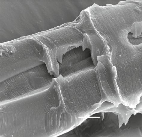
Sericin is a natural product from the silkworm. Sericin is one of the major protein components in the cocoons of Lepidopteron insects. Sericin is a glue-like coating protein surrounded with filament protein, fibroin (Figure 1.1). In manufacturing silk, sericin is a waste product from the degumming process. The silk sericin is classified into two types based on the feeding source of the silkworms: mulberry and non-mulberry sericin. The mulberry silkworm, Bombyx mori, is a well-known source of commercial silk production. This worm is a completely domesticated species that feeds on mulberry leaves. B. mori had long been developed for an indoor cultivation for the silk industry, whereas non-mulberry silkworm or wild silkworm is the group that feeds on other leaves such as oak leaves and castor oil leaves. Most of the non-mulberry silkworms cannot be reared indoors for their whole life cycles. The well-known non-mulberry silkworms are Antheraea, Samia ricini (or Philosamia ricini), and Cricula trifenestrata. Antheraea is a genus of silkworm that feeds on oak leaves and produces “tasar” silk, such as Antheraea assamensis (producing muka silk), Antheraea mylitta, Antheraea pernyi, and Antheraea yamamai. S. ricini produces the famous “eri” silk. In the wild environment, S. ricini feeds on castor oil plant leaves. C. trifenestrata is a wild silkworm producing “cricula” silk. The diversity of silkworm sources (genus, species, and diet) may produce distinct sericin characteristics.
1.2 Localization of Silk Sericin
Sericin is located at several sites of silkworms and cocoons. In the mulberry silkworm, B. mori, it has been reported that sericin is present in three components including silk gland, cocoon, and floss (Gamo et al. 1977; Kikkawa 1953; Yamada 1978). For non-mulberry silkworms, sericin is also secreted in the cocoon peduncle (Dash et al. 2006). The silk gland is the site that produces sericin. In a histological study, sericin was found to be mainly synthesized in the middle and posterior of the silk gland. Sericin protein is then sent to anterior silk glands via the lumen for secretion and cocoon construction (Consortium 2008; Kikkawa 1953; Yamanouchi 1922).
Sustainable Uses of Byproducts from Silk Processing, First Edition. Narendra Reddy and Pornanong Aramwit. © 2021 WILEY-VCH GmbH. Published 2021 by WILEY-VCH GmbH.
Structure and Properties
1.3 Molecular Mass of Sericin
The molecular mass of sericin has been observed using sodium dodecyl sulfate and polyacrylamide gel electrophoresis (SDS-PAGE). The diversity pattern of sericin was investigated from various extraction sites, extraction methods, species, and strains. Figure 1.2 shows the molecular mass of sericin from different extraction methods using SDS-PAGE.
1.3.1 Middle Silk Gland Sericin
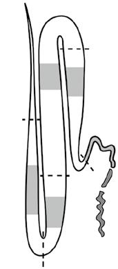
The middle silk gland (MSG, Figure 1.3) is a synthesis site for sericin in silkworm. Sericin obtained from MSG is known as native sericin. The MSG sericin measured by gel electrophoresis was found in intact bands and various protein sizes. Sericin extracted from the silk gland of mulberry silkworm was identified with three sizes of sericin including 130, 210, and 220 kDa as shown in Figure 1.4 (Sprague 1975). The study of sericin extracted from four MSG sections, including the anterior, middle to anterior, middle, and posterior sections, found five different sizes of sericin polypeptides. Two polypeptides, 177 and 134 kDa, were isolated from the anterior MSG. The middle section of the MSG had two polypeptides (309 and 145 kDa). One polypeptide (80 kDa) was found in the posterior section of the MSG (Gamo et al. 1977). Therefore, various sericin polypeptides were observed in mulberry sericin extracted from the MSG in the range between 80 and 309 kDa.
1.3.2 Mulberry Cocoon sericin
Cocoon sericin has been isolated, and the molecular mass of the protein was studied. Multiple sericin polypeptides have been extracted by several approaches including temperature, pressure, urea, acid, and alkali solution. In 1980, Tokutake reported that sericin
15k V X2, 000 10 μ m 080705
Sericin
Fibroin
Figure 1.1 Scanning electron microscope (SEM) of a silk filament that contains fibroin and sericin.
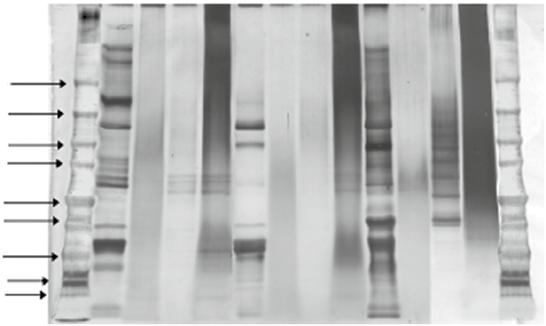
Figure 1.2 Sodium dodecyl sulfate and polyacrylamide gel electrophoresis (SDS-PAGE) of sericin extracted from white, green, and yellow cocoons using the following methods: urea solution (U), high temperature–high pressure (H), citric acid solution (A), and sodium carbonate solution (B). Different silk strains with various extraction methods show different molecular weights ranging from 10 to >225 kDa. Source: Reprinted with permission from Aramwit et al. (2010a).
Figure 1.3 Diagrammatic representation of the silk gland in the mature larvae of silkworms. Shadowed parts in the figure indicate the section used for the extraction of silk proteins, fibroin, and sericin. A, anterior gland; AM, anterior section in the middle gland; MM, middle section in the middle gland; PM, posterior section in the middle gland; P, posterior gland. Source: Reprinted with permission from Gamo et al. (1977). © 1977 Elsevier.
and fibroin protein could be separated by the precipitation of sericin in acidic conditions (pH 5.5). Later, the gel filtration approach was used to separate the precipitated sericin into five fractions (Tokutake 1980). This result suggests that sericin comprises a variety of forms. In 1982, Gamo and colleagues reported the molecular size of sericin proteins obtained by boiling with an alkali solution for extracting the protein from the cocoon. SDS-PAGE
Origin
Origin
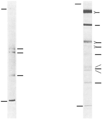
210 000–220 000
000 75 000–78 000
000–68 000
Figure 1.4 Separation of the protein components of sericin by sodium dodecyl sulfate and polyacrylamide gel electrophoresis on slabs of (a) 4% acrylamide and 0.1% bisacrylamide; (b) 8% acrylamide and 0.2% bisacrylamide. Source: Reprinted with permission from Aramwit et al. (2010a).
analysis revealed the molecular size of sericin in three different bands including the sizes >226.5, 226.5, and 218.8 kDa (Gamo 1982). In 2002, Takasu et al. compared the molecular mass between cocoon sericin and sections of the MSG. Cocoon proteins were extracted by saturated aqueous lithium thiocyanate solution. The four cocoon proteins were identified from SDS-PAGE analysis and named after the similar sizes found in subparts of the MSG. The two close bands around 250 kDa were named sericin A, as a reference to the specific size found in the anterior of the MSG. The fraction of 400 kDa was named sericin M, which was abundantly found in the middle of the MSG. The molecular size 150 kDa was named sericin P; this protein was the lowest expressed and found only in the posterior subpart of the MSG (Takasu et al. 2002). Moreover, the observation of sericin patterns was different depending on the extraction methods. In 2010, Aramwit et al. compared the extracted sericin protein pattern from four different extraction methods, including urea, autoclave (high heat and high pressure), acidic solution, and alkaline solution. Clear bands were observed with urea extraction, which found sericin in various sizes from 10 to >225 kDa. Sericin extracted by autoclave showed smear patterns ranging from 20 to 150 kDa. Acid and alkaline extraction solutions revealed band patterns mixing between clear bands and smears between 50–150 kDa and 15–75 kDa, respectively. In addition, both acid and alkaline extractions shared a clear band pattern at 50–70 kDa (Aramwit et al. 2010b). From all this information, it is evident that the extraction method affected the size and pattern of cocoon sericin proteins and is related to its biological properties. Additionally, the different silkworm strains gave different patterns of sericin proteins. The study of B. mori from three strains based on the pigment color (different concentrations of
flavonoids and carotenoids in cocoons), white shell, greenish shell, and yellow shell (Figure 1.5), had variations in sericin molecular mass. Urea-extracted sericin revealed the clear bands in all strains, but different size ranges for the white shell and yellow shell strains with proteins ranging from 10 to >225 kDa, while the greenish shell strain had mass ranging from 10 to 150 kDa. The autoclave method showed smear bands ranging from 50–150, 35–100, and 35–75 kDa for the white shell, greenish shell, and yellow shell strains, respectively. For acid and alkali extraction methods, all strains also displayed different bands within the range of 35–150 and 15–75 kDa, respectively (Aramwit et al. 2010b). This suggests that not only the sericin extraction method but also the strain of silkworms affected the molecular mass structure of extracted-sericin proteins.
1.3.3 Non-mulberry Cocoon and Peduncle Sericin
Non-mulberry cocoon sericin has been studied in S. ricini, A. assamensis, and A. mylitta. In 2004, Ahmad et al. observed non-mulberry sericin at 66 kDa from S. ricini and A. assamensis (Ahmad et al. 2004). In 2007, Dash et al. reported a 70 kDa sericin extracted from A. mylitta (Dash et al. 2007). Isolated sericin from non-mulberry cocoons’ molecular mass revealed a range between 66 and 70 kDa, which is smaller than observations for mulberry sericin.
The differences in the sericin extraction methods had dissimilar protein patterns in nonmulberry cocoon sericin from S. ricini, A. assamensis, and A. mylitta. The autoclave technique showed smear bands in all sericin species. The high temperature and high pressure from this extraction method might degrade all types of sericin protein. For the urea extraction method, there appeared a diversity of sericin protein sizes. S. ricini sericin was detected at the size higher than 300 kDa and in the range of 200–250 kDa. A. assamensis was observed in two bands with a size >250 kDa and at approximately 90 kDa. For A. mylitta, three bands were revealed with sizes of 250, 200, and 70 kDa (Sahu et al. 2016). These non-mulberry sericins detected by this study are composed of a high molecular mass in between 200 and 300 kDa, which was close to the high molecular mass of mulberry sericin. A similar study was performed comparing five extraction methods: urea, autoclave, conventional, acidic solutions, and alkaline solutions in A. assamensis and S. ricini. A. assamensis showed smeared bands in urea, autoclave, and conventional methods, whereas acidic and alkali solutions displayed a clear band at 75 kDa. For S. ricini, a clear band at 75 kDa was revealed
(a) (b)
(c)
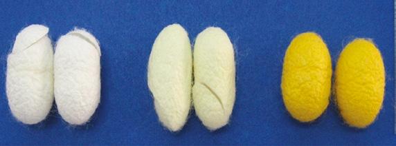
Figure 1.5 Physical appearance of silk cocoons, while shell (a), green shell (b), and yellow shell (c).
1 Sericin: Structure and Properties 6 along with smeared protein in urea, conventional, and alkali solutions. Smears with no intact bands of sericin proteins were observed from the acid and autoclave extraction methods (Kumar and Mandal 2017). These data showed that cocoon sericin protein patterns were different depending on their species and extraction methods. Therefore, the cocoon sericin protein might have a diversity of structures.
The peduncle is a strong filament in a ring form for attaching the non-mulberry cocoon to the branches of a tree. Sericin extracted from the peduncle of A. mylitta had a single band detected at 200 kDa (Dash et al. 2006). The size of this protein is similar to that observed for the MSG sericin extraction of A. mylitta (Dash et al. 2009). This sericin protein might have a major role in the action of the A. mylitta peduncle.
1.4 Layers of Sericin
The microstructure of silk gland sericin has been observed in silkworm. Histological studies have shown that sericin can be clearly divided into three distinct parts. Sericin I is the inner layer connected to fibroin. Sericin II is the middle layer and is the most abundant type. Sericin III is the outer layer, which covers the outside and is mostly mucous (Kikkawa 1953).
Cocoon sericin could be separated into three layers based on its solubility properties from extraction methods such as temperature, pressure, urea, acid, or alkali solution. Three layers have been divided into outer, middle, and inner layers, which are connected to fibroin. The amino acid composition of each layer has been defined differently. The pattern of amino acid residues (mol%) was used to identify the type of sericin protein. Fifteen amino acids have been identified. Four residues, including serine, threonine, glycine, and aspartic acid, were present at higher levels in all three layers (Shaw and Smith 1951).
1.5 Sericin Amino Acid Components
The amino acid composition of sericin has been reported from parts of silkworms and cocoons, including the silk gland, cocoon, floss, and peduncle. The percentage of amino acid contents in the total amino acid component (mol%) could indicate the individual structure of each sericin. The application of percent amino acid components was used as a reference for the distinction between sericin and fibroin silk protein (Gamo et al. 1977).
1.5.1 Silk Gland of Mulberry Sericin
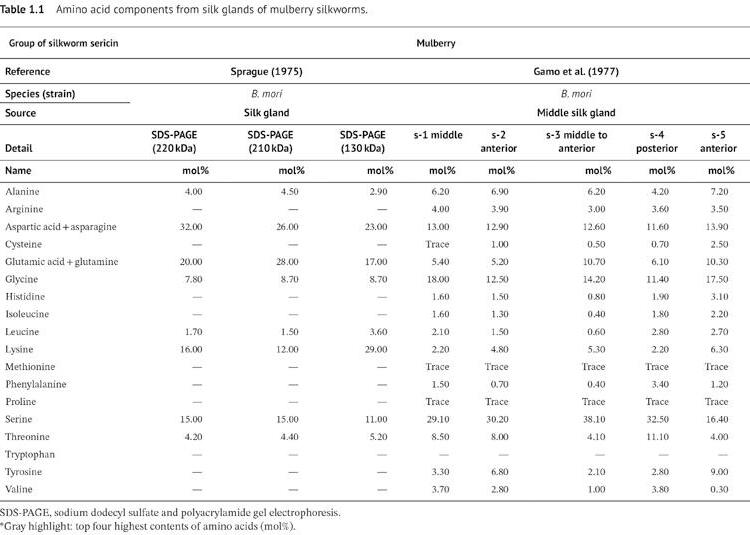
The amino acid residue composition in the silk gland of mulberry silkworm is revealed in Table 1.1. Most of the common amino acid composition (>10 mol%), which was found in all extraction methods, is serine and aspartic acid (including asparagine). In 1975, Sprague reported the amino acid content of sericin with three different molecular masses, including 220, 210, and 130 kDa. The four major amino acid residues, including aspartic acid, glutamic acid, lysine, and serine, had averages of 27, 22, 19, and 14 mol%, respectively (Sprague 1975). In 1977, Gamo et al. reported the amino acid composition of sericin
1 Sericin: Structure and Properties
from different sections of the MSG, including the middle (fraction: s-1), anterior (fraction: s-2 and s-5), middle to anterior (fraction: s-3), and posterior (fraction: s-4) sections (Gamo et al. 1977). Serine residues had an average of 30 mol% in all sections. The exception is the anterior MSG fraction s-5, which secreted serine content of 16 mol%. This fraction (s-5) contains serine in amounts that are half that of the other fractions. The minor component amino acids of all fractions were glutamic acid (average 15 mol%) and aspartic acid (average 13%). Threonine was found as a high percentage residue in fractions s-1, s-2, and s-4, on average 9%, while fraction s-3 and s-5 had a glutamic acid-rich component, on average 10%, instead of threonine residue. This data meant that sericin at different sections of the silk gland presented unique characteristics of sericin structure.
1.5.2 Sericin from Mulberry Cocoons
The amino acid composition of cocoon sericin was studied from various extraction methods, species, and strains. Amino acid components from mulberry silk sericin were different (Table 1.2). In mulberry cocoon sericin, the highest three amino acid components commonly found were serine (average 32 mol%), glycine (average 17 mol%), and aspartic acid (average 16 mol%). The less abundant sericin amino acid is threonine (average 8 mol%). However, the amino acid composition of sericin is different based on the extraction methods. Sericin from the acid precipitation method was found to have alanine-rich residues (15 mol%) instead of threonine. Alanine richness is not commonly found in other extraction methods (Tokutake 1980). Different sericin fractions showed differences in amino acid contents, such as the fraction sericin A (13 mol%) having glutamic acid richness. Meanwhile, the other fractions, sericin M and sericin P, showed high threonine contents at an average of 11 mol% (Takasu et al. 2002). The strain of silkworm did not show differences in the ratio of the amino acid residues by the autoclave extraction method (Aramwit et al. 2009). Cocoon sericin containing high amounts of serine, glycine, and aspartic acid is common. However, the different extraction methods may affect some amino acid contents, such as glutamic acid, threonine, and alanine. In addition, strains of silkworms revealed the different amino acid components from alkali extraction methods (Table 1.2). The alkaline extractions from three different silkworm strains found different components. The fourth top amino acid composition was found to be glutamic acid rich in Chul 3/2 and Chul 4/2 strains (average 7 mol%), whereas Chul 1/1 had threonine (7 mol%) (Aramwit et al. 2010a). The major amino acid residues of cocoon sericin were similar. Only some less common amino acid components such as threonine, glutamic acid, and alanine were different among extraction methods and strains. The modification of sericin structure by chemical solution was supported by the study of the properties of sericin being improved (Teramoto et al. 2004). Therefore, the extraction method interferes with the sericin structure.
Floss is a soft filament that covers the mulberry silk cocoon of B. mori. Its function is to protect and hang the cocoon on a tree branch. Floss sericin has been extracted and tested for the amino acid content (Table 1.2) (Yamada 1978). Three major amino acid residues were serine (40 mol%), glycine (18 mol%), and aspartic acid (10 mol%). The serine of floss sericin revealed around twofold higher percentage content when compared to cocoons. The differences in amino acid composition in floss and cocoons may reflect their specific properties and functions.
Table 1.2 Amino acid composition of sericin obtained from the cocoons of mulberry silkworms.
Group of silkworm sericin Mulberry silkworm sericin
Reference Yamada (1978) Tokutake (1980) Gamo (1982) Takasu (2002) Teramoto et al. 2004 Aramwit (2009)
Species (strain) B. mori (mandarina) B. mori (shugetsu × Hosho) B. mori
Source Floss Cocoon Cocoon Cocoon Cocoon Cocoon Cocoon Cocoon Cocoon Cocoon Cocoon Cocoon
Table 1.2 (Continued)
Group of silkworm sericin Mulberry silkworm sericin
Reference Yamada (1978) Tokutake (1980) Gamo (1982) Takasu (2002) Teramoto et al. 2004 Aramwit (2009)
Species (strain) B. mori (mandarina) B. mori (shugetsu × Hosho) B.
Source Floss Cocoon Cocoon Cocoon Cocoon Cocoon Cocoon Cocoon Cocoon Cocoon Cocoon Cocoon
1.2 (Continued)
Reference
et al. (2010)
Table
Structure and Properties
1.5.3 Sericin from Non-mulberry Cocoons
The cocoon sericin from non-mulberry silkworm has been reported from two species, A. mylitta (Dash et al. 2007, 2008) and C. trifenestrata (Yamada and Tsubouchi 2001). The amino acid analysis revealed three major components, including serine, glycine, and threonine (Table 1.3). Interestingly, glutamic acid, which has a high content in mulberry sericin, is less observed in non-mulberry sericin. The serine component in cocoon sericin of A. mylitta averages 19 mol%, which is lower than that in mulberry sericin (average 32 mol%). However, C. trifenestrata serine showed 40 mol%, which is higher than mulberry cocoon sericin but similar to the floss of mulberry sericin. Non-mulberry cocoon sericin showed a
Group of silkworm sericin
Non-mulberry
Reference Dash (2006) Dash (2006) Dash (2007)
Species (strain)
Dash
Source Peduncle Cocoon Cocoon Cocoon Cocoon
Detail Peduncle sericin
Table 1.3 Amino acid composition of sericin obtained from non-mulberry silkworms.
1.5 Sericin Aino cid oAponents 13 very low percent molecular content of aspartic acid, but a high component of threonine (average 12 mol%). The minor amino acid component in A. mylitta is histidine (average 12 mol%), and it is tyrosine in C. trifenestrata (7 mol%). Cysteine and phenylalanine were not found in non-mulberry sericin. From the amino acid composition of non-mulberry cocoon sericin and mulberry cocoon sericin, it seems that the structure of sericin is different and may lead to different structures and biological properties.
The peduncle, which has a function in hanging A. mylitta cocoons on the tree, had an amino acid composition that was high in glycine (24 mol%), serine (21 mol%), glutamic acid (11 mol%), and threonine (10 mol%) (Dash et al. 2006). The amino acid content is similar to the cocoon sericin of A. mylitta, which has no aspartic acid residue. A major component of glutamic acid was observed, which was different from the cocoon part that had a low mol% component. The amino acid composition of the peduncle sericin was different from the cocoon sericin. Therefore, the peduncle sericin may have a special property specific to its function.
Overall, a high serine content (>10 mol%) was found in all parts of the silkworm for all species and extraction methods. The other major amino acids, glycine, threonine, aspartic acid (including asparagine), glutamic acid (including glutamine), lysine, histidine, and tyrosine, were found to vary in each part. The different amino acid components of sericin might be related to its structure, property, and function (Figures 1.6 and 1.7).
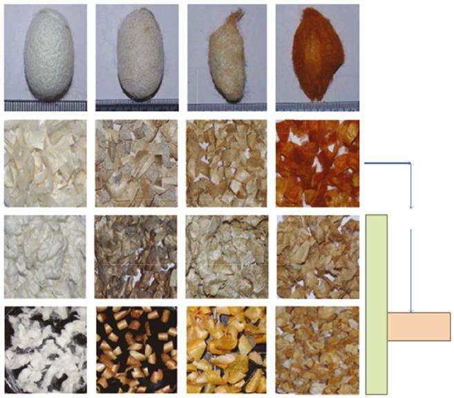
Figure 1.6 Physical appearance of mulberry and non-mulberry silk cocoons before and after degumming with different methods of sericin isolation. Source: Reprinted with permission from Sahu et al. (2016).
A. assamaS. ricini
A. mylitta
B. mori
Cocoons
Structure and Properties
Fresh (no degumming)
Urea degummed
Autoclave degummed
Fresh (no degumming)
Urea degummed
Autoclave degummed
Fresh (no degumming)
Urea degummed
Autoclave degummed
Fresh (no degumming)
Urea degummed
Autoclave degummed
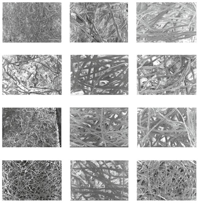
Figure 1.7 Scanning electron microscope (SEM) images of the cocoons of mulberry and nonmulberry silkworms. The cocoons are observed before (50×) and after degumming (100×) using urea and autoclave degumming methods. Scale bar represents 100 μm. Source: Reprinted with permission from Sahu et al. (2016).
1.6 Sericin Gene
Sericin proteins are translated from sericin genes, which are produced from MSG cells. Several studies have identified and characterized the sericin genes from MSG cells. The sericin genes were differently expressed in the MSG subparts (including anterior, middle, and posterior) and the stages of the silkworm larvae. Based on sericin transcripts, B. mori sericin genes could be separated into three types: sericin 1 (ser1), sericin 2 (ser2), and sericin 3 (ser3). These genes have a specific purpose for their localization as described in Chapter 11 under the B. mori genome (Dong et al. 2015).
The ser1 gene was first identified by Okamoto et al., in 1982. Two sericin mRNAs extracted from the MSG were discovered at lengths of 11.0 and 9.6 kb. The mRNA complementary sequences represent similar genomic diriboxynucleic acid (DNA), which determined five exons with 114 bp internal in the repetitive region consisting of approximately 60 repeats. The repetitive region was composed of 38 amino acids which had high residues of serine
mytitta
anama
mori
mori
mori
rkin
rkin
A. anama
A. anama
A. mytitta
A. mytitta
(40 mol%), aspartic acid (17 mol%), glycine (15 mol%), and threonine (10 mol%) (Okamoto et al. 1982). The composition of this region was similar to the amino acid component from the crude sericin protein extracted from the MSG. In 1986, Michaille et al. identified four variable sizes of sericin mRNAs that were compatible with 24 kb genomic DNA, including 10.5, 9.0, 4.0, and 2.8 kb (Michaille et al. 1986). The two sericin mRNAs had similar lengths between 10.5 and 9.0 kb and 11.0 and 9.6 kb, respectively, as reported by Okamoto (1982). In 1997, Garal et al. analyzed 4.0 kb of ser1 transcripts and combined the ser1 gene sequence from mRNA and genome sequences, which were previously reported by Okamoto and Michaille (Garel et al. 1997; Michaille et al. 1986; Okamoto et al.1982). The summarized ser1 gene sequence revealed 23 kb with nine exons and eight introns that were elucidated (Garel et al. 1997). The four major transcripts from the ser1 gene: 10.5, 9.0, 4.0, and 2.8 kb, were different by the absence of different exons (Michaille et al. 1986; Michaille et al. 1990). The variation of MSG sericin mRNA splicing was reported due to the production from alternative splicing of the sericin gene. The transcription of sericin was expressed in various types depending on the silkworm larval development stage (Ishikawa and Suzuki 1985). The silkworm larval stage was detected in various types of sericin transcripts in MSG cells (Couble et al. 1987). This suggests that the developmental stage induced different splicing of sericin genes in the cells. In addition, the amino acid compositions revealed in sericin M (middle of MSG) (Takasu et al. 2002) were similar to the ser1 protein (Tsubouchi et al. 2005).
The ser2 gene, another sericin gene discovered in MSG, was first identified with two encoding matured mRNAs at lengths 5.4 and 3.1 kb by Couble et al. in 1987 (Couble et al. 1987). In 1990, Michaille et al. reported two groups of ser2 transcripts, one of 3.1 kb and a variable-sized one between 5.0 and 6.4 kb (Michaille et al. 1990). In 2009, Kludkiewicz et al. reported two ser2 mRNAs containing 5.7 and 3.1 kb (Kludkiewicz et al. 2009). The ser2 proteins were predicted to have 1740 and 882 amino acid residues, which were identified as 230 and 120 kDa. The entire ser2 gene from the genomic DNA sequence database (Accession number GQ381286) is composed of 13.54 kb with 13 exons and 12 introns. The different mature mRNA sequences were observed by the deletion of the repetitive region at the exon 9a position. This evidence confirmed its generation by alternative splicing (Kludkiewicz et al. 2009). From these results, the ser2 transcript revealed the identical sizing at the small length of 3.1 kb and the polymorphism of the long transcript gene in the range of 5.0–6.4 kb from alternative splicing at a gene exon.
The ser3 gene was mainly obtained in the floss and outer layer of a silkworm cocoon. Takasu and colleagues identified 4.9 kb of ser3 gene transcript. The genomic length of the ser3 gene revealed 6.575 kb with three exons and two introns observed. Therefore, the ser3 protein consists of 1271 amino acid residues with two regions of motif sequences, 86 amino acid residues with 10 repeats, and 18 amino acid residues with 18.5 repeats. The estimated size of ser3 protein was 120 kDa (Takasu et al. 2007).
Non-mulberry A. yamamai had the sericin genes identified from the transcriptomic study of the last instar silk gland. Five sericin genes were discovered including AySrn1, AySrn2, AySrn3, AySrn4, and AySrn, with transcription size variation between 3.8 and 5.5 kb. The AySrn gene characters are different from B. mori sericins. All of the AySrn genes have short two exons with 22–28% of serine residues (Zurovec et al. 2016). The lower amount of exons in the sericin gene and its amino acid components are different from sericin genes in B. mori (3–12 exons). Therefore, the sericin proteins from different silkworm species appear to have different effects on their functions.
1 Sericin: Structure and Properties 16
The ser genes transcribe differently depending on the silkworm larval stages. The silkworm larval (instar) is developed in five stages, and then the mature stage is started to create a cocoon. The ser genes were differentially expressed by the specific instar stage. The development of B. mori instar stage has been influenced by the sericin gene expressions. In the last silkworm stage (the fifth instar), the transcripts of ser1 and ser3 started increasing at day 4 of the fifth instar, whereas ser2 transcripts were highly expressed since the third instar and decreased rapidly on day 4 of the fifth instar. This suggests that the expression of sericin genes is specific and dependent on the silkworm development stage. Moreover, sericin transcripts have investigated the expression in the fifth instar of each MSG subpart, including the anterior, middle, and posterior sections. The ser1 transcripts were highly expressed in the middle and posterior sections of MSG cells. The ser3 transcript was expressed at the anterior and middle MSG cells (Takasu et al. 2010, 2007). The ser2 transcripts were detected faintly in middle MSG and highly expressed at anterior MSG cells (Takasu et al. 2010). Therefore, these three sericin genes might contain some specific purpose which are beneficial for the different developmental stages of the silkworm.
1.7 Sericin Structure
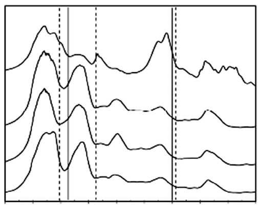
The secondary structure of sericin has been observed by Fourier transform infrared spectroscopy (FTIR). It has been reported that the silkworm species and isolation method affected to the sericin structure. Aramwit et al. reported B. mori cocoon sericins from four isolation methods, including autoclaving, urea, acidic, and alkaline solutions, and mainly found random coil and β-sheet structures (Figure 1.8). However, the urea extraction
Amide I Amide II Amide III
process
Figure 1.8 FTIR spectra of SS obtained from various extraction methods: acidic, alkaline, urea, and heat processes. Source: Reprinted with permission from Aramwit et al. (2010a). © 2010 John Wiley & Sons.
1.7 Seririn SeriSreS 17 method found different peaks related to the urea solution, which suggests that urea might be integrated into the sericin structure. Therefore, the urea used in sericin extraction possibly affected the sericin protein structure and function (Aramwit et al. 2010a).
The structure of sericin from different silkworm species (B. mori, A. assamensis, and S. ricini) and cocoon extraction methods (urea, conventional, autoclaved, acidic, and alkaline) has been studied by Kumar et al. (Figure 1.9) (Kumar and Mandal 2017). The secondary structure of sericin protein was classified based on the percentage of α-helix, β-sheet, turns, and random coils. The sericin structures were variable among species and extraction methods. All three species had a similarly high percentage of β-sheet and random coil when obtained using the urea extraction method. The mulberry silkworms, B. mori, sericin extraction with an acidic solution showed a higher percentage of α-helix and turns compared to the sericin structure from the urea extraction method. Meanwhile, the conventional and alkali methods resulted in the absence of the β-sheet, whereas the autoclaved method resulted in an absence of α-helix. In contrast, non-mulberry silkworm sericins of
220 kDa
160 kDa
120 kDa
100 kDa
90 kDa
80 kDa
70 kDa
60
220
30 kDa
20 kDa 10 kDa
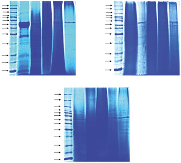
Figure 1.9 Molecular weight distribution of sericin extracted from cocoons using different extraction methods in sodium dodecyl sulfate and polyacrylamide gel electrophoresis (SDS-PAGE). 10% SDS-PAGE gel showing bands of (A) protein ladder and sericin extracted from (a) BoAbyx Aori, (b) ntheraea assaAensis, and (c) PhioosaAia ricini using (B) urea degradation, (C) acid degradation, (D) autoclaved, (E) conventional method, and (F) alkali degradation. Source: Reprinted with permission from Kumar and Mandal (2017).
A. assamensis and S. ricini from the urea extraction method did not result in α-helix in the structure. The β-sheet structure of sericin extracted from A. assamensis was absent in all extraction methods. However, S. ricini sericin had a variety of percentages (Figure 1.10) (Kumar and Mandal 2017). The differences in the α-helix structure between mulberry and non-mulberry sericins might be from the different gene sequences of each species. This evidence may lead to a different function of each sericin protein among species. The variable structures of sericin from different extraction methods may cause the different properties and biological activities of sericin proteins.

Figure 1.10 FTIR spectra of sericin extracted from the cocoons of BoAbyx Aori, ntheraea assaAensis, and PhioosaAia ricini using (a) urea degradation, (b) autoclaving, (c) acid degradation, (d) conventional method, and (e) alkali degradation. Source: Reprinted with permission from Kumar and Mandal (2017). © 2017 Elsevier.
1.8 SericinProperties
The predicted structures of three B. mori sericin gene sequences from ser1, ser2, and ser3 have been observed to have a repetitive region of the sericin protein. The repetitive region of the ser1 protein is composed of 38 amino acids with approximately 60 repeats, and a 40% serine residue component was revealed in the high β-sheet content (Garel et al. 1997). The repetitive sequence of the ser2 gene consists of 15 amino acids rich in charged residues, including lysine, aspartic acid, glutamic acid, and arginine at this region that formed the β-sheet structure of ser2 proteins (Michaille et al. 1990). The amino acid components appearing in MSG ser2 proteins were different from those found in cocoon ser1 and ser3 proteins. The ser2 proteins produced from the MSG stopped expression before the silkworm contracted the cocoon. This showed that there was very little ser2 protein left in the cocoon. Therefore, ser2 proteins were not observed in the cocoon isolate (Kludkiewicz et al. 2009; Takasu et al. 2010). The repetitive region of the ser3 gene is composed of 86 repeating amino acids and another 8 repeating amino acids with 45% sericin residue composition. The ser3 repetitive regions were predicted to contain lower formations of the β-sheet structure than the ser1 structure (Takasu et al. 2007). The different amino acid components of each repeated sequence were formed by different structures and properties of each ser protein. This data suggests that the different ser genes produced the specific protein properties appropriate for each sericin layer.
1.8 Sericin Properties
1.8.1 Biophysical Properties
1.8.1.1 Water Solubility
Sericin can be dissolved in hot water and could be precipitated after exposure to cold water (Sprague 1975). The water solubility of sericin was explained by the correlation with its amino acid content. Amino acid compositions of both mulberry and non-mulberry sericin have relatively high levels of serine, glycine, and threonine. The polar hydroxyl side chain residue of serine and threonine accounting over 30% in its total amino acid profile resulted in sericin presenting strong hydrophilicity (Padamwar and Pawar 2004). The correlation between sericin structure and its water-soluble properties has been studied in mulberry (B. mori) and non-mulberry (A. mylitta, A. assamensis, and S. ricini) sericins by circular dichroism spectroscopy. The secondary structures of sericin were determined to be random coils, β-sheet, and low α-helix content. In aqueous solution, sericin rapidly changed from random coil to β-sheet. However, an FTIR study showed that the sericin powder was mostly in the random coil and α-helix conformation (Sahu et al. 2016). This evidence shows that the β-sheet conformation, which is largely seen in aqueous sericin, is the solubilized form in water. Therefore, β-sheet formation is one of the factors of sericin imparting its watersoluble properties. Additionally, temperature is also a factor that changes the sericin structure. Sericin forms an insoluble structure at high water temperatures. At low water temperatures, sericin converts from random coil to β-sheet. This property is beneficial for gel formation and can be useful for biomaterial applications (Zhu et al. 1998).
As previously discussed in the gene sequence information, the repetitive regions of the ser genes (ser1, ser2, and ser3) from B. mori sequences have been shown to have high
1 Sericin: Structure and Properties
contents of β-sheet formation. The high number of repetitive regions makes it possible to increase the hydrophilic property of sericin protein. However, ser3 protein, which has a lower content of β-sheet formation than ser1 protein, was reported to be more hydrophilic than ser1 protein in a hydrophobicity prediction study (Garel et al. 1997; Takasu et al. 2007). This data suggests that the prediction method used may not be directly applicable to evaluate sericin protein properties. Therefore, to characterize sericin property, several techniques may be needed to collect the information that would be required for finding new applications.
1.8.1.2 Gelation
The gelation property of sericin has been observed under various conditions, depending on sericin solution concentration, temperature, and pH. The study of mulberry sericin from B. mori has revealed that gelation formed rapidly at a high concentration of sericin solution. The gelation rate was elevated at a high temperature (40 °C) and decreased at a lower temperature. Gel setting time was faster at pH 6 and became slower at a higher pH. During the gelation process, the strength of sericin increased, whereas the surface tension decreased. The secondary structure of sericin in the gelation process changed from random coil to β-sheet structure (Zhu et al. 1998; Zhu et al. 1995). This evidence suggests that sericin gelation is a thermoreversible process.
The property of sericin gelation was applied to the sericin protein as a biomaterial crosslinked with various types of polymers, such as biopolymers (polysaccharides; cellulose) (Wang et al. 2017), synthetic polymer (polyvinyl alcohol) (Aramwit et al. 2010b), or self-assembled (hydrogel) (Zhang et al. 2019). The network crosslinking between sericin and polymers was generally formed by covalent bonding at the polar functional groups of sericin amino acids (hydroxyl, carboxyl, and amino). The crosslinking could be produced by chemical and physical techniques. Chemical crosslinks were performed using agents such as glutaraldehyde (Nayak et al. 2012; Wang et al. 2017) and genipin (Aramwit et al. 2013, 2010; Wang et al. 2015). For physical crosslinks, ultraviolet (UV) light was used for photo-crosslinking bonds (Qi et al. 2018). These results demonstrated that sericin is capable of forming a gel with various polymers and several crosslinking techniques.
Gelation was also reported in non-mulberry sericin, A. mylitta. Similar to mulberry sericin, gel formation was observed in the crosslinking between A. mylitta sericin and polymers. This non-mulberry sericin was bonded with polyvinyl alcohol via a glutaraldehyde crosslinking agent (Mandal et al. 2011). Likewise, the natural polymer (cellulose) was reported to crosslink with sericin by the dual crosslinking agents, glutaraldehyde, and aluminum chloride (Nayak et al. 2014). Therefore, sericin from all sources could be efficiently used as a biomaterial application for future medicines and cosmetics.
1.8.1.3 Thermal
Stability
Thermogravimetric analysis tests the mass stability of sericin according to time and temperature change. Sericins extracted from mulberry (B. mori) and non-mulberry (A. mylitta, A. assamensis, and S. ricini) silkworms have been studied for its stability as related to temperature. The non-mulberry sericins were reported to present more stability than mulberry sericin. The highest stability was for sericin from S. ricini (Figure 1.11) (Sahu et al. 2016). This property suggests that sericins from different species have different structures and properties.
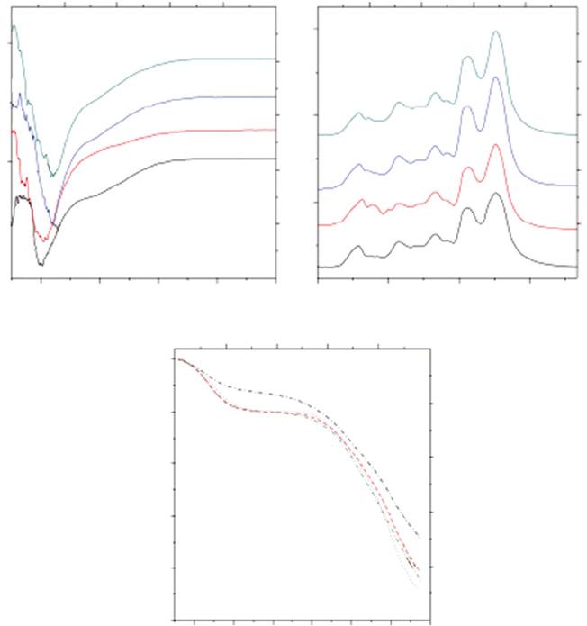
Figure 1.11 (a) Circular dichroism (CD) spectra of 0.1% w/v sericin solution from cocoons of different species: (A) Ayoitta, (B) assaAensis, (C) eririnr, (D) BoAbyx Aori; (b) FTIR spectrum of sericin powders from the various species: (A) . Ayoitta, (B) . assaAensis, (C) . eririnr, (D) B. Aori; (c) thermogravimetric analysis (TGA) curves of lyophilized sericin powders of (A) Ayoitta, (B) assAa, (C) eririnr, (D) B. Aori. Source: Reprinted with permission from Sahu et al. (2016). Licensed under CC BY 4.0.
1.8.1.4 Ultraviolet (UV) protection
Sericin has shown the ability to protect cells from UV radiation. The study of the photoprotective properties of B.mori reported that sericin was effective in reducing skin oxidative stress (Zhaorigetu et al. 2003), inhibiting UVB-induced apoptosis (Dash et al. 2008), and absorbing UVC radiation (Kiro et al. 2017). Non-mulberry sericin from A. assamensis and S. ricini was reported to increase cell viability against both UVA and UVB more than B. mori sericin (Kumar et al. 2018). The A. assamensis sericin enhanced collagen production from both UVA and UVB radiation, while the B. mori sericin enhanced protection only from UVA (Kumar and Mandal 2019). This information seems to indicate that non-mulberry sericins have photoprotective properties that are better than those of mulberry sericin.