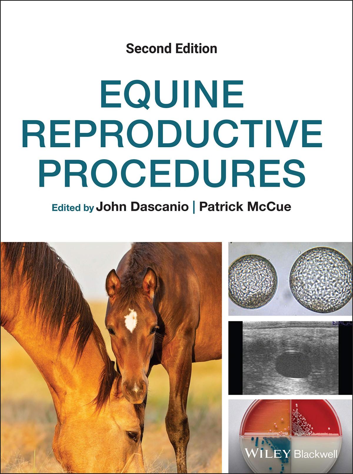
Equine Reproductive Procedures
Second Edition
Edited
by John Dascanio, VMD
Diplomate ACT and ABVP (Equine)
Senior Associate Dean for Academic and Student Affairs
Professor of Theriogenology
School of Veterinary Medicine
Texas Tech University Amarillo, Texas, United States
Patrick McCue, DVM, PhD
Diplomate ACT
Iron Rose Ranch Professor of Equine Reproduction
Colorado State University
Equine Reproduction Laboratory Fort Collins, Colorado, United States
This edition first published 2021 © 2021 John Wiley & Sons, Inc.
Edition History
John Wiley and Sons, Inc. (1e, 2014)
All rights reserved. No part of this publication may be reproduced, stored in a retrieval system, or transmitted, in any form or by any means, electronic, mechanical, photocopying, recording or otherwise, except as permitted by law. Advice on how to obtain permission to reuse material from this title is available at http://www.wiley.com/go/permissions.
The right of John Dascanio and Patrick McCue to be identified as the authors of the editorial material in this work has been asserted in accordance with law.
Registered Offices
John Wiley & Sons, Inc., 111 River Street, Hoboken, NJ 07030, USA
John Wiley & Sons Ltd, The Atrium, Southern Gate, Chichester, West Sussex, PO19 8SQ, UK
Editorial Office
111 River Street, Hoboken, NJ 07030, USA
For details of our global editorial offices, customer services, and more information about Wiley products visit us at www.wiley.com.
Wiley also publishes its books in a variety of electronic formats and by print‐on‐demand. Some content that appears in standard print versions of this book may not be available in other formats.
Limit of Liability/Disclaimer of Warranty
The contents of this work are intended to further general scientific research, understanding, and discussion only and are not intended and should not be relied upon as recommending or promoting scientific method, diagnosis, or treatment by physicians for any particular patient. In view of ongoing research, equipment modifications, changes in governmental regulations, and the constant flow of information relating to the use of medicines, equipment, and devices, the reader is urged to review and evaluate the information provided in the package insert or instructions for each medicine, equipment, or device for, among other things, any changes in the instructions or indication of usage and for added warnings and precautions. While the publisher and authors have used their best efforts in preparing this work, they make no representations or warranties with respect to the accuracy or completeness of the contents of this work and specifically disclaim all warranties, including without limitation any implied warranties of merchantability or fitness for a particular purpose. No warranty may be created or extended by sales representatives, written sales materials or promotional statements for this work. The fact that an organization, website, or product is referred to in this work as a citation and/or potential source of further information does not mean that the publisher and authors endorse the information or services the organization, website, or product may provide or recommendations it may make. This work is sold with the understanding that the publisher is not engaged in rendering professional services. The advice and strategies contained herein may not be suitable for your situation. You should consult with a specialist where appropriate. Further, readers should be aware that websites listed in this work may have changed or disappeared between when this work was written and when it is read. Neither the publisher nor authors shall be liable for any loss of profit or any other commercial damages, including but not limited to special, incidental, consequential, or other damages.
Library of Congress Cataloging-in-Publication Data
Names: Dascanio, John J. (John Joseph), 1961– editor. | McCue, Patrick M., editor. | Dascanio, John J. (John Joseph), 1961– Equine reproductive procedures.
Title: Equine reproductive procedures / edited by John Dascanio, Patrick McCue.
Description: 2nd edition. | Hoboken, NJ : Wiley-Blackwell, 2021. | Preceded by Equine reproductive procedures / John J. Dascanio, Patrick M. McCue. 2014 . | Includes bibliographical references and index.
Identifiers: LCCN 2020026465 (print) | LCCN 2020026466 (ebook) | ISBN 9781119555988 (cloth) | ISBN 9781119555971 (adobe pdf) | ISBN 9781119555933 (epub)
Subjects: MESH: Horses–physiology | Reproduction–physiology | Reproductive Techniques, Assisted–veterinary | Breeding
Classification: LCC SF768.2.H67 (print) | LCC SF768.2.H67 (ebook) | NLM SF768.2.H67 | DDC 636.1–dc23
LC record available at https://lccn.loc.gov/2020026465
LC ebook record available at https://lccn.loc.gov/2020026466
Cover Design: Wiley
Cover Images: (horses) Suzanne Sylvester / S. Sylvester Photography, LLC (right) courtesy of Patrick McCue
Set in 9.5/12.5pt STIXTwoText by SPi Global, Pondicherry, India
10 9 8 7 6 5 4 3 2 1
List of Contributors xiii
Preface xv Par t I Non-Pregnant Mare 1
1 Reproductive Evaluation 3
Patrick M. McCue
2 Teasing 7
Patrick M. McCue
3 Tail Wrap and Preparation/Washing of the Perineum 11
John J. Dascanio
4 Placement of a Tail Rope 17
John J. Dascanio
5 Perineal Conformation Evaluation 21
John J. Dascanio
6 Removal of a Persistent Hymen 25
John J. Dascanio
7 Palpation of the Reproductive Tract 27
Patrick M. McCue
8 Ultrasound Evaluation 31
Patrick M. McCue
9 Prediction of Ovulation 37
Patrick M. McCue
10 Speculum Examination of the Vagina 41
John J. Dascanio
11 Digital Examination of the Vagina/Cervix 45
Sofie Sitters
12 Uterine Culture Collection: Swab/Brush 49
John J. Dascanio
13 Antimicrobiotic Sensitivity Testing 53
Jillian Bishop and Patrick M. McCue
14 Microbiology: Microbial Culture 55
Patrick M. McCue and Jillian Bishop
15 Microbiology: Gram Stain 59
Jillian Bishop and Patrick M. McCue
16 qPCR Assay for the Diagnosis of Infectious Endometritis 61
Ryan A. Ferris and Patrick M. McCue
17 Uterine Cytology Collection: Swab/Brush 67
John J. Dascanio and Ryan A. Ferris
18 Uterine Culture/Cytology: Low Volume Lavage 73
John J. Dascanio
19 Endometrial Biopsy 77
Patrick M. McCue
20 Hysteroscopic Examination of the Uterus 81
Patrick M. McCue
21 Endometrial Cyst Removal 85
John J. Dascanio
22 Chromosomal Analysis 89
Terje Raudsepp
23 Endocrinological Examination 97
Patrick M. McCue
24 Laparoscopic Examination of the Female Reproductive Track with Ovarian Biopsy 99
Dean A. Hendrickson
25 Evaluation of the Mammary Gland 103
Patrick M. McCue
26 Antisperm Antibody Testing 107
Patrick M. McCue
27 Starch Granule Test for the Evaluation of Oviductal Patency 109
Sofie Sitters and John J. Dascanio
28 Fluorescent Microspheres Test for Evaluation of Oviductal Patency 111
Sofie Sitters and John J. Dascanio
29 Oviductal Flush Procedure for the Evaluation of Oviductal Patency 113
Sofie Sitters and Patrick M. McCue
30 Prostaglandin E2 Treatment for Blocked Oviducts 117
Patrick M. McCue, Sofie Sitters and Charles F. Scoggin
31 Prostaglandin E1 Treatment for Blocked Oviducts 119
Patrick M. McCue and Charles F. Scoggin
32 Hysteroscopic Hydrotubation of the Oviducts 121
Charles F. Scoggin
33 Transvaginal Ovarian Biopsy 123
Claire E. Card
34 Uterine Lavage 127
John J. Dascanio
35 Uterine Infusion 131
John J. Dascanio
36 Therapeutics for Infectious Endometritis 133
Ryan A. Ferris
37 Bacterial Biofilm and Endometritis 137
Ryan A. Ferris
38 Latent Uterine Bacterial Infections 141
Morten Rønn Petersen and Anders Miki Bojesen
39 Acupuncture in Mare Reproduction 143
Kristina Lu and Lauren Javernick
40 Restraint for Breeding 149
John J. Dascanio
41 Natural Service: Pasture Breeding 151
Patrick M. McCue
42 Natural Service: In-hand Breeding 153
Charles F. Scoggin
43 Breeding Stitches 157
Charles F. Scoggin
44 Reinforcement Breeding 159
Charles F. Scoggin
45 Breeding with Fresh or Cooled Semen 161
John J. Dascanio and Patrick M. McCue
46 Insemination Through a Vaginal Speculum 165
Patrick M. McCue
47 Breeding with Frozen Semen 167
John J. Dascanio
48 Deep Horn Insemination 171
John J. Dascanio
49 Hysteroscopic (Low Dose) Insemination 173
Patrick M. McCue
50 Transvaginal Oocyte Pickup 177
Jennifer N. Hatzel
51 Packaging and transpor t of oocytes 183
Jennifer N. Hatzel
52 Intracytoplasmic Sperm Injection 187
Jennifer N. Hatzel and JoAnne Stokes
53 Caslick Operation or Vulvoplasty 191
John J. Dascanio
54 Contagious Equine Metritis Testing 195
John J. Dascanio
55 Management of Seasonal Anestrus: Artificial Photoperiod 199
Patrick M. McCue
56 Management of Seasonal Anestrus: Hormone Therapy 201
Patrick M. McCue
57 Hormone Therapy in Cycling Mares 205
Patrick M. McCue
58 Estrus Suppression 211
Patrick M. McCue
59 Intrauterine Devices for Estrus Suppression 215
Carlos M. Gradil
60 Immunocontraception 221
Patrick M. McCue
61 Harvesting and Shipping Ovaries for Oocyte Recovery 223
Patrick M. McCue
Part II Pregnant Mare 225
62 Embryo Collection 227
Patrick M. McCue
63 Embryo Evaluation 231
Patrick M. McCue
64 Embryo Biopsy 235
Patrick M. McCue
65 Embryo Packaging for Cooled Transpor t 237
Patrick M. McCue
66 Embryo Cryopreservation 241
Patrick M. McCue
67 Non-Surgical Embryo Transfer 245
Patrick M. McCue
68 Autogenous Transfer of ICSI-Derived Embryos 249
Jennifer N. Hatzel
69 Palpation per Rectum of the Pregnant
Reproductive Tract 253
Sofie Sitters
70 Ultrasound Examination of the Pregnant
Reproductive Tract 257
Patrick M. McCue
71 Speculum Examination of the Pregnant Mare 263
Sofie Sitters
72 Determination of Fetal Sex at 55–200 days 265
Richard D. Holder
73 Assessment of Late-Term Fetal Well-being 273
John J. Dascanio
74 Combined Thickness of the Uterus and Placenta 277
Patrick M. McCue
75 Placentitis: Diagnosis and Treatment 281
Justin W. McNaughten and Margo L. Macpherson
76 Doppler Ultrasonography 287
John J. Dascanio
77 Prediction of Fetal Age 291
John J. Dascanio
78 Twin Reduction: Manual Technique 295
John J. Dascanio
79 Twin Reduction: Transvaginal Aspiration 299
John J. Dascanio
80 Twin Reduction: Cranio-Cervical Dislocation 303
Karen Wolfsdorf
81 Twin Reduction: Thoracic Compression 307
Patrick M. McCue
82 Twin Reduction: Transabdominal Fetal Cardiac Puncture 309
John J. Dascanio
83 Elective Termination of Pregnancy 313
John J. Dascanio
84 Abortion Diagnostic Evaluation: Sample Collection in the Field and Submission 317
Julie T. Cecere and John J. Dascanio
85 Endocrine Evaluation of Pregnancy 319
Patrick M. McCue
86 Treatment of Vaginal Varicosities 323
John J. Dascanio
87 Manual Correction of Uterine Torsion 327
John J. Dascanio
88 Preparation for Foaling 331
John J. Dascanio
89 Assessment of Mammary Gland Secretions to Predict Foaling 335
John J. Dascanio
90 Assessment of pH of Mammary Gland Secretions to Predict Foaling 339
Patrick M. McCue
91 Monitoring of Peri-Parturient Mares Using Video and Web Cameras 343
Ryan A. Ferris
92 Foaling Alert: Vulvar Device 345
John J. Dascanio
93 Positional Labor Alert Devices 347
Ryan A. Ferris
94 Induction of Parturition 351
John J. Dascanio
95 Normal Foaling 355
John J. Dascanio
96 Premature Separation of the Placenta 361
Patrick M. McCue
97 Dystocia Management 365
Patrick M. McCue and Sofie Sitters
98 Dystocia Correction 369
John J. Dascanio
99 Fetotomy 375
John J. Dascanio
100 Epidural 381
John J. Dascanio
101 Induction of Lactation to Create a Nurse Mare 383
Pouya Dini and Peter F. Daels
102 Screening to Prevent Neonatal Isoerythrolysis 385
Patrick M. McCue
Part III Postpartum Mare 387
103 Evaluation of Colostrum Specific Gravity 389
Patrick M. McCue
104 Evaluation of Colostrum Quality: Brix
Refractometry 393
Patrick M. McCue
105 Colostrum Banking 395
Patrick M. McCue
106 Obtaining Milk from the Mare 399
John J. Dascanio
107 Placental Evaluation 401
Patrick M. McCue
108 Removal of Retained Placenta 407
John J. Dascanio
109 Abdominocentesis 413
John J. Dascanio
110 Uterine Prolapse Treatment 415
John J. Dascanio
111 Buhner Needle Placement of a Perivulvar
Suture 419
John J. Dascanio
Part IV Stallion 423
112 Breeding Soundness Evaluation 425
Patrick M. McCue
113 Training the Stallion to Use a Phantom 429
John J. Dascanio
114 Missouri Artificial Vagina 433
John J. Dascanio and Lynda M. J. Miller
115 Colorado Model Artificial Vagina 437
Patrick M. McCue
116 Roanoke Artificial Vagina 441
John J. Dascanio
117 Hannover Artificial Vagina 445
Sofie Sitters
118 Teaser Stallions 447
Charles F. Scoggin
119 Standing Semen Collection 449
John J. Dascanio
120 Collection of Semen Using an Open-Ended Artificial Vagina 453
Patrick M. McCue
121 Chemical Ejaculation 455
Patrick M. McCue
122 Preparation of a Jump Mare or a Mare for Natural Cover 457
John J. Dascanio
123 Washing the Penis 459
John J. Dascanio
124 Evaluation of Sexual Behavior 463
John J. Dascanio and Sue M. McDonnell
125 Enhancement of Sexual Interest and Response 467
Sue M. McDonnell
126 Management of Ejaculation Difficulty 471
Sue M. McDonnell
127 Calibrated Spectrophotometer Evaluation of Sperm Concentration 475
John J. Dascanio
128 Hemocytometer Evaluation of Sperm Concentration 479
John J. Dascanio
129 NucleoCounter® Evaluation of Sperm Concentration and Viability 483
Patrick M. McCue
130 Visual Evaluation of Sperm Motility 489
Patrick M. McCue
131 Computer-Assisted Sperm Analysis 491
John J. Dascanio and Lynda M. J. Miller
132 Eosin-Nigrosin Staining in the Evaluation of Sperm 495
Julie T. Cecere
133 Evaluation of Sperm Morphology 499
Leonardo Brito
134 Determination of Daily Sperm Output 505
Patrick M. McCue
135 Measurement of Testicular Size and Estimation of Daily Sperm Output 507
Patrick M. McCue
136 Wet Mount Evaluation of Sperm 511
John J. Dascanio
137 Diff-Quik® Evaluation of Round Cells and Sperm 513
John J. Dascanio and Lynda M. J. Miller
138 Electron Microscopy of Constituents of Seminal Ejaculates 515
D. N. Rao Veeramachaneni
139 Bacterial Culture 519
John J. Dascanio
140 Evaluation of pH and Osmolarity of Semen 521
Patrick M. McCue
141 Alkaline Phosphatase: A Marker for Ejaculation 523
Patrick M. McCue
142 Diagnosis and Management of Urospermia 525
John J. Dascanio
143 Hemospermia 529
Patrick M. McCue
144 Semen Extenders and Sperm Media 533
Patrick M. McCue
145 Preparation of Semen for Cooled Transport 537
Etta A. Bradecamp
146 Packing Semen for Cooled Transport 541
Etta A. Bradecamp
147 Longevity Testing of Sperm 547
John J. Dascanio
148 Calculation of g Force for Centrifuging Semen 549
John J. Dascanio
149 Centrifugation of Semen: Standard Technique 551
Etta A. Bradecamp
150 Centrifugation of Semen: Cushion Technique 557
Etta A. Bradecamp
151 Centrifugation of Semen: Selection of Motile Sperm Using a Single Layer Colloid Technique 561
Etta A. Bradecamp
152 Use of a SpermFilter 563
Marco A. Alvarenga and Lorenzo G. T. M. Segabinazzi
153 Semen Freezing 567
Patrick M. McCue
154 Refreezing Semen 573
Patrick M. McCue
155 Thawing Frozen Semen 577
John J. Dascanio
156 Semen Processing for Intracytoplasmic Sperm Injection 581
Patrick M. McCue
157 Preparing a Vapor Shipper 585
Julie T. Cecere and Paul Loomis
158 Loading a Vapor Shipper 589
Julie T. Cecere and Paul Loomis
159 Maintaining a Long-Term Frozen Semen Storage Tank 591
John J. Dascanio
160 International Transpor t of Frozen Semen 593
Paul Loomis
161 Harvesting and Shipping Testes 597
John J. Dascanio
162 Epididymal Sperm Recovery 599
John J. Dascanio and Lynda M. J. Miller
163 Endoscopic Examination of the Urethra 603
Patrick M. McCue
164 Endoscopic-Guided Cannulation of the Seminal Vesicles 607
Patrick M. McCue
165 Palpation and Ultrasonography of the Testis, Epididymis, and Spermatic Cord 609
Sofie Sitters
166 Palpation and Ultrasonography of the Accessory Sex Glands 615
Sofie Sitters
167 Radiographic Examination of the Penis 621
Patrick M. McCue
168 Testicular Biopsy and Aspiration 623
John J. Dascanio
169 Assessment of Sperm Plasma Membrane Integrity and Viability: Propidium Iodide/ SYBR-14 627
Amanda I. Glazar, and Patrick M. McCue
170 Sperm Chromatin Structure Assay 629
Charles Love
171 Hypo-Osmotic Swelling Test 633
Patrick M. McCue
172 Assessment of Sperm Acrosomal Status: PE-PNA 635
Amanda I. Glazar and Patrick M. McCue
173 Assessment of Sperm Mitochondrial Function: JC–1 and Rhodamine 123 637
Amanda I. Glazar, and Patrick M. McCue
174 Equine Viral Ar teritis Testing 639
G. Reed Holyoak and Udeni B. R. Balasuriya
175 Diagnostic Endocrinology: Baseline Hormone Levels 645
Patrick M. McCue
176 Diagnostic Endocrinology: GnRH Stimulation Tests 647
Patrick M. McCue
177 Diagnostic Endocrinology: hCG Stimulation Test 649
Patrick M. McCue
178 Diagnostic Endocrinology: Estrogen Conjugate Assay 653
Patrick M. McCue
179 Diagnostic Tests for Cryptorchidism 655
Patrick M. McCue
180 Probang Apparatus 659
Julie T. Cecere
181 Support Apparatus for Paraphimosis 663
John J. Dascanio
182 Equine Castration Techniques 667
Stacy Anderson
183 Stallion Acupuncture 671
Rhonda A. Rathgeber
Part V Newborn Foal 675
184 Birth Resuscitation 677
Patrick M. McCue
185 Handling and Restraint 681
Undine Christmann
186 Madigan Foal Squeeze Technique 687
John Madigan
187 Evaluation of Passive Transfer 691
Patrick M. McCue
188 Colostral Administration via Naso-Gastric Intubation 695
John J. Dascanio
189 Sedation 699
Undine Christmann
190 Intravenous Catheter Placement 701
Undine Christmann
191 Plasma Therapy in the Foal 707
Undine Christmann
192 Acupuncture in the Neonate 711
Peter Morresey
193 Routine Care 713
Patrick M. McCue
194 Foal Rejection 719
Patrick M. McCue
195 Fostering a Foal onto a Nurse Mare 723
Peter F. Daels and Pouya Dini
196 Entropion 725
John J. Dascanio
197 Umbilical Ultrasound 727
Undine Christmann
198 Management of Soft Tissue Laxity 733
Stacy Anderson
199 Management of Angular Limb Deformities 735
Stacy Anderson
200 Management of Congenital Flexural Limb Deformities 739
Stacy Anderson
Part VI Appendices 745
Appendix A Society for Theriogenology Stallion Reproductive Evaluation Form 745
Appendix B Mare Breeding Soundness Evaluation Form 749
Appendix C Formulary for Equine Reproduction 751
Appendix D Foaling Kit and Associated Equipment and Supplies 759
Index 761
List of Contributors
Marco A. Alvarenga DVM, PhD School of Veterinary Medicine São Paulo State University Botucatu, Brazil
Stacy Anderson DVM, MVSc, PhD, DACVS-LA College of Veterinary Medicine Lincoln Memorial University Harrogate, TN, USA
Udeni B. R. Balasuriya BVSc, MS, PhD, FSLCVS Department of Pathobiological Sciences School of Veterinary Medicine Louisiana State University Baton Rouge, LA, USA
Jillian Bishop BS, Pharm D, BCPS University of Kansas Health System Lexexa, KS, USA
Anders Miki Bojesen DVM, PhD, DECVPS Department of Veterinary and Animal Science University of Copenhagen Copenhagen, Denmark
Etta A. Bradecamp DVM, DACT, DABVP (Equine) Rood and Riddle Equine Hospital Lexington, KY, USA
Leonardo Brito DVM, PhD, DACT Stgenetics Middleton, WI, USA
Claire E. Card DVM, PhD, DACT Department of Large Animal Clinical Sciences Western College of Veterinary Medicine University of Saskatchewan Saskatchewan, Canada
Julie T. Cecere DVM, MS, DACT Department of Small Animal Clinical Sciences Virginia‐Maryland College of Veterinary Medicine Blacksburg, VA, USA
Undine Christmann Dr.Med.Vet., MS, MPH, PhD, DACVIM College of Veterinary Medicine Lincoln Memorial University Harrogate, TN, USA
Peter F. Daels DVM, PhD, DACT, DECAR Faculty of Veterinary Medicine University of Gent Merelbeke, Belgium
John J. Dascanio VMD, DACT, DABVP (Equine) School of Veterinary Medicine Texas Tech University Amarillo, TX, USA
Pouya Dini DVM, PhD, DECAR, DACT Department of Population Health and Reproduction School of Veterinary Medicine University of California‐Davis Davis, CA, USA
Ryan A. Ferris DVM, MS, DACT Summit Equine Gervais, OR, USA
Amanda I. Glazar PhD Kynectiv, Inc. Evergreen, CO, USA
Carlos M. Gradil DVM, MS, PhD, DACT Cummings School of Veterinary Medicine University of Massachusetts Veterinary and Animal Sciences Amherst, MA, USA
Jennifer N. Hatzel DVM, MS, DACT Equine Reproduction Laboratory Colorado State University Fort Collins, CO, USA
Dean A. Hendrickson DVM, MS, DACVS Department of Clinical Sciences College of Veterinary Medicine and Biomedical Sciences Colorado State University Fort Collins, CO, USA
Richard D. Holder DVM
Hagyard Equine Medical Institute Lexington, KY, USA
G. Reed Holyoak DVM, PhD, DACT Department of Veterinary Clinical Sciences Oklahoma State University Stillwater, OK, USA
Lauren Javernick DVM
Hagyard Equine Medical Institute Lexington, KY, USA
Paul Loomis MS Select Breeders Services Chesapeake City, MD, USA
Charles Love DVM, PhD
Large Animal Clinical Sciences, College of Veterinary Medicine and Biomedical Sciences, Texas A&M University, College Station, TX, USA
Kristina Lu VMD, DACT
Hagyard Equine Medical Institute Lexington, KY, USA
Margo L. Macpherson DVM, MS, DACT College of Veterinary Medicine University of Florida Gainesville, FL, USA
John Madigan DVM, MS, DACVIM, DACAW School of Veterinary Medicine University of California‐Davis Davis, CA, USA
Patrick M. McCue DVM, PhD, DACT Equine Reproduction Laboratory
Colorado State University Fort Collins, CO, USA
Sue M. McDonnell PhD, CAAB, Hon DACT
New Bolton Center School of Veterinary Medicine University of Pennsylvania Kennett Square, PA, USA
Justin W. McNaughten BVMS, DACT Rhinebeck Equine LLP Rhinebeck, NY, USA
Lynda M. J. Miller DVM, PhD, DACT College of Veterinary Medicine
Lincoln Memorial University Harrowgate, TN, USA
Peter Morresey BVSC, MVM, MACVSc, DACT, DACVIM, CVA Rood and Riddle Equine Hospital Lexington, KY, USA
Morten Rønn Petersen DVM, PhD, DACT
The Fertility Clinic University Hospital Copenhagen Faculty of Health and Medical Sciences University of Copenhagen Copenhagen, Denmark
Rhonda A. Rathgeber DVM, PhD Hagyard Equine Medical Institute Lexington, KY, USA
Terje Raudsepp MS, PhD
Department of Veterinary Integrative Biosciences College of Veterinary Medicine and Biomedical Sciences Texas A&M University College Station, TX, USA
Charles F. Scoggin DVM, MS, DACT Rood and Riddle Equine Hospital Lexington, KY, USA
Lorenzo G. T. M. Segabinazzi DVM, MS School of Veterinary Medicine São Paulo State University Botucatu, Brazil
Sofie Sitters DVM Amsterdam, The Netherlands
JoAnne Stokes BS Equine Reproduction Laboratory Colorado State University Fort Collins, CO, USA
D. N. Rao Veeramachaneni BVSc, MScVet, PhD
Animal Reproduction and Biotechnology Laboratory Colorado State University Fort Collins, CO, USA
Karen Wolfsdorf DVM, DACT
Hagyard Equine Medical Institute Lexington, KY, USA
Preface
This second edition of the Equine Reproductive Procedures book has been substantially updated with 39 new chapters, and a multitude of new diagnostic procedures and therapeutic protocols added to existing chapters. The overall goal has remained the same, which is to provide equine professionals with practical clinical information on basic and advanced techniques in the field of equine reproduction. It is our hope that veterinary students, graduate students, researchers in the field of equine reproduction and veterinarians in private practice will find the material in this book useful in their collective work.
The second edition is divided into sections on the Non‐Pregnant Mare, Pregnant Mare, Postpartum Mare, Stallion and Newborn Foal. A total of 40 authors and co‐authors contributed to the development of the book. In the first edition, we noted that approximately 90% of reproductive procedures used in the horse industry are nearly identical throughout the world. The other 10% varies according to the training and experience of the individual, geographic
region, and availability of supplies and facilities. Hopefully we will all continue to learn throughout our careers from the knowledge and experience of others. Consequently, while the objective of this book is to provide examples of how reproductive procedures may be performed, it should not be construed that these are the only or the optimal methods to achieve diagnostic or therapeutic goals.
Again, we would like to thank the staff at Wiley Blackwell for their professional assistance in the development of this second edition. We would also like to acknowledge our many mentors and colleagues for providing academic and clinical guidance over the years. We owe our professional careers to their collective wisdom. Most importantly, we would also like to thank our families for their unwavering support. Thank you, Linda, Sarah, and Gabriel Dascanio! Thank you, Diane, Daniel, and Kelly McCue!
John Dascanio
Patrick McCue
Part I
Non-Pregnant Mare
Reproductive Evaluation
Patrick M. McCue
Equine Reproduction Laboratory, Colorado State University, USA
Introduction
The goals of a mare reproductive evaluation or breeding soundness examination (BSE) are to identify known or potential reproductive abnormalities and to evaluate the potential of a mare to become pregnant and carry a foal successfully to term. Mare reproductive examinations are performed in open (non‐pregnant) mares prior to the onset of the breeding season, in problem mares during the breeding season, in barren mares at the end of the breeding season, as well as in mares with a history of embryonic loss, abortion, or other reproductive problems, or as part of a pre‐purchase examination. The goal of this chapter is to provide an overview of the mare breeding soundness evaluation. Details on specific examinations will be covered in other chapters.
Equipment and Supplies
Obstetrical sleeve (non‐sterile), obstetrical lubricant (non‐sterile), tail wrap, metal bucket and plastic bag liner, non‐irritant soap, roll cotton, vaginal speculum, obstetrical lubricant (sterile), obstetrical sleeve (sterile), uterine culture device, culture transport system (optional), uterine cytology device, glass slides, uterine biopsy forceps, formalin.
Examination Technique
Identification
All mares should be properly identified, and the breed, registration name, registration number, and date of birth recorded. Photographs should be taken or accurate drawings of markings and tattoos recorded.
Reproductive History
A complete breeding history should be obtained, including current reproductive status (maiden, open, barren, pregnant, or foaling), number of cycles bred during the last season, date of last breeding, breeding technique used (artificial insemination, natural cover, or pasture breeding), number of stallions, date of last foal, number of previous foals, and any previous history of abnormal estrous cycles, uterine infections, embryonic loss, or abortion.
Physical Examination
A general physical examination should be performed to assess whether the mare has the capacity to carry a foal to term. The evaluation should include, but is not limited to, examination of the oral cavity, eyes, and the respiratory, cardiac, and musculoskeletal systems. In addition, diet and body condition should be evaluated.
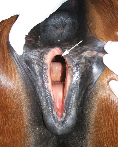
Perineal Conformation
See also Chapter 5. The external genitalia (vulva) should be evaluated for conformation and muscular tone. The optimal perineal conformation consists of a vulva in a nearly vertical position with two thirds of the vulva ventral to the brim of the pelvis. The muscular tone of the vulva should be sufficient to prevent or minimize aspiration of air into the vestibule or vagina. Horizontal sloping of the vulva secondary to recession of the anus or poor muscular tone to the labia of the vulva may predispose the mare to an ascending infection of the uterus.
Estrous Detection
See also Chapter 2. The mare should be exposed to a stallion with good libido to evaluate estrous cycle stage. Adequate time should be taken to allow shy or nervous mares to express behavioral estrus. When teasing a mare with a foal, the foal must be restrained, and the mare may need to be restrained with a twitch before signs of estrus are exhibited.
Palpation Per Rectum
See also Chapter 7. The entire reproductive tract, including the cervix, uterus, and ovaries, should be thoroughly and systematically examined by palpation per rectum. The tone of the uterus and cervix, size and consistency of ovarian follicles, and the presence of a recent ovulation or a corpus hemorrhagicum should be recorded. The presence of abnormal ovarian, parovarian (i.e., fimbrial cysts), or uterine structures should be recorded.
Ultrasonography Per Rectum
See also Chapter 8. Manual palpation should be followed by a systematic ultrasound evaluation of the entire reproductive tract. Ultrasound is used in broodmares to visualize structures in the reproductive tract that cannot be palpated or differentiated on palpation per rectum, and in the early diagnosis of pregnancy, diagnosis of twins, and evaluation of potential ovarian or uterine pathology.
Vaginal Speculum Examination
See also Chapter 10. A vaginal speculum examination is performed to evaluate the anatomy of the vagina and the external os of the cervix. Speculum examination is useful in determination of the stage of the estrous cycle (via cervical morphology and vaginal mucous membrane changes), and detection of urine pooling and the presence of cervical/vaginal inflammation or discharge.
Digital Examination of the Cervix
See also Chapter 11. After the speculum examination is completed, the cervix should be examined manually for patency and the presence of abnormalities, such as adhesions, lacerations, or other cervical defects.
Uterine Culture
See also Chapter 12. Culture of the uterine lumen is usually performed in conjunction with cytology for the diagno-
sis of endometritis. Endometritis can be suspected in mares that exhibit an abnormally short estrous cycle, vaginal or cervical discharge, inflamed cervix on speculum examination, and free fluid in the uterus during diestrus detected on ultrasound.
Endometrial Cytology
See also Chapter 17. Cytologic evaluation of the uterus involves the collection and interpretation of cells lining the uterus (endometrium) and within the uterine lumen. Cytology is used in conjunction with culture and biopsy in the diagnosis of endometritis. Advantages of endometrial cytology for the diagnosis of endometritis include the ease of sample collection, low cost, and rapid availability of results.
Endometrial Biopsy
See also Chapter 19. Endometrial biopsy involves collection of a small sample of the uterine lining (endometrium) for histologic evaluation. It is primarily used as an aid in the diagnosis of uterine disease and as a prognostic indicator of the ability of a mare to carry a foal to term. An endometrial biopsy can also be used as the sample source for culture and cytologic evaluation.
Other Tests
The standard examination procedures in the mare BSE may not identify the cause of subfertility. Consequently, other examinations may be indicated (Table 1.1).
Additional Comments
Interpretation of the results of a mare BSE should take into account the mare’s age, reproductive history, breed, stallion, breeding management, and other factors. Ultimately the goals are to determine the potential for fertility and detect abnormalities that may be associated with reduced fertility. Management and therapeutic options may be outlined to help optimize a successful outcome. It is important to emphasize that a mare BSE is only an evaluation of potential fertility and that the true assessment of fertility is the ability of the mare to conceive and carry a foal to term.
Table 1.1 Diagnostic tests that may be performed in addition to the standard tests in a mare breeding soundness evaluation.
Test
Chromosome analysis (karyotype)
Hormone analysis
Hysteroscopy
Laparoscopy
Low volume lavage
Oviductal flush
Oviductal patency test
Oviductal PGE2 application
Ovarian biopsy
Test breed
Indication
Evaluate numeric or structural changes in chromosomes
Evaluate pituitary and/or ovarian endocrine function. Most commonly used to evaluate corpus luteum function and in the detection of an ovarian granulosa cell tumor
Direct visualization of the interior of the uterus to detect intrauterine adhesions and other localized lesions, as well as inflammation and fibrosis
Direct visualization of the serosal surface of the ovary, oviduct, uterus, and abdominal cavity. Also used in ovarian biopsy, evaluation of oviductal patency, and the application of prostaglandin E2 (PGE2) to the oviductal surface
Collection of uterine samples for culture, cytology, and other evaluations (e.g., polymerase chain reaction). The effluent fluid may also be evaluated for clarity and pH
Performed by laparotomy, laparoscopy, or via videoendoscopy; used both diagnostically and therapeutically in suspected cases of oviductal blockage
Deposition of fluorescent microbeads or starch granules onto the surface of the ovary or possibly within the infundibulum of the oviduct and subsequent examination of the uterine lumen for passage of the test material is used diagnostically to evaluate oviductal patency
Direct application of PGE2 can be used diagnostically and therapeutically in suspected cases of oviductal blockage
Laparoscopic collection of ovarian tissue for histologic evaluation may be used in the diagnosis of ovarian pathology
Breeding to a highly fertile stallion can be used diagnostically to help determine if the mare is a cause of subfertility or infertility
Further Reading
LeBlanc MM, Lopate C, Knottenbelt D, Pascoe R. 2003. The Mare. Equine Stud Farm Medicine and Surgery. London: Elsevier, pp. 113–213.
McCue PM. 2008. The problem mare: management philosophy, diagnostic procedures, and therapeutic options. J Eq Vet Sci 28: 619–26.
Patrick M. McCue
Equine Reproduction Laboratory, Colorado State University, USA Teasing
Introduction
The 21‐day equine estrous cycle can be divided into two phases: estrus and diestrus, based on sexual receptivity to a stallion. Estrus is the period during which a mare is sexually receptive to the advances of a stallion. Behavioral estrus is stimulated by increasing levels of estradiol produced by the developing dominant follicle in the absence of progesterone. The average length of estrus has been reported to be 6.5 days, with a range of 4.5–8.9 days. Ovulation typically occurs 24–48 hours before the end of estrus. An increase in progesterone from the developing corpus luteum is responsible for the cessation of behavioral estrus. Behavioral diestrus largely overlaps the physiological luteal phase associated with high levels of progesterone produced by the corpus luteum.
Equipment and Supplies
Stallion, mare, lead shank, helmet, facilities (teasing rail, fence, chute, stall).
Technique
● A mare should be teased with a stallion that exhibits good libido in order to successfully evaluate estrous cycle stage.
● Adequate time should be taken to allow shy or nervous mares to express behavioral estrus.
● Knowledge of the mare’s previous behavioral patterns may be helpful.
● In addition, observation of the mare’s behavior with other horses in a pasture or paddock may indicate when a mare is in estrus.
Individual Teasing
An individual mare should be exposed to a stallion for an interval of time that is long enough for her to show estrus or diestrus types of behavior. It is preferable to have both the mare and stallion restrained for individual teasing; however, if only one handler is present, it is better to have the stallion restrained with the mare loose so that abnormal behaviors in the stallion may be corrected. Mares that remain indifferent may need to be teased longer, teased with a different stallion, or may just show more subtle signs. Mares may be reluctant initially and yet later show frank estrus behavior (i.e., “break down”). Sometimes full behavioral estrus is only expressed within a few hours of ovulation. It is also not unusual for a mare to fail to show signs of estrus while being directly teased to a stallion, and then break down as the stallion leaves. Mares may also display estrus at the mere sound or sight of a stallion. Mares with a foal by their side may be reluctant to display estrus behavior readily as they are concerned for their foal’s safety.
Group Teasing
A stallion may be used to tease more than one mare concurrently if he is brought to the edge of a pen or turned out adjacent to a group of mares. Mares are allowed to approach the stallion at will in such a teasing program. However, some mares will not approach the stallion and will not express estrus when teased as part of a group. It may be necessary to tease such mares individually.
It is often not very efficient to tease mares as a group, since often the only mares that come to the fence or tease rail are assertive mares in heat or mares that want to attack the stallion. One may not be able to determine the heat status of mares that remain a distance from the stallion. It is generally more effective, but certainly more time
Equine Reproductive Procedures, Second Edition. Edited by John Dascanio and Patrick McCue.
consuming, to tease mares individually. Mares that are less dominant mares or further away from the fence or tease rail should still be observed for estrus behavior.
Behavioral Responses
Common behavioral responses of mares in estrus and diestrus are listed in Boxes 2.1 and 2.2 and shown in Figures 2.1 and 2.2.
Box 2.1 Common Behavioral Characteristics of Mares in Estrus
Tail raised and arched or deviated to one side
Rhythmic eversion of the labia and exposure of clitoris (“winking”)
Passive urination
Ears relaxed and either held forward or in a neutral position
Rear limbs slightly abducted (i.e., wide‐based stance)
Stifles and hocks flexed
Lowering of the pelvis (i.e., “squatting”)
Leaning into fence or gate
Vocalization (squealing)
Calm behavior; does not try to bite or strike stallion
Box 2.2 Common Behavioral Characteristics of Mares in Diestrus
Tail held down or aggressively switched from side to side
Ears pinned back
Aggressive toward advances of the stallion
Biting at the stallion
Attempt to move away from the stallion
Squealing or vocalization
Calm behavior; does not try to bite or strike stallion
It should be noted that expression of estrus does not always indicate that a mare is in the follicular phase of the estrous cycle. Seasonally anovulatory mares, ovariectomized mares, and pregnant mares have all been reported to occasionally show signs of estrus when teased with a stallion. This may be due to submissive behavior or a lack of progesterone.
Records
Maintaining an accurate record of teasing behavior will be helpful when monitoring the estrous cycle of a mare. Notations can be made as to whether or not the mare exhibits overt, subtle, or no signs of estrus throughout a cycle (Table 2.1).
Table 2.1 Abbreviations for responses of a mare to a stallion (i.e., teasing behavior). Abbreviation
into or going out of heat
+ Indifferent
Figure 2.1 Mare in estrus (i.e., teasing in heat). Note the base-wide stance, raised tail, and urination.
Figure 2.2 Mare in diestrus (i.e., out of heat) on far side of teasing wall. Note the ears and aggressive stance.
Silent Estrus
Maiden mares may not show heat well and foaling mares may not show heat at all unless the foal is restrained and/ or safely away from the stallion. Subordinate mares may be inhibited from expressing estrus in the presence of a dominant mare. In addition, a mare may have a preference for, or an aversion toward, an individual stallion. Mares with “silent estrus” may have lower concentrations of estradiol 17 β than mares expressing normal estrus.
Further Reading
Ginther OJ. 1979. Reproductive Biology of the Mare: Basic and Applied Aspects. Ann Arbor, MI: McNaughton and Bunn, pp. 59–68.
Additional Comments
There are many systems used for teasing mares, including chutes, rails, fences, pens, and paddocks. Keys to successful teasing are patience, persistence, and knowing the behavioral characteristics of each mare. Consequently, it is advantageous for the same individual(s) to tease mares each day, so that slight variations in individual responses can be recognized.
McCue PM, Scoggin CF, Lindholm ARG. 2011. Estrus. In: McKinnon AO, Squires EL, Vaala WE, Varner DD (eds). Equine Reproduction, 2nd edn. Ames, IA: Wiley Blackwell, pp. 1716–27.
Tail Wrap and Preparation/Washing of the Perineum
John J. Dascanio
School of Veterinary Medicine, Texas Tech University, USA
Introduction
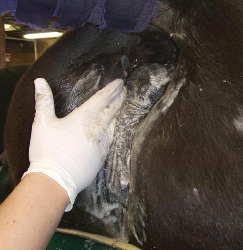
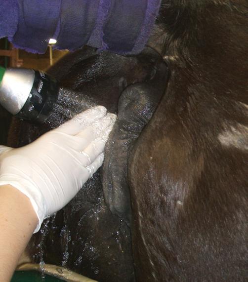
The mare’s perineum should be washed prior to internal reproductive procedures and in preparation for foaling, to remove gross debris and to reduce bacterial numbers. In addition, the perineum is washed prior to perineal surgery such as the placement of a Caslick suture (vulvoplasty). The perineum is also washed as part of the minimum contamination breeding technique to reduce contamination of the uterus during natural cover or artificial insemination.
Equipment and Supplies
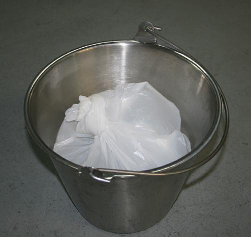
Tail wrap, tail rope, non‐irritant soap, roll cotton, stainless steel bucket, plastic bag/bucket liner, paper towels, examination gloves.
Placement of a Tail Wrap
● A tail wrap should be placed prior to washing of the perineum. This may be a reusable neoprene wrap, Velcro® wrap, gauze wrap, flexible elastic wrap (Vetrap™), or an obstetrical sleeve.
● A reusable neoprene Velcro® wrap is cost effective and quick in application. It should be washed regularly with a disinfectant soap and dried before reuse. This is especially important if the wrap is used with different mare populations. The prevention of disease transmission is difficult with the reuse of this wrap without sanitation; thus, if there is a suspicion of such, a disposable tail wrap should be used.
● A gauze wrap is placed on the tail using one of two techniques. With both techniques the wrap goes from the base of the tail to a level just below the ventral
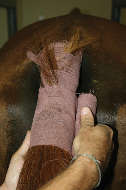
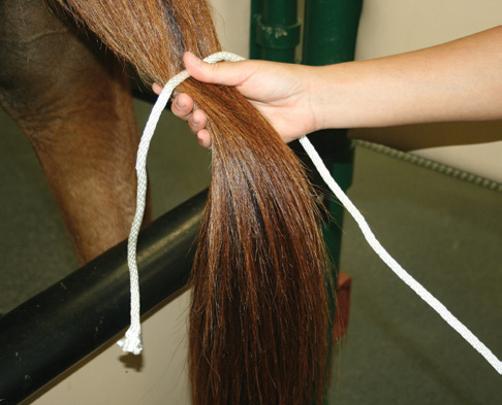
commissure of the vulva. Technique one is to start at the top of the tail and with every circumferential wrap, flip some tail hairs upward so that they become trapped between successive layers (Figure 3.1). This prevents the tail wrap from slipping down the tail. This tail wrap
Figure 3.1 Gauze tail wrap with tail hairs flipped up to prevent sliding of the wrap down the tail.


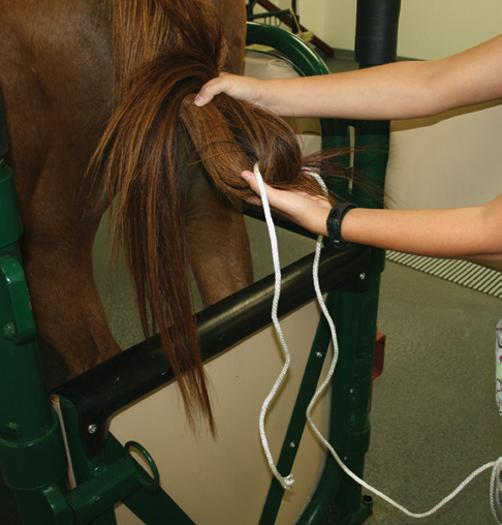
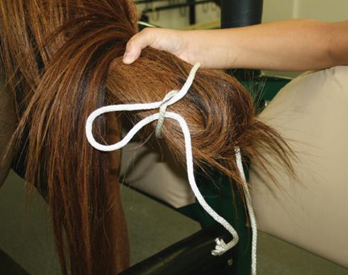
must be unwound to remove. The second type of gauze wrap starts at about the level of the ventral vulvar commissure (Figure 3.2). A small piece of gauze is left hanging and the wrap placed around the tail, moving upward to the tail base. Hair should not be included between layers, with each layer overlapping the previous slightly. Once at the tail base, the gauze is twisted 360 degrees so that the twisted part is on the dorsal tail, while the ventral aspect of the wrap remains flat and untwisted. This allows the twist to bite into the tail hair on the dorsum, preventing slippage of the wrap, while the flat underneath aspect prevents blood occlusion. The wrap is continued downward and is twisted upon every circumferential wrap ending at the point where the wrap began. The small hanging piece of gauze from the beginning is tied to the remaining wrap to prevent the wrap from coming loose. This wrap may be pulled from the top to slide down the tail and removed without having to unwind the wrap when the procedure is completed. This wrap acts like a Chinese finger trap.
● If an obstetrical sleeve is used for a tail wrap, the tail is placed entirely inside the sleeve. The sleeve may be split at the tail base and tied together to close the sleeve around the tail base, or elastic adhesive tape or an elastic band and clamp may be used to wrap around the sleeve at the tail base to secure it. This type of tail wrap is useful for mares with diarrhea to prevent soiling of the tail hairs.
● If a flexible elastic wrap is used such as Vetrap™, the wrap may either be used to wrap the entire tail or the upper portion (Figure 3.3). The entire tail is wrapped for a dystocia or a fetotomy to prevent tail hairs from interfering with the procedure. To place the entire tail in a wrap, fold the tail hairs so that the hairs do not extend beyond the tail stump. Wrap the elastic wrap around the tail/tail hairs so that no hair is visible along the entire tail length. This in effect creates a “club” with the tail wrapped completely.
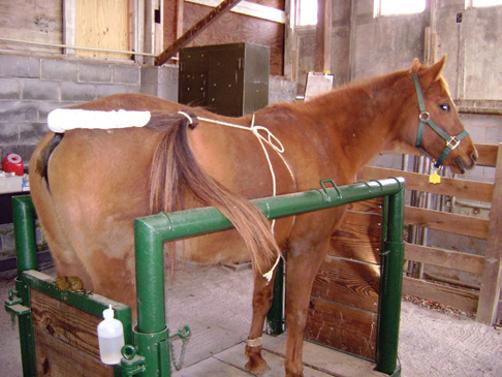
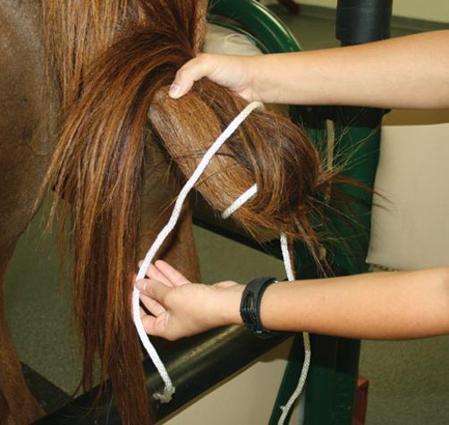
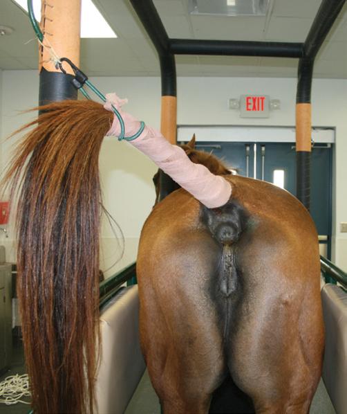
● After wrapping, the tail is held to the side so that the perineum can be washed or the tail is tied to the mare (Figure 3.4).
Figure 3.2 Gauze tail wrap twisted 360 degrees on the dorsum of the tail while the ventral aspect is wide.
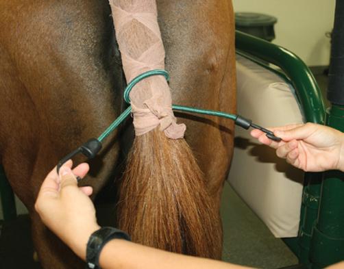
Figure 3.3 Elastic tail wrap encompassing the entire tail.
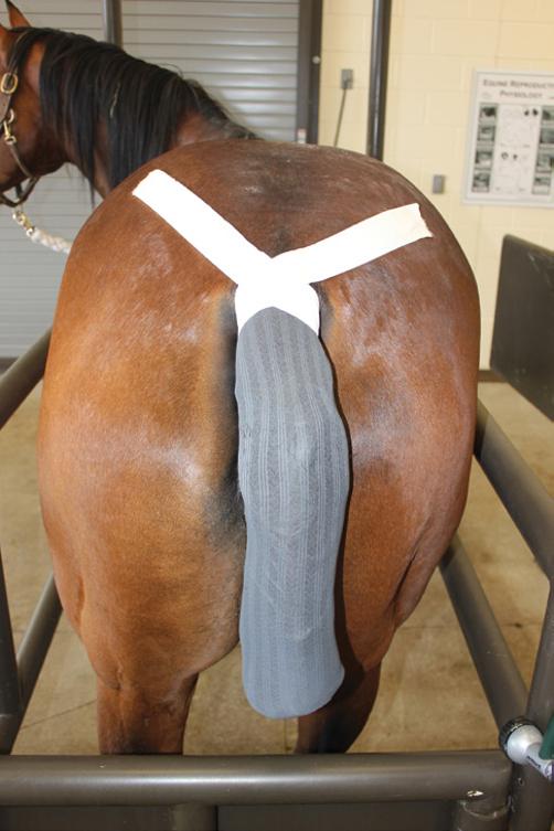
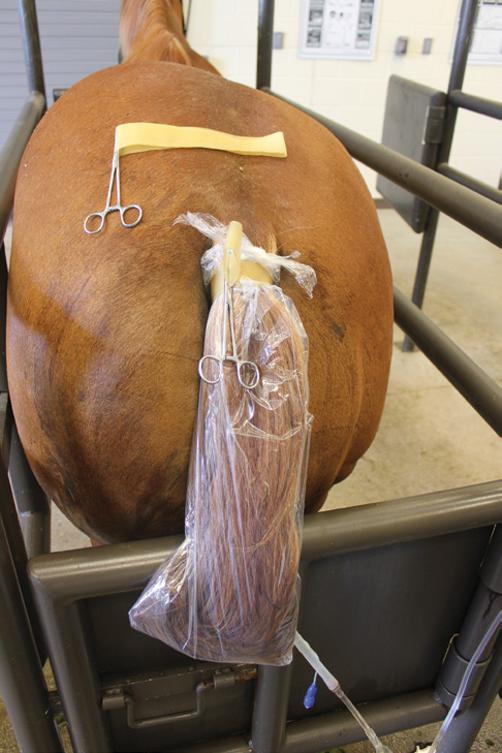
● Alternative tail wrap techniques include use of a sock held in place by 5 cm (2 inch) medical tape (Figure 3.5) or a plastic bag (75 × 25 cm (30 × 10 inches)) held in place by 2.5 cm (1 inch) rubber tubing and a clamp
Placement
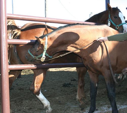
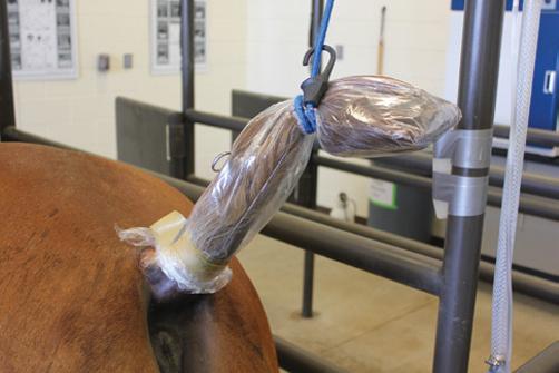
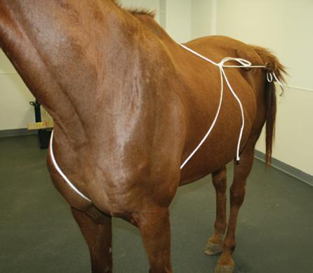
(Figure 3.6). The wrapped tail can be held out of the way by using an elastic cord (Figure 3.7) or being tied to the mare as previously described.
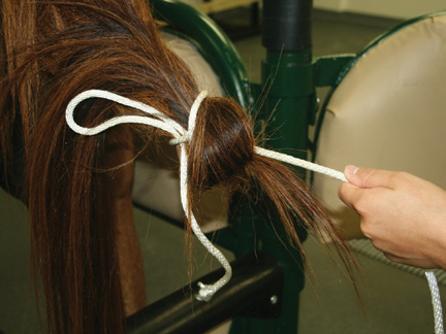
Figure 3.4 Tail wrap tied to the mare with quick release knots.
Figure 3.5 Tail wrap using a sock and secured with medical tape.
Figure 3.6 Tail wrap using a plastic bag and secured with rubber tubing and a clamp. An extra clamp is shown above the tail.
Figure 3.7 Wrapped tail held out of the way using an elastic cord.