Emergency Management of the Hi‐Tech Patient in Acute and Critical Care
Editors
Ioannis Koutroulis, MD, PhD, MBA
Attending Physician, Emergency Medicine
Children’s National Hospital
Assistant Professor of Pediatrics, Emergency Medicine, Genomics and Precision Medicine
George Washington University School of Medicine and Health Sciences
Nicholas Tsarouhas, MD
Medical Director, CHOP Transport Team
Attending Physician, Emergency Medicine
Children’s Hospital of Philadelphia
Professor of Clinical Pediatrics
Perelman School of Medicine at the University of Pennsylvania
Associate Editors
Richard J. Lin, MD
Attending Physician, Critical Care Medicine
Clinical Director, Progressive Care Unit
Associate Professor of Clinical Anesthesiology, Critical Care and Pediatrics, Perelman School of Medicine at the University of Pennsylvania
Jill C. Posner, MD, MSCE, MSEd
Attending Physician, Emergency Medicine
Children’s Hospital of Philadelphia
Professor of Clinical Pediatrics
Perelman School of Medicine at the University of Pennsylvania
Michael Seneff, MD
Director, Intensive Care Unit
Associate Professor of Anesthesiology and Critical Care Medicine
George Washington University Hospital
George Washington University School of Medicine and Health Sciences
Robert Shesser, MD
Professor and Chair
Department of Emergency Medicine
George Washington University School of Medicine and Health Sciences
This edition first published 2021 © 2021 John Wiley & Sons Ltd
All rights reserved. No part of this publication may be reproduced, stored in a retrieval system, or transmitted, in any form or by any means, electronic, mechanical, photocopying, recording or otherwise, except as permitted by law. Advice on how to obtain permission to reuse material from this title is available at http://www.wiley.com/go/permissions.
The right of Ioannis Koutroulis, Nicholas Tsarouhas, Richard J. Lin, Jill C. Posner, Michael Seneff, and Robert Shesser to be identified as the authors of the editorial material in this work has been asserted in accordance with law.
Registered Office(s)
John Wiley & Sons Ltd, The Atrium, Southern Gate, Chichester, West Sussex, PO19 8SQ, UK
Editorial Office
9600 Garsington Road, Oxford, OX4 2DQ, UK
For details of our global editorial offices, customer services, and more information about Wiley products, visit us at www.wiley.com.
Wiley also publishes its books in a variety of electronic formats and by print‐on‐demand. Some content that appear in standard print versions of this book may not be available in other formats.
Limit of Liability/Disclaimer of Warranty
The contents of this work are intended to further general scientific research, understanding, and discussion only and are not intended and should not be relied upon as recommending or promoting scientific method, diagnosis, or treatment by physicians for any particular patient. In view of ongoing research, equipment modifications, changes in governmental regulations, and the constant flow of information relating to the use of medicines, equipment, and devices, the reader is urged to review and evaluate the information provided in the package insert or instructions for each medicine, equipment, or device for, among other things, any changes in the instructions or indication of usage and for added warnings and precautions. While the publisher and authors have used their best efforts in preparing this work, they make no representations or warranties with respect to the accuracy or completeness of the contents of this work and specifically disclaim all warranties, including without limitation any implied warranties of merchantability or fitness for a particular purpose. No warranty may be created or extended by sales representatives, written sales materials or promotional statements for this work. The fact that an organization, website, or product is referred to in this work as a citation and/or potential source of further information does not mean that the publisher and authors endorse the information or services the organization, website, or product may provide or recommendations it may make. This work is sold with the understanding that the publisher is not engaged in rendering professional services. The advice and strategies contained herein may not be suitable for your situation. You should consult with a specialist where appropriate. Further, readers should be aware that websites listed in this work may have changed or disappeared between when this work was written and when it is read. Neither the publisher nor authors shall be liable for any loss of profit or any other commercial damages, including but not limited to special, incidental, consequential, or other damages.
Library of Congress Cataloging‐in‐Publication Data
Names: Koutroulis, Ioannis, 1980– editor.
Title: Emergency management of the hi-tech patient in acute and critical care / senior editors, Ioannis Koutroulis, Nicholas Tsarouhas ; editors, Richard J. Lin, Jill C. Posner, Michael Seneff, Robert Shesser.
Description: Chichester, West Sussex ; Hoboken, New Jersey : Wiley-Blackwell, 2021. | Includes bibliographical references and index.
Identifiers: LCCN 2020028239 (print) | LCCN 2020028240 (ebook) | ISBN 9781119262923 (cloth) | ISBN 9781119262954 (adobe pdf) | ISBN 9781119262985 (epub)
Subjects: MESH: Emergency Treatment–instrumentation | Emergency Medicine–instrumentation | First Aid–instrumentation | Critical Care–methods
Classification: LCC RC86.8 (print) | LCC RC86.8 (ebook) | NLM WB 26 | DDC 616.02/5–dc23
LC record available at https://lccn.loc.gov/2020028239
LC ebook record available at https://lccn.loc.gov/2020028240
Cover Design: Wiley
Cover Image: © Donald Iain Smith/iStock/Getty Images
Set in 9.5/12.5pt STIXTwoText by SPi Global, Pondicherry, India
Contents
List of Contributors ix
Preface xv
Acknowledgments xvii
Section I Gastro-intestinal Devices 1
1 Using and Troubleshooting Enteral Feeding Devices 3
Courtney E. Nelson and Thane A. Blinman
2 Gastrointestinal Diversions: Colostomies, Ileostomies, Mucous Fistulas, and Spit Fistulas 21
Ellen G. Szydlowski and Peter Mattei
3 Management of the Bariatric Surgery Patient in the Emergency Depar tment 27
Megan Lavoie and Joy Collins
4 Transjugular Intrahepatic Portosystemic Shunt 37
Heather House and Anne Marie Cahill
Section II Central Catheters 43
5 Indwelling Central Venous Catheter Devices 45
Anna Weiss
6 Vascular Access for Hemodialysis 57
Sarah Fesnak, Xenia Morgan, and Kimberly Windt
7 Peritoneal Dialysis Catheters 63
Jamie Lovell
Section III ENT and OMFS Devices 71
8 Or thopedic Devices 73
Carmelle Wallace, Andrew Tyler, and Keith D. Baldwin
9 Spine Devices 95
Susan E. Nelson and Keith D. Baldwin
10 Facial Distractors: Cranial Vault, Midface, and Mandibular 109
Francesca Drake and Phuong D. Nguyen
11 Management of Facial Fractures in Adults and Children 117
Sameer Shakir and Phuong D. Nguyen
12 Cranial Orthotics and Ear Molding 129
Meg Ann Maguire and Phuong D. Nguyen
13 Ear, Nose, and Throat Devices 137
Eva Delgado and Adva Buzi
Section IV CNS Devices 157
14 Cerebral-ventricular Shunts 159
Panagiotis Kratimenos, Angela Burd, and Chima Oluigbo
15 Initial Evaluation and Management of Patients with Neurosurgical Devices 169
Peter J. Madsen, Chariton Moschopoulos, and Benjamin C. Kennedy
Section V Miscellaneous Devices 183
16 Ophthalmic Devices 185
Marlet Bazemore and William P. Madigan
17 Breast Implants 189
R. Jason VonDerHaar and Liza C. Wu
18 Cutaneous Delivery Systems/Patches 203
Desiree M. Seeyave
19 Dental Emergencies Involving Oral Hardware 209
Chisom O.A . Agbim and Ioannis Koutroulis
Section VI Wound Management Devices 215
20 Emergency Management of Patients with Negative Pressure Wound Therapy Devices (NPWTD) 217
David Yamane and Tenagne Haile-Mariam
21 Emergency Management of Patients with Wound Infections 233
David Yamane and Tenagne Haile-Mariam
Section VII GU Devices 235
22 Emergency Management of Patients with Genitourinary Prostheses 237
Katie Wagner, Michael Phillips, and Kelly Chiles
Section VIII Cardiac Devices 245
23 Management of Prosthetic Valve Complications 247
Massoud Kazzi and Guenevere V. Burke
24 Emergency Management of Patients with Vascular Occlusion Devices, Stents, or Filters 257
Amy Caggiula
25 The Emergency Depar tment Approach to the Patient with an Implantable Mechanical Circulatory Suppor t Device 267
Robert Shesser
26 Emergency Management of the Patient with an Implantable Pacemaker or Defibrillator 277
Michael J. O’Neal
Section IX Pulmonary Devices 289
27 Invasive Ventilation: A Discussion of Equipment and Troubleshooting 291
Pelton A . Phinizy, Joseph J. Bolton, and John F. Tamasitis
28 Respiratory Medication Devices 313
Natalie Napolitano and James B. Fink
29 Secretion Clearance Devices 325
Amanda J. Nickel, Daniel Dawson, and Oscar H. Mayer
30 Non-invasive Ventilation 337
Oscar H. Mayer and Anthony Mozzone
31 Tracheostomy Tubes 347
Joanne Stow, Allison E. Boyd, and Jerry Cabrera
Index 373
List of Contributors
Chisom O.A. Agbim, MD, MSHS
Division of Emergency Medicine
Children’s National Medical Center
Washington, DC USA
Keith D. Baldwin, MD, MSPT, MPH
Department of Orthopedic Surgery
Perelman School of Medicine at the University of Pennsylvania Philadelphia, PA USA and Department of Orthopedic Surgery
Children’s Hospital of Philadelphia Philadelphia, PA USA
Marlet Bazemore, MD, MPH
Division of Ophthalmology
Children’s National Hospital Washington, DC, USA and
George Washington University School of Medicine and Health Sciences Washington, DC, USA
Thane A. Blinman, MD, FACS
Division of General and Thoracic Surgery
Department of Surgery
Children’s Hospital of Philadelphia Philadelphia, PA, USA
and Perelman School of Medicine at the University of Pennsylvania Philadelphia, PA, USA
Joseph J. Bolton, MBA, RRT-NPS Department of Respiratory Care Services
Children’s Hospital of Philadelphia Philadelphia, PA USA
Allison E. Boyd, MSN, CRNP Division of Otolaryngology Department of Surgery
Children’s Hospital of Philadelphia Philadelphia, PA USA
Angela Burd, DNP, CPNP-AC Hospital Medicine Division
Children’s National Hospital Washington, DC USA
Guenevere V. Burke, MD Department of Emergency Medicine
George Washington University School of Medicine and Health Sciences
Washington, DC USA
List of Contributors x
Adva Buzi, MD
Attending Physician
Division of Otolaryngology Department of Surgery
Children’s Hospital of Philadelphia Philadelphia, PA USA and Perelman School of Medicine at the University of Pennsylvania Philadelphia, PA USA
Jerry Cabrera, RRT Smith’s Medical Gary, IN USA
Amy Caggiula, MD Department of Emergency Medicine
George Washington University School of Medicine and Health Sciences Washington, DC USA
Anne Marie Cahill, MBBch, BAO Division of Interventional Radiology Department of Radiology
Children’s Hospital of Philadelphia Philadelphia, PA, USA and
Perelman School of Medicine at the University of Pennsylvania Philadelphia, PA USA
Kelly Chiles, MD Urology Private Practice Washington, DC USA
Joy Collins, MD
Division of General and Thoracic Surgery Department of Surgery
Children’s Hospital of Philadelphia
Philadelphia, PA USA and
Perelman School of Medicine at the University of Pennsylvania Philadelphia, PA USA
Daniel Dawson, RRT-NPS Department of Respiratory Care Services
Children’s Hospital of Philadelphia Philadelphia, PA USA
Eva Delgado, MD Division of Emergency Medicine Department of Pediatrics
Children’s Hospital of Philadelphia Philadelphia, PA, USA
Francesca Drake, RNFA, BSN, CNOR Division of Plastic and Reconstructive Surgery
Children’s Hospital of Philadelphia Philadelphia, PA USA
Sarah Fesnak, MD Division of Emergency Medicine Department of Pediatrics
Children’s Hospital of Philadelphia Philadelphia, PA USA and
Perelman School of Medicine at the University of Pennsylvania Philadelphia, PA USA
James B. Fink, PhD, RRT, FAARC, FCCP Aerogen Pharma Corporation San Mateo, CA, USA and
Rush Medical School Chicago, IL USA
and Texas State University
Round Rock, TX USA
Tenagne Haile‐Mariam, MD
Department of Emergency Medicine
George Washington University School of Medicine and Health Sciences
Washington, DC USA
Heather House, MD
Division of Emergency Medicine
Department of Pediatrics
Children’s Hospital of Philadelphia Philadelphia, PA USA and Perelman School of Medicine at the University of Pennsylvania Philadelphia, PA USA
Massoud Kazzi, MD
Department of Emergency Medicine
George Washington University School of Medicine and Health Sciences
Washington, DC USA and Critical Care Medicine
Washington Adventist Hospital
Takoma Park, MD USA
Benjamin C. Kennedy, MD
Department of Neurosurgery
Perelman School of Medicine at the University of Pennsylvania Philadelphia, PA USA and
Division of Neurosurgery
Children’s Hospital of Philadelphia Philadelphia, PA USA
List of Contributors
Panagiotis Kratimenos, MD, PhD
Division of Neonatal-Perinatal Medicine
Children’s National Hospital Washington, DC, USA and
George Washington University School of Medicine and Health Sciences Washington, DC, USA
Megan Lavoie , MD, FAAP
Division of Emergency Medicine
Department of Pediatrics
Children’s Hospital of Philadelphia Philadelphia, PA USA and
Perelman School of Medicine at the University of Pennsylvania Philadelphia, PA USA
Jamie Lovell, MD
Division of Emergency Medicine
Department of Pediatrics
Cincinnati Children’s Hospital Medical Center Cincinnati, OH USA and
University of Cincinnati College of Medicine Cincinnati, OH USA
William P. Madigan, MD
Division of Ophthalmology
Children’s National Hospital Washington, DC, USA and
Uniformed Services University of the Health Sciences Bethesda, MD, USA and
George Washington University School of Medicine and Health Sciences Washington, DC, USA
List of Contributors
Peter J. Madsen, MD
Division of Neurosurgery
Children’s Hospital of Philadelphia Philadelphia, PA, USA and
Department of Neurosurgery
Perelman School of Medicine at the University of Pennsylvania Philadelphia, PA, USA
Meg Ann Maguire, MS, RN, CRNP, CPNP-PC
Division of Plastic and Reconstructive Surgery
Children’s Hospital of Philadelphia Philadelphia, PA, USA
Peter Mattei, MD
Division of General, Thoracic and Fetal Surgery
Children’s Hospital of Philadelphia Philadelphia, PA USA and
Perelman School of Medicine at the University of Pennsylvania Philadelphia, PA USA
Oscar H. Mayer, MD
Division of Pulmonary and Sleep Medicine
Children’s Hospital of Philadelphia Philadelphia, PA USA and
Department of Pediatrics
Perelman School of Medicine at the University of Pennsylvania Philadelphia, PA USA
Xenia Morgan, CRNP
Hemodialysis Unit
Division of Nephrology
Children’s Hospital of Philadelphia Philadelphia, PA USA
Chariton Moschopoulos, MD Division of Neurology
Boston Children’s Hospital Harvard Medical School Boston, MA USA
Anthony Mozzone, BA, CRT Promptcare Respiratory King of Prussia, PA USA
Natalie Napolitano, MPH, RRT, RRTNPS, FAARC
Department of Respiratory Care Services
Children’s Hospital of Philadelphia Philadelphia, PA USA
Courtney E. Nelson, MD
Division of Emergency Medicine
Department of Pediatrics
AI DuPont Hospital for Children Wilmington, DE, USA and
Sidney Kimmel Medical College at Thomas Jefferson University Philadelphia, PA, USA
Susan E. Nelson, MD, MPH
Department of Orthopedic Surgery University of Rochester Medical Center Rochester, NY USA
Phuong D. Nguyen, MD
Division of Plastic and Reconstructive Surgery
Children’s Hospital of Philadelphia Philadelphia, PA USA
Amanda J. Nickel, MSc, RRT-NPS, RRT-ACCS
Department of Respiratory Care Services
Children’s Hospital of Philadelphia Philadelphia, PA USA
Chima Oluigbo, MD
Department of Neurosurgery
Children’s National Hospital Washington, DC, USA and
George Washington University School of Medicine and Health Sciences Washington, DC, USA
Michael J. O’Neal, MD
Department of Emergency Medicine University of San Francisco San Francisco, CA USA
Michael Phillips, MD Department of Urology
George Washington University School of Medicine and Health Sciences Washington, DC USA
Pelton A. Phinizy, MD
Division of Pulmonary and Sleep Medicine
Children’s Hospital of Philadelphia Philadelphia, PA USA and Department of Pediatrics
Perelman School of Medicine at the University of Pennsylvania Philadelphia, PA USA
Desiree M. Seeyave, MB.BS
Department of Emergency Medicine & Hospitalist Services
Children’s Hospital of Georgia Emergency Department Augusta, GA USA
Sameer Shakir, MD Division of Plastic Surgery
Children’s Hospital of Philadelphia Philadelphia, PA USA
Joanne Stow, MSN, APRN, PPCNP-BC, CORLN Division of Otolaryngology Department of Surgery
Children’s Hospital of Philadelphia Philadelphia, PA, USA
Ellen G. Szydlowski, MD Division of Emergency Medicine Department of Pediatrics
Children’s Hospital of Philadelphia Philadelphia, PA USA and
Perelman School of Medicine at the University of Pennsylvania Philadelphia, PA USA
John F. Tamasitis, RRT-NPS
Children’s Hospital Home Care
Children’s Hospital of Philadelphia King of Prussia, PA USA
Andrew Tyler, MD, PhD Department of Orthopedic Surgery
Vanderbilt University Medical Center Nashville, TN USA
R. Jason VonDerHaar, MD Department of Surgery
Indiana University Health Indianapolis, IN USA and
University of Indiana School of Medicine Indianapolis, IN USA
Katie Wagner, BS
George Washington University School of Medicine and Health Sciences Washington, DC USA
List of Contributors
Carmelle Wallace, MD, MPH
Division of Emergency Medicine
Department of Pediatrics
University of Alabama at Birmingham Birmingham, AL
USA
Anna Weiss, MD, MSEd
Division of Emergency Medicine
Department of Pediatrics
Children’s Hospital of Philadelphia Philadelphia, PA USA and Perelman School of Medicine at the University of Pennsylvania Philadelphia, PA USA
Kimberly Windt, MSN, RN, CNN
Hemodialysis Unit
Division of Nephrology
Children’s Hospital of Philadelphia Philadelphia, PA
USA
Liza C. Wu, MD
Department of Surgery
Hospital of the University of Pennsylvania and the Children’s Hospital of Philadelphia Philadelphia, PA, USA and
Division of Plastic Surgery
Perelman School of Medicine at the University of Pennsylvania Philadelphia, PA USA
David Yamane, MD
Department of Emergency Medicine
George Washington University Hospital and
Department of Anesthesia and Critical Care Medicine
George Washington University Hospital Washington, DC USA
Preface
The idea of writing this book was born in early 2016; we knew that the road would be bumpy yet exciting! We are thrilled to introduce the first edition of our manual to assist health care providers in managing our very unique patients with various hardware devices. These “hi‐tech” devices help our patients breathe, walk, hear, and do a host of other essential activities of daily living. Importantly, when these patients seek emergency care due to device malfunction, infections, or other complications, it is crucial that the medical provider know how to manage both the patient and the device.
It is often challenging for the emergency/critical care clinician to troubleshoot and, sometimes, even identify the problems with many of these hi‐tech devices. Moreover, specialized consultation is not always readily available. It is quite challenging for providers to recall complex diagnostic and therapeutic algorithms for patients with specialized equipment, thus increasing the risk for morbidity and mortality. This guide provides a stepwise approach to acute presentations of patients with clinical hardware, focusing on specific instructions for initial evaluation and management. Our intent is that this endeavor will assist health care providers in both community settings and academic centers in the treatment of complex patients with “hi‐tech” equipment.
It is our sincere hope that this book will improve the quality of care to our unique patient population who utilize specialized hardware. Our goal is to provide our readers with an overview of the basic approach to clinical scenarios of device malfunction and related complications of the most commonly used medical devices. However, as it was impossible to include all devices in this book, the practitioner should make every effort to ensure they have the most accurate information for each patient’s hardware.
We want to thank our fellow editors, authors, and contributors, who gave their time selflessly so that we could produce this book. Also, we would like to thank our teachers, mentors, and colleagues, from whom we learn every day. Most importantly, the real source of our inspiration, our patients and their families, deserve our eternal heartfelt gratitude.
Ioannis and Nick
Acknowledgments
I would like to thank Nick and all the editors of the book, as, without them, this effort would not have been possible. Nick’s support was really amazing, and his help with some of the problems we faced during the creation of the book really made a difference. As a great clinician, researcher, and educator, Nick’s contribution was vital for the whole project. Jill, Mike, Rich, and Rob went above and beyond to have all the chapters completed on time and make sure the content was appropriate. Thank you all!
I want to dedicate this book to my mother who, despite all the difficulties, never left my side. She has been my inspiration all these years. Also, to Dr. Levinsky, who discussed the initial idea for the book with me. And finally, to all the people in my life who made a difference, thank you!
Ioannis Koutroulis
I’d like to first acknowledge, Ioannis, the creator of the book and our persevering leader. Through thick and thin, Ioannis remained upbeat and encouraging. Ioannis stayed enthusiastic, despite delays as a result of our heavy administrative and clinical workloads due to sustained high volumes and acuities in our emergency departments (EDs), inpatient areas, and intensive care units. With an eternally positive attitude, he even respectfully pushed us through a pandemic toward the completion of the “Hi-Tech Book” project. Thank you, Ioannis! My most genuine love and appreciation goes to my family – wife Debbie, my son Christopher, and my daughter Nicole. The importance of their continual support of my long ED shifts, constant meetings, and endless “homework” cannot be understated. They have forever tolerated “Daddy” in his study … reading, writing, typing, answering pages, making phone calls, etc. Finally, a special thank you to my mother who, with little education and even less money, somehow managed to raise three pretty good kids. Love you, mom.
Nick Tsarouhas
To my parents, who raised me to believe I can do anything. To my mentors, who have showed me the way.
To my patients, who have inspired and humbled me. And to my husband and daughters, my loves and light.
Jill C. Posner
Section I
Gastro-intestinal Devices
Using and Troubleshooting Enteral Feeding Devices
Courtney E. Nelson1,2 and Thane A. Blinman3,4
1 Division of Emergency Medicine, Department of Pediatrics, AI DuPont Hospital for Children, Wilmington, DE, USA
2 Sidney Kimmel Medical College at Thomas Jefferson University, Philadelphia, PA, USA
3 Division of General and Thoracic Surgery, Department of Surgery, Children’s Hospital of Philadelphia, Philadelphia, PA, USA
4 Perelman School of Medicine at the University of Pennsylvania, Philadelphia, PA, USA
Introduction
Enteral feeding devices deliver nutrition directly to the stomach and/or small intestine for patients with anatomic or physiological feeding impairments. Common indications for enteral feeding devices include feeding and swallowing dysfunction, severe gastroesophageal reflux, malnutrition, neurological disorders, and prolonged ventilation. Given the breadth of indications for enteral feeding devices, a clinician in any setting, and particularly those in the emergency department, is likely to encounter these devices on a daily basis. These are simple devices with a simple purpose, but their dysfunction is highly disruptive and worrisome to patients and their caregivers. This chapter will teach you how to manage the simplicity of a working enteral feeding device and navigate the intricacies of an unruly device.
Equipment/Device
Enteral feeding devices can be categorized into temporary and long-term devices. Furthermore, the name for each device comes from its origin (nose, mouth, or stomach) and terminus (stomach or small intestine) (Table 1.1). Nasal and oral tubes are temporary and work well for patients with transient feeding difficulties. Gastrostomy tubes (G-tube) and jejunostomy tubes (J-tube) are ideal for more long-term or permanent enteral nutrition. Enteral feeding devices are sized in French (Fr) units, which is the outer diameter of the tube in millimeters multiplied by three. A 9 Fr tube, for example, has an outer diameter of 3 mm. Tube lengths are usually given in centimeters and vary widely from very short low profile “button” type tubes to very long naso-jejunal (NJ) tubes.
Emergency Management of the Hi-Tech Patient in Acute and Critical Care, First Edition. Edited by Ioannis Koutroulis, Nicholas Tsarouhas, Richard J. Lin, Jill C. Posner, Michael Seneff, and Robert Shesser. © 2021 John Wiley & Sons Ltd. Published 2021 by John Wiley & Sons Ltd.
Table 1.1 Enteric feeding devices.
OriginDestination Tube
Temporary feeding devices
Nose (naso-)Stomach (gastric)
Duodenum
Jejunum
Mouth (oro-)
Long-term feeding devices
PercutaneousStomach
Jejunum
Naso-gastric
Naso-duodenal
Naso-jejunal
AbbreviatedPlacement
NG ND NJ
Bedside
Oro-gastricOGBedside
Gastrica
Gastro-jejunostomy
Jejunostomy
G-tube GJ
J-tube
Surgically or endoscopically
Fluoroscopically
Fluoroscopically
a Commonly, these tubes are called “PEG tubes”; however, a percutaneous endoscopic gastrostomy is a procedure and not a specific tube.
Temporary Feeding Devices
Naso-gastric (NG), naso-duodenal (ND), NJ, and oro-gastric (OG) feeding tubes are used for short-term enteral feeding, defined as that less than 12 weeks. OG tubes are reserved for patients in the intensive care unit and rarely seen in the emergency department. Temporary feeding tubes are typically constructed from polyurethane or silicone-based polymers, both of which are flexible, reasonably durable, minimally reactive biologically, and, for most, immunologically inert. Polyurethane tubing has the added benefit of being made with a water-activated lubricant to ease insertion and increased durability. Depending on the manufacturer, NG, NJ, ND, and OG tubing may come with weighted tips, radiopaque indicators, stylets, and/or magnets to help with placement. Common pediatric tube size for feeding is a 6–8 Fr and for adults a 12–14 Fr.
Gastric Decompression Devices
Similar to the temporary feeding devices, there are NG and OG tubes used for decompression and lavage. These devices are larger than feeding tubes: 8–10 Fr for children and 12–14 Fr for adults. Decompression devices are divided into single and double lumen tubes. Single lumen tubes, such as the Levin tube, are used more frequently in the emergency department or intensive care unit for intermittent decompression. Single lumen tubes should not be placed on continuous suction because they can adhere to the stomach wall and cause tissue damage. A double lumen tube is the preferred decompression device because it has both a large lumen for suction or irrigation and a small lumen (typically blue in color) that vents the large lumen. This small lumen serves as a pop-off valve for the device to prevent excessive suctioning. There are two common types of double lumen tubes: Salem sumps and Replogle tubes. Salem sumps are preferred in an emergency setting because they have several suction holes along the side of the tubing for rapid efficient suctioning, whereas Replogle tubes have suction holes only at the most distal end of the tube.
When using a double lumen tube, it is critical that the small lumen be kept to room air to adequately vent the large lumen. It should not be clamped, used for suction, or used for irrigation. Finally, the proximal end of a double lumen decompression device must be kept above waist-height, otherwise the gastric contents may reflux into the small lumen.
Long-term Feeding Devices
G-tubes are used for long-term or permanent enteric feeding. G-tubes are divided into standard adjustable length tubes and low-profile (i.e. button) tubes. When caring for a patient with a long-term feeding device, it is imperative that you know the type of tube the patient has, how the tube was placed, and how to use the tube in order to adequately care for your patient.
Basic G-tube Anatomy
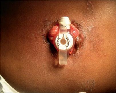
G-tubes are made of silicone, polyurethane, or, rarely, latex rubber to provide the flexibility and durability needed for long-term feeding. They serve as a direct pathway to the gut. G-tubes are made up of one to three ports on the most proximal end, followed by a tube or shaft that carries nutrition to the gastrointestinal (GI) tract, and then a balloon or nonballoon retention device on the distal end (Figure 1.1). In some tubes, there are separate feeding and medication ports. Only in balloon G-tubes is there a port that is used to expand the retention balloon. In addition, standard G-tubes have an external retention device with air vents and feet that hold it 1–2 mm above the skin surface to prevent skin breakdown and keep the stoma site clean and dry.
There are advantages and disadvantages to balloon and nonballoon G-tubes. The benefit of balloon G-tubes is that they can be replaced at home; however, they are not as well tolerated as nonballoon retention devices because of the size of the balloon. Furthermore, balloon retention devices need to be changed more frequently than nonballoon G-tubes (every three months compared to every six months, respectively). The main disadvantage of nonballoon G-tubes is that every tube change has to be done by a medical professional.
Standard G-Tube
Standard G-tubes are adjustable length tubes that have an external bolster, which sits on the skin and can be moved up and down to adequately secure the tube in a patient of any size (Figure 1.1 a and b). These are particularly helpful in patients with increased soft tissue or in patients with a projected weight gain where a low-profile tube with a fixed shaft length may not fit properly. Standard G-tubes can be placed surgically or endoscopically.
Standard G-tubes are placed surgically using the Stamm procedure or via a laparoscopic approach. You will know your patient had the Stamm procedure if he or she has a 6–8 cm midline incision on examination. During the procedure, the surgeon dissects down to the anterior wall of the stomach. The G-tube is placed directly into the stomach via an anterior
Figure 1.1 Standard G-tubes and low-profile G-tubes. (a) Standard GJ tube with three ports: balloon port, jejunal port, and gastric port. (b) Standard G-tube with three ports: medication port, gastric port, and balloon port. (c) Low-profile tubes with both nonballoon and balloon retention devices.
incision. The balloon is inflated and used to pull the stomach against the inner abdominal wall to determine the best location for the percutaneous exit of the tubing. Once this incision is made, the tubing is pulled through the abdominal wall and the tube is anchored in place with sutures. A standard G-tube placed surgically will have a well-healed tract within four weeks.
Standard G-tubes are placed endoscopically by using the percutaneous endoscopic gastrostomy(PEG) technique. Of note, the term “PEG” is used inaccurately in medical vernacular to refer to all kinds of G-tubes, but a PEG is actually the procedure and not a type of tube. During a PEG procedure, an endoscope is used to transilluminate the stomach and identify the stoma site. A needle is then inserted through the skin into the stomach with a guidewire that is pulled up through the esophagus and out of the mouth. This guidewire is then used to guide the G-tube into the stomach. A small incision is made, and the G-tube is pulled through the stomach and abdominal walls and secured in place by the internal and external bolsters alone.
There are two main advantages to a surgically placed G-tube compared to a PEG procedure. First, a mature tract forms in 4 weeks with a laparoscopic procedure compared to 6–12 weeks with a PEG procedure. Second, a surgically placed G-tube provides direct
External retention device
Balloon port
Feeding port
Medication port
G-tube shaft
Non-balloon internal retention device
Balloon internal retention device
Standard G-tubes
C. Low profile G-tubes
Balloon port
Balloon port
visualization of the anatomy, whereas, with an endoscopically placed G-tube, there is always the risk of a bowel perforation if a portion of bowel is caught between the abdominal wall and the gastric wall during G-tube placement.
Low-Profile G-Tube
Low-profile G-tubes have a port that sits flush with the skin surface (Figure 1.1c). They are more easily hidden than the standard G-tubes simply by the nature of their size, and patients tend to prefer them for this reason. In addition, the smooth surface of a G-tube port site without tubing is less prone to accidental dislodgement compared to standard G-tubes. However, there are drawbacks to a low-profile tube. First, external tubing has to be attached in order to deliver a feed, which creates one additional step and an additional piece of equipment that can malfunction. Second, low-profile tubes cannot be adjusted to accommodate increased abdominal wall thickness and must be replaced with a tube that has a longer shaft length when there are signs of abdominal wall compression.
Although they were not designed to be placed primarily, low-profile G-tubes can be placed laparoscopically. In pediatric surgical practice, the laparoscopic primary G-button gastrostomy is now widely performed. In this approach, one trocar is placed through the umbilicus and another through a small incision in the left upper quadrant. A stitch is placed in the anterior wall of the stomach and passed through the trocar in the left upper quadrant. Once the suture material is outside the abdomen, the trocar is removed and the anterior wall is pulled through the initial trocar site. The stomach and abdominal walls are sutured together. The gastrostomy is made in the portion of stomach wall that is exposed. The appropriate button is then placed in the gastrostomy and sutured in place. Similar to a surgically placed standard G-tube, a low-profile G-tube tract matures in four weeks.
Jejunal Tubes
NJ, gastro-jejunal (GJ), and J-tubes are ideal for patients with gastric dysmotility, severe gastroesophageal reflux, recurrent emesis, and those at risk for pulmonary aspiration. The jejunum is fed continuously and at a lower rate compared to bolus feeds given through a G-tube. Whereas the NJ and J-tubes provide direct access to the jejunum, GJ tubes are a hybrid with both gastric and jejunal ports. The gastric port is used for medications or venting the stomach, while the jejunal port is used for continuous enteral nutrition. J-tubes are secured to the abdominal wall with an internal retention device in the jejunum, while GJ tubes have an internal retention device within the gastric cavity and a jejunal extension that passes through the G-tube and bypasses the stomach. Jejunal extensions carry the added risk of tube migration, volvulus around the extension tubing, and higher rates of tube clogging secondary to smaller tube size. Percutaneous J-tubes are rarely used because of the thinness of the jejunal wall and increased risk of complications.
NJ and GJ tube placement requires fluoroscopic or endoscopic guidance for placement. Percutaneous J-tubes are placed surgically.
Indications
Enteral feeding devices are indicated when a patient has a functional GI tract but cannot safely or adequately feed by mouth. This includes patients with a swallowing dysfunction or neurologic disorder. Similarly, patients on a ventilator or those with severe reflux require enteral feeding to prevent aspiration. Finally, patients with inadequate nutrition or in a hypermetabolic state (i.e. cardiac disease, renal disease, or pulmonary disease) may not be able to meet their nutritional demands with oral feeding alone and require enteral feeding as supplementation. The indication for enteral nutrition and duration of feeding needed determine the type of tube recommended. Contraindications specific to each feeding device are listed in Table 1.2.
Management
Routine Care
Enteral feeding devices require daily care to ensure the tube is patent and to protect the surrounding skin from irritation. All feeding devices need to be flushed with room temperature water following each feed or medication administration to prevent clogging. The tubes should also be monitored for tube deterioration that indicates the tube needs to be changed: discoloration, foul smell, and tube deformity. NG and OG tubes need to be monitored for pressure necrosis at the point of insertion and retaped as needed. Similarly, G-tubes and GJ tubes can cause pressure ulcers if the tissue between the internal and external retention devices is compressed too tightly. Standard G-tubes should be turned regularly and evaluated to ensure the external retention device sits 1–2 mm above the skin surface without creating a dimple in the skin. Finally, internal balloon retention devices should be checked regularly to confirm the appropriate amount of fluid is in the balloon to prevent tube dislodgement.
Routine Replacement
Temporary Tubes
NG and OG tubes can be placed and replaced by home nursing or properly trained family members (Table 1.3). NG/OG tubes should be replaced approximately every 7–10 days.
NG/OG feeding tubes G-tubes
● Maxillofacial disorders
● Esophageal or oropharyngeal tumors or trauma
● Laryngectomy
● Confirmed skull or cervical spine injury above C4
● Clotting dysfunction
● Ingestion of corrosive substance
● Severe gastroesophageal reflux
● Gastric dysmotility
● Gastric outlet obstruction
Discuss with appropriate consulting service prior to NG/OG placement
J-tubes
● Ascites
● Crohn’s disease
● Immunosuppression
Table 1.2 Enteric feeding device contraindications.
Table 1.3 NG tube insertion.
Supplies
● Nasogastric tube
● Sterile water
● 50 mL catheter tip syringe
● Tape to secure tubing
Stepwise procedure
1) Position patient sitting upright with neck midline; avoid hyperextension.
2) Lubricate the NG tip with sterile water. Avoid jelly as it will affect the pH.
3) Direct the tube into one of the nostrils and, keeping the tube horizontal, aim the tube directly posterior. Ask the patient to swallow, as this will help guide the tube into the esophagus by closing the epiglottis.
4) Once the tube passes through the nasopharynx, have the patient lean forward and bend his/her chin while continuing to swallow which will further push the tube down the esophagus.
5) Continue to pass the tube until you reach the predetermined tube depth.
6) Stop and remove the tube if the patient has any signs of respiratory distress.
7) Attach a 50 ml syringe and aspirate contents to the tube.
8) Test aspirate on pH paper, any value below 4.0 is considered gastric contents.
9) Secure the tube by taping to the nose and face.
Properly sizing the tubing is necessary prior to placement. When choosing a tube size, most feedings can be given with a 6 or 8 Fr tube in pediatric patients and a 12–14 Fr tube in adult patients. Appropriate tube insertion depth is classically measured by taking the feeding tube and measuring from the tip of the nose to the ear lobe and finally to the xyphoid. Several pediatric studies have found that this measurement may underestimate tube depth and result in tubes that terminate in the distal esophagus and pose an aspiration risk. Therefore, in pediatric patients, a better measurement is either using published age-related height-based measurements or by measuring from the tip of the nose to the ear lobe and then to the midpoint between the xyphoid and umbilicus. For OG tubes, the measurement should start at the mouth. Once the appropriate tube size and depth of insertion are determined, the same steps may be followed for either NG or OG tube placement, substituting the nasal passages for the oropharynx in the place of OG placement (Table 1.3). Finally, NG/OG tube placement is an uncomfortable procedure, and patients should be treated with topical lidocaine either as 4% lidocaine spray, 2% lidocaine jelly, or nebulized 4% lidocaine prior to the procedure.
NG/OG tube placement can be verified by the aspiration of gastric contents with a pH < 4.0. However, be aware that medications can change the gastric pH, and in a patient with reflux, an esophageal aspirate may have the same pH as a gastric aspirate. Likewise, the aspiration of “gastric fluid” does not confirm gastric placement alone because there is fluid within the bronchial tree and distal esophagus that resembles gastric fluid. In addition, lack of fluid aspirate can lead to falsely believing the tube is not in the stomach when the tube collapses or is above the fluid level. Finally, auscultation is an unreliable method of determining NG/OG tube placement because the sound of an NG tube in the thorax may
transmit to the upper abdomen. X-ray confirmation remains the gold standard for NG/OG tube placement in both adult and pediatric patients.
Bedside placement of ND and NJ feeding tubes is still controversial; however, there are increasingly more studies supporting this practice. Most research has focused on the placement of ND tubes. ND tube placement is similar to NG tube placement; however, the patient is kept in the right-lateral decubitus position. Several adjunctive measures have been described including the use of promotility agents and gas insufflation to promote tube position past the pylorus. Of note, these techniques are better described in the adult patients and less so in pediatric patients. All post-pyloric feeding tubes should be confirmed with an X-ray prior to use.
G-tubes and GJ tubes
G-tubes may be replaced by a caregiver following the first tube change and at least four weeks from initial tube placement (Table 1.4). Typically, a gastrostomy tube is changed every three to four months. G-tube replacement should be confirmed with the aspiration of gastric contents and/or pH testing. The gold standard for G-tube confirmation is a fluoroscopic dye study whereby dye is injected through the G-tube port and a radiograph is taken to verify dye positioning in the stomach. If there is any trauma to the G-tube site or if the tube is considered an immature tube (<4 weeks from placement), aspiration of gastric
Table 1.4 Gastrostomy-tube replacement.
Supplies
● G-tube low profile button with extension tubing
Or
● Traditional G-tube
● Luer slip tip syringe to inflate balloon
● Larger catheter tip syringe to prime and flush tubing
Optional:
● G-tube port stylet
Stepwise procedure
1) Deflate the G-tube gastric balloon with a 10 ml syringe.
2) Gently remove the G-tube by holding the port site and steadily pulling it back.
3) Keep stoma patent with a Foley catheter (do not exceed the G-tube size).
4) Remove the G-tube from packaging and check balloon by filling it with tap water (do not fill with normal saline as this will degrade balloon and do not use air as it will not provide adequate tension on the balloon).
5) Deflate balloon prior to tube insertion.
6) Insert the G-tube stylet, if one is provided.
7) Lubricate the tube with sterile jelly (do not use petroleum jelly as it will degrade tubing).
8) Direct the G-tube into the stoma and apply steady pressure.
9) Stop and reposition if you meet resistance.
10) Once the G-tube external base is resting on the skin surface, inflate the balloon.
11) Confirm positioning by pulling gently on the port site.
contents and pH testing are inadequate, and the tube site should be verified with a fluoroscopic dye study.
GJ tube must be placed by interventional radiology under fluoroscopy to ensure proper placement for both the initial placement and any subsequent tube replacement. GJ tubes are replaced every six months.
Complications/Emergencies
Tube Dislodgment
Tube dislodgment is a common emergency department chief complaint in both adults and children. This can occur because of coughing, gagging, pulling on the tubing, or getting the tubing caught around an object. In all cases, stop the feeding and inquire how long the patient can maintain his or her blood sugar without feeding. Hypoglycemia is a common complication for patients who are accustomed to receiving continuous feeds, and an infusion of dextrose-containing IV fluids is commonly needed while awaiting feeding tube replacement. Replacement follows an algorithm based on the type of enteric feeding device and the duration since its initial placement (Figure 1.2). Unfortunately, tube replacement is not without risk, and the astute provider must be aware of clinical signs of an improperly positioned tube and how to best verify tube placement.
NG tube replacement is a simple procedure, and some patients may even replace their own NG tubes nightly; however, it is not without risk. Complication rates range from 1 to 2% in adults and up to 20–40% in pediatric patients, with higher rates seen in neonatal patients. Complications with improper NG tube placement include pneumonia if the tube is placed in the lungs and peritonitis if the tube perforates the bowel. Given this high complication rate, all NG tube replacements in the emergency department setting should be confirmed with a radiograph. Auscultation, enzyme testing, and pH testing are unreliable.
G-tube placement is an equally common emergency department procedure, and complication rates range from 0.6 to 20%, with higher rates seen in immature tubes and tubes with traumatic dislodgement. The definition of an immature tube is debated in the literature. Most studies define immature tubes as those less than four to six weeks from placement; however, some studies define immature tubes as those less than six months from placement. There is an inverse relationship between the age of the tube, and, therefore, G-tube tract healing, and the complication rate. In addition, patients who are symptomatic postreplacement are more likely to have a G-tube complication. Complications include gastric outlet obstruction and intraperitoneal tube placement.
All G-tube replacements can be completed at the bedside; however, the person performing the procedure and the method of checking placement are dependent on the age of the tube and the patient’s presenting symptoms. The first tube change postoperatively is the most critical and should be completed by the subspecialty service responsible for the tube’s placement. In addition, any stoma site with significant trauma or stoma that is difficult to identify should be evaluated by general surgery. Uncomplicated mature G-tubes can be replaced at the bedside by the emergency department team, and placement should be confirmed with gastric aspirate and pH testing alone. For patients with immature tracts,
Displaced enteric feeding device
1. Determine the type of tube
2. Assess patient’s glycemic control off of feeding and determine need for intravenous dextrose containing fluids
3. Keep stoma patent with a Foley catheter
Oral or nasal fe eding tube
Bedside replacement
See Table 2
Consult interventional radiology for placement under fluoroscopy
Gastrostomy tube
Standard or low profile gastrostomy tube
Immature tract < 4 weeks
Call team responsible for initial placement
Mature tract > 4 weeks
Bedside replacement
See table 3
trauma to the stoma site, or symptoms following G-tube replacement, contrast-enhanced radiograph is needed to confirm G-tube placement prior to use. Extravasation of contrast dye on imaging and failure to fill the stomach indicates that the tube is improperly positioned.
All transgastric jejunal tubes must be replaced by interventional radiology under fluoroscopic guidance.
Peristomal Skin Irritation
Patients can present with G-tube erythema for a variety of reasons. While the presence of skin irritation can be highly distressing to patients and caregivers, the cause is commonly nonurgent. However, it is vital that providers have a healthy differential in order to distinguish severe causes of peristomal irritation from those that are less severe.
Naso-jejunal/Gastro-jejunal
Figure 1.2 Algorithm of the displaced enteric feeding device.
Peristomal Leakage
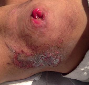
Peristomal leakage of gastric contents is seen with most G-tubes. Diabetes, malnutrition, and poor wound healing can increase the likelihood and amount of leakage secondary to poor approximation of skin tissue around the tubing. In addition, a tightly secured retention device, noted by dimpling of the skin, can cause an inflammatory reaction and lead to increased leakage of gastric contents.
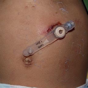
Skin irritation from peristomal leakage can be distinguished from infection by the color, which is a faint pink instead of the deep red color of cellulitis (Figure 1.3a). Likewise, the skin is not tender. Finally, crusting around the tube site, that is, dried formula and gastric juices, should easily wipe away.
Treatment options for peristomal leakage include skin barrier creams such as zinc oxide and antacid treatment to decrease the acidity of the gastric contents. If the stoma appears too large for the tubing, do not increase the size of the tube. A larger stoma site is not because the patient grew or gained weight. The stoma size increases secondary to repetitive trauma from the tube moving within the stoma. Increasing the tube size will only stretch the stoma further and lead to greater leakage of gastric contents. Do not make this common mistake. Instead, remove the tube and allow the stoma to shrink in size over the next several hours. A stoma can close within as little as 24 hours, so a smaller catheter should be left in place to maintain patency of the stoma. Once the stoma has decreased to the appropriate size, place the original sized G-tube into the site.
Stomal Cellulitis
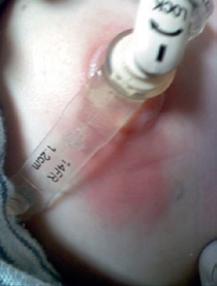
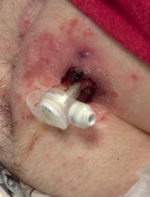
Stomal cellulitis is distinguished from skin irritation by the deeper red color and spreading erythema around a G-tube site with significant pain to touch (Figure 1.3b). Patients with poor wound healing and an immunocompromised state are at increased risk for cellulitis. Pathogens are typically skin flora including beta-hemolytic streptococci and Staphylococcus aureus.
Figure 1.3 (a) Peristomal leakage notable for dried crusted skin without surrounding erythema. (b) Peristomal cellulitis distinguished from simple leakage by the deeper erythematous skin extending from the G-tube site. (c) Peristomal candidiasis distinguished from cellulitis by its satellite lesions.
(a)
(b)
(c)
In a well-appearing child with otherwise no systemic symptoms, a first-generation cephalosporin is adequate to treat streptococcal infection. If a patient is a known methicillin-resistant S. aureus carrier or appears ill, coverage should include agents that treat methicillin-resistant Staphylococcus aureus (MRSA) based on local antibiograms. The tube does not need to be removed in the setting of stomal cellulitis. If there is fluctuance around the tube, an ultrasound should be obtained to evaluate for a peristomal abscess. Peristomal abscesses will require bedside incision and drainage and broad-spectrum antibiotic coverage.
Stomal Candidiasis
Stomal candidiasis is much less common than bacterial infections of the stoma. Patients with candidiasis should be well appearing and have typical satellite lesions around the stomal site (Figure 1.3c). Similar to other forms of candidiasis, treatment with topical antifungal agents alone (nystatin or clotrimazole) is sufficient.
Necrotizing Fasciitis
Necrotizing fasciitis of the stoma is an exceedingly rare but life-threatening complication. The patient will have erythematous, edematous, and tender skin with bullae. The lesion will rapidly expand, and the patient will look toxic. Similar to all infections, those patients with poor wound healing, diabetes, and malnutrition are at greatest risk. Necrotizing fasciitis is a surgical emergency that requires immediate surgical evaluation, wound debridement, and intravenous antibiotic treatment.
Stomal Bleeding
Bleeding at the stomal site is one of three things: hypergranulation tissue, mucosal irritation with or without prolapse, and upper GI bleeding. Distinguishing between the three is important because, while, to the patient they may all be an emergency, the severity and treatment are dramatically different.
A granuloma is a well-circumscribed, pearly piece of tissue adherent to the stoma (Figure 1.4a). It presents as chronic, low-grade bleeding. Its cause is unknown, but it is thought to arise from repetitive trauma from the G-tube rubbing against the stoma. Hypergranulation tissue is of low risk but causes significant distress among patients and caregivers. Treatment is largely topical, including 0.1% triamcinolone cream, commercially available granuloma-reducing agents, and salt packing. These agents are not without risk; specifically, triamcinolone can cause skin thinning, systemic absorption, and may precipitate a fungal infection. Silver nitrate application, kenalog injections, electrocautery, and G-tube site revision are used in more persistent cases.
Another cause of chronic mild stomal bleeding is mucosal irritation. This too is caused by repetitive movement of the tube within the stoma and can be quickly resolved with properly sizing the tube. In addition, gauze and tape can be used to better secure the tube in position.
Finally, acute stomal bleeding is either prolapsed stomal tissue or upper GI bleeding. Prolapsed gastric tissue has a deeper red color compared to the color of a granuloma, and it is acute not chronic (Figure 1.4b). This distinction is important because silver nitrate would injure the gastric mucosa and should not be used in the setting of gastric tissue
Figure 1.4 (a) Granuloma notable for its pearly color and irregular shape. (b) Prolapse distinguished from granuloma by the deeper erythema and more uniform shape.
prolapse. Prolapse can be treated with the application of salt or sugar to shrink the gastric tissue and then firm and steady pressure to direct the tissue back into the stomal site. If this is unsuccessful, general surgery should be consulted. Significant stomal site bleeding without prolapsed tissue, skin irritation, or granuloma development is upper GI bleeding, until proven otherwise, and should be evaluated by endoscopy.
Clogged Tubing
A clogged feeding tube is one of the most common causes for enteric feeding device malfunction. Residue from medications and formula build up over time and ultimately can lead to complete occlusion of the tube lumen. Resins and bulking agents are contraindicated through any enteric feeding device as they both can lead to obstruction of the tubing. Likewise, all medications and formula administrations should be followed by a 20 ml flush to prevent blockage.
The management of a clogged feeding tube depends on the type of tubing. An NG or OG tube should simply be replaced. Likewise, a G-tube in a well-healed tract with no trauma should also be replaced if simple declogging measures do not remove the obstruction. Every effort should be made to release the obstruction for GJ and NJ tubes, as the placement of both of these requires fluoroscopic guidance.
Most feeding tube obstructions can be flushed with a 60 ml syringe. First, try pumping air into the tubing to break apart the clot. If that does not work, the best irrigant is warm water. Carbonated beverages and colas have been studied and are inferior. Finally, if warm water does not remove the obstruction, then a mixture of pancreatic enzymes dissolved in a bicarbonate solution can be used. The mixture is left in the feeding tubing for two to three minutes, and then flushed through with warm water. One option is to mix a pancrelipase tablet with 650 mg of bicarbonate in 10 ml of water. If neither of these treatments is successful, a contrast-enhanced radiograph should be ordered to confirm tube placement,
and alternative diagnoses such as buried bumper or G-tube displacement should be considered.
Ulceration
Ulcerations from enteric feeding devices can be at the proximal and distal ends of the tubing. For both NG and G-tubes, the pressure of the device against the nasal ala and abdominal wall, respectively, can lead to local superficial bleeding. Bleeding that comes directly from a tube aspirate is more indicative of GI tract bleeding. In the case of an NG tube, the tubing can irritate the lining of the esophagus and develop into esophageal ulceration. For a G-tube, the pressure of the internal retention device against the stomach lining can form an ulcer. Superficial ulcerations can be treated with tube repositioning, but internal ulcerations require tube removal to allow for healing.
Peritonitis
Peritonitis in a patient with an enteric feeding device is caused by an improperly placed tube, until proven otherwise. In the case of NG tube placement, the tube perforates the bowel wall; and in the case of G-tube placement, the tube can be improperly placed in the peritoneum. Patients may initially be asymptomatic but will progress to diffuse abdominal tenderness, rebound, and sepsis. All NG tubes should have radiographic confirmation of their placement. For G-tubes, patients with immature tracts, trauma to the tract, or any difficulty placing the G-tube should have a contrast-enhanced radiograph to confirm tube placement. Some argue that if a patient is observed receiving a feeding without difficulty, the tube is likely properly positioned. However, patients with multiple comorbidities may not be able to show discomfort. One must have a heightened level of suspicion and err on the side of caution when confirming NG and G-tube replacements because while complications are rare, they can be life-threatening.
Gastric Outlet Obstruction
Gastric outlet obstruction is a significant complication, but the insightful physician will be able to identify the problem and treat it within moments. Obstruction is caused by the retention balloon blocking the pylorus either because the tube migrated to the pylorus in the case of a standard G-tube or because the balloon is overfilled in the case of a low-profile button. Patients will present with abdominal pain, nausea, feeding intolerance, and nonbilious emesis. A contrast-enhanced radiographic study that shows dye filling the small intestine but sparing the stomach confirms the diagnosis (Figure 1.5). Treatment is relatively anticlimactic and includes deflating the internal balloon, repositioning it away from the pylorus and reinflating it or simply reducing the amount of fluid in the retention balloon itself.
Buried Bumper Syndrome
Buried bumper syndrome (BBS) is a rare but life-threatening complication of children with G-tubes. BBS is defined as the presence of an embedded internal fixation device into the gastric mucosa of the abdominal wall. This is typically caused by securing the external retention device too tightly to the skin surface and thus narrowing the space between the internal and