1 Possible Traces and Clues of Early Life Forms
Marie-Christine MAUREL
ISYEB-CNRS-MNHN, Sorbonne University, Paris, France
1.1. Introduction
“The pivotal question of the living ... is that of morphogenesis.”
Claude Bernard, 1878
Since its formation 4.5 billion years ago, the Earth has occupied a unique place in the solar system. The presence of liquid water, the first cradle of life, on 70% its surface is one of the major features of its appearance: 90% of life’s history has taken place in water and we still ask ourselves numerous questions about its formation today.
Around 3.85 billion years ago, mineral matter and organic matter intertwined to produce, by chemical reactions, the amino acids of proteins, the constituents of the nucleic acids RNA and DNA (nitrogenous bases, sugars, phosphates), and all sorts of blocks that contributed to the design of the matrix of living things. Less than a billion years after the accretion of our planet, the elementary building blocks of biological molecules were thus formed on the primitive Earth. Today, life is omnipresent, covering the entire planet and its systems, at all latitudes, including our skin and our digestive tract, inhabited by thousands of bacteria species.
The Explosion of Life Forms, coordinated by Georges CHAPOUTHIER and Marie-Christine MAUREL. © ISTE Ltd 2020.
There is a very wide variety of shapes, from rod-shaped bacilli to spherical shells, as well as helical, spiral or star-shaped structures, to name but a few of the forms observed in microscopic bacteria, that are living either in isolation, in filamentous association, in symbiosis, etc. The diversity of shapes and sizes is also observed in viral particles (Adriaenssens et al. 2018) and in multicellular organisms such as humans, snails, ferns, geckos, etc.
And diversity also manifests itself over time: nowadays, we no longer find pithecanthropes, lepidodendrons, tyrannosaurs, ammonite trilobites, etc.
Are the varieties observed today the only ones possible? Are there other paths, other formats, other modes that have not (yet) been explored by nature? Or by our own understanding? Or by our technological limits?
The total number of living species is estimated at 1012, and only 105 of these have been identified to date (Locey and Lennon 2016). We only know 2–10% of the species that exist today, which represents 1/1,000 of the species that have existed for 3.85 billion years (Mora et al. 2011).
These data alone justify the weakness of our generalizations.
1.2. Have “things”
always been as they are today?
What prebiotic chemical reactions can reasonably be simulated in the laboratory? The attributes of today’s living organisms (structures, perennial and hereditary metabolic pathways) have passed through billions of years of planetary, geological and environmental events. Natural selection, by not retaining what was becoming inadequate, has amplified new attempts and taken the place of what was left vacant by recent extinctions, thus favoring innovation. This is how new species settle into diversity.
It is worth recalling that Jean-Baptiste Lamarck, in Philosophie zoologique (1809), was the first to develop a physical theory of living beings, that is to say, an organization of the subject driven by a series of physical processes. He considered that the simplest beings were formed in an appropriate environment in response to physical-chemical laws. According to Lamarck, the living beings resulting from this spontaneous generation adapted to the environment and thus became more “complex”.
The invention of the microscope in the 21st Century revealed that all living organisms are made up of cells of different shapes. Since then, the cell has been
regarded as the fundamental structural and functional biological unit of all living beings.
A student of Justus von Liebig, Moritz Traube, developed an “artificial mineral cell” from copper and potassium ferrocyanide in 1867. He observed the growth and budding of structures that resembled cell-like forms like those observed by Robert Hooke in 1665.
Then, Stéphane Leduc, inventor of the term “synthetic biology” (1912), declared at the beginning of the 20th Century, despite the craze for chemistry at the time: “Why is it less acceptable to try to find out how to make a cell than to make a molecule?”
Later, in the 1920s, the Soviet biochemist Alexander Oparin and the English geneticist John Burdon Sanderson Haldane proposed that elementary bricks of life, formed from gaseous elements in the atmosphere, were deposited in the primitive ocean, creating a “prebiotic soup” rich in assorted molecules that, when assembled, formed protocells or coacervates. From then on, all sorts of microspheres, micelles, protobionts and other models of single cells were imagined to represent simple compartments.
In order to “come alive”, the first processes were therefore able to take place in micro-environments that were capable of keeping the different components close to each other, thus promoting their interactions. Initially, this could be in the hollow of a rock or on clay dust, then in tiny organic vesicles, a kind of small bag that fills with molecules until it splits in two.
Coacervates were synthesized in a laboratory in the 1930s by Bungenberg de Jong. Proteinoids (Fox 1988), microspheres, marigranules and other compartmentalizing structures were obtained in order to mimic early cell forms.
In 1951, the young biochemist Boris Pavlovitch Belooussov, who worked in the Biophysics Laboratory of the Ministry of Health of the USSR, wanted to create an inorganic reaction that was similar to the energy-producing reaction in all aerobic organisms. Such a pathway, called the “Krebs cycle” (or “citric acid cycle”, abundant in lemons), works in living cells to break down sugars and produce energy. Belooussov mixed bromate ions BrO3- with citric acid C6H8O7 in the presence of ceric ions Ce4+ in an acidic medium. He hoped to reduce cerium (4+) to cerium (3+), which would have caused a discoloration in the solution. However, his observations were quite different, since he noticed a periodic succession of colors and discolorations of the reaction medium: Belooussov had just discovered the first oscillating reaction by chance. Ten years later, Anatol Zhabotinsky confirmed these
observations, which had previously received little credit, and in turn described these oscillating, colored and “budding” waves, which earned the two authors the Lenin Prize for the now famous Beloousov-Zhabotinsky reaction. These observations inspired Heinz von Foerster’s (1961) and Henri Atlan’s (1972) theories on selforganization, Francisco Varela’s autopoiesis (1974) and Ilya Prigogine’s (1947) “thermodynamics far from equilibrium”, with related speculations on the origins of life.
1.3. Fossil traces?
Despite laboratory experiments and increasingly sophisticated techniques, we are faced with the absence of traces of the past, irrefutable “evidence” of the first moments. Indeed, down-to-earth, strictly geological considerations make it difficult to explore morphological evidence of the past. Volcanoes, tectonic plates, metamorphism and massive meteorite bombardments occurred during the Hadean period1 (4.5 to 4 billion years ago), during which warm, iron-laden oceans were formed, the Archean period2 (4 to 2.5 billion years ago), and the billions of years that preceded us.
Geological records provide direct evidence of the presence of primitive life on Earth in four main ways: through microfossils, stromatolites (literally “layers of stone”, from the Greek: stroma, “carpet”, and lithos, “stone”), molecular biomarkers, and stable isotope ratios. However, the interpretation of the samples and in situ analyses is still under discussion, as Archean rocks are scarce and poorly preserved.
Traces of ancient life may be hidden in fossil remains, which careful excavations are trying to discover. Fossils are rare because the fossilization process takes a long time, and when traces remain, they must escape decomposition and destruction, only to reappear later through soil erosion. Of the evidence left by ancient organisms, 99% is recent, dating back 545 million years. They include leaves, footprints, shells, bones, teeth, and spores or skeletons of fish or dinosaurs, most often preserved by sedimentation.
However, micropaleontologists refer to persistent traces of microorganisms in the form of stromatolites that existed almost 4 billion years ago (Schopf 1993; Allwood et al. 2006).
So, what happened in the first billion years of Earth’s history that led to the appearance of organisms that are similar to today’s bacteria?
1 Hadean: comes from the name Hades, which refers to the Greek god of the underworld.
2 Archean: from the ancient Greek Αρχή/Arkhē, which means “beginning, origin”.
Today, microbial communities build laminated “stone mats” in two ways. The cyanobacteria’s method is to trap fine sediments with a sticky film of mucus that each cell secretes, then cement the sediment grains with calcium carbonate precipitated in water. Because living cyanobacteria are photosynthetic, they move towards the light, so the bacterial mat always remains on the outside of the stromatolite. The cyanobacteria’s second method of constructing stromatolites is the precipitation of their own carbonate structure in water, with the incorporation of sediments (Awramik 1994).
Corrugated forms, resulting from probable laminations, have been observed in the south-west of Greenland at sites dating back 3.8 billion years. In the Isua region, the outcrops of ancient sedimentary rocks show forms in the metamorphosed minerals that resemble layers of stromatolites 1 to 4 cm high, produced by microorganisms that would have developed at that time, as suggested by the presence of yttrium (Y), a rare earth typical of sediments deposited in a shallow marine environment. However, these results have been contested by some, who instead point to formations of tectonic origin (Nutman 2016).
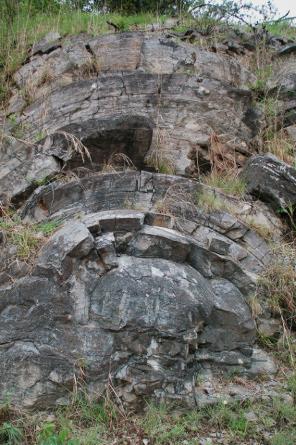
Elsewhere, in the Pilbara region (Warrawoona rock formation) in Western Australia, small sedimentary cushions, or domes, aged at 3.5 billion years old can be found. Early life environments in the Pilbara Craton also included shallow marine sedimentary environments and hydrothermal regions. Other such formations, dating back 2.5 billion years, can be found in the Transvaal in South Africa.
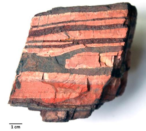
Figure 1.1. Stromatolites dating back 2.5 billion years, observed in the Transvaal in South Africa. Courtesy of Pierre Thomas (2016). For a color version of this figure, see www.iste.co.uk/chapouthier/life.zip
This variety of environments, combined with the variety of stromatolite forms found, would indicate a biosphere that was already diverse 3 to 4 billion years ago.
Biodiversity originates from the origins of life itself.
A major contribution highlights a point that is rarely addressed in the study of the metabolism of early stromatolites. Indeed, the hypothesis that cyanobacteria formed fossil stromatolites by mineralization and lithification of microbial mats assumes that oxygenic photosynthesis is a very old process, one that was active more than 3 billion years ago. However, the surface of the Earth was predominantly anoxic in the Archean period, containing less than 1% of today’s oxygen concentration. The increase in oxygen in the ocean, as a result of cyanobacterial photosynthesis, gradually turned it into an oxidizing environment, whereas it was initially reductive. This means that the oldest stromatolites are the product of phototrophic anoxygenic microorganisms, which do not produce oxygen. Supporting this hypothesis, researchers have recently observed that phototrophic sulphooxidizing alpha-proteobacteria are responsible for the precipitation of aragonite, the main calcium carbonate constituting the stromatolites of Lake Dziani Dzaha in Mayotte (Gérard et al. 2018).
In addition, other anaerobic methanotrophic microbial communities were present prior to the oxygenation of the Earth’s atmosphere. Using sulfate ions or organic sulfur to oxidize methane, these microorganisms that lived in sedimentary lake environments left traces in the Tumbiana formation, aged at 2.7 billion years old (Lepot et al. 2019).
The anoxygenic photosynthesis forming the first stromatolites was carried out by phototrophic anoxygenic microorganisms and would therefore have occurred before oxygen photosynthesis.
1.4. Geochemical elements confirming these recent results
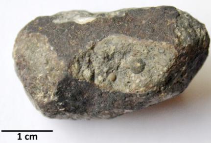
The vast majority of the world’s iron is known as Banded Iron Formation (BIF). Archean banded-iron deposits are marine sedimentary rocks that are very rich in iron and today account for 90% of the iron mined in the world.
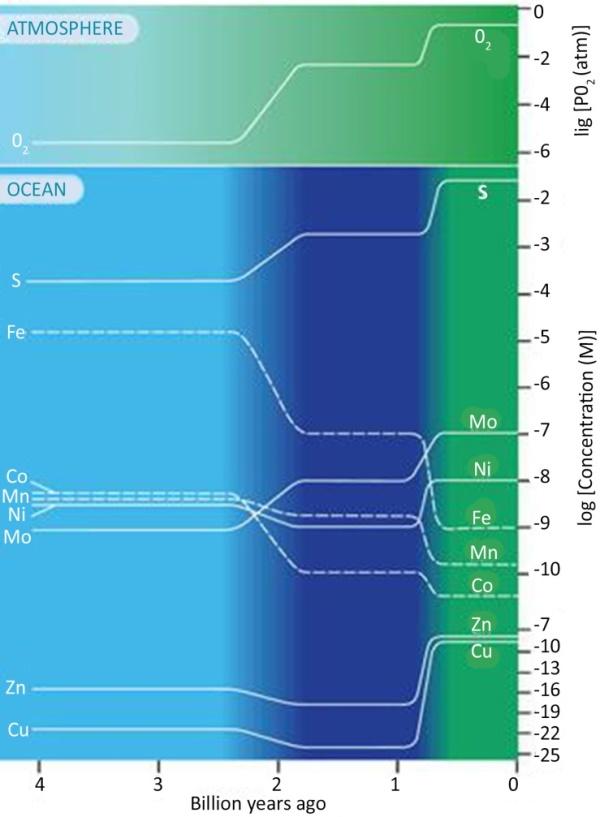
Figure 1.2. The figure shows changes in the abundance of elements over time, mainly sulfur (S) and iron (Fe). The color gradations indicate a transition from anoxic oceans, e.g. low in sulfur, before 2.4 billion years (light blue) to oceans rich in H2S between 1.8 billion and 800 million years (dark blue), and then to complete oxygenation of the oceans (green). Courtesy of Ariel Anbar (2008). For a color version of this figure, see www.iste.co.uk/chapouthier/life.zip
Figure 1.3. Banded Iron Formation (BIF). Courtesy of Pierre Thomas (2011). For a color version of this figure, see www.iste.co.uk/chapouthier/life.zip
The ocean and the Earth’s surface were without oxygen 4 to 2.5 billion years ago.
The weathering of minerals from iron-rich continents produced ferrous ions (Fe2+) that were soluble in water, and therefore particularly mobile, and which were able to spread into the oceans. Volcanic activity at hydrothermal springs may also have contributed to the presence of ferrous ions in solution.
Oxygenation of the oceans by the oxygenic photosynthesis of cyanobacteria, up to about 2.4 billion years ago, caused soluble ferrous iron (Fe2+) to disappear by oxidation into insoluble ferric iron (Fe3+), which precipitated as magnetite and hematite.
When most of the reduced forms of iron were oxidized in the Paleoproterozoic era, sedimentation of banded iron deposits became rare. As a result, the O2 content first increased in the oceans, then in the atmosphere, becoming toxic to anaerobic organisms. This was the Great Oxidation or “Oxygen Catastrophe”.
Given that sea iron precipitated in an insoluble form (Fe3+) in the Archean era, the sea water of that time contained iron in solution, in a soluble form (Fe2+). This proves that the sea of that time was reduced, as was the overlying atmosphere.
Another type of photosynthesis, which is rare and little known, could explain an abundant precipitation of iron oxide: photoferrotrophy, a process where iron provides electrons.
Photoferrotrophy is a photosynthesis (less energy efficient than conventional photosynthesis) that oxidizes the iron (Fe2+) of FeO into iron (Fe3+) of Fe2O3; it can be written in a very simplified way: 2 FeO + H2O + photons → Fe2O3 + 2 H+ + 2 e-
The H+ ions and e- electrons are then used by mechanisms, similar to those of classical photosynthesis, to synthesize carbohydrates from CO2; this metabolism requires the presence of iron (Fe2+) in the environment and leads to the massive precipitation of hematite Fe2O3
Life is determined by the environment and, as multiple environments coexist, the origins of life and biodiversity coincide and evolve together.
1.5. Compartmentalization of resources and primary biomass
In September 1969, a fireball exploded in the sky over Murchison, Australia, followed by a shower of meteorite fragments gathered in a few days. Extraterrestrial amino acids and hydrocarbons in the Murchison meteorite were quickly identified by David Deamer (1985), who showed that organic compounds in the meteorite could also assemble into membranes. Since then, many molecules of biological interest have been identified in other meteorites (Callahan et al. 2011).
Figure 1.4. Carbonaceous chondrite. Courtesy of Pierre Thomas (2016). For a color version of this figure, see www.iste.co.uk/chapouthier/life.zip
Deamer and Barchfeld (1982), Deamer and Pashley (1989), Deamer (1997), Dworkin et al. (2001), will be pioneers in the experimental study of organic compounds synthesized in space. Entering into the planetary atmosphere of the primitive Earth, they mixed with endogenous species, some of them amphiphilic, with polar and non-polar groups on the same molecule. They spontaneously self-assemble to form more complex bimolecular structures which, in turn, form membrane vesicles.
The self-assembly of amphiphilic compounds, lipids forming spherical vesicles, called “liposomes”, are capable of capturing macromolecules. The lipid bilayer is sufficiently permeable to allow exchanges with ionic and polar compounds from outside the compartment, allowing polymerization reactions, a kind of protometabolism, within these vesicles (Zepik et al. 2007). Hydrothermal sites are good candidates for the realization of such prebiotic evolution on the primitive Earth. Vesicles formed on mineral surfaces capture and produce diverse molecular systems. Each vesicle represents a protocell, a kind of chemical microreactor (Damer and Deamer 2015).
Laboratory simulations show that such vesicles easily encapsulate functional macromolecules, including nucleic acids and enzymes. RNA-type polymers are synthesized non-enzymatically in the laboratory from mononucleotides in lipid environments. RNA-type polymers identified by nanopore were analyzed by standard enzyme labeling methods, followed by gel electrophoresis. Chemical activation of the mononucleotides is not required. Instead, the synthesis of phosphodiester bonds is stimulated by the chemical potential of fluctuating anhydrous and hydrated conditions, with heat providing activation energy during the dehydration. In the final hydration step, the RNA-type polymer is encapsulated in lipid vesicles. This process provides the model for a possible first step in the evolution towards an RNA world (Rajamani et al. 2008).
Toppozini and her colleagues (2013) used X-ray diffraction to analyze mixtures of dehydrated self-assembled lipid multilamellar structures in the presence of a layer of mononucleotides such as adenosine monophosphate (AMP). The multilamellar structures are organizational structures, so condensation reactions of the mononucleotides into RNA-like polymers can thus occur (De Guzman et al. 2014; Misuraca et al. 2017).
Cell blanks were obtained by assembling phospholipids, amphiphilic molecules that interact through a hydrophilic head (which likes water) and a hydrophobic tail (which avoids any interaction with water). This structure allows organization in water in the form of vesicles. Researchers have shown that prebiotic compounds, such as polyprenyl phosphate, combine in water to form oriented double layers (Nakatani et al. 2014).
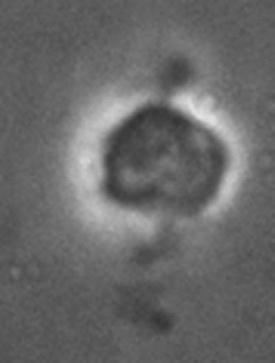
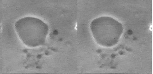
Jeff Errington (2013) followed the development of the Bacillus subtilis L-form (wall-free) bacterium and a liposome under the same conditions. Both bacteria and liposome split, fuse together and produce offspring in the form of small vesicles, which are morphologically very similar (see Figure 1.5). The unusual mode of proliferation of these bacteria by budding and fission could serve as a model for the in vitro simulation of vesicles or primitive cells. The binary fission mechanism provides insight into how early cell life forms were able to proliferate. Later in evolution, the invention of the protective cell wall may have been responsible for the expansion of bacterial life on Earth.
a)
b)
Figure 1.5. a) Proliferation of L-Bacillus subtilis. Courtesy of Jeff Errington. b) Liposome budding. Phospholipid vesicles in the presence of oleic acid. Courtesy of Primož Peterlin (2009)
Luisi and his collaborators were pioneers in the study of the processes by which lipid vesicles could develop and even reproduce (Walde et al. 1994a, 1994b; Luisi 2002, 2016). For example, it has been shown that when oleoyl anhydride was exposed to an aqueous buffer at alkaline pH, the anhydride was slowly hydrolyzed and the resulting oleic acid assembled into membrane vesicles. The vesicles can grow by the addition of fatty acids in the environment and then divide into smaller vesicles that grow, compete for resources, and even have a simple version of a feedback cycle that can regulate growth (Chen and Szostak 2004; Mansy and Szostak 2008; Adamala and Szostak 2013a, 2013b; Engelhart et al. 2016; Jimbo et al. 2016).
What can we deduce from these observations regarding our knowledge of the reproduction of the ancestors of today’s bacteria?
Briers et al. (2012) studied the gap between the first protocell and the hypothetical cell ancestor named “Luca”. Although there are striking coincidences between the two behaviors, the similarities observed still deserve long studies to confirm an ancestral morphological link and the understanding of the origins of unicellular life.
1.6. Rebuilding a living cell: a wide range of possibilities explored, from the mineral to the organic
Today, we speak of “chemical gardens” and “metallic vegetation” to refer to self-organized chemical figures that evoke plant diversity. When a metal salt is brought into contact with a basic aqueous solution of silicate, carbonate or phosphate, patterns called “flowers” are produced in the laboratory (Haudin et al. 2018). Technological innovations are suggested by these natural mineral productions which flourish today in the field of biomimicry. They find multiple biophysical, robotic, electronic, automatic, aeronautical, architectural, etc. applications, with the purpose of compensating for our weaknesses and repairing handicaps. Let us note here that mimicry is not the prerogative of modern technologies. A spider can be mistaken, even by the greatest arachnologist, for a fragment of a dead leaf.
The mechanobiology of biological membranes has become a growing field of research, particularly in the field of synthetic biology, towards artificial cells with genetic circuits and reaction cascades. Maximizing the modularity of their design and their flexibility is made possible by encapsulating them in liposomes, allowing chemical reactions to take place in well isolated environments. Such minimal
synthetic cells, called “synells”, were designed by MIT researchers3, who founded Synlife in 2017. They are governed by external signals and communication between liposomes, which can be fused in a controlled manner (Adamala et al. 2016).
Artificial life, in silico, a new paradigm between the machine and the living, serves as a model for the design of mineral “creatures”, organoids, automata (mechanical or bionic), micro-robots with metabolisms (Hamada et al. 2019), humanoids, etc., which would “emerge” from nanotechnology. The pairing of the brain and machine aims at the illusory perfection of the human species aspiring, as described by Aldous Huxley in 1932 in Brave New World, to impose its path to evolution.
1.7. Conclusion
Seeking to understand how life appeared on Earth also means looking for the fossil traces of the first cells, which inevitably leads to the question of how big the smallest living organism is (implied: the smallest being the simplest, therefore the first to appear on Earth). In connection with genomics work, Craig Venter (director of the Institute for Genomic Research – TIGR4 – in Rockville, Maryland) and Hamilton Smith (who won the Nobel Prize in 1978 for the co-discovery of restriction enzymes) have removed the genes of Mycoplasma genitalium one by one, reputed to have the smallest known genome. The aim is to obtain a viable organism with the minimum number of genes, Mycoplasma laboratorium, making it possible to develop, in fine, the smallest living organism possible: “The goal is to fundamentally understand the components of the most basic living cell” (Singer 2007). This approach raises one of the most delicate problems in contemporary biology: what consequences can the manufacture of such objects have on the future of humanity, the biosphere, the environment and the planet? Is there a risk of unbalancing ecosystems?
Producing organisms that are “outside the norm”, in the technical sense, employs us to think about the standardization of criteria for living things, the quality of life, and lastly, the normativity of living things (what does “being normal” mean?). In 1995, Jean-Pierre Séris declared that it was impossible to set standards of vital value: “Uniformity, the reproduction of that which is identical has a meaning and a technical value. It has no biological value. It is biological nonsense ...”
3 MIT: Massachusetts Institute of Technology.
4 TIGR: The Institute for Genomic Research.
1.8. Acknowledgements
Our thanks go to Ariel Anbar, Jeff Errington, Primož Peterlin, Pierre Thomas and Peter Walde, who kindly provided us with the figures and photographs (Figures 1.1 to 1.5), and to Jacques Vergne and Louis Ter-Ovanessian for their careful reading.
1.9.
References
Adamala, K., Szostak, J.W. (2013). Competition between model protocells driven by an encapsulated catalyst. Nature Chemistry, 5(6), 495–501.
Adamala, K.P., Martin-Alarcon, D.A., Guthrie-Honea, K.R., Boyden, E.S. (2016). Engineering genetic circuit interactions within and between synthetic minimal cells. Nature Chemistry, 9, 431–439.
Adriaenssens, E.M., Sullivan, M.B., Knezevic, P., van Zyl, L.J., Sarkar, B.L., Dutilh, B.E., Alfenas Zerbini, P., Lobocka, M., Tong, Y., Brister, J.R., Moreno Switt, A.I., Klumpp, J., Karam Aziz, R., Barylski, J., Uchiyama, J., Edwards, R.E., Kropinski, A.M., Petty, N.K., Clokie, M.R.J., Kushkina, A.I., Morozova, V.V., Duffy, S., Gillis, S., Rumnieks, J., Kurtböke, I., Chanishvili, N., Goodridge, L., Wittmann, J., Lavigne, R., Bin Jang, H., Prangishvili, D., Enault, F., Turner, D., Poranen; M.M., Oksanen, H.M., Krupovic. M. (2020). Taxonomy of prokaryotic viruses: 2018–2019 update from the ICTV Bacterial and Archaeal Viruses Subcommittee. Archives of Virology, 165, 1253–1260.
Allwood, A.C., Walter, M.R., Kamber, B.S., Marshall, C.P., Burch, I.W. (2006). Stromatolite reef from the early Archaean era of Australia. Nature, 441, 714–718.
Anbar, A.D. (2008). Elements and evolution. Science, 322, 1481–1483.
Atlan, H. (1972). L’Organisation biologique et la théorie de l’information. Hermann, Paris.
Awramik, S.M. (1994). The history and significance of stromatolites. In Early Organic Evolution: Implications for Mineral and Energy Resources, Schidlowski, M. et al. (eds). Springer, Berlin, 435–449.
Bernard, C. (1878). Leçons sur les phénomènes de la vie communs aux animaux et aux végétaux. Librairie Baillière et fils, Paris.
Briers, Y., Walde, P., Schuppler, M., Loessner, M.J. (2012). How did bacterial ancestors reproduce? Lessons from L-form cells and giant lipid vesicles. Bioessays, 34, 1078–1084.
Bungenberg de Jong, H.G. (1936). La coacervation. Les coacervats et leur importance en biologie. Hermann & cie, Paris.
Callahan, M.P., Smith, K.E., Cleaves II, H.J., Ruzicka, J., Stern, J.C., Glavin, D.P., House, C.H., Dworkin, J.P. (2011). Carbonaceous meteorites contain a wide range of extraterrestrial nucleobases. PNAS, USA, 108(34), 13995–13998.
Chen, I.A., Szostak, J.W. (2004). Membrane growth can generate a transmembrane pH gradient in fatty acid vesicles. PNAS, USA, 101(21), 7965–7970.
Damer, B., Deamer, D. (2015). Coupled phases and combinatorial selection in fluctuating hydrothermal pools: a scenario to guide experimental approaches to the origin of cellular life. Life (Basel), 5(1), 872–887.
Deamer, D.W. (1985). Boundary structures are formed by organic components of the Murchison carbonaceous chondrite. Nature, 317, 792–794.
Deamer, D.W. (1997). The first living systems: a bioenergetic perspective. Microbiol. Mol. Biol. Rev., 61, 230–261.
Deamer, D.W., Barchfeld, G.L. (1982). Encapsulation of macromolecules by lipid vesicles under simulated prebiotic conditions. J. Mol. Evol., 18, 203–206.
Deamer, D.W., Pashley, R.M. (1989). Amphiphilic components of carbonaceous meteorites. Orig. Life Evol. Biosph., 19, 21–33.
De Guzman, V., Vercoutere, W., Shenasa, H., Deamer, D. (2014). Generation of oligonucleotides under hydrothermal conditions by non-enzymatic polymerization. J. Mol. Evol., 78(5), 251–262.
Dworkin, J.P., Deamer, D.W., Sandford, S.A., Allamandola, L.J. (2001). Self-assembling amphiphilic molecules: synthesis in simulated interstellar/precometaryices. Proc. Natl. Acad. Sci. USA, 98, 815–819.
Engelhart, A.E., Adamala, K.P., Szostak, J.W. (2016). A simple physical mechanism enables homeostasis in primitive cells. Nat. Chem., 8(5), 448–53.
Errington, J. (2013). L-form bacteria, cell walls and the origins of life. Open Biol, 3, 120–143. Fox, S.W. (1988). The Emergence of Life. Basic Books Publishers, New York.
Gérard, E., De Goeyse, S., Hugoni, M., Agogué, H., Richard, L., Milesi, V., Guyot, F., Lecourt, L., Borensztajn, S., Joseph, M.B., Leclerc, T., Sarazin, G., Jézéquel, D., Leboulanger, C., Ader, M.K. (2018). Role of alphaproteobacteria and cyanobacteria in the formation of stromatolites of Lake Dziani Dzaha (Mayotte, Western Indian Ocean). Frontiers in Microbiology, 9.
Haldane, J.B.S. (1929). The origin of life. The Rationalist Annual, 3–10.
Hamada, S., Yancey, K.G., Pardo, Y., Gan, M., Vanatta, M., An, D., Hu, Y., Derrien, T.L., Ruiz, R., Liu, P., Sabin, J., Luo, D. (2019). Dynamic DNA material with emergent locomotion behavior powered by artificial metabolism. Science Robotics, 4(29).
Haudin, F., Brau, F., De Wit, A. (2018). La chimie génératrice de forme : végétation métallique et jardins chimiques [Online]. Revue Arts et Sciences V2 N1. Available at: https://www.openscience.fr/Numero.
Jimbo, T., Sakuma, Y., Urakami, N., Ziherl, P., Imai, M. (2016). Role of inverse-cone-shape lipids in temperature-controlled self-reproduction of binary vesicles. Biophysical Journal, 110, 1551–1562.
Leduc, S. (1912). La biologie synthétique. Poinat, Paris.
Lepot, K., Williford, K.H., Philippot, P., Thomazo, C., Ushikubo, T., Kitajima, K., Mostefaoui, S., Valley, J.W. (2019). Extreme 13C depletions and organic sulfur content argue for S-fueled anaerobic methane oxidation in 2.72 Ga old stromatolites. Geochimica et Cosmochimica Acta, 244, 522–547.
Locey, K.J., Lennon, J.T. (2016). Scaling laws predict global microbial diversity. PNAS, 113(21), 5970–5975.
Luisi, P.L. (2002). Toward the engineering of minimal living cells. The Anatomical Record, 268, 208–214.
Luisi, P.L. (2016). The Emergence of Life. Cambridge University Press, Cambridge.
Mansy, S.S., Szostak, J.W. (2008). Thermostability of model protocell membranes. PNAS, USA, 105(36), 13351–13355.
Misuraca, L., Natali, F., Da Silva, L., Peters, J., Demé, B., Ollivier, J., Seydel, T., LauxLesourd, V., Haertlein, M., Zaccai, G., Deamer, D., Maurel, M.C. (2017). Mobility of a mononucleotide within a lipid matrix: A neutron scattering study. Life (Basel), 7(1).
Mora, C., Tittensor, D.P., Adl, S., Simpson, A.G.B., Worm, B. (2011). How many species are there on Earth and in the ocean? PLoS Biol., 9(8).
Nakatani, Y., Ribeiro, N., Streiff, S., Gotoh, M., Pozzi, G., Désaubry, L., Milon, A. (2014). Search for the most ‘primitive’ membranes and their reinforcers: a review of the polyprenyl phosphates theory. Orig. Life Evol. Biosph., 44(3), 197–208.
Nutman, A.P., Bennett, V.C., Friend, C.R.L., Van Kranendonk, M.J., Chivas, A.R. (2016). Nature, 537, 535–538.
Oparin, A.I. (1938). The Origin of Life. MacMillan, New York.
Peterlin, P., Arrigler, V., Kogej, K., Svetina, S., Walde, P. (2009). Growth and shape transformations of giant phospholipid vesicles upon interaction with an aqueous oleic acid suspension. Chem. Phys. Lipids, 159(2), 67–76.
Prigogine, I. (1947). Étude thermodynamique des phénomènes irréversibles. Dunod, Paris.
Rajamani, S., Vlassov, A., Benner, S., Coombs, A., Olasagasti, F., Deamer, D. (2008). Lipidassisted synthesis of RNA-like polymers from mononucleotides. Orig. Life Evol. Biosph., 38, 57–74.
Schopf, J.W. (1993). Microfossils of the Early Archean Apex chert: New evidence of the antiquity of life. Science, 260(5108), 640–646.
Séris, J.-P. (1994). La technique. PUF, Paris.
Singer, E. (2007). Craig Venter’s genome. MIT Technology Review, September 4.
Thomas, P. (2011). Les fers rubanés (Banded Iron Formation) de l’Archéen de Barberton, groupe de Fig Tree (-3,26 à -3,22 Ga), Afrique du Sud [Online]. Available at: https://planet-terre.ens-lyon.fr/image-de-la-semaine/ Img364-2011-10-10.xml.