
Gregory Y. Lauwers
Visit to download the full and correct content document: https://ebookmass.com/product/gastrointestinal-pathology-correlative-endoscopic-and -histologic-assessment-1st-edition-gregory-y-lauwers/

More products digital (pdf, epub, mobi) instant download maybe you interests ...

Endoscopic diagnosis and treatment in urinary bladder
pathology : handbook of endourology 1st Edition Geavlete
https://ebookmass.com/product/endoscopic-diagnosis-and-treatmentin-urinary-bladder-pathology-handbook-of-endourology-1st-editiongeavlete/
Gastrointestinal and Liver Pathology: A Volume in the Series: Foundations in Diagnostic Pathology 3rd Edition
Amitabh Srivastava
https://ebookmass.com/product/gastrointestinal-and-liverpathology-a-volume-in-the-series-foundations-in-diagnosticpathology-3rd-edition-amitabh-srivastava/
Sleisenger and Fordtran's Gastrointestinal and Liver Disease Review and Assessment 10th Edition Emad Qayed
https://ebookmass.com/product/sleisenger-and-fordtransgastrointestinal-and-liver-disease-review-and-assessment-10thedition-emad-qayed/
Sleisenger and Fordtran's Gastrointestinal and Liver Disease Review and Assessment 11th Edition Emad Qayed
https://ebookmass.com/product/sleisenger-and-fordtransgastrointestinal-and-liver-disease-review-and-assessment-11thedition-emad-qayed/
Endoscopic Surgery of the Orbit 1st Edition Raj Sindwani
https://ebookmass.com/product/endoscopic-surgery-of-theorbit-1st-edition-raj-sindwani/
Invertebrate Pathology 1st Edition Andrew F. Rowley
https://ebookmass.com/product/invertebrate-pathology-1st-editionandrew-f-rowley/
Endoscopic Craniosynostosis Surgery: An Illustrated Guide to Endoscopic Techniques David F. Jimenez
https://ebookmass.com/product/endoscopic-craniosynostosissurgery-an-illustrated-guide-to-endoscopic-techniques-david-fjimenez/
Mesenteric Principles of Gastrointestinal Surgery: Basic and Applied Science Coffey, John Calvin, Sehgal, Rishabh, Walsh, Dara Mesenteric Principles Of Gastrointestinal Surgery: Basic And Applied Science
Coffey https://ebookmass.com/product/mesenteric-principles-ofgastrointestinal-surgery-basic-and-applied-science-coffey-johncalvin-sehgal-rishabh-walsh-dara-mesenteric-principles-ofgastrointestinal-surgery-basic-and-applied-scie/
Atlas of Endoscopic Ultrasonography, 2nd Edition Frank G. Gress
https://ebookmass.com/product/atlas-of-endoscopicultrasonography-2nd-edition-frank-g-gress/
Gastrointestinal Pathology
Gastrointestinal Pathology
Correlative Endoscopic and Histologic Assessment
Edited by
Gregory Y. Lauwers, MD, FAGPS
Senior Member and Director, Gastrointestinal Pathology Service Department of Pathology, Moffitt Cancer Center Professor, Departments of Pathology and Cell Biology and Oncologic Sciences University of South Florida Tampa, FL, USA
Michael B. Wallace, MD, MPH
Consultant – Gastroenterology and Hepatology
Fred C. Andersen Professor of Medicine, Mayo Clinic School of Medicine Jacksonville, FL, USA
Associate Editor:
Till S. Clauditz, MD
Associate Professor, Head of Gastrointestinal Pathology Service Department of Pathology with Section Molecular Pathology and Cytology University Medical Center Hamburg-Eppendorf, Hamburg, Germany
This edition first published 2021 © 2021 John Wiley & Sons Ltd
All rights reserved. No part of this publication may be reproduced, stored in a retrieval system, or transmitted, in any form or by any means, electronic, mechanical, photocopying, recording or otherwise, except as permitted by law. Advice on how to obtain permission to reuse material from this title is available at http://www.wiley.com/go/permissions.
The right of Gregory Y. Lauwers and Michael B Wallace to be identified as the authors of the editorial material in this work has been asserted in accordance with law.
Registered Offices
John Wiley & Sons, Inc., 111 River Street, Hoboken, NJ 07030, USA
John Wiley & Sons Ltd, The Atrium, Southern Gate, Chichester, West Sussex, PO19 8SQ, UK
Editorial Office
9600 Garsington Road, Oxford, OX4 2DQ, UK
For details of our global editorial offices, customer services, and more information about Wiley products visit us at www.wiley.com.
Wiley also publishes its books in a variety of electronic formats and by print-on-demand. Some content that appears in standard print versions of this book may not be available in other formats.
Limit of Liability/Disclaimer of Warranty
The contents of this work are intended to further general scientific research, understanding, and discussion only and are not intended and should not be relied upon as recommending or promoting scientific method, diagnosis, or treatment by physicians for any particular patient. In view of ongoing research, equipment modifications, changes in governmental regulations, and the constant flow of information relating to the use of medicines, equipment, and devices, the reader is urged to review and evaluate the information provided in the package insert or instructions for each medicine, equipment, or device for, among other things, any changes in the instructions or indication of usage and for added warnings and precautions. While the publisher and authors have used their best efforts in preparing this work, they make no representations or warranties with respect to the accuracy or completeness of the contents of this work and specifically disclaim all warranties, including without limitation any implied warranties of merchantability or fitness for a particular purpose. No warranty may be created or extended by sales representatives, written sales materials or promotional statements for this work. The fact that an organization, website, or product is referred to in this work as a citation and/or potential source of further information does not mean that the publisher and authors endorse the information or services the organization, website, or product may provide or recommendations it may make. This work is sold with the understanding that the publisher is not engaged in rendering professional services. The advice and strategies contained herein may not be suitable for your situation. You should consult with a specialist where appropriate. Further, readers should be aware that websites listed in this work may have changed or disappeared between when this work was written and when it is read. Neither the publisher nor authors shall be liable for any loss of profit or any other commercial damages, including but not limited to special, incidental, consequential, or other damages.
Library of Congress Cataloging-in-Publication Data applied for
HB ISBN: 9780470658369
Cover Design: Wiley
Cover Images: © Gregory Y. Lauwers and Michael B. Wallace
Set in 9.5/12.5pt STIXTwoText by SPi Global, Pondicherry, India
10 9 8 7 6 5 4 3 2 1
Contents
List of Contributors vii
1 General Principles of Biopsy Diagnosis of GI Disorders 1
Herbert C. Wolfen, Michael B. Wallace, Naohisa Yahaghi and Yutaka Saito
2 Esophagus Inflammatory Conditions 11
Anthony R. Mattia, Gregory Y. Lauwers, Michael B. Wallace and Till S. Clauditz
3 Epithelial Metaplastic, Polypoid, and Neoplastic Conditions of the Esophagus 35
Till S. Clauditz, Gregory Y. Lauwers, Michael B. Wallace and Anthony R. Mattia
4 Inflammatory Disorders of the Stomach 73
Till S. Clauditz , Michael B. Wallace and Gregory Y. Lauwers
5 Polyps of the Stomach 99
K. Kim, Till S. Clauditz, Jun Haeng Lee and Gregory Y. Lauwers
6 Gastric Neoplastic Conditions: Precursor Lesions and Early Gastric Cancer 125
Till S. Clauditz and Gregory Y. Lauwers
Gastric Neoplastic Conditions: Lymphoid Lesions of the Stomach 142
Mounir Trimeche and Laurence de Leval
7 Inflammatory and Miscellaneous Conditions of the Small Intestine 161
Ian Brown and Michael B. Wallace
8 Polyps of the Small Intestine 195
Ian Brown and Michael B. Wallace
9 Epithelial and Nonepithelial Neoplasms of the Small Intestine 209
Ian Brown, Michael B. Wallace and Till S. Clauditz
10 Inflammatory Conditions of the Colon 235
Tze Sheng Khor, Till S. Claudtiz, Bence Kővári, Gregory Y. Lauwers, Michael B. Wallace and Priyanthi Kumarasinghe
11 Polyps of the Large Intestine 307
Christophe Rosty, Michael B. Wallace and Till S. Clauditz
12 Epithelial Neoplasms of the Large Bowel 321
Christophe Rosty, Michael B. Wallace and Till S. Clauditz
13 Inflammatory Conditions of the Anus 337
Thomas Arnason and Michael B. Wallace
14 Polyps and Neoplastic Lesions of the Anus 349
Thomas Arnason, Michael B. Wallace and Till S. Clauditz
Index 369
List of Contributors
Thomas Arnason
Queen Elizabeth II Health Sciences Centre and Dalhousie University
Halifax
Nova Scotia
Canada
Ian Brown
Envoi Specialist Pathologists
Brisbane Australia
Till S. Clauditz
Department of Pathology
University-Medical-Center
Hamburg Germany
Tze Sheng Khor
PathWest Laboratory Medicine
Queen Elizabeth II Medical Centre
Nedlands
Western Australia
Australia
K. Kim
Samsung Medical Center
Sungkyunkwan University School of Medicine
Seoul South Korea
Bence Kővári
Department of Pathology
University of Szeged
Szeged
Hungary
Priyanthi Kumarasinghe
Department of Anatomical Pathology
PathWest, QE II Medical Centre
Univ. of Western Australia
Perth
Australia
Gregory Y. Lauwers
Moffitt Cancer Center
Tampa Florida
USA
Jun Haeng Lee
Samsung Medical Center
Sungkyunkwan University School of Medicine
Seoul
South Korea
Laurence de Leval
Institute of Pathology University of Lausanne Lausanne
Switzerland
Anthony R. Mattia
Newton-Wellesley Hospital Newton, MA
USA
Christophe Rosty Envoi Specialist Pathologists
Brisbane
Queensland
Australia
Yutaka Saito
National Cancer Center
Tokyo Japan
Mounir Trimeche
Institute of Pathology University of Lausanne Lausanne
Switzerland
Michael B. Wallace
Mayo Clinic
Jacksonville
Florida
USA
Herbert C. Wolfen Mayo Clinic
Jacksonville Florida
USA
Yahaghi Keio University Cancer Center
Tokyo Japan
Naohisa
General Principles of Biopsy Diagnosis of GI Disorders
Herbert C. Wolfen1, Michael B. Wallace1, Naohisa Yahaghi2 and Yutaka Saito3
1 Mayo Clinic, Jacksonville, Florida, USA
2 Keio University Cancer Center, Tokyo, Japan
3 National Cancer Center, Tokyo Japan
Tissue sampling of the gastrointestinal tract at the time of endoscopy is the cornerstone of many gastrointestinal diagnoses. The development of a flexible endoscope and the subsequent ability to directly acquire tissue under optical guidance has been one of the most important advancements in the field of gastroenterology throughout its history. Although tissue sampling can be performed through nonendoscopic devices, the ability to directly correlate precise locations and target biopsies to specific areas of disease is critical to our ability to diagnose and further understand gastrointestinal pathology. Many of the advancements in our understanding of the basic pathology and molecular biology of gastrointestinal disease can be directly attributed to our ability to acquire tissue for histological, molecular, and genetic analyses. An excellent example is our deep understanding of the molecular pathology of colorectal cancer development from normal colonic epithelium to adenoma to colorectal cancer, a discovery made possible because of colonoscopic access to precursor lesions such as adenomatous polyps and early cancers.
In this chapter, we will review general principles of tissue acquisition at the time of endoscopy including the following topics:
● Endoscopic equipment for obtaining tissue including endoscopic accessory channels, biopsy forceps, snare devices, needle aspiration and cytology brush.
● General principles of optimal sampling technique.
● Methods of tissue preparation in the endoscopy laboratory to optimize diagnostic accuracy.
● The role of endoscopic ultrasound (EUS)-guided fineneedle aspiration cytology.
Endoscopic Equipment for Tissue Sampling
Modern endoscopic equipment can be divided in two general categories: the endoscope that allows access to the gastrointestinal tract and accessory devices that are typically passed through the working channel of the endoscope to directly acquire tissue, including biopsy forceps, snares, fine-needle aspiration devices, and cytology brushes. Recent developments in tissue sampling include devices that are capable of wide-field, often definitive, endoscopic resection of early neoplasia and invasive carcinoma.
A modern endoscope is a remarkably robust and versatile instrument including a light source, optical lenses with a video capture device, image processing, and display equipment, and importantly for the purposes of tissue acquisition, an accessory channel ranging from 1 to 6 mm (typically 3–4 mm), which allows passage of devices for mechanical collection of tissue (Figures 1.1 and 1.2).
There is a general trade-off between the diameter of the instrument and the ease and comfort with which it can be passed through the natural orifices of the body such as the mouth and anus. In general the larger the outer diameter, the larger the accessory channel is to accommodate larger instruments for tissue acquisition. A fundamental limitation of most flexible endoscopes, as opposed to surgical instruments, is that all accessories pass through a single access point of the endoscope. As compared to surgical instruments with multiple access points, the endoscopic devices do not typically allow triangulation to acquire a large bulk tissue or resect entire organs. For this reason, most tissue is sampled through pinch forceps, needle
aspiration, or wire loop snare devices. More recently, electrosurgical needles and other cutting tools have been developed, which have allowed wide-field resection of tissues of virtually any diameter (Figure 1.3).
Pinch Biopsy Forceps
The flexible pinch biopsy forceps have been one of the most versatile of all instruments for tissue acquisition. These typically involve a flexible steel cable and lever device with two sharp-edged cups, which can be opened and closed to acquire tissue (Figure 1.4).
Standard endoscopic sampling typically acquires tissue from the mucosa and occasionally a submucosal depth of the intestinal wall; however, large-capacity forceps as well as multiple sampling including “bite on bite” allow sampling of the deeper layers. Pinch biopsy forceps come in multiple sizes from very small instruments such as a pediatric forceps, which can be passed through very small working channels. Recent development of very tiny forceps makes it possible to pass them through special endoscopes into the bile or pancreas duct and to pinch biopsy outside of the traditional gastrointestinal lumen (Figure 1.5).
Studies comparing jumbo forceps to standard forceps have generally not shown significant advantages of larger capacity forceps. A limitation of most forceps is the inability to sample tissue in the submucosa routinely. This is highlighted in studies looking for Barrett’s esophagus after the surface epithelium has been ablated. Biopsy forceps can remove tissue with mechanical closure alone or with electrocautery (“hot biopsy”) although the use of hot biopsy has diminished significantly due to increased risks of complication and tissue damage in the biopsy specimen.
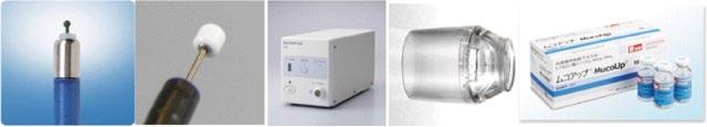
Figure 1.3 (a) Tools for performing endoscopic resection including endoscopic submucosal dissection (ESD). Source: Zeon Medical. (b) Standard and insulated tip electrocautery knives for incision and dissection. Source: © 2017 Korean Society of Gastrointestinal Endoscopy. (c) CO2 insufflator for luminal distension, which is preferred to air given rapid reabsorption. Source: Olympus. (d) Distal attachment hood to facilitate maintaining view within the submucosal space. Source: Fujifilm medical. (e) Injection fluid (hyaluronic acid; Mucoup [Johnson and Johnson]) for submucosal lifting. Source: Gut and Liver.
Jet B knife Zeon medical (a)
Figure 1.1 Endoscope with control handle and tip. The tip contains a light source, imaging window, and accessory channel through which various tissue acquisition devices can be passed.
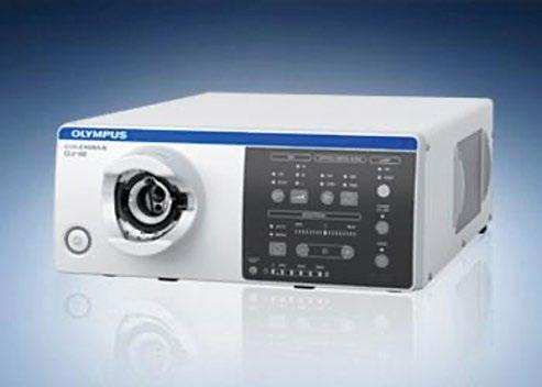
Figure 1.2 Endoscopic processor, which converts the light captured from the endoscope tip into a visible image for display. Source: Olympus America, Inc. With permission.
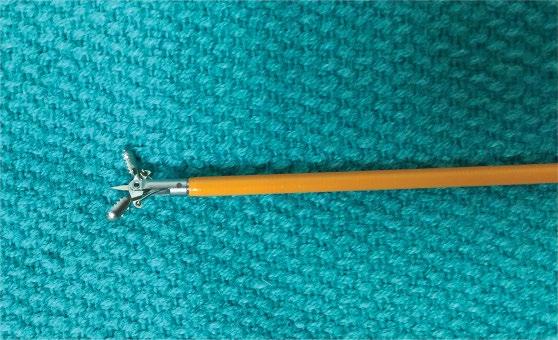
Figure 1.4 Endoscopic biopsy forceps in the open position. The needle-like pin in the center holds the tissue in place so one to two samples can be obtained per pass.
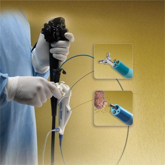
Figure 1.5 Micro biopsy forceps <1 mm in diameter, which can be passed through specialized endoscopes into the bile duct, pancreas duct, or via 19-gauge needles for extraluminal tissue sampling. Source: Boston Scientific Corporation with permission.
Endoscopic Snare Devices
Endoscopic snare devices have also been widely used for resection of polypoid as well as flat lesions throughout the gastrointestinal tract. They have been remarkably versatile and effective over the past four decades. Endoscopic snares typically involve a metallic wire, which may braided or monofilament (Figure 1.6).
A wire loop is generally constrained within a small caliber plastic catheter. At the distal end of the catheter, the wire loop can be opened to various sizes to grasp and resect polyps of different sizes. Typical sizes include loops 5–30 mm in diameter. There are numerous different shapes including

Figure 1.6 Endoscopic snare for polypectomy. The wire loop is extended in the open position outside the plastic sheath. When closed, the wire loop is strangulated and resects the polyp tissue.
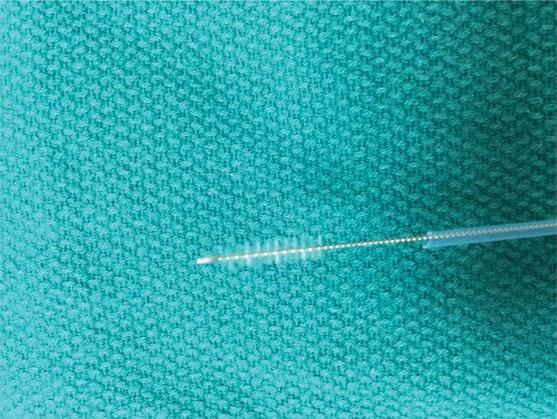
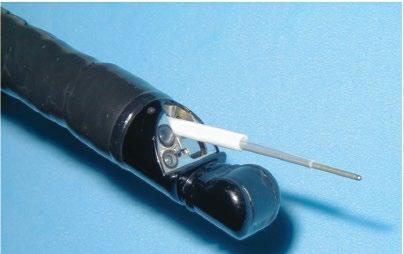
Figure 1.7 Endoscopic cytology brush. Note the abrasive brush extended beyond the protective plastic sheath.
oval, hexagonal, and asymmetric “duck bill.” Snares also come in various degrees of stiffness, which allow resection of lesions of many shapes and sizes. Tissue can be resected with mechanical closure alone (so-called “cold snare”) or with mechanical plus electrosurgical cutting (“hot snare”). Recent studies suggest that cold snare is associated with lower risk of bleeding and bowel wall injury.
Endoscopic Brush Cytology
Abrasive brush cytology has been used in many different fields of tissue sampling. Typical endoscopic brush is constrained within a plastic catheter similar to endoscopic snares (Figure 1.7).
After passing through the accessory channel of the endoscope, the abrasive brush is exposed and rubbed against the area of tissue sampling. This is most commonly applied to obtain specimens for microbiology, particularly fungal specimens. These have also been widely applied in the biliary tract for sampling of suspicious biliary strictures. More recent advances include a highly abrasive wide-area tissue sampling system that has been recently studied and Barrett’s esophagus as an alternative to pinch forceps, as well as nonendoscopic abrasive sponge sampling, which has the potential to offer inexpensive population-based screening for esophageal neoplasia (Figure 1.8).
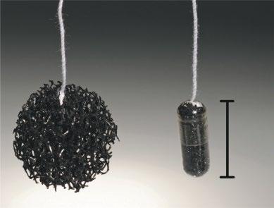
Figure 1.8 Nonendoscopic abrasive cytology brush. Source: From Kadri, S.R., Lao-Sirieix, P., O’Donovan, M. et al. (2010). Acceptability and accuracy of a non-endoscopic screening test for Barrett’s oesophagus in primary care: cohort study. BMJ 341: c4372. doi: https://doi.org/10.1136/bmj.c4372. Open access article.
Endoscopic Fine-Needle Aspiration (FNA) Devices
The most common application of FNA devices in endoscopy is endoscopic ultrasound-guided tissue sampling (EUS-FNA). The development of endoscopic ultrasound in the 1970s and 1980s significantly expanded the reach of endoscopic tissue sampling into the pancreas and other extraluminal organs such as lymph nodes and now virtually any organ within reach of the proximal or distal GI tract lumen (Figure 1.9).
Other needle aspiration devices include biliary sampling needles often used in conjunction with forceps and brush sampling, or so-called “triple sampling.” EUS-FNA devices come in various sizes from 19- to 25-gauge. These devices are typically attached to a handle, which allows the endoscopist to puncture and make to-and-fro movements within the target lesion and also apply negative pressure. With various methods, both cytologic and histologic material can be obtained through these needle devices. Recent efforts to develop Tru-Cut devices have been met with variable success.
Tissue Processing in the Endoscopy Laboratory
An excellent recent review on the topic of tissue acquisition has been published by the American Society for gastrointestinal endoscopy. For most routine tissue sampling the biopsy material is placed in formalin and subsequently embedded in paraffin for histological analysis. Other specific preparations include sampling for microbiological evaluation, molecular or genetic testing, and electromicroscopy. These
(a)
(b)
Figure 1.9 (a) and (b) Endoscopic ultrasound endoscope and guided fine-needle aspiration (EUS-FNA) device.
2 cm
special samples should be handled according to local institutional guidelines. In general, samples that are obtained for culture should be obtained prior to placing forceps into formalin as formalin residue can destroy or inactivate live microbial tissue.
Non-oriented Samples
Most pinch biopsy samples are placed directly in formalin without orientation. This is also true for small polyps where the likelihood of invasive cancer is very low. The sample is placed directly into a small formalin jar. The needle or forceps should be rinsed if it comes in direct contact with formalin as subsequent contact with living tissue can cause chemical injury.
Oriented Samples
Increasingly, endoscopic resection is being used for definitive oncologic removal of precancerous and invasive lesions. In these cases it is advantageous to orient the specimen so that precise staging as well as margin assessment can be performed. A typical situation is in endoscopic resection of early neoplasia in the esophagus such as Barrett’s esophagus. In this case a sample is frequently obtained through endoscopic mucosal resection. Large pieces of tissue, typically 1–2 cm in diameter, can be obtained to a depth of the submucosa. In this case the sample should be removed from the patient in a way that does not damage or distort the tissue. This is typically performed by suctioning the tissue into a distal attachment cap/hood and then removing the endoscope. Alternative retrieval devices include a modified snare with a protective net. These tissues should then be immediately oriented at the bedside typically by placing them on a paraffin or similar firm block. The tissue should be flattened typically with the mucosal side up and the edges carefully pinned so that the tissue remains flat (Figure 1.10).
Cytology Samples
Cytology samples obtained from either brush or fine-needle aspiration can be processed in a variety of ways. Most commonly these are prepared as thin smear on glass slides, which can be evaluated either immediately as an air-dried sample stained with a modified Romanowsky stain, or as an alcohol-fixed slide evaluated with Papanicolaou staining. It is often helpful to place additional excess material in a cytological preservative or formalin. Special handling is required for samples where lymphoproliferative disease is considered. Typically these include cytological preservatives such

Figure 1.10 Pinned and oriented resection tissue from Barrett’s esophagus-associated neoplasia. Note the mucosal side facing up and the lateral margins pinned to prevent curling of the edges.
as Roswell Park Memorial Institute (RPMI) medium that allow subsequent flow cytometry. As a practical matter, placement in such a preservative should be considered even when likelihood is low since cells preserved in such solution can always be examined with routine cytological methods; however, cells that are placed in formalin or alcohol cannot be evaluated for flow cytometry. The use of rapid on-site evaluation (ROSE) cytology has been shown in many studies to increase diagnostic yield and reduce the need for repeated procedures.
Culture Samples
Samples obtained for culture should be placed in a sterile specimen container. Sterile saline maybe needed to prevent drying of the specimen. Attention should be paid to minimize contamination although it is recognized that the endoscope and the organs throughout which it is passed are not sterile and is not possible to obtain a purely sterile access into the gastrointestinal lumen. Contamination with oropharyngeal organisms or colonic organisms is not uncommon.
Specific Organ Sampling Methods
Esophagus
Diagnostic Sampling
Indications for diagnostic sampling of the esophagus include Barrett’s esophagus and other suspected neoplastic disorders, inflammatory disorder such as eosinophilic esophagitis, gastroesophageal reflux disease, and infectious esophagitis.
The most common indication in Western countries for tissue sampling from the esophagus is likely Barrett’s esophagus. There are established guidelines for sampling of the esophagus. These have traditionally been based on the notion that early neoplasia is not visible endoscopically and thus random sampling of the mucosa should be performed to ensure adequate detection of early neoplasia. The most widely used standard is the so-called Seattle protocol. The American Society for Gastrointestinal Endoscopy (ASGE) guideline for tissue sampling calls for surveillance of nondysplastic Barrett’s esophagus in four quadrants every 2 cm with a large-capacity forceps for the entire Barrett’s mucosa. Patients with established low-grade dysplasia sampling should be more intensive with four-quadrant biopsies every 1–2 cm. For patients who choose to undergo surveillance for high-grade dysplasia, sampling every 1 cm should be performed; however, more recent evidence suggests that patients with established low-grade and high-grade dysplasia should likely undergo therapy to eradicate the Barrett’s esophagus. Advances in endoscopic imaging, such as highdefinition narrow-band imaging, confocal laser endomicroscopy, and chromoendoscopy, have significantly increased the yield of biopsy and allow much more targeted sampling although these have not yet replaced the need for random biopsy. These advances are likely to reduce the need for random biopsies and focus more on targeted sampling.
For eosinophilic esophagitis the ASGE recommends two to four biopsies from the proximal esophagus and two to four biopsies from the distal esophagus. Biopsy should also be obtained from the gastric antrum and duodenum when diffuse eosinophilic gastroenteritis is suspected.
For suspected infectious esophagitis, multiple biopsies from the margin and base of a visualized ulcer should be obtained and the sample should be sent for standard histology as well as immunohistochemical and possibly viral cultures and PCR. For candidal esophagitis, multiple biopsies of the affected area as well as cytology brushings may be complementary to biopsy.
Therapeutic Sampling
Increasingly, endoscopic resection methods are being applied to early neoplasia the esophagus, particularly Barrett’s esophagus and early squamous cell carcinoma. These are largely confined to tumors suspected to be T1 or nodular high-grade dysplasia. Endoscopic mucosal resection (EMR) methods generally involve an EMR device such as a modified band ligator followed by snare resection of the pseudopolyp (Figure 1.11–1.13).
Other methods include endoscopic submucosal dissection (ESD) in which the lateral margins of the suspected area are incised with electrocautery knife followed by dissection along the submucosal plane to obtain an en bloc specimen. EMR devices can typically obtain samples 1–2 cm in diameter into

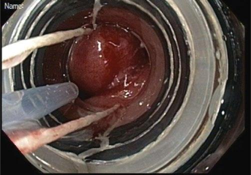
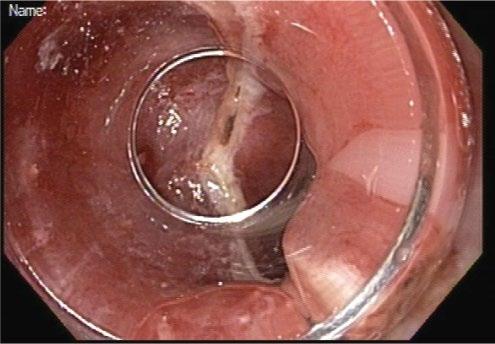
Figure 1.12
Multiband mucosectomy device. The black rubber bands are mounted on the outside of a plastic cap. The tissue is suctioned into the cap and a band deployed to create a pseudopolyp, which is then removed by snare.
Figure 1.13 Area of resection at 6–12 o’clock includes the entire region of suspected neoplasia at 9 o’clock. The remaining tissue at the 9 o’clock area represents intact deep submucosa and muscularis propria.
Figure 1.11 Barrett’s esophagus with flat neoplasia (9 o’clock) for targeted endoscopic resection.
the depth of the mid-submucosa. Endoscopic submucosal dissection can obtain samples of any lateral diameter and generally to the base of the submucosa.
One major advantage of these techniques is the ability to perform wide-field or en bloc resection and orient the sample. Samples should be retrieved without causing trauma to the tissue, preferably by removal through the endoscopic cap or through a snare-net as opposed to suctioning via the accessory channel, which can traumatize or fragment the tissue. Once retrieved the tissue should be handled as with other endoscopic resection specimens by orienting the specimen and pinning it on a fixed material such as a paraffin block. En bloc specimens can be oriented in terms of the oral and anal side of the lesion and assessed for lateral and deep margins. For resections that are performed piecemeal such as with multiband mucosectomy, the lateral margins cannot be accurately assessed and so complete resection relies on the endoscopic inspection intraluminally. The specimens should still be oriented and assessed for the deep margin.
Stomach
Diagnostic Sampling
Major indications for diagnostic sampling of the stomach include assessment for Helicobacter pylori infection, diagnosis of gastritis, metaplastic atrophic change, gastric polyps, and suspected neoplasia, particularly in the setting of gastric ulceration or early gastric cancer.
H. pylori Sampling Biopsy is one of several recommended methods for H. pylori sampling that also includes urease breath testing and stool testing for H. pylori antigen. When using endoscopic tissue sampling, there are two general methods including non-histological testing of the tissue for the presence of urease using the traditional Campylobacterlike organism test (CLO-test). In this case, the tissue should be placed in the standard agar well and visually inspected for a pH change following the instructions for use in this product. Histological sampling can also be performed with one of two methods. One method is to take three biopsies including one from the angularis corpus antrum junction, one from the greater curvature of the corpus, and one from the greater curvature of the antrum. Alternatively, the updated Sydney protocol may be followed, which includes five biopsies including one from the antrum lesser curve, antrum greater curve, gastric corpus lesser curve, and greater curve, and one from the angularis of the stomach.
Environmental Metaplastic Atrophic Gastritis (EMAG) Current guidelines recommend 7–12 biopsies including 4-quadrant biopsies including an antrum, 2 from the angularis, 4 from the corpus, and 2 from the cardia.
Gastric Polyps Gastric polyps are very frequently encountered, particularly in patients who are on chronic proton pump inhibitor therapy. Current ASGE guidelines for management of gastric polyps suggest that polyp should be sampled by biopsy. Fundic gland polyps larger than 1 cm should be removed by polypectomy. Hyperplastic polyps larger than 5 mm should be removed by polypectomy, and all adenomatous polyps should be removed by polypectomy. In patients who have numerous polyps, particularly where endoscopic inspection is highly consistent with fundic gland polyps, the largest of the polyps should be removed by polypectomy and representative sampling performed of smaller polyps.
Ulcer Disease Because of the potential for neoplasia in the setting of ulcers, numerous biopsies should be obtained from the base as well as the margins of the ulcer to exclude malignancy. Cytology may also be helpful. Sampling for concurrent H. pylori infection should be performed as suggested above.
Therapeutic Sampling
Endoscopic resection in early gastric cancer is now widely performed throughout the world. The endoscopic resection methods include endoscopic mucosal resection typically with a cap-assisted device as in the esophagus, or injection of a submucosal agent such as saline followed by snare resection of the lifted tissue. Lesions larger than 1–2 cm should generally be removed en bloc by endoscopic submucosal dissection when early gastric cancer is suspected. Tissue should be handled in the same manner as discussed above in esophagus.
Small Intestine
Major indications for tissue sampling of the small intestine include celiac disease as well as resection of early neoplasia.
Diagnostic Sampling
Currently established protocols for sampling for celiac disease recommend four to six biopsies from the duodenum including the duodenal bulb and distal duodenum. All other suspicious areas should be sampled using routine biopsy technique. Sampling of the papilla should be performed with caution as biopsy in this area can cause pancreatitis. When a suspected neoplastic lesion is seen in the region of the papilla, it is preferred to take a biopsy that does not immediately injure the orifice of the bile duct or the pancreas duct.
Therapeutic Sampling
Adenomatous polyps of the duodenum are handled in a similar manner to adenomatous lesions anywhere in the gastrointestinal tract. Endoscopic mucosa resection can be
performed with injection followed by snare. There appears to be higher risks of bleeding and perforation associated with endoscopic resection of duodenal lesions. This is particularly true were using cap-assisted devices where the thin wall of the duodenum is suctioned into the cap leading to inadvertent full-thickness resection.
Special attention is required for resection of lesions involving the ampulla of Vater. In these cases, typically the lesion is resected followed by placement of a stent in the pancreatic and bile duct orifice to prevent stricturing of these and acute pancreatitis.
Another special situation is polypoid lesions in the distal small intestine, which can now be accessed with deep enteroscopy methods. Polyps are generally removed in the same manner as polypoid lesions elsewhere in the gastrointestinal tract by snare polypectomy. Special circumstances include numerous polyps such as those developed in patients with Peutz–Jeghers syndrome. These can be particularly large and numerous. It may not be possible to resect and retrieve all tissues. Because of the laborious process of deep enteroscopy, it is often not feasible to extract the endoscope with each large polypoid tissue. Thus diagnostic sampling can be performed by biopsy of the lesion or fragmentation of the lesion with a snare followed by complete therapeutic excision of the lesion.
Colon
Major indications for diagnostic sampling of the colon include detection of both overt and microscopic colitis and surveillance for dysplasia in inflammatory bowel disease.
Perhaps the most commonly performed tissue sampling in the field of gastroenterology involves removal of colorectal polyps and tissue sampling of more advanced colorectal neoplasia.
Diagnostic Sampling
In patients with chronic diarrhea and suspected microscopic colitis, random biopsies should be taken throughout the colon including at least two biopsies from the right, transverse, descending, and sigmoid colon. For limited sampling the flexible sigmoidoscopy is also a reasonable approach. In this case at least two biopsies should be obtained from the sigmoid and descending colon as well as transverse colon if this can be reached with the sigmoidoscope.
In the setting of inflammatory bowel disease, biopsy sampling should be performed to establish the diagnosis and to assess the extent. For the initial diagnosis, the American Society for gastrointestinal endoscopy recommends two biopsies from each of five sites including the ileum and rectum. Surveillance of inflammatory bowel disease is a special circumstance and is evolving. Traditionally, random biopsies
were obtained throughout the colon. Current guidelines recommend biopsy of each colonic segment with four-quadrant biopsies every 10 cm from the cecum to the rectum for a minimum total of 33 biopsy samples. In cases where the entire colon is not affected, four samples should be obtained every 10 cm of the affected areas. More recently, it has been shown that using dye spray such as indigo carmine or methylene blue can identify areas of dysplasia with high accuracy and thus guide directed sampling without the need for random biopsy. Biopsies should however also be assessed from each colonic segment to assess for inflammation.
Therapeutic Sampling
Endoscopic resection methods of flat and lateral spreading colorectal polyps has expanded rapidly over last 10 years. Most noninvasive neoplastic lesions of the colon can now be removed through advanced endoscopic methods such as EMR or ESD avoiding the need for surgery. Lesions less than 2 cm can typically be removed en bloc with endoscopic mucosal resection involving injection of a fluid cushion under the lesion followed by snare resection. Where there is suspected early (mucosal or superficial submucosal) invasive carcinoma, en bloc resection should be performed using either EMR or ESD depending on the size of the lesion. The tissue processing should be the same as for other neoplastic lesions with orientation of the specimen and pinning of the specimen to assess the lateral and deep margins (Figure 1.14).
The final histological analysis allows for detailed staging, as well as matching the corresponding histological and endoscopic findings; note the lavender line overlays indicating sites of advanced neoplasia. The close collaboration between pathologists and endoscopists further improves the accuracy of each procedure (Figure 1.15).
Summary
The role of pathology in gastrointestinal endoscopy remains critical. Gastroenterologists should have a thorough knowledge of optimal methods of tissue removal and initial processing to allow optimal diagnosis and therapeutic results. Advances in endoscopic resection have allowed complete resection of many early cancers but require closer cooperation between the endoscopist and pathologist to ensure the tissue is properly handled and staged. With EUS-FNA, the direct interaction of pathologist with rapid on-site cytological evaluation has led to higher accuracy, and reduced the need for repeat procedures. Finally, advances in imaging will continue to improve the targeting of tissue sampling and reduce the need for low-yield random sampling methods.
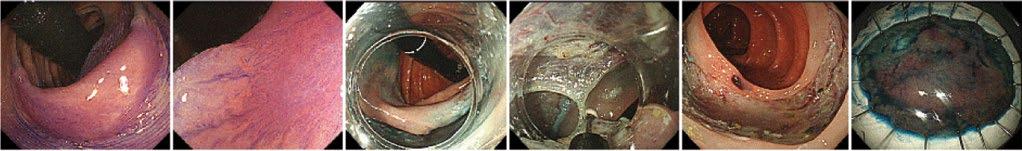
Figure 1.14 Endoscopic submucosal dissection (ESD) procedure. A flat neoplastic lesion is seen in panel 1–2 after staining with cresyl violet. The margins are incised (panel 3) with the endoscope retroflexed (black tube). The submucosal plane is dissected with a needle knife (panel 4). The final resection site (panel 5) and corresponding resection specimen prepared for pathology processing (panel 6).
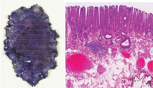
Figure 1.15 Endoscopically resected en bloc well-differentiated adenocarcinoma 28 × 17 mm with superficial submucosal invasion and no lymphovascular invasion and negative horizontal and vertical margins. This sample meets criteria for endoscopic curative resection.
Figure 1.16 Large mediastinal lymph node sampled with EUS-FNA. The needle is seen entering from the upper right of the screen into the tumor in the center of the image.
Further Reading
Al-Haddad, M. and Eloubeidi, M.A. (2008). Diagnostic and therapeutic applications of endoscopic ultrasound-guided punctures. Dig. Dis. 26 (4): 390–397.
Basford, P., George, R., Nixon, E. et al. (2014). Endoscopic resection of sporadic duodenal adenomas: comparison of endoscopic mucosal resection (EMR) with hybrid endoscopic submucosal dissection (ESD) techniques and the risks of late delayed bleeding. Surg. Endosc. 28 (5): 1594–1600.
da Cunha Santos, G., Boerner, S.L., and Geddie, W.R. (2011).
Maximizing the yield of lymph node cytology: lessons learned from rapid onsite evaluation of image- and endoscopic-guided biopsies of hilar and mediastinal lymph nodes. Cancer Cytopathol. 119 (6): 361–366.
Eloubeidi, M.A., Tamhane, A., Jhala, N. et al. (2006). Agreement between rapid onsite and final cytologic interpretations of EUS-guided FNA specimens: implications for the endosonographer and patient management. Am. J. Gastroenterol. 101 (12): 2841–2847.
Falk, G.W., Rice, T.W., Goldblum, J.R., and Richter, J.E. (1999). Jumbo biopsy forceps protocol still misses unsuspected cancer in Barrett’s esophagus with high-grade dysplasia. Gastrointest. Endosc. 49 (2): 170–176.
Gupta, N., Mathur, S.C., Dumot, J.A. et al. (2012). Adequacy of esophageal squamous mucosa specimens obtained during endoscopy: are standard biopsies sufficient for postablation surveillance in Barrett’s esophagus? Gastrointest. Endosc. 75 (1): 11–18.
Hikichi, T., Irisawa, A., Bhutani, M.S. et al. (2009). Endoscopic ultrasound-guided fine-needle aspiration of solid pancreatic masses with rapid on-site cytological evaluation by endosonographers without attendance of cytopathologists. J. Gastroenterol. 44 (4): 322–328.
Humphris, J.T.J., Kwok, A., and Katelaris, P.H. (2007). Cold snare polypectomy for diminutive polyps: an assessment of the risk of incomplete removal of small adenomas. Gastrointest. Endosc. 69: AB207.
Ichise, Y., Horiuchi, A., Nakayama, Y., and Tanaka, N. (2011). Prospective randomized comparison of cold snare polypectomy and conventional polypectomy for small colorectal polyps. Digestion 84 (1): 78–81.
Ikematsu, H., Yoda, Y., Matsuda, T. et al. (2013). Long-term outcomes after resection for submucosal invasive colorectal cancers. Gastroenterology 144 (3): 551–559. quiz e514.
Johanson, J.F., Frakes, J., and Eisen, D. (2011). Computerassisted analysis of abrasive transepithelial brush biopsies increases the effectiveness of esophageal screening: a multicenter prospective clinical trial by the EndoCDx Collaborative Group. Dig. Dis. Sci. 56 (3): 767–772.
Kadri, S.R., Lao-Sirieix, P., O’Donovan, M. et al. (2010). Acceptability and accuracy of a non-endoscopic screening test for Barrett’s oesophagus in primary care: cohort study. BMJ 341: c4372. https://doi.org/10.1136/bmj.c4372.
LeBlanc, J.K., Emerson, R.E., Dewitt, J. et al. (2010). A prospective study comparing rapid assessment of smears and ThinPrep for endoscopic ultrasound-guided fineneedle aspirates. Endoscopy 42 (5): 389–394.
Levy, M.J., Reddy, R.P., Wiersema, M.J. et al. (2005). EUSguided trucut biopsy in establishing autoimmune pancreatitis as the cause of obstructive jaundice. Gastrointest. Endosc. 61 (3): 467–472.
Mann, N.S., Mann, S.K., and Alam, I. (1999). The safety of hot biopsy forceps in the removal of small colonic polyps. Digestion 60 (1): 74–76.
Monkemuller, K.E., Fry, L.C., Jones, B.H. et al. (2004). Histological quality of polyps resected using the cold versus hot biopsy technique. Endoscopy 36 (5): 432–436.
Phoa, K.N., van Vilsteren, F.G., Weusten, B.L. et al. (2014). Radiofrequency ablation vs endoscopic surveillance for patients with Barrett esophagus and low-grade dysplasia: a randomized clinical trial. JAMA 311 (12): 1209–1217.
Qumseya, B.J., Wang, H., Badie, N. et al. (2013). Advanced imaging technologies increase detection of dysplasia and neoplasia in patients with Barrett’s esophagus: a metaanalysis and systematic review. Clin. Gastroenterol. Hepatol. 11 (12): 1562–1570. e1561–e1562.
Sakamoto, T., Matsuda, T., Nakajima, T., and Saito, Y. (2012). Efficacy of endoscopic mucosal resection with circumferential incision for patients with large colorectal tumors. Clin. Gastroenterol. Hepatol. 10 (1): 22–26.
Sharaf, R.N., Shergill, A.K., Odze, R.D. et al. (2013). Endoscopic mucosal tissue sampling. Gastrointest. Endosc. 78 (2): 216–224.
Trier, J.S. (1971). Diagnostic value of peroral biopsy of the proximal small intestine. N. Engl. J. Med. 285 (26): 1470–1473.
Vogelstein, B., Fearon, E.R., Hamilton, S.R. et al. (1988). Genetic alterations during colorectal-tumor development. N. Engl. J. Med. 319 (9): 525–532.
Wang, K.K. and Sampliner, R.E. (2008). Updated guidelines 2008 for the diagnosis, surveillance and therapy of Barrett’s esophagus. Am. J. Gastroenterol. 103 (3): 788–797.
Watanabe, T., Itabashi, M., Shimada, Y. et al. (2012). Japanese Society for Cancer of the Colon and Rectum (JSCCR) guidelines 2010 for the treatment of colorectal cancer. Int. J. Clin. Oncol. 17 (1): 1–29.
Wu, L., Li, P., Wu, J. et al. (2012). The diagnostic accuracy of chromoendoscopy for dysplasia in ulcerative colitis: meta-analysis of six randomized controlled trials. Color. Dis. 14 (4): 416–420.
Esophagus Inflammatory Conditions
Anthony R. Mattia1, Gregory Y. Lauwers2, Michael B. Wallace3 and Till S. Clauditz4
1 Newton-Wellesley Hospital, Newton, MA, USA
2 Moffitt Cancer Center, Tampa, Florida, USA
3 Mayo Clinic, Jacksonville, Florida, USA
4 Department of Pathology, University-Medical Center, Hamburg, Germany
Normal Histology, Variations
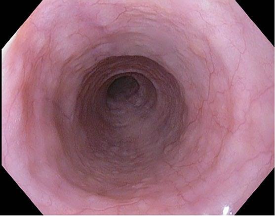
The esophagus has a pearly white and smooth surface under which a network of small vessels is seen (Figure 2.1) except for the distal esophageal sphincter where prominent longitudinal and palisading vessels (exceeding 100 mm in diameter) are observed and used to define the esophagogastric junction.
Microscopic Features
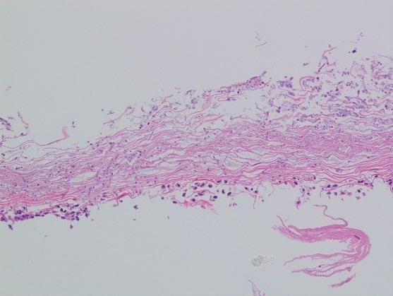
The esophageal mucosa is composed of a non-keratinizing flat stratified squamous epithelium, lamina propria, and muscularis mucosae (Figure 2.2; Table 2.1). The squamous epithelium is 10–20 cell layers thick measuring 300–500 μm in thickness. The prickle cells contain a large quantity of cytoplasmic glycogen, which stains positively with periodic acid–Schiff (PAS). The basal proliferative zone, the cuboidal or polyhedral basal and parabasal cells account for 15% or less of the epithelial thickness. A small number of specialized cell types, including T lymphocytes, Langerhans cells, endocrine cells, and melanocytes are also present in the deep/parabasal epithelium. The lamina propria consists of loose connective tissue with scattered inflammatory cells, vessels, nerves, and small mucous glands. The lamina propria papillae normally extend into the epithelium for 1/3–1/2 of its thickness. The muscularis mucosae is a thick layer of smooth muscle bundles oriented longitudinally. In the distal 1–2 cm of the esophagus, the basal zone, papillae, and intraepithelial cells may be more prominent, and include rare eosinophils. Esophageal mucosal biopsies typically include the squamous epithelium and scant lamina propria. The muscularis mucosae is seldom present, and better identified in esophageal mucosal resection specimens.
Infectious Esophagitis
Definition, General Features, Predisposing Factors
Infection is a common cause of esophagitis, particularly in immunocompromised patients but can also occur in apparently immunocompetent hosts with certain predisposing conditions. Fungi (Candida species) and viruses are responsible for most cases of infectious esophagitis, and dual infections may be encountered. Bacterial, mycobacterial, and parasitic esophagitis (e.g. Chagas disease due to Trypanosoma cruzi) are rarely encountered in esophageal biopsy material.
Fungal Esophagitis
Fungal infection due to Candida albicans is the most common cause of infectious esophagitis.
Predisposing conditions are broad and include HIV infection/AIDS, other immunosuppressed conditions such as organ transplant, prolonged corticosteroid therapy as well as other conditions such as diabetes, malignancy, antibiotic therapy, acid-suppressive therapy, and pregnancy. Infection due to Candida tropicalis and Candida (Torulopsis) glabrata has also been described, as well as infection due to other fungi such as Histoplasma capsulatum and Aspergillus species.
Viral Esophagitis
Herpes simplex (HSV) is the most common etiology of viral esophagitis, and also occurs primarily in the setting of immunosuppression, such as solid organ and bone marrow transplantation, underlying malignancy, chemotherapy, and HIV infection (AIDS). These patients may also have disseminated infection at the time of diagnosis. It can also develop in healthy adults including pregnant women and
children. Both HSV types 1 and 2 can infect the esophagus with type 1 being most common. Infection by varicellazoster virus (VZV) may also rarely occur in immunocompromised hosts.
Cytomegalovirus (CMV)-induced esophagitis also typically occurs in the immunosuppressed population, particularly in the setting of HIV infection/AIDS, organ transplantation, chemotherapy, corticosteroid therapy, and other forms of long-term immunosuppression. CMV may rarely cause esophagitis in immunocompetent patients, especially in the elderly.
Bacterial Esophagitis
Clinically significant bacterial esophagitis occurs almost exclusively in immunocompromised patients. Secondary bacterial colonization of areas of prior esophageal injury and ulceration is more common. Bacterial invasion of squamous mucosa or the deeper layers is required to establish a diagnosis of primary bacterial esophagitis. Gram-positive bacteria are the most common causative organisms (including Staphylococcus aureus, Staphylococcus epidermidis, Streptococcus viridans, and beta-hemolytic streptococci).
Mycobacterial Esophagitis
The rare mycobacterial esophagitis typically occurs in the setting of advanced immunosuppression. Both Mycobacterium tuberculosis complex and Mycobacterium avium complex can be encountered.
Parasitic Esophagitis
Chagas disease involving the esophagus is an important cause of esophageal dysfunction (dysphagia) in endemic regions, such as Latin America. However, the organism and its pathologic effects are typically not identified on biopsy specimens.
Clinical Features and Endoscopic Characteristics
Epithelium:
Non-keratinizing squamous epithelium
Glycogen rich
10–20 cells thick (300–500 μm)
Basal proliferative zone up to 15%
Few scattered parabasal T lymphocytes and other specialized cells
Lamina propria:
Papillary length 1/3 to 1/2 of epithelial thickness
Muscularis mucosae
Clinical and endoscopic features are specific to each condition. For candidal esophagitis, symptoms vary from none (especially in patients with mild disease from chronic inhaled steroids) to severe dysphagia and odynophagia in immunosuppressed patients. Candida appears as a white, cottage cheese-like exudate, which is partially adherent to the epithelium (Figure 2.3). HSV and CMV both cause ulcerations. HSV ulcers are typically small, 1–3 mm, whereas CMV ulcers may be both deep and wide. Biopsy yield for the virus is highest from the edge of HSV ulcers and from the center of CMV ulcers. In practice, both are usually obtained. Other infections’ etiologies may have nonspecific findings. The inflammatory
Figure 2.1 Endoscopic appearance of distal esophagus with normal squamous mucosa.
Figure 2.2 Normal esophagus squamous mucosa with normal stratified non-keratinizing epithelium.
Table 2.1 Normal esophageal mucosa.
reaction of tuberculous esophagitis often imparts a radiologically detectable mass-like lesion, whereas mucosal changes include linear ulcers and induration that preferentially affects the mid-portion of the esophagus. Fistula formation and perforation may occur. Chagas disease presentation is similar to that of achalasia with manometric findings of a poorly relaxing lower esophageal sphincter and an aperistaltic esophagus body.
Microscopic Features
Candida
The biopsies typically show an active esophagitis with a mixed inflammatory infiltrate of neutrophils, lymphocytes, and eosinophils. Neutrophils are often present in small, superficial clusters associated with parakeratosis or squamous debris, which may be a diagnostic clue. Prominent intraepithelial neutrophils may be associated with abscesses, erosions, or ulcerations. The inflammatory response may be minimal in severely immunocompromised patients.
Yeast forms of C. albicans and pseudohyphae (with refractile cell walls) are usually present in superficial desquamated keratin debris (Figure 2.4). The yeast are 3–5 μm in diameter basophilic oval forms while the, nonseptate sausage-like pseudohyphae are 3–5 μm in diameter and are arranged perpendicular to the epithelial surface. Occasional septate hyphae can be present. The presence of psuedohyphal and hyphal forms correlates with active infection. Most infections are superficial with isolated, intraepithelial fungal forms, and organisms in
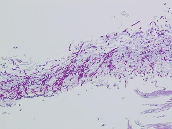
detached squamous cells. However, in immunocompromised hosts, deeper tissue invasion within ulcers and erosions may be seen. Candida are often conspicuous in routinely stained sections, although their appearance is enhanced with periodic acid–Schiff (PAS, Figure 2.5) or Grocott methenamine silver (GMS) stains. In Candida (Torulopsis) glabrata infections, only small budding yeast forms are seen, which may mimic histoplasmosis.
Candida can colonize preexisting ulcers or damaged mucosa of any etiology, and in such cases the possibility of dual infection or pathology should be considered. Also, when Candida yeast forms are present only in desquamated keratin debris unassociated with inflammation or endoscopic findings, carry over from an oral infection should be suspected.
Figure 2.3 Endoscopic appearance of white exudate typical of Candida albicans in a patient on chronic inhaled corticosteroid.
Figure 2.4 Debris of slough off superficial squamous epithelium with fungal hyphae and acute inflammation.
Figure 2.5 PAS stain of Figure 2.4 illustrating the fungal elements of Candida albicans
Cytologic brushings are also very useful in the diagnosis of Candida esophagitis, as the organisms are readily identified on Papanicolaou stained smears.
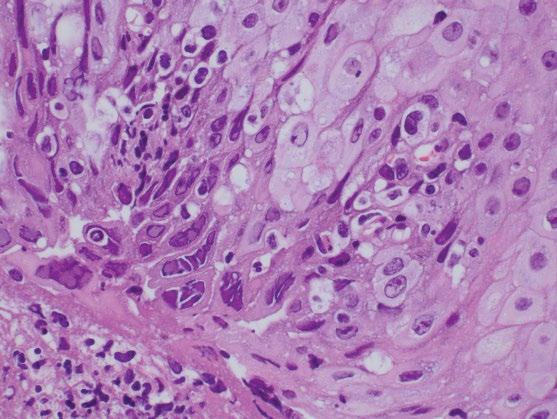
Herpes Simplex (HSV)
If HSV infection is suspected, in addition to biopsies for histology, fresh tissue can be sent for viral culture to confirm the diagnosis and identify strains that may be resistant to acyclovir. HSV infection is associated with ulceration and a mixed inflammatory infiltrate of intraepithelial neutrophils, eosinophils, and lymphocytes, as well as aggregates of macrophages that can be a diagnostic clue. HSV infects squamous cells of intact or denuded epithelium and is best identified at the edge of an ulcer and in cells within the ulcer slough. Therefore, optimal histologic diagnosis requires sampling of the ulcer edge rather than the ulcer bed. The typical viral cytopathic effects include multinucleation, ground glass nuclei, and dense intranuclear eosinophilic inclusions with a thickened nuclear membrane and a clear halo (Cowdry type A inclusion bodies) (Figure 2.6). Viral inclusions and multinucleated cells are not always identifiable in biopsies, and ancillary immunohistochemistry (IHC) staining for HSV may be required. Concomitant infection of Candida, cytomegalovirus, or bacteria can exist, particularly in immunocompromised patients. Varicella Zoster Virus has been associated with esophagobronchial fistula formation.
Cytomegalovirus (CMV)
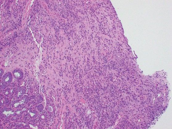
Culture is not used routinely, and does not distinguish the simple presence of CMV from active infection; however, culture may be useful for identifying drug resistance. Ulceration and granulation tissue formation with associated acute inflammation is common. In contrast to HSV
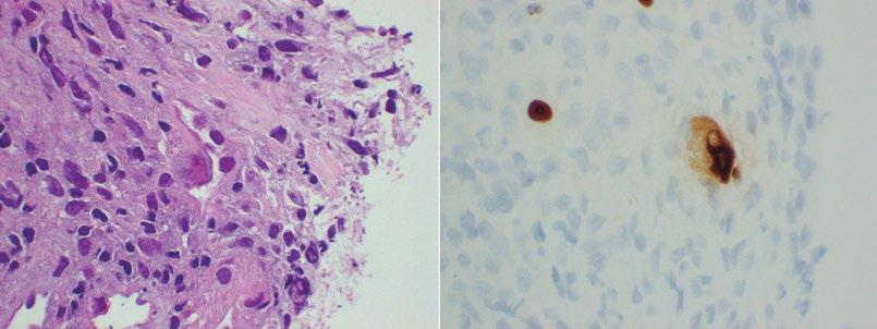
esophagitis, CMV rarely infects the squamous epithelium, and preferentially involves endothelial cells, stromal cells, and glandular epithelium. Therefore, biopsies of the ulcer bed should preferentially be performed when CMV esophagitis is suspected (Figure 2.7). CMV cytopathic effect is characterized by nuclear enlargement with classic “owl eye” large intranuclear inclusions, and granular, eosinophilic cytoplasmic inclusions (Figure 2.8a). Smaller, atypical intranuclear inclusions may be present and are more subtle, simulating activated fibroblasts. These infected cells are best identified by immunohistochemical staining (Figure 2.8b). CMV may coexist with HSV and Candida infection in some cases.
Bacterial Esophagitis
Sheets of bacteria are typically present with associated necrosis and mucosal erosion, highlighted by a tissue Gram stain. Inflammation may be scant or absent in neutropenic patients.
Mycobacterial Esophagitis
Histologic features include subepithelial and mural necrotic and non-necrotic granulomas with fibrosis and chronic inflammation. Acid fast bacilli can be recognized on Ziehl–Neelsen stain in approximately 2/3 of cases.
Immunohistochemical Studies and Molecular Features
IHC staining for HSV (HSV 1 and 2 cocktail stain) may be useful for suspected infections if diagnostic inclusions are not readily identified on H&E sections. Similarly, IHC for CMV is useful to highlight infected cells without typical CMV morphology, and may be more sensitive than light microscopy. PCR for M. tuberculosis may be useful in selected cases.
Figure 2.6 Multinucleated basal keratinocytes typical of HSV esophagitis.
Figure 2.7 Erosive esophagitis with prominent granulation and atypical cellular elements.
Differential Diagnosis
Clinical
Inflammatory changes of the esophagus can be seen in many conditions, most commonly acid reflux disease. Behcet’s disease may present with focal ulcers, as can pillinduced (associated with an impacted pill, which may or may not be present during exam) ulcers.
Microscopic
The differential diagnosis of Candida esophagitis includes reflux esophagitis, drug injury, glycogenic acanthosis, ectopic sebaceous glands, and other infections.
The differential diagnosis of HSV esophagitis includes other etiologies of mucosal ulceration, including other infections, Crohn’s disease, HIV-associated ulceration, and drug injury. In some cases, the cytologic atypia induced in the squamous epithelium by the viral cytopathic effects of HSV may be difficult to distinguish from squamous dysplasia/carcinoma, as well as radiation esophagitis.
Prognosis, Evolution, and Clinical Management
Most patients present with dysphagia or odynophagia and undergo upper endoscopy with biopsy. Infectious etiologies are treated with appropriate antimicrobials (e.g. fluconazole for candida), typically with high response rates. In immunosuppressed patients, including those on chronic inhaled steroids, frequent retreatment, or chronic suppressive treatment, may be necessary after full efforts to restore immune function.
Gastroesophageal Reflux Disease
Definition, General Features, Predisposing Factors
The chronic regurgitation of gastroduodenal juice into the esophagus causes gastroesophageal reflux disease (GERD). The mucosal injury results from the effects of refluxed gastric acid, bile, pepsin, and duodenal contents that overwhelm the normal protective antireflux barriers, such as esophageal acid clearance and mucosal resistance. Risk factors and predisposing conditions include obesity, diet, sedentary lifestyle, tobacco and alcohol use, hiatus hernia, reduced lower esophageal sphincter tone, loss of esophageal peristaltic function, gastric hypersecretory states, and delayed gastric emptying. Symptomatic GERD is prevalent worldwide, with considerable geographic variation. Prevalence estimates are approximately 20% in North America and Europe, with only East Asia showing estimates <10%.
Clinical Features and Endoscopic Characteristics
There are several endoscopic classifications of gastroesophageal reflux disease (Figure 2.9). The Los Angeles Classification divides esophagitis into four grades:
Grade A: one or more mucosal breaks, each no longer than 5 mm.
Grade B: at least one mucosal break more than 5 mm long, but not continuous between the tops of 2 mucosal folds.
(a) (b)
Figure 2.8 High-magnification evaluation of Figure 2.7 demonstrated (a) endothelial CMV infection (b) confirmed by immunohistochemistry.
Grade C: at least one mucosal break that is continuous between the tops of 2 or more mucosal folds, but which is not circumferential.
Grade D: a circumferential mucosal break, erosions and ulceration, often resulting in scarring and stenosis if the reflux is recurrent.
Mucosal bridges and esophagogastric fistulae may be encountered.
The endoscopic findings in reflux esophagitis have been classified into three main stages: active, healing, and scarring. Relapse and healing occur repeatedly in patients with gastroesophageal reflux disease (GERD), and a mixture of the stages is often seen in biopsy specimens.
Microscopic Features
The typical features of GERD are basal cell layer hyperplasia, papillary elongation, dilated intercellular spaces (intercellular edema/spongiosis), and intraepithelial inflammation, including neutrophils, eosinophils, and mononuclear cells (Table 2.2; Figures 2.10, 2.11). However, none of these features is specific for this diagnosis. The proliferative epithelial changes are more common than inflammatory infiltration. Basal cell hyperplasia is diagnosed when the thickness of the basal layer exceeds 15% in a well-oriented section. Papillary elongation, defined as extending >50% of the epithelial thickness, should also be only assessed in well-oriented sections. Finally, in minimal GERD, dilated intercellular spaces in the basal and parabasal areas may be the only finding. Esophageal squamous mucosa normally contains few or no inflammatory cells and the inflammatory infiltrate in GERD is highly variable. The presence of occasional
Table 2.2
Histologic features of GERD.
Basal cell hyperplasia (>15%)
Papillary elongation (>50%)
Dilated intercellular spaces (spongiosis)
Intraepithelial inflammation
Neutrophils
Eosinophils
Mononuclear cells
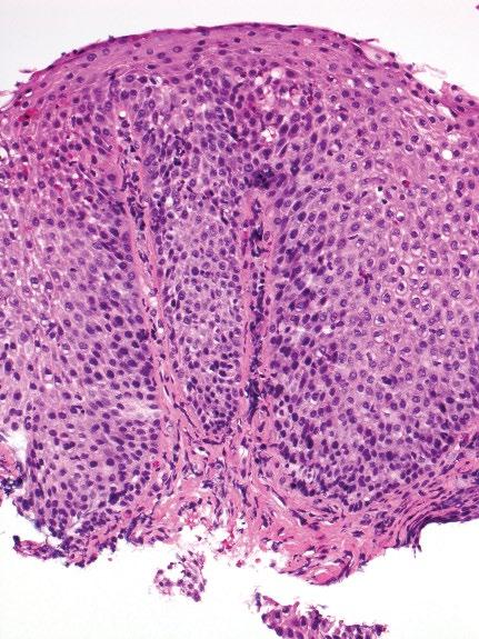
Figure 2.10 Characteristic appearance of low-power view of reflux esophagitis with basal cell hyperplasia, elongation of papillae, lymphocytic and eosinophilic infiltrate.
eosinophils is typical of GERD, although large numbers of intraepithelial eosinophils mimicking eosinophilic esophagitis may be identified. Notably, eosinophils are not found in all GERD biopsies and can be seen in asymptomatic patients. Neutrophils may be present, and are more numerous in areas of erosion and ulceration. Lymphocytes, often with irregular nuclear contours (squiggle cells), are usually scattered through the epithelium. Finally, severe inflammation with ulceration may be associated with inflammatory pseudotumor formation, with marked atypia of the reactive stromal cells.
In practice, the report should provide a descriptive summary of the histologic features and injury patterns present, as well as their severity. A note can be added to state that the findings are compatible with reflux injury pattern in the appropriate clinical setting.
Figure 2.9 Endoscopic appearance of erosive esophagitis in a patient with gastroesophageal reflux disease.

Differential Diagnosis
Clinical
Clinically, erosive esophagitis from GERD is typically in the lower third of the esophagus and contiguous with the gastroesophageal junction as opposed to other inflammatory or infectious etiologies that may occur more proximally. The presence of a normal distal esophagus with more proximal inflammation virtually excludes GERDassociated inflammation. Typical symptoms are heartburn and regurgitation. Ph- metry (monitoring the acid reflux events by a wired or wireless sensor in the distal esophagus) can be used in challenging cases but is rarely necessary with obvious symptoms and when esophagitis is seen.
Microscopic
As none of the histologic features are specific for GERD, other etiologies such as infection, eosinophilic esophagitis, iatrogenic injuries, Crohn’s disease, and inflammatory skin disorders involving the esophagus may enter into the differential diagnosis.
The presence of large numbers of neutrophils in the superficial epithelium should suggest the possibility of fungal infection or superinfection, and special stains (PAS-diastase, GMS) should be performed to exclude Candida organisms. Large numbers of intraepithelial eosinophils raising the possibility of eosinophilic esophagitis and correlation with clinical findings and PPI treatment status are required (see Section “Eosinophilic Esophagitis”). Numerous intraepithelial lymphocytes, if predominant, may suggest an alternative diagnosis such as lymphocytic esophagitis or lichen planus-associated esophagitis (see Sections “Lymphocytic Esophagitis” and “Dermatological Diseases Involving the Esophagus”).
Ulceration with regenerative squamous epithelial atypia may be difficult to distinguish from in situ-squamous cell carcinoma and dysplasia. The regenerating epithelial cells have round nuclei with minimally thickened nuclear membranes; fine chromatin; and large, irregular nucleoli, and generally have abundant cytoplasm. Further, maturation toward the epithelial surface can usually be found. In difficult cases, additional biopsies should be requested after medical treatment of the esophagitis. However, in some cases, inflammatory pseudotumors with marked atypia of the reactive stromal cells will need to be differentiated from poorly differentiated carcinoma or sarcoma. Cytokeratin immunohistochemistry (IHC) may be helpful when trying to distinguish between reparative reactions and malignancy.
Less commonly, activated lymphocytes in areas of ulceration may appear atypical and raise the suspicion for lymphoma. In some cases, immunohistochemistry and/or flow cytometry may be necessary to assess for clonality.
Prognosis, Evolution, and Clinical Management
GERD management is based on the severity and potential for complications and largely consists of lifestyle modification, anti-acid medications or surgery. Most nonerosive or minimally erosive (Los Angeles Grade A) responds to a combination of lifestyle modifications and low-dose (single daily proton pump inhibitor [PPI] such as omeprazole) anti-acid therapy. Lifestyle modifications include weight loss, avoiding large meals with two to three hours of bedtime (where gravity no long prevents “upward” reflux), and minimizing specific refluxogenic foods (alcohol, mint, fatty food, chocolate, caffeine). PPI therapy is highly effective, even for severe erosive GERD but may require twice-daily therapy. More than 95% of patients will heal erosive disease on PPI therapy.
Surgical therapy is aimed at restoring the lower esophageal sphinter pressure zone and angle of His, and reducing hiatal hernias. Surgery is also highly effective with more than 80% of individuals having sustained elimination or reduction in GERD symptoms and use of PPI. The major side effect of surgery is retention of gas in the stomach (“gas-bloat syndrome”) due to the inability to naturally belch swallowed air.
Eosinophilic Esophagitis
Definition, General Features, Predisposing Factors
Eosinophilic esophagitis (EOE) is a chronic, immunemediated or antigen-mediated inflammatory condition characterized by symptomatic esophageal dysfunction and
Figure 2.11 High-power magnification of reflux esophagitis with basal cell hyperplasia, spongiosis, and eosinophilic infiltrate.
eosinophil predominant mucosal inflammation. EOE represents a type 2 (Th2) helper T cell and cytokine-mediated disorder involving genetic predisposition, environmental exposures, and allergic background. Food-based antigens appear to be the dominant mediators of EOE, although aeroallergens may also act as triggers in susceptible patients. Since it was first established as a distinct clinicopathologic entity in 1993, EOE has become an increasingly recognized cause of dysphagia and esophageal dysfunction, particularly among patients refractory to medical treatment of gastroesophageal reflux disease.
EOE is reported worldwide, with highest prevalence in the United States and Western Europe. It occurs more frequently in children and young adults, although it is increasingly recognized in older adults, and is more common in males (male-to-female ratio of 3–4 : 1). A history of other allergic conditions may be present, such as asthma and atopic dermatitis, as well as peripheral eosinophilia, which are more common in children.
Current diagnostic criteria for EOE include a combination of clinical and pathologic features (Table 2.3).
Table 2.3 Definition of eosinophilic esophagitis and diagnostic criteria.
Symptoms related to esophageal dysfunction
Eosinophil-predominant inflammation on esophageal biopsy, characteristically consisting of a peak value of 15 eosinophils per high-power field (EOS/HPF)
Mucosal eosinophilia is isolated to the esophagus and persists after a PPI trial
Secondary causes of esophageal eosinophilia excluded (Table 2.5)
A response to treatment (dietary elimination; topical corticosteroids) supports the diagnosis of EOE, but is not required.
Clinical Features and Endoscopic Characteristics
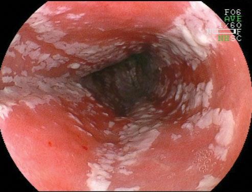
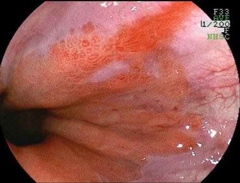
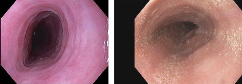
Eosinophilic esophagitis (EOE) is increasing in prevalence and often seasonal with spikes during typically airborne allergy season. It presents with dysphagia to solids, classically with pills sticking. Other atopic symptoms are common. At endoscopy, the esophagus has white punctate spots (eosinophilic microabscess), rings, longitudinal furrows (Figure 2.12), and desquamates easily with insufflation or dilation. In late stages, the esophagus may be very narrowed and poorly distensible.
Microscopic Features
Esophageal biopsies are required to diagnose EOE. Eosinophils are generally absent from the esophageal squamous epithelium in normal individuals. The histologic hallmark is the presence of an increased number of eosinophils within the squamous epithelium (Figure 2.13).
Although there is no exact threshold number that establishes a diagnosis, 15 eos/HPF (peak count) in at least one biopsy is considered a “minimum” threshold.
Eosinophilia may be patchy, and it is recommended that two to four biopsies from at least two locations in the esophagus should be taken to maximize diagnostic yield. The distribution of eosinophilia is important, as they may be identified in both proximal and distal biopsies or be more prominent in proximal biopsies. Practically, only intact eosinophils with visible nuclei should be counted and eosinophilic granules should not be included, although degranulated eosinophils may be a useful clue.
Other histologic findings may be present although none of these should be considered pathognomonic. These features include moderate-marked reactive basal hyperplasia,
(a)
(b)
Figure 2.12 Endoscopic appearance of eosinophilic esophagitis (a) with so-called feline esophagus appearance and (b) with extensive white microabcesses. Subtle circular rings and longitudinal furrows also present.
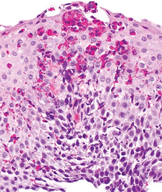
eosinophilic microabscesses (superficial aggregates of 4 eosinophils), superficial layering of eosinophils, and subepithelial fibrosis. The presence of subepithelial fibrosis is also associated with eosinophils in the lamina propria and correlates with fibro-stenotic complications of EOE, but may not be identifiable depending on biopsy depth. Other inflammatory cells, such as lymphocytes and mast cells, can also be increased. When reporting the cases, a note is useful to summarize additional relevant histologic features and to provide an overall assessment of the case based on all available biopsies (including prior biopsies, if applicable). For example, active esophagitis with intraepithelial eosinophils (peak count of # per HPF) followed by a note. When large numbers of eosinophils are present (e.g. >40 per HPF), an exact count may be difficult and it is appropriate to report the peak count as such (e.g. “peak count of >40 per HPF”).
Immunohistochemical Studies and Molecular Features
Currently, the examination of esophageal mucosal biopsies by hematoxylin and eosin (H&E) stained sections, with clinical correlation, remains the most reliable diagnostic test for EOE. The diagnostic value of ancillary studies in
distinguishing eosinophilic esophagitis from gastroesophageal reflux disease and other conditions remains unproven.
Immunohistochemical stains targeting eosinophil peroxidase, major basic protein, and eosinophil-derived neurotoxin, have been reported to enhance detection of eosinophils compared to evaluation of H&E sections, but these stains are not widely used in routine clinical practice. A recent study suggested that the presence of intra-squamous IgG4 deposits by immunohistochemistry may be a useful adjunctive marker for EOE.
Molecular analyses of esophageal tissues, such as gene expression analysis of chemokines such as eotaxin-3 (CCL26), and transcriptome analysis show promise to improve diagnosis and clinical monitoring, and to guide patient-specific therapy.
Differential Diagnosis
Clinical
Dysphagia and rings in the esophagus can occur from any inflammatory condition. Even eosinophils may be present in GERD alone. EOE typically requires a confirmatory biopsy from the upper and lower esophagus showing eosinophilic infiltrates. Desquamative conditions that should be excluded include bullous pemphigoid and lichen planus. These typically require deep biopsies and special immunofluorescence staining; thus, communication between endoscopist and pathologist is critical.
Microscopic
The histologic appearance of EOE is not specific, and mucosal infiltration by eosinophils is a component of a variety of other esophageal inflammatory conditions (Table 2.4).
GERD and PPI-responsive esophageal eosinophilia exhibit considerable histologic overlap with EOE. In an untreated patient, fewer numbers of eosinophils with distal predominance may be suggestive of GERD. Correlation with response to PPI therapy is paramount.
In eosinophilic gastroenteritis, obtaining biopsy specimens from the stomach and duodenum and other clinical symptoms such as nausea, vomiting, and diarrhea may be helpful in establishing a diagnosis.
The presence of granulomas is helpful in diagnosing esophageal involvement by Crohn’s disease, but they are uncommon. Eosinophils may also be admixed with neutrophils and lymphocytes, and involvement of other parts of the GI tract is typically present.
Infections are recognized by the identification of viral inclusions (such as HSV), and fungal or parasitic organisms. Mixed inflammation with neutrophils may also be present. Rare cases of HSV esophagitis occurring in a background of EOE have been described.
Figure 2.13 Characteristic histology of eosinophilic esophagitis with basal cell hyperplasia, marked eosinophilic infiltrate with eosinophilic microabscesses.
Table 2.4 Endoscopic and histologic features of the major causes of infectious esophagitis.
OrganismEndoscopic featuresMorphological features
Candida Focal or confluent white plaques
Herpes simplex Superficial ulcers, multiple
CMV Single deep ulcer, linear ulcers
Parakeratosis with neutrophils
Colonization of ulcers
Budding yeast, pseudohyphae, occasional septate hyphae
Superficial invasion typical
Edge of ulcer—inclusions in squamous cells and/or squamous debris of ulcer slough
Multinucleation, ground glass nuclei, dense eosinophilic nuclear inclusions (Cowdry type A)
Base of ulcer—inclusions in endothelial and stromal cells
Intranuclear (“owl’s eye”) and granular cytoplasmic inclusions
Atypical inclusions common
Table 2.5 Conditions associated with esophageal eosinophilia in the differential diagnosis of EOE.
GERD
PPI-responsive esophageal eosinophilia
Eosinophilic gastrointestinal diseases
Crohn’s disease
Infection (Candida, HSV, parasites)
Hypereosinophilic syndrome
Achalasia
Drug hypersensitivity injury
Vasculitis
Pemphigus
Connective tissue diseases
Graft versus host disease (GVHD)
In drug-induced esophageal eosinophilia, there is a temporal relationship and resolution when the offending agent is withdrawn.
Ancillary Studies (if Applicable)
IHC for virus (HSV) and/or special stains for fungi (GMS, PAS-diastase) are indicated if infection is suspected.
Prognosis, Evolution, and Clinical Management
EOE is initially treated with proton pump inhibitors. There is emerging evidence that PPIs affect specific inhibition of the chemokine, eotaxin, which blunts eosinophil infiltration. It may be difficult to distinguish GERD-associated eosinophilia from EOE in this subgroup. For those that do not respond to PPI, options include identification and avoidance of food antigens (typically through a Six Food
Ancillary studies
PAS-diastase, GMS stains
IHC
Elimination Diet; SFED) or suppression with topical corticosteroids, which can be from a swallowed metered dose inhaler, or from peroral viscous budesonide. For late-stage, fibrotic disease, repeated, carefully monitored esophageal dilation is effective.
Lymphocytic Esophagitis
Definition, General Features, Predisposing Factors
This uncommon and poorly characterized condition is histologically defined as the presence of dense lymphocytic infiltrates involving the esophageal mucosa without concomitant evidence of significant active inflammation. It is described in approximately 1/1000 esophageal biopsies, predominantly affecting older females (median age 63 years). This controversial entity lacks clearly defined clinical associations in adults, and likely represents a nonspecific reaction pattern to a wide variety of mucosal insults (Table 2.6), including GERD, medications, infection, motility disorders, and other immune-mediated disorders. In the pediatric population, lymphocytic esophagitis has been described in association with Crohn’s disease and celiac disease.
Clinical and Endoscopic Characteristics
About 75% of patients present with esophageal symptoms, most commonly dysphagia or odynophagia, which may raise the suspicion of eosinophilic esophagitis. Similarly, the endoscopic impression may suggest eosinophilic esophagitis, with the presence of rings, furrows, plaques, and strictures.
IHC
Table 2.6 Conditions associated with esophageal lymphocytosis (lymphocytic esophagitis).
GERD
Medications
Infection
Motility disorders
Immune-mediated disorders (e.g. CVID)
Lichen planus/lichenoid esophagitis
Crohn’s disease (pediatric)
Celiac disease (pediatric)
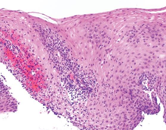
Figure 2.14 Characteristic histology of lymphocytic esophagitis.
Microscopic Features
Recently proposed histologic criteria include the presence of dense lymphocytic infiltrates involving esophageal squamous mucosa with marked spongiosis in the absence of significant numbers of neutrophils or eosinophils (Figure 2.14). The findings are most pronounced in the peripapillary epithelium. There is no required or established “minimum” count or density of intraepithelial lymphocytes, and the pattern and distribution of the lymphocytic epithelial injury is most important.
Immunohistochemical Studies and Molecular Features
Ancillary testing, including the subtyping of the intraepithelial CD3, CD4, and CD8 T cells is generally noncontributory and not required for the diagnosis. A subset of cases with CD4 predominant T cells and dysmotility has been reported.
Differential Diagnosis
The clinical and endoscopic differential diagnosis includes EOE, although by definition eosinophils are rare or absent. If neutrophils are present, special stains for candida may be warranted. Intraepithelial lymphocytes may be prominent in lichen planus esophagitis/lichenoid esophagitis, although the additional presence of a band-like lymphocytic infiltrate in the lamina propria and necrotic keratinocytes is a distinguishing feature. Characteristic direct immunofluorescence findings of lichen planus DIF may or may not be demonstrated. Lymphocytosis may also be associated with esophageal involvement by Crohn’s disease in children, although other histologic and clinical features of Crohn’s disease are typically present, particularly elsewhere in the gut.
Prognosis
Follow-up clinical and biopsy data are limited, although in some patients sequential biopsies have demonstrated persistent lymphocytosis, suggesting a chronic process.
Drug-Induced Esophagitis
Definition, General Features, Predisposing Factors
Medication-related injury to the esophageal mucosa (“pill esophagitis”) is an increasingly recognized complication of orally administered drugs. Mucosal injury is typically caused by prolonged direct contact and local caustic effects of the offending agent, although mechanisms of toxicity may be complex and multifactorial. Numerous drugs have been implicated (Table 2.7). Most cause a nonspecific injury pattern, and the diagnosis is suggested by the clinical history. Elderly patients and women are most commonly affected. The elderly often have multiple underlying risk factors, and the female predominance is attributed to the more common use of certain medications in this group, such as iron supplements and bisphosphonates. Risk factors include pills taken with little or no water or while recumbent, underlying disorders of esophageal motility (e.g. diabetes, strictures, achalasia) and disorders of local anatomy causing external compression (enlarged left atrium, aortic aneurism). However, many patients do not exhibit abnormal esophageal function or other risk factors.
Clinical and Endoscopic Characteristics
Typically, there is a temporal relationship between the onset of symptoms and ingestion of a potentially injurious drug. Common symptoms include sudden-onset chest
Table 2.7 Medications implicated in causing esophagitis and histologic features.
Medication Histologic features
NSAIDs Nonspecific, ± crystalline debris
Antibiotics (e.g. tetracyclines/ doxycycline, clindamycin)
Doxycycline—vascular injury
Potassium chlorideNonspecific
Iron
Bisphosphonates (e.g. alendronate)
Bluish-brown crystalline debris, Fe stain +
Nonspecific, ± crystalline debris and multinucleated squamous cells
Quinidine Nonspecific
Resins (e.g. kayexalate, bile acid sequestrants)
Kayexalate:
Basophilic crystals with “fish scales,” AFB (black), PAD, Diff-quick bile acid sequestrants:
Orange-black crystals, lack “fish scales,” AFB (yellow)
MycophenolateIncreased apoptosis (GVHD-like)
Taxanes Mitotic arrest, ring mitoses
pain, odynophagia and/or dysphagia that occur within a few hours or days after taking the medication. Severe reflux injury is often an initial diagnostic concern. The chest pain may be pleuritic in nature, mimicking a pulmonary embolism, or may suggest an acute cardiac syndrome. Hematemesis and perforation are rare complications.
Endoscopically, most lesions occur in the middle third of the esophagus, at the level of the aortic arch, although any level may be involved. Findings include erythema, erosions, ulcers (including “kissing” ulcers), and strictures. Discrete erosions in this region, away from the Z-line, are an important clue suggesting a drug-related etiology (Figure 2.15). Occasionally, a diffuse mucosal sloughing pattern has been described.
Microscopic Features (Table 2.7)
Biopsies most commonly show nonspecific esophagitis characterized by erosions, ulcers, active inflammation with neutrophils and occasionally eosinophils, surface epithelial damage, and granulation tissue. Adjacent mucosa is relatively normal. A careful search of the exudate and ulcer bed for refractile or polarizable foreign material is warranted, which may be an important diagnostic clue. However, in most cases the clinical history is necessary to make the diagnosis.
Some medications are associated with additional characteristic histologic features. Erosions and ulcers associated
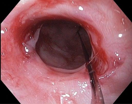
Endoscopic
with doxycycline injury often exhibit a distinct capillary/ vascular injury with degenerative changes and possible fibrin thrombi. In iron pill-associated mucosal injury, bluish-brown crystalline iron debris is found adjacent to the surface epithelium, admixed with the inflammatory exudate, or within granulation tissue (Figure 2.16). The iron can be highlighted by a Prussian Blue stain, which may also demonstrate crystalline debris in the lamina propria or underlying vessels. Although iron is well recognized as a caustic agent in the esophagus and elsewhere in the GI tract, some cases may represent secondary deposition in preexisting lesions. Alendronate has been associated with multinucleated squamous cells, in addition to the presence of polarizable crystalline material.
Kayexalate (sodium polystyrene sulfonate) in sorbitol can be recognized by the presence of characteristic lightly basophilic, refractile, nonpolarizable crystals that display a mosaic pattern that resembles fish scales. The crystals can be highlighted by acid fast (black), PAS/Alcian blue and Diff-Quik stains. Bile acid sequestrants (e.g. colestipol, colesevelam, cholestyramine) may be recognized by their orange-black crystals, which lack a striated, fish-scale appearance and may appear pale yellow on acid fast stain.
Mycophenolate, used in solid organ transplantation, is associated with increased apoptosis of esophageal squamous epithelial cells, resembling GVHD. Taxane-based chemotherapy leads to striking mitotic arrest with ring mitoses, along with epithelial necrosis or ulceration.
Differential Diagnosis
The major histologic differential diagnostic consideration is infection, particularly fungal (Candida) or viral (HSV, CMV), which may need to be excluded with appropriate histochemical and immunohistochemical stains or
Figure 2.15
appearance of inflammatory stricture of the esophagus with sloughing mucosa.
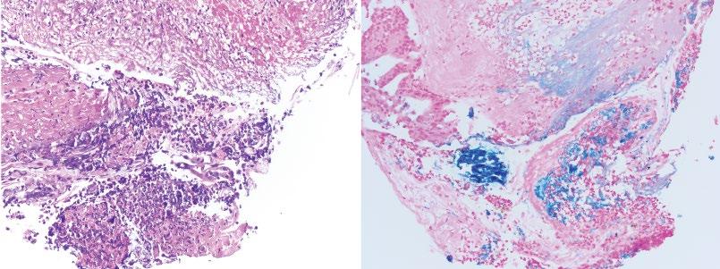
recognized by characteristic viral cytopathic effect. In rare cases, exuberant granulation tissue and excessive exudates mimic the gross appearance of a neoplasm. If eosinophils are prominent, reflux or eosinophilic esophagitis may enter into the differential diagnosis. GVHD should be excluded if prominent apoptosis is present, such as with mycophenolate injury. Finally, lichen planus involvement of the proximal esophagus with stricture formation can also mimic drug injury. In all of these considerations, the clinical context is critical to the evaluation of the histopathologic findings.
Prognosis, Evolution and Clinical Management
Most cases are self-limited and resolve without complications. Discontinuing the suspected caustic agent and supportive management, such as PPI therapy, hydration, sucralfate, or topical anesthetics, typically results in an uneventful recovery. Local epinephrine may be needed for active bleeding. Dilatation may be required if strictures develop, and surgical management is rarely needed for severe complications such as perforation. Most patients can resume their medications with recommendations for proper administration, which will assist in avoiding recurrent injury.
Sloughing Esophagitis
Definition, General Features, Predisposing Factors
Sloughing esophagitis is characterized by extensive superficial squamous mucosal necrosis or desquamation, associated with endoscopic plaques imparting a sloughing
appearance to the mucosa. Sloughing esophagitis has also been included under the terms esophagitis dissecans superficialis and acute necrotizing esophagitis, which includes broader inclusion criteria and more diverse causes of esophageal injury. It occurs in middle-aged to older adults (median age 56 years), frequently with debilitating comorbidities and who are commonly on multiple medications, especially central nervous system depressants and drugs known to cause esophageal mucosal injury. It most likely represents a form of direct contact injury, rather than an ischemic injury, and may represent a unique pattern of drug-induced esophagitis. Acute necrotizing esophagitis (“black esophagus”) is a rare condition that arises in debilitated patients with severe comorbidities such as hypoperfusion, sepsis, diabetic ketoacidosis, and malignancy, and results from the combining effect of ischemia and corrosive injury of gastric contents. A rare case occurring in a child who was associated with a history of vomiting has also been reported.
Clinical and Endoscopic Characteristics
Dysphagia, odynophagia, chest pain, heartburn, vomiting or cough are reported in 45% of patients, include. The middistal esophagus is most commonly involved. Endoscopy reveals white plaques or membranes, and there may be sloughing of the epithelium as well as other nonspecific findings such as erythema, erosions, rings, or webs. In black esophagus, the sloughing necrotic membrane may appear blackened (Figure 2.17). The clinical presentation is distinctive with bleeding and a characteristic diffuse circumferential black mucosal discoloration of the distal esophagus with an abrupt transition at the gastroesophageal junction.
Figure 2.16 (a) Iron tablet erosive esophagitis with fibrinopurulent debris. (b) The Prussian Blue iron stain is diagnostic.