First edition published 2012 by John Wiley & Sons Ltd
All rights reserved. No part of this publication may be reproduced, stored in a retrieval system, or transmitted, in any form or by any means, electronic, mechanical, photocopying, recording or otherwise, except as permitted by law. Advice on how to obtain permission to reuse material from this title is available at http://www.wiley.com/go/permissions.
The right of Alfonso J. Cruz‐Jentoft and John E. Morley to be identified as the author(s) of the editorial material in this work has been asserted in accordance with law.
Registered Office(s)
John Wiley & Sons, Inc., 111 River Street, Hoboken, NJ 07030, USA
John Wiley & Sons Ltd, The Atrium, Southern Gate, Chichester, West Sussex, PO19 8SQ, UK
Editorial Office
9600 Garsington Road, Oxford, OX4 2DQ, UK
For details of our global editorial offices, customer services, and more information about Wiley products visit us at www.wiley.com.
Wiley also publishes its books in a variety of electronic formats and by print‐on‐demand. Some content that appears in standard print versions of this book may not be available in other formats.
Limit of Liability/Disclaimer of Warranty
The contents of this work are intended to further general scientific research, understanding, and discussion only and are not intended and should not be relied upon as recommending or promoting scientific method, diagnosis, or treatment by physicians for any particular patient. In view of ongoing research, equipment modifications, changes in governmental regulations, and the constant flow of information relating to the use of medicines, equipment, and devices, the reader is urged to review and evaluate the information provided in the package insert or instructions for each medicine, equipment, or device for, among other things, any changes in the instructions or indication of usage and for added warnings and precautions. While the publisher and authors have used their best efforts in preparing this work, they make no representations or warranties with respect to the accuracy or completeness of the contents of this work and specifically disclaim all warranties, including without limitation any implied warranties of merchantability or fitness for a particular purpose. No warranty may be created or extended by sales representatives, written sales materials or promotional statements for this work. The fact that an organization, website, or product is referred to in this work as a citation and/or potential source of further information does not mean that the publisher and authors endorse the information or services the organization, website, or product may provide or recommendations it may make. This work is sold with the understanding that the publisher is not engaged in rendering professional services. The advice and strategies contained herein may not be suitable for your situation. You should consult with a specialist where appropriate. Further, readers should be aware that websites listed in this work may have changed or disappeared between when this work was written and when it is read. Neither the publisher nor authors shall be liable for any loss of profit or any other commercial damages, including but not limited to special, incidental, consequential, or other damages.
The CIP date for ISBN: 9781119597872 has been applied.
Set in 10/12pt TimesNewRomanMTStd by SPi Global, Pondicherry, India
10 9 8 7 6 5 4 3 2 1
16 Measurements of Muscle Mass, Equations and Cut‐off
Marjolein Visser and Laura Schaap
17 Deuterated Creatine Dilution to Assess Muscle Mass (D3‐Cr Muscle Mass) in Humans: Methods, Early Results, and Future
Peggy M. Cawthon and William J. Evans 18
Laura Orlandini, Yves Rolland and Matteo Cesari
20 Biomarkers for Physical Frailty and Sarcopenia: A “Two‐Body
Anna Picca, Riccardo Calvani and Emanuele Marzetti
21
Charlotte Beaudart, Jean‐Yves Reginster, Olivier Bruyère and Anton Geerinck
Tommy Cederholm, Jürgen M. Bauer and Alfonso J. Cruz‐Jentoft 24 Beta‐hydroxy‐beta‐methylbutyrate
Francesco Landi, Riccardo Calvani, Anna Picca and Emanuele Marzetti 25
Francesco Landi, Graziano Onder and Rolland Yves
Stany Perkisas, Keliane Liberman, Ivan Bautmans and Maurits Vandewoude
Kristina Norman
List of Contributors
Hidenori Arai
National Center for Geriatrics and Gerontology, Obu, Aichi, Japan
Jürgen M. Bauer
Center for Geriatric Medicine and Network Aging Research, Heidelberg University, Heidelberg, Germany
Ivan Bautmans
Frailty in Ageing Research Department, Vrije Universiteit Brussel, Brussels, Belgium
Charlotte Beaudart
Division of Public Health, Epidemiology and Health Economics, WHO Collaborating Center for Public Health aspects of musculo‐skeletal health and ageing, University of Liège, Liège, Belgium
Yves Boirie
Université Clermont Auvergne, Unité de Nutrition Humaine, Clermont‐Ferrand, France
INRA, UMR 1019, Unite de Nutrition Humaine, CRNH, Clermont‐Ferrand, France
CHU Clermont‐Ferrand, Service de Nutrition Clinique, Clermont‐Ferrand, France
Olivier Bruyère
Division of Public Health, Epidemiology and Health Economics, WHO Collaborating Center for Public Health aspects of musculo‐skeletal health and ageing, University of Liège, Liège, Belgium
Riccardo Calvani
Department of Geriatrics, Neurosciences and Orthopedics, Teaching Hospital “Fondazione Policlinico A. Gemelli” at the Catholic University of the Sacred Heart, Rome, Italy
Peggy M. Cawthon
California Pacific Medical Center, Research Institute, San Francisco, CA, USA
Department of Epidemiology and Biostatistics, University of California, San Francisco, CA, USA
Tommy Cederholm
Theme Ageing, Karolinska University Hospital, Stockholm, Sweden
Clinical Nutrition and Metabolism, Department of Public Health and Caring Sciences, Uppsala University, Uppsala, Sweden
Matteo Cesari
LIST OF CONTRIBUTORS
Dipartimento di Scienze Cliniche e di Comunità, University of Milan, Milan, Italy; Unità di Medicina Interna ad Indirizzo Geriatrico, IRCCS Istituti Clinici Scientifici Maugeri, Milan, Italy
Liang‐Kung Chen
Center for Geriatrics and Gerontology, Taipei Veterans General Hospital, Taipei, Taiwan
Department of Geriatric Medicine, School of Medicine, National Yang Ming University, Taipei, Taiwan
Aging and Health Research Center, National Yang Ming University, Taipei, Taiwan.
Antonio Cherubini
Geriatria, Accettazione Geriatrica e Centro di ricerca per l’invecchiamento IRCCS INRCA, Ancona, Italy
Paul Coen
Translational Research Institute for Metabolism and Diabetes, Florida Hospital, Orlando, FL, USA
Alfonso J. Cruz‐Jentoft
Servicio de Geriatría, Hospital Universitario Ramón y Cajal (IRYCIS), Universidad Europea de Madrid, Madrid, Spain
Richard Dodds
AGE Research Group, Newcastle University Translational and Clinical Research Institute, Newcastle, UK
Michael Drey
Department of Medicine IV, University Hospital, Ludwig Maximilian University Munich, Munich, Germany
Gustavo Duque
Department of Medicine‐Western Health, Melbourne Medical School, University of Melbourne, St Albans, Melbourne, VIC, Australia
Australian Institute for Musculoskeletal Science (AIMSS), University of Melbourne and Western Health, St Albans, Melbourne, VIC, Australia
William J. Evans
Department of Nutritional Sciences and Toxicology, University of California, Berkeley, CA, USA
Division of Geriatrics, Duke University Medical Center, Durham, NC, USA
Luigi Ferrucci
Translational Gerontology Branch, National Institute on Aging, Baltimore, MD, USA
Beatrice Gasperini
Department of Geriatrics and Rehabilitation, Santa Croce Hospital, Azienda Ospedaliera Ospedali Riuniti Marche Nord, Fano, Italy
Anton Geerinck
Division of Public Health, Epidemiology and Health Economics, WHO Collaborating Center for Public Health aspects of musculo‐skeletal health and ageing, University of Liège, Liège, Belgium
Bret H. Goodpaster
Translational Research Institute for Metabolism and Diabetes, Florida Hospital, Orlando, FL, USA
Christelle Guillet
Unité de Nutrition Humaine, CHU Clermont‐Ferrand, Service de Nutrition Clinique, CRNH Auvergne, INRA, Université Clermont Auvergne, Clermont‐Ferrand, France
Sandra Helmers
Assistance Systems and Medical Device Technology, Carl von Ossietzky University Oldenburg, Oldenburg, Germany
Ben Kirk
Department of Medicine‐Western Health, Melbourne Medical School, University of Melbourne, St Albans, Melbourne, VIC, Australia
Australian Institute for Musculoskeletal Science (AIMSS), University of Melbourne and Western Health, St Albans, Melbourne, VIC, Australia
Francesco Landi
Department of Geriatrics, Neurosciences and Orthopedics, Teaching Hospital “Fondazione Policlinico A. Gemelli” at the Catholic University of the Sacred Heart, Rome, Italy
Thomas F. Lang
UC San Francisco, San Francisco, CA, USA
Keliane Liberman
Frailty in Ageing Research Department, Vrije Universiteit Brussel, Brussels, Belgium
Federica Macchi
Department of Medicine, Geriatric Division, University of Verona, Verona, Italy
Emanuele Marzetti
Università Cattolica del Sacro Cuore, Institute of Internal Medicine and Geriatrics, Rome, Italy
Department of Geriatrics, Neurosciences and Orthopedics, Teaching Hospital “Fondazione Policlinico A. Gemelli”, Catholic University of the Sacred Heart, Rome, Italy
Beatriz Montero‐Errasquín
Servicio de Geriatría, Hospital Universitario Ramón y Cajal (IRYCIS), Madrid, Spain
LIST OF CONTRIBUTORS x
John E. Morley
Division of Geriatric Medicine, Saint Louis University School of Medicine, St. Louis, MO, USA
Nicole Nori
Department of Medicine, Geriatric Division, University of Verona, Verona, Italy
Kristina Norman
Department of Nutrition and Gerontology, German Institute of Human Nutrition Potsdam‐Rehbrücke, Nuthetal, Germany
Charité – Universitätsmedizin Berlin, Corporate Member of Freie Universität Berlin, Humboldt‐Universität zu Berlin and Berlin Institute of Health, Berlin, Germany
Graziano Onder
Department of Geriatrics, Catholic University of the Sacred Heart, Rome, Italy
Laura Orlandini
Health Care of the Older Person, Nottingham University Hospitals NHS Trust, Nottingham, UK
Stany Perkisas
Geriatric Medicine, Medical School, Department Geriatrics, University of Antwerp, Antwerp, Belgium
Mark D. Peterson
Department of Physical Medicine and Rehabilitation, University of Michigan‐Medicine, Ann Arbor, MI, USA
Mathew Piasecki
Clinical, Metabolic and Molecular Physiology, MRC Versus Arthritis Centre for Musculoskeletal Ageing Research, Nottingham Biomedical Research Centre, University of Nottingham, Nottingham, UK
Anna Picca
Department of Geriatrics, Neurosciences and Orthopedics, Teaching Hospital “Fondazione Policlinico A. Gemelli” at the Catholic University of the Sacred Heart, Rome, Italy
Steven Phu
Department of Medicine‐Western Health, Melbourne Medical School, University of Melbourne, St Albans, Melbourne, VIC, Australia
Australian Institute for Musculoskeletal Science (AIMSS), University of Melbourne and Western Health, St Albans, Melbourne, VIC, Australia
Jean‐Yves Reginster
Division of Public Health, Epidemiology and Health Economics, WHO Collaborating Centre for Public Health aspects of musculoskeletal health and ageing, University of Liège, Liège, Belgium
Andrea P. Rossi
Department of Medicine, Geriatric Division, University of Verona, Verona, Italy
Avan Aihie Sayer
AGE Research Group, Newcastle University Translational and Clinical Research Institute, Newcastle, UK
Laura Schaap
Department of Health Sciences, Faculty of Science, Vrije Universiteit Amsterdam, Amsterdam, The Netherlands
José A. Serra
Chair Geriatric Department, Hospital General Universitario Gregorio Marañón, Facultad de Medicina. Universidad Complutense, CIBER‐FES, Madrid, Spain
Cornel Sieber
Institute for Biomedicine of Aging, Friedrich‐Alexander‐University of Erlangen‐Nürnberg, Nuremberg, Germany
Department of Medicine, Kantonsspital Winterthur, Winterthur, Switzerland
Maurits Vandewoude
Geriatric Medicine, Medical School, Department Geriatrics, University of Antwerp, Antwerp, Belgium
Marjolein Visser
Department of Health Sciences, Faculty of Science, Vrije Universiteit Amsterdam, Amsterdam, The Netherlands
R. Visvanathan
Adelaide Geriatrics Training and Research with Aged Care (G‐TRAC Centre), Discipline of Medicine, Adelaide Medical School, University of Adelaide, South Australia, Australia
National Health and Medical Research Council Centre of Research Excellence on Frailty and Healthy Ageing, University of Adelaide, South Australia, Australia
Aged & Extended Care Services, The Queen Elizabeth Hospital, Central Adelaide
Local Health Network, Adelaide, South Australia, Australia
Stefano Volpato
Department of Medical Sciences, University of Ferrara, Ferrara, Italy
Stéphane Walrand
Université Clermont Auvergne, Unité de Nutrition Humaine, Clermont‐Ferrand, France
INRA, UMR 1019, Unite de Nutrition Humaine, CRNH, Clermont‐Ferrand, France
Jean Woo
Department of Medicine & Therapeutics, Chinese University of Hong Kong, Hong Kong, China
Solomon Yu
LIST OF CONTRIBUTORS
National Health and Medical Research Council Centre of Research Excellence in Frailty and Healthy Ageing, University of Adelaide, Adelaide, South Australia, Australia
Yves Rolland
INSERM Unit 1027; Université de Toulouse; Gérontopole, Centre Hospitalier Universitaire de Toulouse, Toulouse, France
Mauro Zamboni
Department of Medicine, Geriatric Division, University of Verona, Verona, Italy
Marta Zampino
Intramural Research Program, National Institute on Aging, Baltimore, MD, USA
Jesse Zanker
Department of Medicine‐Western Health, Melbourne Medical School, University of Melbourne, St Albans, Melbourne, VIC, Australia
Australian Institute for Musculoskeletal Science (AIMSS), University of Melbourne and Western Health, St Albans, Melbourne, VIC, Australia
Preface
Since the original coining of the term sarcopenia in 1988, there has been a rapid increase in the development of scientific approaches to its pathophysiology, definition (together with ethnic appropriate cut‐offs), and management. This was highlighted when sarcopenia was established as a muscle disease with its own ICD‐CM diagnosis code (ICD‐10‐CMM62.84). Primary sarcopenia (age related) is of central interest to geriatricians, nutritionists, gerontologists, epidemiologists, biologists, physical and occupational therapists, and all health professionals who provide care for older persons. Secondary sarcopenia has become an increasingly important, treatable side effect of chronic diseases, e.g. congestive heart failure or chronic obstructive pulmonary disease, in many persons.
Since the first edition of Sarcopenia some eight years ago, there have been major advances in the understanding of the basic science concepts of how aging interacts with muscles to alter its function. This has been coupled with an increased knowledge in methodology to measure muscle mass and function. There has been a realization that the decline in function due to muscle loss is the hallmark in the development of sarcopenia. This has led to more sophisticated definitions of the disease and a recognition that these definitions require ethnic‐specific definitions. While the primary treatment of sarcopenia relies on resistance and other exercises together with nutritional approaches, a large number of pharmacological agents to treat sarcopenia are under development. These exciting and rapid changes have led us to produce a second edition of this book.
This new edition remains a clear and precise reference work for all those health professionals, exercise physiologists, and researchers interested in understanding the complexity of sarcopenia. This book provides the state of art of the complexity involved in the biological aspects of age‐related muscle wasting alongside the direct effects of disease on muscles. It explores the rapidly increasing epidemiological knowledge demonstrating the devastating effects of sarcopenia on health outcomes and quality of life of individuals. It explores in detail the modern diagnostic and management approaches to recognizing and improving outcomes in individuals with sarcopenia. To do this we have assembled a wide range of authors from around the world, who are experts in this topic area. We also focus on primary and secondary prevention of sarcopenia as important approaches to enhance the quality of life in older persons. This book represents a state‐of‐the‐art textbook, with a comprehensive approach to sarcopenia. We hope it will be a valuable reference tool to all those who are interested in this topic. Our authors have taken complex topics and written about them in a clear way allowing access to the knowledge for those starting out in the field, as well as expert researchers and clinicians who are interested in recognizing and treating sarcopenia.
Alfonso J. Cruz‐Jentoft and John E. Morley
CHAPTER 1
Definitions of Sarcopenia
Alfonso J. Cruz‐Jentoft1, Beatriz Montero‐Errasquín2 and John E. Morley3
1Servicio de Geriatría, Hospital Universitario Ramón y Cajal (IRYCIS), Universidad Europea de Madrid, Madrid, Spain
2Servicio de Geriatría, Hospital Universitario Ramón y Cajal (IRYCIS), Madrid, Spain
3Division of Geriatric Medicine, Saint Louis University School of Medicine, St. Louis, MO, USA
SARCOPENIA: BIRTH AND FIRST STEPS
Irving Rosenberg is credited to have coined the term sarcopenia (from the Greek roots – sarx = flesh and ‐penia = low, meaning “poverty of flesh”) in 1988 to describe the striking age‐related decline in lean body mass and its potential functional significance [1].
Methods to estimate muscle mass (or lean body mass) were increasingly available, as were epidemiological studies using such techniques. Based on these parameters, sarcopenia was operationally defined as a gradual loss of muscle mass. For instance, Baumgartner used a definition based on appendicular skeletal muscle mass estimated by dual‐energy x‐ray absorptiometry (DXA), corrected for height, and defined sarcopenia as being two standard deviations below sex‐specific means of healthy young persons (18–40 years) of a reference population [2]. Longitudinal studies confirmed that a progressive reduction in muscle mass was present in both males and females [3]. Muscle mass declines at approximately 1–2% per year after the age of 50 years. Sarcopenia, when defined as a severe muscle mass loss (two standard deviations below healthy young populations), is present in 5–13% of persons of 60–70 years old and 11–50% of those over 80 years [4].
While the definition of sarcopenia based on a reduced muscle mass alone served the scientific community fairly well, it was less satisfying for clinicians, the pharmaceutical industry, and regulatory agencies. Unlike bone mineral density, measures of muscle mass have not been widely adopted by clinicians. Regulatory agencies have failed to accept that restoration of muscle mass is a valid reason to allow a drug to be approved for use. Also, many crucial aspects of sarcopenia are missed by the simplistic use of muscle mass as a measure, which has shown to be a weak predictor of outcomes; and the link between muscle mass, muscle function (defined by muscle strength and power),
physical performance, and other downstream outcomes is not linear [5–8]. The fact that all clinical measures of muscle mass are in fact estimations and have a wide range of measurement error may partially explain this situation [9]. Research has also showed that loss of muscle strength is two to five times faster than loss of muscle mass and is associated with changes in muscle quality (defined as intramuscular fat) and is more predictive of outcomes [3, 8].
GROWTH AND ADOLESCENCE OF SARCOPENIA
In the first decade of the twenty‐first century, the relevance of muscle function was so clear that different lines of action were proposed, including the use of different terms to name the condition. Dynapenia and kratopenia were suggested as alternative terms to describe the loss of muscle strength and power [8, 10], and myopenia as an alternative for universal skeletal muscle wasting [11]. However, six different international consensus definitions published at the end of the decade all proposed redefining sarcopenia by adding the loss of muscle mass to the loss of muscle function, with slightly different approaches [10, 12–16].
European Working Group on Sarcopenia in Older People and Asian Working Group on Sarcopenia
To date, this is the most widely cited definition and the only definition that was endorsed by a range of international scientific societies (European Geriatric Medicine Society [EuGMS], European Society for Clinical Nutrition and Metabolism [ESPEN], International Association of Geriatrics and Gerontology‐European Region [IAGG‐ER], International Academy on Nutrition and Aging [IANA]) [13]. The European Working Group on Sarcopenia in Older People (EWGSOP) defined sarcopenia as a syndrome characterized by progressive and generalized loss of skeletal muscle mass and strength, with a risk of adverse outcomes such as physical disability, decreased quality of life, and increased mortality. According to the EWGSOP criteria, diagnosis of sarcopenia required documentation of low muscle mass plus documentation of either low muscle strength or low physical performance. With the aim of encouraging the assessment of sarcopenia in all patients and all health‐care settings, the EWGSOP provided a wide range of tools that made the assessment feasible even in settings with limited resources, including a suggested algorithm for case finding based on physical performance (usual gait speed) as the easiest and most reliable first step to begin sarcopenia screening in clinical practice. However, the EWGSOP found no evidence to recommend cut‐off points for each of the parameters used in the definition. The EWGSOP also suggested dividing sarcopenia into categories (primary or age‐related and secondary sarcopenia), and sarcopenia staging to reflect the severity of the condition.
The EWGSOP initiative was strongly supported by the Asian Working Group on Sarcopenia (AWGS) [15]. This group collected the best available evidence of sarcopenia research from Asian countries to establish the consensus for sarcopenia diagnosis
and take an extra step forward by proposing gender‐specific cut‐off values for muscle mass estimation with DXA or bioimpedance analysis, handgrip strength, and usual gait speed. In addition to sarcopenia screening for community‐dwelling older people, the AWGS recommended sarcopenia assessment in certain clinical conditions and health‐care settings to facilitate implementing sarcopenia in clinical practice.
European Society for Clinical Nutrition and Metabolism Special Interest Groups
A special interest group on cachexia–anorexia in chronic wasting diseases was created in ESPEN, and the definition, assessment, and staging of cachexia were identified as a priority [12]. In the first consensus paper published by this group on the definition of cachexia and pre‐cachexia, the need of criteria for the differentiation between cachexia and other conditions associated with low muscle mass lead to a cooperation with the ESPEN special interest group on nutrition in geriatrics. Diagnosis of sarcopenia was defined by the combined presence of a low muscle mass (a percentage of muscle mass ≥2 standard deviations below the mean measured in young adults of the National Health and Nutrition Examination Survey [NHANES] population) and low gait speed.
Society for Sarcopenia, Cachexia and Wasting Disorders (SSCWD)
This organization conveyed American and European researchers to develop a definition of sarcopenia that could be a meaningful surrogate for clinically useful endpoints, allow for treatments, include only measurements longitudinally linked to meaningful outcomes, and have definable cut‐off points based on the data [10]. Deviating from the other groups, it was decided that “sarcopenia with limited mobility” would be the preferred term to define persons with a need for therapeutic interventions. Sarcopenia with limited mobility was defined as a muscle loss associated with a slow walking speed, with an approach that mirrored that proposed by ESPEN. The limitation in mobility should not be clearly attributable to the direct effect of specific diseases such as peripheral vascular disease, or central or peripheral nervous system disorders, dementia, or cachexia. This group left the question open of whether sarcopenia as a term should be limited to use in older persons or used as a general term for adults of any age.
International Working Group on Sarcopenia (IWGS)
A group of American and European geriatricians and scientists from academia and industry, some of them involved in other definitions, met in Italy at the end of 2009, to arrive at a consensus definition of sarcopenia. Sarcopenia was defined as the age‐associated loss of skeletal muscle mass and function [14]. It should be considered in all older patients who present with observed declines in physical function, strength, or overall health, and especially in those who are bedridden, cannot independently rise from a chair, or who have a slow gait speed. A reduced muscle mass would confirm sarcopenia in this clinical setting.
Foundation for the National Institutes of Health
A few years later, an American initiative led by the Foundation for the National Institutes of Health (FNIH) Biomarkers Consortium used a different approach, mostly based on the pooled analysis of epidemiological studies, to define sarcopenia [16]. This initiative compiled data from nine studies in community‐dwelling older persons, with a pooled sample of 26 625 participants, to identify sex‐specific cut‐off points for low muscle mass (estimated by the appendicular lean mass adjusted for body mass index) and low muscle strength (measured as grip strength). These cut‐off points were shown to be associated with functional limitations (including slow gait speed, used as a component of other definitions).
Both the AWGS and the FNIH tried to overcome a major limitation of the EWGSOP definition – it did not recommend explicit cut‐off points for the parameters included in the definition – by proposing precise references to define normality for each variable. However, all definitions at this time agreed on the overall concept of sarcopenia as a compound of low muscle mass and reduced muscle function, defined by muscle strength, reduced physical performance, or both. The role of muscle quality, although mentioned in some initiatives, was still quite unclear.
Some important milestones derived from these definitions have been, among others, the recognition of sarcopenia as an independent condition with an ICD‐10‐CM code in 2016 [17], the development of the first clinical guideline for the condition [18], and the involvement of the European Medicines Agency in initiatives to develop a framework for drug development [19].
MATURITY OF SARCOPENIA: RECENT DEFINITIONS
A decade later, the EWGSOP met again, with a wider academic support (adding the endorsement of International Osteoporosis Foundation [IOF] and European Society for Clinical and Economic Aspects of Osteoporosis, Osteoarthritis and Musculoskeletal Diseases [ESCEO] to ESPEN and EuGMS) to review and update the 2010 definition and reflect the advances in scientific, epidemiological, and clinical knowledge, and to facilitate the implementation of sarcopenia in mainstream clinical practice [20]. The updated consensus definition, named EWGSOP2, states that a person with low muscle strength and low muscle mass or quality will be diagnosed with sarcopenia. When sarcopenia impairs physical performance measures, it will be staged as severe sarcopenia. Sarcopenia is now understood as an organ (skeletal muscle) failure or insufficiency [21] that may appear acutely (in the setting of an acute disease or sudden immobility) or have a more chronic course. It aligns with the new function‐centered model proposed by the World Health Organization that focuses on intrinsic capacity (defined as a composite of all physical and mental capacities of an individual) [22].
The three main advances of the EWGSOP2 definition result from new insights: (i) sarcopenia is no longer considered primarily as a geriatric syndrome but as a muscle condition with an ICD‐CM diagnosis code (ICD‐10‐CM M62.84); (ii) loss of muscle quality is introduced as a new diagnostic criterion; and (iii) muscle strength is
recognized as the best predictor of health outcomes. This definition intends to introduce sarcopenia in wide‐stream clinical practice as well, by offering a simple diagnostic algorithm. The AWGS has also published and updated definition [23]. Australia and New Zealand have opted to endorse the EWGSOP definitions [24]. Similar definitions focusing on the loss of strength or function in combination with a loss of lean mass have been published by the Society of Sarcopenia, Cachexia and Muscle wasting [25] and by the International Conference of Frailty and Sarcopenia Research [18]. Both also strongly recommended resistance exercise and the major treatment modality.
On the American side, the Sarcopenia Definition and Outcomes Consortium (SDOC) was funded by the National Institute on Aging (NIA) in 2015 with additional support of the FNIH. The SDOC aim is to develop evidence‐based diagnostic cut‐off points for lean mass and/or muscle strength that enable identification of people at risk or mobility disability as a target population of potential function‐promoting therapies [26]. As in the FNIH initiative, the SDOC is again using an epidemiological approach using several cohorts, mostly in the United States but also in Europe, in order to accumulate data from a large number of subjects and be able to calculate an algorithm predictive of sarcopenia outcomes. The project was completed in August 2019, and the final document with recommendations published in 2020 [27].
Sarcopenia is now extending well beyond older age, with recent initiatives trying to define sarcopenia within organ diseases [28] and even in pediatrics [29].
A global (European, Asian, American, and Australia/New Zealand) initiative is now in process to try to come to a consensus on an operational definition of sarcopenia that would finish this long trip.
NEW PLAYERS: BONE, FAT, AND MUSCLE
There are specific aspects not contemplated in current definitions of sarcopenia, as the role of fat, bone, or both which are still far to be settled [30]. They are discussed in detail in other chapters of this book. However, it seems pertinent to mention some aspects that are related to general definitions here.
Osteoporosis is a skeletal condition closely linked to sarcopenia, a condition that has been named osteosarcopenia. The coexistence of both conditions seems to increase the risk of falls and other outcomes associated with each condition alone [31]. There is still some discussion if osteosarcopenia should be defined by body composition (i.e. low muscle mass and low skeletal mass) or if muscle function should be part of the definition [32, 33].
Sarcopenia and obesity also coexist frequently in the so‐called sarcopenic obesity [34–37]. Increases in muscle fat and body weight have a strong influence in the accuracy and adjustment of most methods that estimate skeletal muscle mass. As in osteosarcopenia, a body composition approach (i.e. low muscle mass plus obesity defined as increased fat or by anthropometry) has coexisted in research with an approach that defines sarcopenia using functional measures, and efforts to refine the definition are under way.
THE FRONTIERS: FRAILTY, CACHEXIA, MALNUTRITION
Frailty, cachexia, and malnutrition are conditions that share some elements with sarcopenia: they are frequent in old age, predict adverse outcomes, and include in some way low muscle mass within their definitions, which may lead clinicians into problems when trying to sort out which condition predominates in a given patient [38, 39].
The Global Leadership Initiative on Malnutrition (GLIM) has proposed a definition of malnutrition that includes reduced muscle mass as one of the three phenotypic diagnostic criteria [40]. Thus, the finding of a low muscle mass with normal muscle function may suggest that malnutrition is present, although this may well be the start toward a malnutrition‐related sarcopenia.
Low muscle mass is also included in the most widely used definitions of cachexia, which also consider the role of low muscle strength [12, 41]. The border between disease‐related sarcopenia and cachexia (a time‐honored term used to describe severe weight loss and muscle wasting associated with severe inflammatory conditions) is quite blurred, usually depending on the degree of inflammation, the underlying pathophysiology, the triggering condition, and even the discipline the practitioner comes from [42].
The links between physical frailty and sarcopenia are addressed in a different chapter. However, it is relevant that the frailty phenotype includes unintentional weight loss (usually associated with muscle wasting), weakness (defined by a low grip strength), and reduced physical performance (slow walking speed) [43], all of them part of the definition of sarcopenia. Both conditions are closely linked, sarcopenia being a player in a relevant portion of cases with physical frailty [44, 45]. International and Asian definitions of physical frailty exist [46, 47].
THE RESEARCH ARENA
The definition of sarcopenia is rapidly evolving, as is true for many other common conditions and specialties [48–50].
Among the most relevant areas of research and debate that are needed to further improve the definition of sarcopenia some may worth mentioning, in no particular order [25, 45]:
• Muscle mass measurements need to be improved from the present estimations to real measures, in order to decide how this parameter is best included in the definitions of sarcopenia [9].
• The role of physical performance (as part of the definition, measure of severity, or upstream outcome) should be clarified [51].
• Cut‐off points that are ethnically appropriate need to be developed.
• Epidemiological studies enriched with complex populations (i.e. those living in nursing homes) are still needed to define the best cut‐off points for each parameter and technique used to define sarcopenia in their capacity to predict outcomes.
• A practical way to separate cachexia, sarcopenia, and malnutrition in clinical practice, in order to improve clinical management, is needed, but may not be feasible in many cases.
• The definition of sarcopenia when it comes as a comorbidity of other major diseases (i.e. liver disease, renal diseases, cancer, major surgery) is currently being addressed by many studies, but still many use the muscle mass paradigm not including function.
• Agreement on which of the many adverse outcomes are more relevant to address sarcopenia both in clinical practice and research would increase the number of patients with the diagnosis and foster research of a wide range of therapies.
• The need and role of simple screening tools [52] compared with muscle mass and function measures need to be established.
• Finally, the concept of sarcopenia within a life course approach needs further refinement. Is sarcopenia an old‐age condition, or should the threshold be moved and extended to younger populations? If so, are the same definitions valid across the life span?
SUMMARY
Sarcopenia was originally defined as age‐related muscle mass. Recent definitions have extended this to include muscle function and muscle quality using different approaches. Current definitions have confirmed the concept that sarcopenia is relevant, frequent, and linked with adverse outcomes, but have not yet been able to extend the diagnosis and management to current clinical practice. The definition of sarcopenia is still work in progress.
2. Baumgartner RN, Koehler KM, Gallagher D, et al. Epidemiology of sarcopenia among the elderly in New Mexico. Am J Epidemiol. 1998;147:755–763.
3. Delmonico MJ, Harris TB, Visser M, et al. Longitudinal study of muscle strength, quality, and adipose tissue infiltration. Am J Clin Nutr. 2009;90:1579–1585.
4. von Haehling S, Morley JE, Anker SD. An overview of sarcopenia: facts and numbers on prevalence and clinical impact. J Cachexia Sarcopenia Muscle. 2010;1(2):129–133.
5. Visser M, Goodpaster BH, Kritchevsky SB, et al. Muscle mass, muscle strength, and muscle fat infiltration as predictors of incident mobility limitations in well‐functioning older persons. J Gerontol Biol Sci Med Sci. 2005;60:324–333.
6. Newman AB, Kupelian V, Visser M, et al. Strength, but not muscle mass, is associated with mortality in the health, aging and body composition study cohort. J Gerontol Biol Sci Med Sci. 2006;61:72–77.
7. Goodpaster BH, Park SW, Harris TB, et al. The loss of skeletal muscle strength, mass, and quality in older adults: the health, aging and body composition study. J Gerontol Biol Sci Med Sci. 2006;61:1059–1064.
8. Clark BC, Manini TM. Sarcopenia = / = dynapenia. J Gerontol Biol Sci Med Sci. 2008;63: 829–834.
9. Evans WJ, Hellerstein M, Orwoll E, Cummings S, Cawthon PM. D3 ‐creatine dilution and the importance of accuracy in the assessment of skeletal muscle mass. J Cachexia Sarcopenia Muscle. 2019;10(1):14–21.
10. Morley JE, Abbatecola AM, Argiles JM, et al. Sarcopenia with limited mobility: an international consensus. J Am Med Dir Assoc. 2011;12:403–409.
11. Fearon K, Evans WJ, Anker SD. Myopenia‐a new universal term for muscle wasting. J Cachex Sarcopenia Muscle. 2011;2:1–3.
12. Muscaritoli M, Anker SD, Argiles J, et al. Consensus definition of sarcopenia, cachexia and pre‐cachexia: joint document elaborated by Special Interest Groups (SIG) ‘cachexia‐anorexia in chronic wasting diseases’ and ‘nutrition in geriatrics. Clin Nutr. 2010;29(2):154–159.
13. Cruz‐Jentoft AJ, Baeyens JP, Bauer JM, et al. Sarcopenia: European consensus on definition and diagnosis: report of the European Working Group on Sarcopenia in Older People. Age Ageing. 2010;39:412–423.
14. Fielding RA, Vellas B, Evans WJ, et al. Sarcopenia: an undiagnosed condition in older adults. Current consensus definition: prevalence, etiology, and consequences. International working group on sarcopenia. J Am Med Dir Assoc. 2011;12(4):249–256.
15. Chen L‐K, Liu L‐K, Woo J, et al. Sarcopenia in Asia: consensus report of the Asian Working Group for Sarcopenia. J Am Med Dir Assoc. 2014;15(2):95–101.
16. Studenski SA, Peters KW, Alley DE, et al. The FNIH sarcopenia project: rationale, study description, conference recommendations, and final estimates. J Gerontol A Biol Sci Med Sci. 2014;69(5):547–558.
17. Anker SD, Morley JE, von Haehling S. Welcome to the ICD‐10 code for sarcopenia. J Cachexia Sarcopenia Muscle. 2016;7(5):512–514.
18. Dent E, Morley JE, Cruz‐Jentoft AJ, et al. International Clinical Practice Guidelines for Sarcopenia (ICFSR): screening, diagnosis and management. J Nutr Health Aging. 2018;22(10):1148–1161.
19. Le Lain R, Ignaszewski C, Klingmann I, Cesario A, de Boer WI, SPRINTT Consortium. SPRINTT and the involvement of stakeholders: strategy and structure. Aging Clin Exp Res. 2017;29(1):65–67.
20. Cruz‐Jentoft AJ, Bahat G, Bauer J, et al. Sarcopenia: revised European consensus on definition and diagnosis. Age Ageing. 2019;48(1):16–31.
21. Cruz‐Jentoft AJ. Sarcopenia, the last organ insufficiency. Eur Geriatr Med. 2020;75(7):1317–1323.
22. Cesari M, Araujo de Carvalho I, Amuthavalli Thiyagarajan J, et al. Evidence for the domains supporting the construct of intrinsic capacity. J Gerontol A Biol Sci Med Sci. 2018;73(12):1653–1660.
23. Chen LK, Woo J, Assantachai P, et al. Asian Working Group for Sarcopenia: 2019 consensus update on sarcopenia diagnosis and treatment. J Am Med Dir Assoc. 2020;21(3):300–307.e2.
24. Zanker J, Scott D, Reijnierse EM, et al. Establishing an operational definition of sarcopenia in Australia and New Zealand: Delphi method based consensus statement. J Nutr Health Aging. 2019;23(1):105–110.
25. Bauer J, Morley JE, Schols AMWJ, et al. Sarcopenia: a time for action. An SCWD position paper. J Cachexia Sarcopenia Muscle. 2019;10(5):956–961.
26. Cawthon PM, Travison TG, Manini TM, et al. Establishing the link between lean mass and grip strength cut‐points with mobility disability and other health outcomes: proceedings of the sarcopenia definition and outcomes consortium conference. J Gerontol A Biol Sci Med Sci. 2020;75(7):1317–1323.
27. Bhasin S, Travison TG, Manini TM, et al. Sarcopenia definition: the position statements of the sarcopenia definition and outcomes consortium. J Am Geriatr Soc. 2020;68(7):1410–1418.
28. Carey EJ, Lai JC, Sonnenday C, et al. A North American expert opinion statement on sarcopenia in liver transplantation. Hepatology 2019;70(5):1816–1829.
29. Ooi PH, Thompson‐Hodgetts S, Pritchard‐Wiart L, Gilmour SM, Mager DR. Pediatric sarcopenia: a paradigm in the overall definition of malnutrition in children? JPEN J Parenter Enteral Nutr. 2020;44(3):407–418.
30. Bauer JM, Cruz‐Jentoft AJ, Fielding RA, et al. Is there enough evidence for osteosarcopenic obesity as a distinct entity? A critical literature review. Calcif Tissue Int. 2019;105(2):109–124.
31. Nielsen BR, Abdulla J, Andersen HE, Schwarz P, Suetta C. Sarcopenia and osteoporosis in older people: a systematic review and meta‐analysis. Eur Geriatr Med. 2018;9(4):419–434.
32. Edwards MH, Dennison EM, Aihie Sayer A, Fielding R, Cooper C. Osteoporosis and sarcopenia in older age. Bone. 2015;80:126–130.
33. He H, Liu Y, Tian Q, Papasian CJ, Hu T, Deng H‐W. Relationship of sarcopenia and body composition with osteoporosis. Osteoporos Int. 2016;27(2):473–482.
34. Baumgartner RN, Wayne SJ, Waters DL, Janssen I, Gallagher D, Morley JE. Sarcopenic obesity predicts instrumental activities of daily living disability in the elderly. Obes Res. 2004;12(12):1995–2004.
35. Donini L. Critical appraisal of definitions and diagnostic criteria for sarcopenic obesity based on a systematic review. Clin Nutr. 2020;39(8):2368–2388.
36. Scott D, Hirani V. Sarcopenic obesity. Eur Geriatr Med. 2016;7(3):214–219.
37. Barazzoni R, Bischoff SC, Boirie Y, et al. Sarcopenic obesity: time to meet the challenge. Clin Nutr. 2018;37(6 Pt A):1787–1793.
39. Ter Beek L, Vanhauwaert E, Slinde F, et al. Unsatisfactory knowledge and use of terminology regarding malnutrition, starvation, cachexia and sarcopenia among dietitians. Clin Nutr. 2016;35(6):1450–1456.
40. Cederholm T, Jensen GL, Correia MITD, et al. GLIM criteria for the diagnosis of malnutrition ‐ a consensus report from the global clinical nutrition community. Clin Nutr. 2019;38(1):1–9.
41. Fearon K, Strasser F, Anker SD, et al. Definition and classification of cancer cachexia: an international consensus. Lancet Oncol. 2011;12(5):489–495
42. Peterson SJ, Mozer M. Differentiating sarcopenia and cachexia among patients with cancer. Nutr Clin Pract. 2017;32(1):30–39.
43. Fried LP, Tangen CM, Walston J, et al. Frailty in older adults: evidence for a phenotype. J Gerontol Biol Sci Med Sci. 2001;56:146–156.
44. Morley JE. Frailty and sarcopenia: the new geriatric giants. Rev Investig Clin. 2016;68(2): 59–67.
46. Dent E, Lien C, Lim WS, et al. The Asia‐Pacific clinical practice guidelines for the management of frailty. J Am Med Dir Assoc. 2017;18(7):564–575.
47. Dent E, Morley JE, Cruz‐Jentoft AJ, et al. Physical frailty: ICFSR international clinical practice guidelines for identification and management. J Nutr Health Aging. 2019;23(9): 771–787.
48. Ram CVS, Giles TD. The evolving definition of systemic arterial hypertension. Curr Atheroscler Rep. 2010;12(3):155–158.
49. Słodki M, Respondek‐Liberska M, Pruetz JD, Donofrio MT. Fetal cardiology: changing the definition of critical heart disease in the newborn. J Perinatol. 2016;36(8):575–580.
50. Louis ED. The evolving definition of essential tremor: what are we dealing with? Parkinsonism Relat Disord. 2018;46(Suppl 1):S87–S91.
51. Beaudart C, Rolland Y, Cruz‐Jentoft AJ, et al. Assessment of muscle function and physical performance in daily clinical practice: a position paper endorsed by the European Society for Clinical and Economic Aspects of Osteoporosis, Osteoarthritis and Musculoskeletal Diseases (ESCEO). Calcif Tissue Int. 2019;105(1):1–14.
52. Malmstrom TK, Miller DK, Simonsick EM, Ferrucci L, Morley JE. SARC‐F: a symptom score to predict persons with sarcopenia at risk for poor functional outcomes. J Cachexia Sarcopenia Muscle. 2016;7(1):28–36.
CHAPTER 2
Epidemiology of Muscle Mass Loss with Age
Marjolein Visser
Department of Health Sciences, Faculty of Science, Vrije Universiteit Amsterdam, Amsterdam, The Netherlands
INTRODUCTION
The development of new body composition methods in the early 1970s and 1980s led to more research on this topic, including the study of differences in body composition between young and older persons. These initial studies were followed by much larger studies covering a wide age range investigating how body composition varied across the life span. Variations in lean body mass and fat‐free mass were described between age groups. These studies served as the important scientific basis for developing the concept sarcopenia. Sarcopenia was originally defined as the age‐related loss of muscle mass [1]. The term is derived from the Greek words sarx (flesh) and penia (loss). The development of this concept further stimulated research in this specific body composition area. More recently, large‐scale studies among older persons have included accurate and precise measurements of skeletal muscle mass. Moreover, these measurements have been repeated over time, enabling the sarcopenia process to be studied.
This chapter will discuss the results of epidemiological studies investigating the age‐related loss of skeletal muscle mass. First, several cross‐sectional studies will be presented comparing the body composition between younger and older persons. Then prospective studies will be discussed investigating the change in body composition with aging. The chapter will conclude with the results of more recent, prospective studies that precisely measured change in skeletal muscle mass in large samples of older persons.
MUSCLE MASS DIFFERENCES AMONG AGE GROUPS
Comparisons among young and older men and women with regard to muscle size have been made in several small studies starting in the 1980s. The results showed that
Figure 2.1 Differences in fat‐free mass and lean mass using different body composition methodologies between men of different age groups. BIA = bioelectrical impedance; DXA = dual‐energy x‐ray absorptiometry. Source: Based on references [7, 8].
healthy women in their 70s had a 33% smaller quadriceps cross‐sectional area as obtained by compound ultrasound imaging compared with women in their 20s [2]. Using the same methodology and age groups, healthy older men had a 25% smaller quadriceps cross‐sectional area [3]. In a study investigating thigh composition using five computed tomography (CT) scans of the total thigh, smaller muscle cross‐sectional areas were observed in older men compared with younger men even though their total thigh cross‐sectional area was similar. The older men had a 13% smaller total muscle cross‐sectional area, 25.4% smaller quadriceps, and 17.9% smaller hamstring cross‐sectional area [4]. Using magnetic resonance imaging of the leg anterior compartment, muscle area was measured in young and older men and women [5]. The older persons had a smaller area of contractile tissue, 11.5% less in women and 19.2% less in men, compared with the young persons. These data, obtained by different body composition technologies, clearly showed a smaller muscle size in older persons compared with young persons. The observed differences in muscle size between age 20 and age 70 suggested a loss of skeletal muscle mass of about 0.26–0.56% per year.
The amount of non‐muscle tissue within the muscle was also assessed using five CT scans of the thigh in 11 older men and 13 young men [4]. Older men had 59.4% more non‐muscle tissue within the quadriceps and 127.3% within the hamstring muscle. In a similar study, the amount of non‐muscle tissue in older men was 81% higher in the plantar flexors as compared with young men [6]. Thus, apart from the smaller muscle size in old age, these studies suggested that the composition of the muscle also changed with aging, leading to less “lean” muscle tissue in old age.
With the greater availability of body composition methods such as bioelectrical impedance and dual‐energy x‐ray absorptiometry (DXA) over time, cross‐sectional data on muscle size in large study samples including a broad age range have been collected. Examples of these studies using lean mass from DXA (the non‐bone, non‐fat soft tissue mass) and fat‐free mass from bioelectrical impedance, presented by 10‐year age groups of men, are presented in Figure 2.1 [7, 8]. Older age groups had a lower
DXA whole body lean (kg)
arm muscle (cm2)
Figure 2.2 Differences in muscle cross‐sectional area and lean mass using different body composition methodologies between men and women of different age groups. DXA = dual‐energy x‐ray absorptiometry; ct = computed tomography; Anthrop. = anthropometry, using arm circumference and triceps skinfold. Source: Based on references [9–11].
total body fat‐free mass, lower total body lean mass, and lower arm and leg lean mass. Figure 2.2 presents the differences in muscle size between 10‐year age groups in men and women. With increasing age group, the data suggested a lower whole‐body lean mass and leg lean mass as assessed by DXA [9], a smaller arm muscle cross‐sectional area (from anthropometric measures [10]), and a smaller calf muscle cross‐sectional area (from peripheral qualitative CT [11]). These cross‐sectional data derived from samples from Italy, Australia, India, Japan, and the United States consistently suggested a decline in muscle size with aging. These data also suggested a steeper decline in muscle size with aging in men compared with women.
Cross‐sectional data from a sample of 72 women aged 18–69 years suggested a strong correlation between age and the amount of low‐density lean tissue as assessed by a CT scan of the mid‐thigh. The density of muscle tissue as assessed by CT is indicative of the amount of fat infiltration into the muscle [12]. Higher age was associated with greater amounts of low‐density lean tissue (correlation coefficient = 0.52 [13]). This result again suggested a greater fat infiltration into the muscle with the increasing age.
These cross‐sectional data, however, should be interpreted carefully as cohort and period effects, and not aging per se, may have caused the observed differences in muscle size and muscle composition between the age groups. For example, well‐known cohort differences in body height, a strong determinant of muscle size, may partly explain the lower muscle mass in older persons compared with younger persons. In addition, period differences in lifestyle (e.g. sports participation, diet, and obesity status) and job demands may have differentially affected muscle size and muscle composition between age groups. Therefore, prospective data are needed within the same individuals to investigate the true change in muscle mass with aging.
CHANGE IN MUSCLE MASS WITH AGING
Forbes was among the first researchers to report prospective data on the age‐related decrease in lean body mass in a small group of adults using potassium40 counting data [14]. The reported decline was −0.41% per year as observed in 13 men and women aged 22–48 years.
Many prospective studies followed using body composition techniques such as bioelectrical impedance, isotope dilution, skinfolds, and underwater weighing to study change in fat‐free body mass and total body water with aging [15–21]. However, due to the body composition methodologies used in these studies, no precise measurement of skeletal muscle mass could be obtained because fat‐free mass and total body water also include lean, non‐muscle tissue such as the visceral organs and bone. Therefore, these studies only provide a crude estimate of the sarcopenia process with aging.
More recent prospective studies have measured the decline in appendicular lean mass using DXA [22–25] and the decline in muscle cross‐sectional area by CT in relatively large samples of older men and women [26, 27]. The characteristics of these studies are presented in Table 2.1. From these studies a precise and accurate estimation of the sarcopenia process can be obtained. The relative annual decline in skeletal muscle mass was estimated to be between −0.65 and −1.39% per year for older men and between −0.61 and −0.80% per year for older women (Figure 2.3). Even in weight‐stable older persons, a decline in appendicular lean mass was observed [24, 25]. In older persons the absolute as well as the relative decline of skeletal muscle mass with aging was generally larger in men compared with women.
Table 2.1 characteristics of prospective studies investigating the age‐related change in skeletal muscle mass in older men and women as assessed by dual‐energy x‐ray absorptiometry (DXA) or computed tomography (ct).
Reference N and sexCountry
221129 men 1178 women united states 70–90 7DXA leg lean mass
23114 man 95 women Japan70–796DXA leg lean mass
24*24 men 54 women united states 60–90 4.7DXAAppendicular lean mass
25*60 men 101 women Italy68–782DXAAppendicular lean mass
26869 men 934 women united states 70–79 5 ct mid‐thigh muscle cross‐sectional area
27 188 men 166 women united Kingdom
*weight‐stable sample. sD = standard deviation.
muscle area
Figure 2.3 Annual decline (%) in skeletal muscle mass in older men and women from prospective studies with follow‐up times from 2 to 7 years.
Moreover, prospective studies show that the relative annual decline in skeletal muscle mass increases with higher age group between the ages 40 and 90 years [23, 28]. For example, the relative 6‐year change in leg lean mass increased from −0.33% in women in their 40s to −0.65% in women in their 70s. For men, these percentages increased from −0.07 to −0.65% [23].
Limited data are available on the prospective change in muscle fat with aging. Data from the Health, Aging and Body Composition Study showed an increase in intermuscular fat at the mid‐thigh of 3.1 cm2 in older men and 1.7 cm2 in older women during the 5‐year follow‐up [29]. This is translated to an annual increase of 9.7% in men and 5.8% in women. This increase was paralleled by a decline in subcutaneous fat at the mid‐thigh and shows specifically the increasing fat infiltration into the muscle tissue with the increasing age. Moreover, data from 99 adult male twins with a mean age of 47.3 years at baseline, showed a decline in the ratio of muscle cross‐sectional area/ functional cross‐sectional area over 15 years as assessed by magnetic resonance imaging (MRI) at the L3–L4 level, indicative of greater fat infiltration into the paraspinal muscles with aging [30].
From these body composition studies it can be concluded that the amount of skeletal muscle mass declines substantially with aging. At the same time, the composition of the muscle changes and a greater fat infiltration into the muscle occurs. It is important to understand the potential impact of these changes on healthy aging.
REFERENCES
1. Rosenberg IH (1997) Sarcopenia: origins and clinical relevance. J Nutr 127(5 Suppl), 990S–991S.
2. Young A, Stokes M, Crowe M (1984) Size and strength of the quadriceps muscles of old and young women. Eur J Clin Invest 14, 282–287.
3. Young A, Stokes M, Crowe M (1985) The size and strength of the quadriceps muscles of old and young men. Clin Physiol 5, 145–154.
4. Overend TJ, Cunningham DA, Paterson DH, Lefcou MS (1992) Thigh composition in young and elderly men determined by computed tomography. Clin Physiol 12, 629–640.
5. Kent‐Braun JA, Ng AV, Young K (2000) Skeletal muscle contractile and noncontractile components in young and older women and women. J Appl Physiol 88, 662–668.
6. Rice CL, Cunningham DA, Paterson DH, Lefcoe MS (1989) Arm and leg composition determined by computed tomography in young and elderly men. Clin Physiol 9, 207–220.
7. Atlantis E, Martin SA, Haren MT, et al. (2008) Lifestyle factors associated with age‐related differences in body composition: the Florey Adelaide Male Aging Study. Am J Clin Nutr 88, 95–104.
8. Das BM, Roy SK (2010) Age changes in the anthropometric and body composition characteristics of the Bishnupriya Maniopuris of Cachar district, Assam. Adv Biosci Biotechnol 1, 122–130.
9. Ito H, Ohshima A, Ohto N, et al. (2001) Relation between body composition and age in healthy Japanese subjects. Eur J Clin Nutr 55, 462–470.
10. Metter EJ, Lynch N, Conwit R, et al. (1999) Muscle quality and age: cross‐sectional and longitudinal comparisons. J Gerontol Biol Sci 54A, B207–B18.
11. Lauretani F, Russo CR, Bandinelli S, et al. (2003) Age‐associated changes in skeletal muscles and their effect on mobility: an operational diagnosis of sarcopenia. J Appl Physiol 95, 1851–1860.
12. Goodpaster BH, Kelley DE, Thaete FL, et al. (2000) Skeletal muscle attenuation determined by computed tomography is associated with skeletal muscle lipid content. J Appl Physiol 89, 104–110.
13. Ryan AS, Nicklas BJ (1999) Age‐related changes in fat deposition in mid‐thigh muscle in women: relationships with metabolic cardiovascular disease risk factors. Int J Obes Relat Metab Disord 23, 126–132.
14. Forbes GB, Reina JC (1970) Adults lean body mass declines with age: some longitudinal observations. Metabolism 19, 653–663.
15. Noppa H, Anderson M, Bengtsson C, et al. (1980) Longitudinal studies of anthropometric data and body composition. Am J Clin Nutr 33, 155–262.
16. Murray LA, Reilly JJ, Choudhry M, Durnin JVGA (1996) A longitudinal study of changes in body composition and basal metabolism in physically active elderly men. Eur J Appl Physiol 72, 215–218.
17. Guo SS, Zeller C, Chumlea WC, Siervogel RM (1999) Aging, body composition, and lifestyle: the Fels Longitudinal Study. Am J Clin Nutr 70, 405–411.
18. Hughes VA, Frontera WR, Roubenoff R, et al. (2002) Longitudinal changes in body composition in older men and women: role of body weight change and physical activity. Am J Clin Nutr 76, 473–481.
19. Kyle UG, Zhang FF, Morabia A, Pichard C (2006) Longitudinal study of body composition changes associated with weight change and physical activity. Nutrition 22, 1103–1111.
20. Dey DK, Bosaeus I, Lissner L, Steen B (2009) Changes in body composition and its relation to muscle strength in 75‐year‐old men and women: a 5‐year prospective follow‐up study of the NORA cohort in Göteborg, Sweden. Nutrition 25, 613–619.
21. Genton L, Karsegard VL, Chevalley T, et al. (2011) Body composition changes over 9 years in healthy elderly subjects and impact of physical activity. Clin Nutr 30, 436–442.
22. Koster A, Ding J, Stenholm S, et al. (2011) Does the amount of fat mass predict age‐related loss of lean mass, muscle strength, and muscle quality in older adults? J Gerontol A Biol Sci Med Sci 66, 888–895.
2: EpIDEmIology of musclE mAss loss wIth AgE 17
23. Kitamura I, Koda M, Otsuka R, et al. (2014) Six‐year longitudinal changes in body composition of middle‐aged and elderly Japanese: age and sex differences in appendicular skeletal muscle mass. Geriatr Gerontol Int 14, 354–361.
24. Gallagher D, Ruts E, Visser M, et al. (2000) Weight stability masks sarcopenia in elderly men and women. Am J Physiol 279, E366–E375.
25. Zamboni M, Zoico E, Scartezzini T, et al. (2003) Body composition changes in stable‐weight elderly subjects: the effect of sex. Aging Clin Exp Res 15, 321–327.
26. Santanasto AJ, Goodpaster BH, Kritchevsky SB, et al. (2017) Body composition remodeling and mortality: the Health Aging and Body Composition study. J Gerontol Med Sci 72, 513–519.
27. Patel A, Edwards MH, Jameson KA, et al. (2018) Longitudinal change in peripheral quantitative computed tomography assessment in older adults: the Hertfordshire Cohort study. Calcif Tissue Int 103, 476–482.
28. Kim KM, Lim S, Oh TJ, et al. (2018) Longitudinal changes in muscle mass and strength, and bone mass in older adults: gender‐specific associations between muscle and bone losses. J Gerontol Med Sci 73, 1062–1069.
29. Delmonico MJ, Harris TB, Visser M, et al. (2009) Longitudinal study of muscle strength, quality, and adipose tissue infiltration. Am J Clin Nutr 90, 1579–1585.
30. Fortin M, Videman T, Gibbons LE, Battié MC (2014) Paraspinal muscle morphology and composition: a 15‐yr longitudinal magnetic resonance imaging study. Med Sci Sports Exerc 46, 893–901.
CHAPTER 3
The Role of Mitochondria in Age‐Related Sarcopenia
Luigi Ferrucci1, Marta Zampino1, Paul Coen2 and Bret H. Goodpaster2
1Intramural Research Program, National Institute on Aging, Baltimore, MD, USA
2Translational Research Institute for Metabolism and Diabetes, Florida Hospital, Orlando, FL, USA
MUSCLES TRANSFORM CHEMICAL ENERGY INTO MECHANICAL ENERGY
Skeletal muscle, one of the largest organs in the human body, undergoes major biological, phenotypic, and functional changes during the aging process. The whole muscle mass declines with aging with a faster rate than the overall fat‐free mass. The decline starts already around the fourth decade of life and accelerates after the age of 70 [1]. The parallel decline of strength exceeds the rate expected from the decline in mass, and this is consistent with profound biological and architectural changes observed in muscles during the aging process both in animal models and in humans [2].
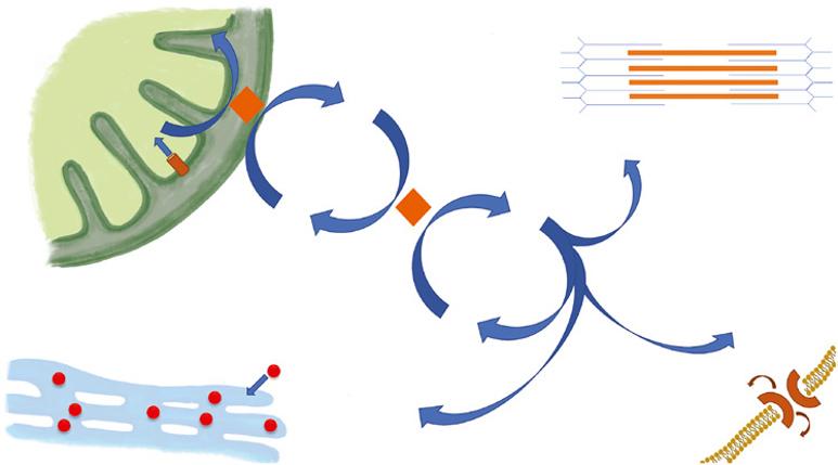
The primary function of muscles is to generate mechanical force, which is essential for the movement of different body parts while accomplishing fundamental functions such as walking, manufacturing and handling objects, moving the eyes, expanding and compressing the lungs, controlling the opening and closing of larynx among many others. Production of mechanical force requires energy that is provided by the hydrolysis of a high‐energy transfer phosphate bond in adenosine 5′‐triphosphate (ATP) to produce adenosine diphosphate (ADP) and inorganic phosphate. The flow of energy continuously matches the energy demand through the phosphocreatine (PCr) shuttle, a system that facilitates transfer of high‐energy phosphate from muscle cell mitochondria to myofibrils (Figure 3.1). Interestingly, more than 95% of creatine in the body is located in striate muscle, where the fluctuation of energy utilization is the highest [3]. Beyond contraction, skeletal muscle health also requires a constant flux of energy to maintain the activity of the sodium/potassium pumps and ensure calcium transport and sequestration in compartments. The energy for these activities is a substantial
Figure 3.1 Phosphocreatine (PCr) shuttle: the ATP generated by the complex V of the electron transport chain converts creatine into PCr in mitochondrial matrix, which in turn allows ADP phosphorylation in the sarcoplasm. The ATP generated will fuel the muscle contraction through interaction with the myosin chains of the sarcomere, the maintenance of membrane, and calcium (Ca2+) sequestration in the sarcoplasmic reticulum. ADP = adenosine diphosphate; ATP = adenosine 5′‐triphosphate; CK = creatine kinase; K+ = potassium; Na+ = sodium.
portion of the total energy consumption in resting muscle, but accounts only for a small percentage of energy utilization during intense contraction [4].
The concentration of ATP in human quadriceps muscles is ~5.5 mM (expressed per 1 kg of whole muscle tissue) [5] and during contraction the rate of ATP hydrolysis increases to ~18 mM/min (moderate intensity) to 55–80 mM/min for submaximal isometric contraction, and as high as 160 mM/min for a dynamic contraction generating maximal power. Thus, in the absence of a fresh supply, the ATP already present could only support 5.5/80 = 0.0685 minute or ~4 seconds of contraction. Hence, efficient and intense production of force in skeletal muscle requires continuous ATP regeneration, which occurs through the hydrolysis of PCr. During a brief exercise the decline of PCr and increase of inorganic phosphorous are the only evident biochemical changes in muscle tissue [6]. Of note, even though PCr functions as an accumulator of chemical energy, its concentration is only fourfold greater than that of ATP and, therefore, could only support contraction for a few more seconds if not continuously recharged by ATP produced by mitochondria. At low levels of exercise, the system can stay stable for prolonged time, but when the exercise becomes intense it overcomes the capacity of energy generation, both aerobically and anaerobically [6]. This is the reason why intense and repeated contractions can be sustained only for a short time, and as the rate of energy production slows down with aging, the time prior to fatigue becomes progressively shorter. Of note, when the contraction ceases, the ATP generated by mitochondria fully recharges PCr that rises back to its pre‐exercise concentration. The rate of PCr recovery is assessed by 31phosphorous magnetic resonance spectroscopy to estimate maximal mitochondrial function [7].