Instant ebooks textbook Flow cytometry of hematological malignancies, 2e (jun 8, 2021)_(1119611253)_
Flow Cytometry of Hematological Malignancies, 2e (Jun 8, 2021)_(111961 1253)_(Wiley-Blackwell).pdf
Claudio
Ortolani
Visit to download the full and correct content document: https://ebookmass.com/product/flow-cytometry-of-hematological-malignancies-2e-jun8-2021_1119611253_wiley-blackwell-pdf-claudio-ortolani/
More products digital (pdf, epub, mobi) instant download maybe you interests ...
Practical Flow Cytometry in Haematology: 100 Worked Examples 1st Edition u2013 Ebook PDF Version
Electrocardiography of Arrhythmias-A Comprehensive Review-A Companion to Cardiac Electrophysiology, 2e (Mar 8, 2021)_(032368050X)_(Elsevier).pdf Mithilesh Kumar Das
All rights reserved. No part of this publication may be reproduced, stored in a retrieval system, or transmitted, in any form or by any means, electronic, mechanical, photocopying, recording or otherwise, except as permitted by law. Advice on how to obtain permission to reuse material from this title is available at http://www.wiley.com/go/permissions.
The right of Claudio Ortolani be identified as the author of this work has been asserted in accordance with law.
Registered Office(s)
John Wiley & Sons, Inc., 111 River Street, Hoboken, NJ 07030, USA
John Wiley & Sons Ltd, The Atrium, Southern Gate, Chichester, West Sussex, PO19 8SQ, UK
Editorial Office
9600 Garsington Road, Oxford, OX4 2DQ, UK
For details of our global editorial offices, customer services, and more information about Wiley products visit us at www.wiley.com.
Wiley also publishes its books in a variety of electronic formats and by print‐on‐demand. Some content that appears in standard print versions of this book may not be available in other formats.
Limit of Liability/Disclaimer of Warranty
The contents of this work are intended to further general scientific research, understanding, and discussion only and are not intended and should not be relied upon as recommending or promoting scientific method, diagnosis, or treatment by physicians for any particular patient. In view of ongoing research, equipment modifications, changes in governmental regulations, and the constant flow of information relating to the use of medicines, equipment, and devices, the reader is urged to review and evaluate the information provided in the package insert or instructions for each medicine, equipment, or device for, among other things, any changes in the instructions or indication of usage and for added warnings and precautions. While the publisher and authors have used their best efforts in preparing this work, they make no representations or warranties with respect to the accuracy or completeness of the contents of this work and specifically disclaim all warranties, including without limitation any implied warranties of merchantability or fitness for a particular purpose. No warranty may be created or extended by sales representatives, written sales materials or promotional statements for this work. The fact that an organization, website, or product is referred to in this work as a citation and/or potential source of further information does not mean that the publisher and authors endorse the information or services the organization, website, or product may provide or recommendations it may make. This work is sold with the understanding that the publisher is not engaged in rendering professional services. The advice and strategies contained herein may not be suitable for your situation. You should consult with a specialist where appropriate. Further, readers should be aware that websites listed in this work may have changed or disappeared between when this work was written and when it is read. Neither the publisher nor authors shall be liable for any loss of profit or any other commercial damages, including but not limited to special, incidental, consequential, or other damages.
Library of Congress Cataloging‐in‐Publication Data
Names: Ortolani, C. (Claudio), author.
Title: Flow cytometry of hematological malignancies / Claudio Ortolani. Description: Second edition. | Hoboken, NJ : Wiley-Blackwell, 2021. | Includes bibliographical references and index. | Description based on print version record and CIP data provided by publisher; resource not viewed.
Identifiers: LCCN 2020028333 (print) | LCCN 2020028334 (ebook) | ISBN 9781119611301 (epub) | ISBN 9781119611271 (Adobe PDF) | ISBN 9781119611257 (cloth)
LC record available at https://lccn.loc.gov/2020028333
LC record available at https://lccn.loc.gov/2020028334
Cover Design: Wiley
Cover Image: courtesy of Claudio Ortolani
Set in 9.5/12pt Minion by SPi Global, Pondicherry, India
10 9 8 7 6 5 4 3 2 1
Foreword to the Second Edition
It has been about a decade since Dr. Claudio Ortolani gave us the first edition of Flow Cytometry in Hematologic Malignancies, and in that time flow cytometry has solidified its place as a routine diagnostic test in the diagnosis and classification of leukemias and lymphomas. Over the decade, dozens of new markers have been discovered, many of which are now a standard part of the flow cytometry arsenal. It is thus timely that Dr. Ortolani has chosen to update his work with this new second edition. As before, this work is extensively researched and prodigiously referenced, with more than 1300 new references not included in the first edition, bringing the total to more than 4500 relevant citations. These references not only cover the new markers, but also include new information about common markers in use for years, and descriptions of the markers covered by the first edition have been expanded to discuss these new advances.
In compiling this book, Dr. Ortolani has maintained the unique structure of the first edition, with the first part of the book oriented around individual antigens, and the second around diseases, supplementing the comprehensive text with characteristic images to illustrate the key points raised. The first section includes detailed information about normal tissue distribution of antigens, and also includes a discussion of some key pitfalls in the interpretation of cytometric findings of many antigens, before discussing pathologic conditions in which the antigens may be found. The disease section has been extensively
revised and now follows the 2016 version of the WHO classification, while also including some newer entities not mentioned in the WHO monograph. In keeping with the changes in classification, this book now includes a much more extensive discussion of phenotypic properties of the different types of T‐cell lymphomas and histiocytic neoplasms than was available previously.
This book will be a valuable resource for anyone interested in flow cytometry and hematologic malignancies. Trainees can easily find detailed phenotypic information about any disease they are learning about, while even an expert, struggling with an unusual case with a phenotype that appears contradictory to an expected diagnosis, can quickly learn whether others have found similar patterns in that diagnosis; moreover, because the citations are so extensive, it is possible simply by considering the numbers of references to a phenotype to assess the weight of evidence for a particular interpretation. Those of us in the field are grateful that Dr. Ortolani took the time to update his work and produce a reference that will be useful for many years to come.
Michael J. Borowitz, MD, PhD Professor of Pathology and Oncology Director of Hematopathology and Flow Cytometry Johns Hopkins Medical Institutions Baltimore, MD, USA
Foreword to the First Edition
Flow cytometry is a crucial tool in the diagnosis of hematolymphoid neoplasms, determining prognosis and monitoring response to therapy. Clinical flow cytometric immunophenotyping, however, is a complex field requiring extensive expertise in normal and abnormal patterns before clinical tests can be appropriately interpreted. Those new to the field are left with the conundrum of how best to achieve this expertise. At particular disadvantage is the resident or clinical fellow seeking to interpret flow cytometric data on a specific patient. The typical flow cytometry reference text is written in an encyclopedic format with extensive narrative that is not conducive to looking up the meaning of unusual test results. Furthermore, the general flow cytometry textbook, although a useful reference, cannot completely cover all the aspects needed to interpret clinical flow cytometry data. Therefore, Flow Cytometry of Hematological Malignancies fills a much needed role in hematopathology and hematology/ oncology. The presentation is oriented toward the diagnostic laboratory in the academic center as well as in the general hospital.
Flow Cytometry of Hematological Malignancies is organized in a novel manner that makes it especially useful for the medical student and residents/fellows still in training, while still providing a valuable resource for hematopathologists, hematologists/ oncologists and experts in the field of clinical flow cytometry. It lists antigens typically studied in clinical flow cytometry laboratories, from CD1 to CD138, followed by a discussion of general as well as flow cytometric features and hematolymphoid neoplasms expressing each antigen. Thus, when interpreting a clinical flow cytometry report, one can easily research an unusual antigen expressed by a leukemia or lymphoma. This pattern
of organization makes more sense than only presenting lists of neoplastic processes and the expected flow cytometric findings. One has to first know the diagnosis on a particular patient before such a reference can be useful. Flow Cytometry of Hematological Malignancies also provides the usual description of typical flow cytometric immunophenotypical findings in the various hematolymphoid neoplasms. This is useful as a reference for panel design as well as diagnosis.
Flow Cytometry of Hematological Malignancies is being published at a time when the field is expanding rapidly and flow cytometry is assuming an even greater role in management of patients with hematolymphoid neoplasia. Dr Ortolani, an outstanding flow cytometrist, possesses extensive expertise in the clinical arena. For over 30 years he was employed in the Clinical Pathology Department of the Venice General Hospital, running one of the first diagnostic flow cytometry units in Italy. His main clinical activity was the diagnosis of hematological neoplasms, with a particular interest in lymphoproliferative diseases. Dr Ortolani has also been very active in teaching flow cytometry in many national and international courses. He has written what I believe to be an outstanding textbook covering the essential aspects of clinical flow cytometry. Dr Ortolani is to be commended for this brilliant contribution that is sure to become a well-used textbook in clinical centers around the world.
Maryalice
Stetler-Stevenson, PhD,
MD
Chief, Flow Cytometry Laboratory National Cancer Institute, National Institutes of Health Bethesda, MD, USA
Foreword to the First Edition
The European Society for Clinical Cell Analysis (ESCCA) is proud to present this volume by Dr Claudio Ortolani. Flow Cytometry of Hematological Malignancies is a benchtop companion for all who are involved in the complex process of characterization and diagnosis of leukemias and lymphomas by immunophenotypical techniques and flow cytometry.
This volume is a useful quick reference text for the matching of CD antigens with malignant hematological diseases, as defined by the WHO 2008 classification, taking into account antibody clones, features and behavior, with particular emphasis on variant forms and unexpected presentations.
After several decades of clinical and laboratory practice in this field, Dr Claudio Ortolani has meticulously prepared this
book under the auspices of the ESCCA. It represents a major achievement for the dissemination of knowledge in one of the most important specialties within clinical cell analysis, as the book aims to improve and standardize the diagnostic process of malignant blood diseases. As a result, communication between clinicians and laboratory operators should benefit!
Bruno Brando ESCCA President Director, Hematology Laboratory and Transfusion Center, Legnano Hospital, Milan, Italy
Preface to the Second Edition
Ten years have passed since the first edition of this book, 10 fruitful years full of new acquisitions in the field of Hematology. Many doubts have been clarified, new questions have been raised, and – above all – new therapeutic targets have been reached. Although molecular biology keeps assuming ever greater importance, especially in the field of acute leukemias and myeloproliferative neoplasms; nevertheless, flow cytometry still remains one of the fundamental pillars of hematopathology and becomes even more and more important in particular areas such as the evaluation of the minimal residual disease and the study of the pharmacodynamic characteristics of experimental therapies with monoclonal antibodies.
In these 10 years, technology has also made substantial progress, and today we have much more efficient tools than before, but also much more complex ones, and consequently the
pre‐analytical and analytical components have become considerably complicated. Even if an attempt to account for these problems has been made, in‐depth treatment of most of them is beyond the scope of this book; they will be dealt with – hopefully – in the next edition, together with the many other topics neglected this time.
Claudio Ortolani MD Adjunct Professor of Laboratory of Diagnostic Cytometry Urbino University Urbino, Italy
The cytometric analysis of hematological malignancies is one of the most difficult applications of flow cytometry, requiring both a good knowledge of hematopathology and good control of the technique. Moreover, the effort of operators is made harder by the continuous evolution of the technology and by the continuous progress in the comprehension of the nature of the diseases. This book was compiled from a series of notes originally intended for people practically involved in the field of diagnostic flow cytometry, and it is an example of what the author would have liked to consult at the beginning of his own career. The goal of this book is to offer the reader a quick and updated source of information on the phenotype of the hematological malignancies recognized by the last WHO classification, with the major exception of Hodgkin lymphoma which because of its peculiar nature is still beyond the limits of flow cytometry, even if things promise to change in the next few years.
The author may have unwittingly sown a number of mistakes and imprecisions, and he will be grateful to all colleagues who report these to him. He also realizes that this book could not have been written without the help of many friends and colleagues. Being unable to cite all of them, the author wants to particularly thank his friend Bruno Brando, current President of the European Society for Clinical Cell Analysis, for the continuous moral and practical support he has given over the years.
Claudio
Ortolani MD Former Director of the Flow Cytometry Unit Clinical Pathology Department Venice General Hospital
Non clustered (or primarily known with other names) antigens
Bcl‐2 Protein, 119
Chemokines and Chemokine Receptors, 121
CRLF2, 128
Cytotoxic Proteins, 129
HLA‐DR, 130
Immunoglobulins, 132
KIR, CD158 isoforms, 136
Myeloperoxidase (MPO), 139
NG2, 140
PCA‐1, 141
ROR‐1, 141
SLAM Molecules and SLAM‐associated Protein (SAP), 142
SOX11, 144
T‐cell Receptor (TCR), 145
Terminal Deoxy‐nucleotidyl‐transferase (TdT), 148
Toll‐like Receptors (TLR), 150
VS38, 151
ZAP‐70, 152
CD1 Antigens
General features
CD1 antigens are a group of five different glycoproteins that weigh 43–49 kD and are encoded by a group of genes situated on the long arm of chromosome 1 [1].
CD1 antigens play a role in the presentation of lipidic and glycolipidic antigens to T and NKT cells [1,2], and can be divided in three groups, namely:
• Group I, encompassing CD1a, CD1b, and CD1c
• Group II, encompassing CD1d
• Group III, encompassing CD1e, which is an intracytoplasmic protein.
CD1 antigens are mainly expressed on cells belonging to T and B lineages and on antigen‐presenting cells (APC).
As for the T lineage, CD1a, CD1b, and CD1c antigens have been demonstrated on the membrane of the cortical or “common” thymocytes [3] and on the membrane of some T‐lymphocyte subsets in cord and neonatal peripheral blood [4]; according to some authors, a low expression of CD1 antigens can be demonstrated in the cytoplasm of T lymphocytes activated in vitro by phytohemagglutinin (PHA) [5]. CD1d has been reported at a high density on common thymocytes, and at a lower density on medullary thymocytes [6].
As for the B lineage, both CD1c and CD1d have been demonstrated on the precursors and on some subsets of mature B lymphocytes. More precisely:
• CD1c has been demonstrated on some subsets of B lymphocytes in the peripheral blood [7,8], in the spleen [7,8], and in the mantle of the germinal center [7]
• CD1c(+) B lymphocytes account for the majority of B cells in tonsils [9], in cord blood [4], in the peripheral blood of
newborns [4], and in the peripheral blood of subjects submitted to autologous or allogeneic bone marrow transplantation during the first year following transplant [8]
• CD1d has been demonstrated on the membrane of bone marrow B precursors [10], on B lymphocytes in peripheral blood [11,12], and on a subset of B lymphocytes in the mantle of the germinal center [11] and in the spleen [13].
As for the APC, CD1a, CD1b, and CD1c antigens have been commonly reported on dendritic cells [6]. Moreover:
• CD1a has been demonstrated on Langerhans cells [14], where it is expressed at an intensity of 1600 molecules per cell [15], on some CD11b(+) CD14(+) mononuclear cells reported in the peripheral blood of burnt subjects and interpreted as Langerhans cell precursors migrating from bone marrow to epidermis [16], on monocytes activated with granulocyte‐macrophage colony‐stimulating factor (GM‐CSF) in vitro [17], and on in vitro monocyte‐derived dendritic cells [18]
• CD1b has been demonstrated on monocytes activated with GM‐CSF in vitro [17] and on a subset of Langerhans cells [19]
• CD1c has been demonstrated on monocytes activated with GM‐CSF in vitro, on Langerhans cells [19], and on a minor subset of myeloid dendritic cells (MDC) characterized by CD11c(++) CD123(±) phenotype [20]
• CD1d has been demonstrated on “resting” monocytes [11], on dendritic cells of the dermis [21], on dendritic cells in peripheral blood [6], and on in vitro monocyte‐derived dendritic cells [21].
CD1d has also been demonstrated in keratinocytes of psoriatic skin [22], in the cells of human scalp hair follicles [22], in the epithelial cells of the gut and other organs [23], and in adipocytes [24].
CD1a, CD1b, CD1c, and CD1d have been demonstrated in the “foam cells” of the atherosclerotic plaque [25].
The cytometric demonstration of molecules belonging to the CD1 family should be performed taking the following points into account:
• cytometric studies have demonstrated that activated T lymphocytes express CD1c on the membrane only when kept at room temperature, and fail to mount the molecule on the surface when kept at +4 or to +37°C [26]
• the expression of CD1a on the surface of the leukemic blasts can fluctuate spontaneously after a short period of incubation in vitro [27].
The antibodies specific for CD1 antigens do not behave in the same way.
It should be kept in mind that CD1a features four different epitopes, the first of which is recognized by clones D47, Na1/34, and L119, the second by clone L404, and the third by clone L504 [28]; it should be noted that CD1a on the cells of B‐cell chronic lymphocytic leukemia (B‐CLL) can be demonstrated only with clones other than OKT6 or Na1/34 [29].
The clones 7C4 and IOT6b recognize different epitopes of CD1b [30].
Diagnostic features
CD1 in myelodysplastic and chronic myeloproliferative diseases
CD1a can be detected by immunohistochemistry (IHC) in the neoplastic cells of the cutaneous localization of chronic myelomonocytic leukemia (CMML); a case is known displaying S‐100(−), CD1a(+), CD4(+), CD56(+), CD123(−), and Langerin/ CD207(−) phenotype, mimicking the phenotype of the indeterminate dendritic cell tumor (IDCT) [31].
CD1a, CD1b, and CD1c are expressed on the membrane of the blast cells in the 20% of cases of chronic myeloid leukemia in blastic crisis (CML‐BC) [27].
The expression of CD1d has been reported on the cells of the juvenile myelomonocytic leukemia (JMML) [32].
CD1 antigens in neoplastic diseases of B‐cell
precursors
In a group of 80 patients affected by childhood B‐cell acute lymphoblastic leukemia (B‐ALL), the expression of CD1d has been demonstrated in 15% of the cases [10]. CD1d expression is significantly associated with pre‐B phenotype, rearrangement of the gene KMT2A (formerly known as MLL), and shorter global survival [10].
CD1 antigens in neoplastic diseases of T‐cell precursors
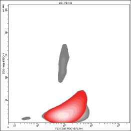
In T‐cell acute lymphoblastic leukemia (T‐ALL) the expression of CD1a antigen has been detected in a percentage of cases ranging from 40% to 50% of cases [27,4565]. CD1a antigen is generally expressed on the cells of T lymphoblastic leukemia/ lymphoma (T‐ALL/LBL) related to the stage of cortical or “common” thymocyte [3,27] (Fig. 1.1). According to the European Group for the Immunological Characterization of Leukemias (EGIL) classification of T‐ALL, CD1a antigen is typically present in the T III form but missing in the T I, T II, and T IV forms [34]. If CD13 is negative, the expression of CD1a is related to good survival [35], while the expression of CD1a together with CD10 is associated with the presence of the t(5;14)(q35;q32) translocation [33,4594,4595].
CD1 antigens in acute myeloid leukemias
The expression of CD1a and of CD1d has been repeatedly reported on the surface of the blasts of the acute myeloid leukemias (AML) [27,32,33]. According to some authors, the expression of CD1a and CD1d is restricted to French–American–British (FAB) subtypes characterized by a monocytic component [32]. In a percentage of cases, the expression of CD1a can be accompanied by the presence of other histiocytic markers (S‐100, CD163, Langerin/CD207) and is interpreted as a sign of histiocytic differentiation [36].
CD3
CD4
CD1a
CD8
SSC (A)
(B) (C)
CD45
Figure 1.1 Peripheral blood from a subject affected by T‐ALL. The blasts (red) express phenotype CD45(dim+) (A), CD1a(+) (B), CD3(+) (heterogeneous) (B), CD4(+) (C), CD8(+) (partial) (D).
Table 1.1 Differential diagnosis of CD5(−) CD10(−) B‐CLPDs
B‐CLPD CD1d CD200
HCL Positive Positive
LPL Dimly positivePositive
MZL Positive Negative
B‐CLPD: B chronic lymphoproliferative disease; HCL: hairy cell leukemia; LPL: lymphoplasmacytic lymphoma; MZL: marginal zone lymphoma.
CD1 antigens in neoplastic diseases of mature B cells
It has been reported that B‐cell prolymphocytic leukemia (B‐PLL) cells express CD1c [9,37], that Burkitt lymphoma (BL) cells do not express CD1c [7], and that hairy cell leukemia (HCL) cells express CD1a [38] and CD1c [9]. The cells of B‐CLL have been reported to express CD1a [9,29], CD1c [7], and CD1d; it is noteworthy that CD1a on B‐CLL cells could be demonstrated only with clones other than OKT6 or Na1/34 [29].
CD1d is usually expressed by the cells of the B‐CLPDs, but at an intensity depending on the type of disease. Its intensity in B‐CLL is typically lower than in normal B cells, but it is progressively higher on MCL, HCL‐variant (HCL‐v), lymphoplasmacytic lymphoma (LPL), splenic marginal zone lymphoma (SMZL), and HCL cells [12]. The combined exploration of CD1d and CD200 seems very promising in the differential diagnosis of CD5(−) CD10(−) B‐CLPDs as well, inasmuch as a recent study has shown that HCL is CD1d(+) CD200(+), LPL is CD1d(−/±) CD200(+), and MZL is CD1d(+) CD200(−) [39] (Table 1.1).
CD1d antigen seems of particular interest in B‐CLL workup, because its intensity of expression and the percentage of CD19(+) CD1d(+) cells are bad prognostic predictors [40,41]; according to some authors [42,43], but not to others [40], CD1d intensity seems to correlate with the absence of somatic hypermutations [43].
Multiple myeloma (MM) cells express CD1d in the early stages but tend to reduce its expression with disease progression [44].
CD1 antigens in neoplastic diseases of mature T and natural killer (NK) cells
The expression of CD1a antigens has been reported in rare cases of peripheral T‐cell lymphoma (PTCL) [45] and in an isolated case of adult T‐cell leukemia/lymphoma (ATLL) [46].
Overexpression of CD1d mRNA has been detected in Sezary’s cells by RT‐PCR [47], but this report has not yet been confirmed by phenotypic studies.
CD1 antigens in neoplasms of histiocytes and dendritic cells
As for the neoplasms of histiocytic and dendritic cells, CD1a, CD1b, and CD1c have been demonstrated with immunohistochemical techniques in the Langerhans cell histiocytosis (LCH) [19,48,49]. One of the most typical features of the pulmonary involvement in LCH is the occurrence of more than 5% of CD1(+) cells in the bronchoalveolar lavage fluid (BALF) [50]. Cells with CD1a(+), CD207(+), CD11b(±), and CD11c(++) phenotype have been detected in the peripheral blood of patients with active LCH [51]. CD1a expression has been reported in an anecdotal case interpreted as acute leukemia of Langerhans cell precursors based on the presence of Birbeck granules and of the ability of blasts to develop dendritic processes when cultured in vitro [52].
CD1a has been demonstrated with immunohistochemical techniques in the IDCT [53], but neither in the follicular dendritic cell sarcoma (FDCS) nor in the interdigitating dendritic cell sarcoma (IDCS) [49].
Partial expression of CD1a and expression of CD1c have been reported on the cells of an exceptional case of leukemia classified as Langerhans cell/dendritic cell leukemia, which occurred in a patient suffering from myelodysplastic syndrome (MDS). This case displayed CD11c(+), CD123(−), CD141(+), CD303(−), CD304(±/−), and CD207/Langerin(+/−) phenotype [54].
CD1 antigens in other pathological conditions
CD1a has been reported on the cells of the so called “indolent T‐lymphoblastic proliferation” (iT‐LBP) [55], an entity not recognized by the 2017 WHO classification (see also the dedicated paragraph).
CD2 Antigen
General features
CD2 is a 45–58 kD glycoprotein belonging to the superfamily of the immunoglobulins, which is encoded by a gene situated on the short arm of chromosome 1. CD2 is an adhesion molecule, constitutes the ligand of the CD58 molecule [56] and interacts with CD48 and CD59 molecules as well [57].
CD2 is normally expressed on thymocytes, on whose membrane it begins to appear at the prothymocyte level [58], and on mature T lymphocytes [59].
Not all mature T lymphocytes co‐express CD2; in the peripheral blood small subsets of T lymphocytes exist that show CD3(+) CD2(−) phenotype and are characterized by the expression of T‐cell receptor (TCR) either with αβ [60] or γδ chains [61].
The expression of CD2 is not restricted to the T lineage. Indeed, it is well known that CD2 is expressed:
• on 70–90% of the NK cells negative for CD3 [62,63], where it is upregulated by activation [64]
• on a minority of follicular dendritic cells (FDC) [65]
• on a subset of mononuclear peripheral cells interpreted as precursors of MDCs [66]
• on a subset of peripheral monocytes characterized by the co‐expression of Fcε receptor (FcεRI) [67]
• on a subset of plasmacytoid dendritic cells (PDCs) [68]
• on a small subset of B cells in fetal liver [69], in fetal bone marrow [69], in thymus [70], in peripheral blood [71], and in the bone marrow of normal subjects [71].
Cytometric features
The staining of peripheral normal lymphocytes with an anti‐CD2 monoclonal antibody (MoAb) generates a positive histogram with a narrow gaussian‐like peak, clearly separated from the negative component, with a channel peak representing the presence of 24 ± 7 E03 ABC (antibody‐binding capacity) [72].
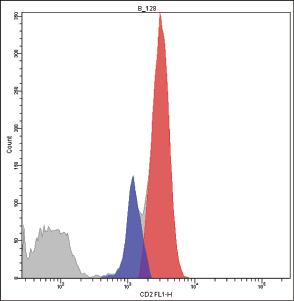
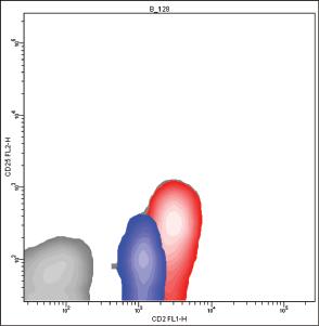
Bimodal histograms can often be seen, especially when immune system activation is ongoing, because a higher number of CD2 molecules is expressed on activated cells [73] (Fig. 1.2).
In these cases, the CD2(bright+) population tends to show higher values of forward and side scatter than the CD2(dim+) population.
CD2(bright+) cells nearly exclusively express CD45R0, while the CD2(dim+) population display either CD45R0 or CD45RA [74]; the staining of CD2(+) CD45RA(+) cells with an anti‐CD2 MoAb generates a histogram with a channel peak representing the presence of 21 ± 4 E03 ABC while the staining of CD2(+) CD45R0(+) lymphocytes with the same MoAb generates a histogram with a channel peak representing the presence of 55 ± 9 E03 ABC [72].
In accordance with the state of chronic activation caused by HIV infection, the lymphocytes of HIV‐infected subjects seem to express a higher amount of CD2 molecules [75], whereas a reduced expression has been documented on the lymphocytes of elderly subjects [76]. According to some authors, the expression of CD2 on NK cells is inhomogeneous, being more intense in the CD16 dim CD56 bright subset than in the CD16 bright CD56 dim subset [77].
Not all the anti‐CD2 MoAbs behave in the same way; some clones are able to inhibit the E‐rosette formation [78], while others are able to activate T lymphocytes in vitro [79].
Diagnostic features
CD2 in myelodysplastic and chronic myeloproliferative diseases
CD2 has been reported in a third of cases of CMML [80].
CD2 in neoplastic diseases of mast cells
CD2 has been reported together with CD22 and CD25 on the neoplastic mast cells in systemic mastocytosis (SM) and acute mast cell leukemia (AMCL) [81–83]. However, an aberrant CD2 (and CD25) expression can be found on mast cells in the bone marrow of subjects without evidence of neoplasms of mast or myeloid stem cell [84].
SSC
CD2
(A)
(B)
CD2
CD25
Figure 1.2 The histogram produced by the cytometric analysis of CD2 is bimodal (A), because the activated CD25(+) lymphocytes (red) express more CD2 molecules than CD25(−) lymphocytes (blue) (B).
CD2 in neoplastic diseases of B‐cell precursors
CD2 has been reported in 1–4% of the globally considered neoplastic diseases of B‐cell precursors [85–87], where it is associated with a worse prognosis [88].
Nonetheless, the expression of CD2 has been detected in just less than 25% of the cases of B‐ALL with KMT2A‐MLLT3 translocation [4565].
CD2 in neoplastic diseases of T‐cell precursors
According to the EGIL classification of T‐lymphoblastic leukemias, CD2 is typically present in the T II, T III, and T IV forms, but is missing in the most immature form, T I [34,89]. In a pediatric group of more than 100 cases of T‐ALL, CD2 has been detected in 80% of the patients [4565].
CD2 is generally expressed by the cases with TCRαβ, but only by some cases with TCRγδ [90,91]; its presence in childhood cases is correlated with an increased probability of maintaining complete remission [92].
CD2 in AML and BPDCN
Depending on the survey, the expression of CD2 has been reported in 3–34% of the observed cases [93–100]. CD2 has been reported:
• on the blasts of pediatric AML‐M2 negative for translocation t(8;21) [101]
• on the promyelocytes of AML‐M3, with predilection for the microgranular variant (AML‐M3v) [99,102,103], and for the presence of the “short” type of the PML‐RARA fusion gene [100,103]
• on both the monocytic and non‐monocytic neoplastic cells of AML‐M4 [80,99,104]
• on the blasts of AML‐M5 [80]
• on the blasts of de novo AML with inv3(q21q26.2) and monosomy 7 [105–107].
The presence of CD2 (and also of CD4, CD7, and CD56) on the blasts of AML is correlated with an increased risk of extramedullary disease (granulocytic sarcoma, and cutaneous, gingival, and meningeal involvement) [108], and with a lower incidence of complete remission [109]. The CD2 expression has been reported in cases of AML with morphological anomalies mimicking the picture of Chediak–Higashi disease (pseudo Chediak–Higashi, PCH) [110], and in some cases of BPDCN, also known as plasmacytoid dendritic cell leukemia (PDCL) [111]. The presence of CD2 on AML‐M3 promyelocytes correlates with the occurrence of thrombotic events [112].
In AML‐M4 with inv(16)/t(16;16), the expression of CD2 is variable, and has been reported as weaker in cases with fusion transcript CBFβ‐MYH11 other than type A [113].
CD2 in neoplastic diseases of mature B cells
Sporadic reports exist signaling the presence of CD2 in isolated cases of B lineage non‐Hodgkin lymphoma [114,115]. Since CD2
has been demonstrated on the surface of normal B lymphocytes [71], it is theoretically possible that these cases constitute a clonal expansion of very infrequent normal B cells rather than an expansion of B‐cell with an aberrant phenotype.
The expression of CD2 has occasionally been demonstrated in the sporadic B‐CLL [71], but it seems particularly frequent in familial B‐CLL, where it appears in 13% of the cases [116]; the demonstration of CD2 on the cells of a patient affected by B‐CLL suggests that clinical investigations should be extended to the relatives as well [116].
Furthermore, CD2 has been demonstrated in some cases of follicular lymphoma (FCL) [71], in some cases of diffuse large B‐cell lymphoma (DLBCL) [71,117–120], in some cases of DLBCL associated with pyothorax (PAL) [121], in some cases of HCL [71], and in a case of MM [122].
CD2 in neoplastic diseases of mature T and NK cells
CD2 is generally expressed on the cells of the neoplastic diseases of mature T and NK cells, but it may also be missing or expressed in an aberrant way. In the peripheral T lymphoma not otherwise specified (PTCLnos), about a third of the cases has been reported to show an aberrant antigen expression [123,124]. An aberrant CD2 expression has been reported with immunohistochemical methods in atypical cutaneous T‐cell infiltrates of subjects affected by mycosis fungoides (MF) [125], and with flow cytometric (FCM) methods on neoplastic lymphocytes of subjects affected by Sézary syndrome (SS) [126], by T‐cell chronic lymphocytic leukemia (T‐CLL) and by ATLL [127].
The CD2 expression is more constant in the cases of angioimmunoblastic T‐cell lymphoma (AITL) [124], while in T‐cell large granular lymphocytic leukemia (T‐LGL) it has been reported either as constant [128] or as variable [129]. The cases of CD8(+) cutaneous T‐cell lymphoma (CD8(+) CTCL) with CD2(+) CD7(−) phenotype show a better prognosis than those with phenotype CD2(−) CD7(+) [130].
In the neoplastic diseases of mature NK cells, CD2 may be missing [131] but it has been reported in most cases of chronic NK cell lymphocytosis (CLPD‐NK/CNKL) [131–133], of aggressive NK cell leukemia (ANKL) [134,135], and of NK lymphoma [136].
CD2 in Hodgkin lymphomas
CD2 has been demonstrated by IHC in the Hodgkin and Reed–Sternberg (HRS) cells of isolated cases of classic Hodgkin lymphoma (CHL), but in most instances, its expression has been judged as aberrant [4626,4627].
Increased expression of CD5 has been documented by FCM on non‐neoplastic T cells in the background of pathological tissues from CHL [4636,4637].
CD2 in neoplasms of histiocytic and dendritic cells
CD2 has been demonstrated by IHC on the membrane of the cells of LCH [137].
CD3 Antigen
General features
CD3 is made up of five different chains, i.e. γ, δ, ε, ζ, and η. Chains
γ, δ, ε, and η are encoded by a gene on the long arm of chromosome 11 [138], while chain ζ, separately clustered as CD247 [139], is encoded by another gene on the long arm of chromosome 1 [140]. In T cells, CD3 transmits the activation signal produced by the engagement of TCR [141,142].
There is evidence that the CD3/TCR complex forms a multimeric array together with the tyrosine‐phosphatase CD45, with a tyrosine‐kinase, and with the CD7, which takes part in signal transmission [143].
The expression of the δ and ε chains is restricted to T lymphocytes, with two important exceptions:
• the fetal and adult activated NK cells, which can contain δ and ε chains in the cytoplasm [144,145]
• the PDCs, in the cytoplasm of which ε chains have been demonstrated [146].
As a rule, γ and ζ chains are present in NK cells as well [147], as either homodimers or heterodimers [148]. In NK cells, both chains are not covalently linked with the transmembrane tail of the CD16, and transmit the signal produced by the linkage between CD16 and the IgG crystallizable fragment [149–151].
Signal transduction by ζ chain is carried out by the intracytoplasmic protein ZAP‐70 [152,153].
During normal T‐cell maturation, the CD3 appears in the cytoplasm at the prothymocyte level but is only expressed on the membrane from the common thymocyte stage on [3,154–159].
During normal T‐cell maturation, the TCR and the CD3 complex are assembled together before they are expressed on the surface [160]; consequently, the TCR is not normally expressed on the membrane in the absence of CD3, and vice versa [161,162].
CD3 has been demonstrated by IHC in the cytoplasm of a subset of Warthin–Finkeldey polykaryocytes, which can be seen in tonsils during the measles prodromal period and are probably derived from T lymphocytes [163].
Cytometric features
Almost all anti‐CD3 monoclonal antibodies are specific for an ε chain epitope [164,165]. The staining of peripheral normal
lymphocytes with an anti‐CD3ε MoAb generates a positive histogram with a narrow gaussian‐like peak, clearly separated from the negative component, with a channel peak representing the presence of 57 ± 7 E03 ABC [72].
Evidence does exist that the number of CD3ε chains is not the same for every T lymphocyte but is particularly high on γδ T cells [166], which express about 116 ± 15 E3 ABC per cell [167].
Among peripheral T lymphocytes, CD3 expression tends to vary depending on the T lymphocyte subset. Evidence does exist that, in comparison to CD8(bright+) T lymphocytes, the CD3 mean fluorescence intensity (MFI) of positive cells is almost twice as intense in CD4(+) T cells, while T CD8(dim+) lymphocytes behave similarly to CD4(+) lymphocytes. This behavior does not depend on cellular dimensions, inasmuch as in CD4(+) lymphocytes the scatter values are even lower than in CD8(bright+) ones [75].
A reduced expression of CD3 has been reported in other cases:
• in alveolar T cells, with a greater negative modulation in CD4(+) cells [168]
• in activated T cells that infiltrate nasal polyps [169]
• in intrathyroidal T lymphocyte subsets in autoimmune thyroid disease [170]
• in intestinal intraepithelial T lymphocytes [171]
• in T cells of patients given OKT3 rescue treatment for transplant rejection [172]
• in T cells of patients with HIV infection [75,173]
• in T cells of aged subjects [76]
• in a minor subset of peripheral T lymphocytes characterized by low CD4 expression, and positivity for CD25 and HLA‐DR [174].
This CD3 downmodulation might be due to the activation state common to the great majority of the reported cases; it is important to bear in mind that a CD3 downmodulation can be caused by apoptosis as well [175].
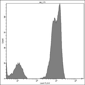
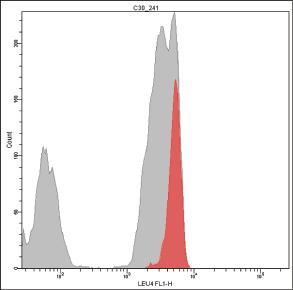
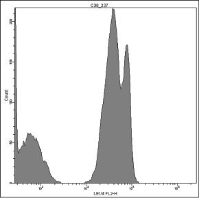
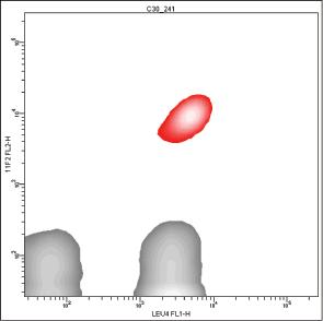
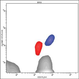
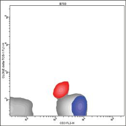
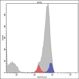
In some cases, the positive histogram can appear with a bimodal shape, mostly due to the presence of a consistent subset of γδ T cells, which actually bear more TCR/CD3ε complexes than αβ T cells on the membrane [167] (Fig. 1.3). In our experience, this behavior does not occur with the γδ T lymphocytes mounting Vδ1/Jδ1 sequences stained by δTCS1 MoAb (Fig. 1.4).
Sometimes it is possible that the bimodality of the CD3(+) peak is due to the presence of a clonal T‐cell population, homogeneously expressing the molecule at an intensity that differs from
Figure 1.3 Pattern of expression of T‐specific CD3 antigen on peripheral T lymphocytes. The positive peak can show a bimodal appearance (A–C), because γδ(+) T lymphocytes (red) express more CD3 molecules than αβ(+) T lymphocytes (B, D).
Figure 1.4 Different patterns of expression of T‐specific CD3 antigen on peripheral γδ(+) T lymphocytes. Vδ1/Jδ1(+) γδ(+) T lymphocytes (red) express CD3 less brightly than the Vδ1/Jδ1(−) γδ(+) T lymphocytes (blue) (A–C). Vδ1/Jδ1(+) γδ(+) T lymphocytes were recognized by MoAb δ‐TCS‐1, and γδ(+) T lymphocytes were recognized by MoAb 11F2.
CD3CD3
other normal residual T cells. This behavior is frequently reported in patients affected by mature T‐cell malignancies [123,176].
In comparison to mature T cells, thymocytes express CD3 with a different intensity; as a rule, most common or cortical CD1(+) CD4(+) CD8(+) thymocytes express low amounts of CD3, while mature or medullar CD1(−) and CD4(+) or CD8(+) thymocytes express the molecule in the same way as mature T lymphocytes [155,177], with a differential higher expression on CD4(+) CD8(−) T cells [75].
A CD4(+) CD8(+) thymocyte subset has been reported expressing high levels of CD3; it is hypothesized that this subset is a late differentiation stage between cortical and medullar thymocytes [178].
As mentioned previously, the CD3 can be looked for both on the membrane and in the cytoplasm of the cell. The demonstration of the intracytoplasmic molecule requires the use of permeabilization techniques which allow intracellular entry of the antibody. Although they could be improved by some optimization procedures [179], such techniques can rely on the use of standardized commercial permeabilizing solutions [180–183].
Great care should be spent in the evaluation of membrane CD3 expression in T‐CLPDs; it is indeed possible that in some cases the neoplastic cells dismiss the antigen after prolonged staining procedures. This possibility is also suggested by the cytograms published in a recent report which demonstrated a bimodal CD3(−)/ CD3(dim+) population in directly stained samples but only a monomodal CD3(−) population in permeabilized ones [184].
MoAb OKT3, SK7/Leu4 and UCHT‐1
The three monoclonal antibodies OKT‐3, SK7/Leu4 and UCHT‐1 recognize CD3ε chains in cells transfected with genes coding for ε and δ chains or for ε and γ chains, but do not recognize CD3ε chains in cells transfected with genes coding for ε chains only [185]. This behavior suggests that the three antibodies recognize a conformational ε chain epitope, depending on the association of ε chain with δ or γ chain, and are not able to detect isolated intracytoplasmic ε chains [185].
Consequently, a negativity for intracytoplasmic ε chains accomplished with one of the aforementioned antibodies is not sufficient proof of ε chain absence, and should be validated using an antibody specific for isolated ε chains, such as SP34 and APA 1/1, or a polyclonal rabbit antiserum raised against a synthetic polypeptide mimicking a sequence on the intracytoplasmic tail of the ε chain [186]. This point is of some practical importance. Given that in thymocyte cytoplasm δ and ε chains are simultaneously expressed from the prothymocyte level onwards [187], these three antibodies are perfectly suitable for demonstrating the intracytoplasmic CD3 antigen in T‐cell malignancies but could miss it in some cases of NK neoplasms. It has been also reported that MoAb UCHT‐1 is able to stain the cerebellar Purkinje cells [188].
MoAb WT31
In the same way as OKT‐3, SK7/Leu4 and UCHT‐1, the MoAb WT31 recognizes CD3ε chains in cells transfected with genes coding for ε and δ chains or for ε and γ chains, but do not recognize CD3ε chains in cells transfected with genes coding for ε chains only [185]. This behavior confirms that, contrary to the original hypothesis [189] and in keeping with successive remarks [164], MoAb WT31 is not specific for a TCRαβ determinant, but binds a conformational epitope on CD3ε chains, and should be considered a bona fide anti‐CD3 antibody.
Nevertheless, it should be stressed that the epitope recognized by MoAb WT31 is particularly accessible to this MoAb in the case of TCRαβ co‐expression; this condition makes MoAb WT31 fit for the presumptive identification of TCRαβ T cells, especially if used in combination with a second antibody specific for the same chain. In this case, the sterical hindrance between the two antibodies blocks the binding between WT31 and the ε chain of T cells bearing TCRγδ, and WT31 behaves like a MoAb specific for TCRαβ only.
In these conditions, the staining of peripheral normal T lymphocytes with the WT31 MoAb generates a histogram with a negative peak encompassing T cells bearing TCRγδ (Fig. 1.5).
The removal of the sterical hindrance allows the WT31 MoAb to bind the ε chain of T cells with TCRγδ, although in a weaker way than TCRαβ T cells. Indeed, if we stain a sample containing a high number of γδ T cells using both the WT31 MoAb and a second MoAb specific for TCRγδ, the WT31 MoAb will generate a histogram with a first positive peak which encompasses γδ negative T cells, and a second positive but intermediate peak which encompasses γδ positive ones (Fig. 1.6).
From a practical point of view, the possibility of sterical hindrance between the WT31 MoAb and another anti‐CD3ε MoAb suggests that a sequential staining procedure should be performed, in which the sample is incubated first with WT31 alone and then with the other anti‐CD3ε antibody.
MoAb T3
The FITC‐conjugated form of T3 displays unexpected behavior [190]. In a multicolor analysis which combines a MoAb specific for TCRγδ (clone 11F2) and a second anti‐CD3ε MoAb (clone SK7), the FITC‐conjugated form of T3 does not recognize γδ T cells (Fig. 1.7).
It is interesting to notice that in this model, T3‐FITC behaves very similarly to WT31, which is shown for comparison (Fig. 1.8).
The anomalous behavior of T3‐FITC is difficult to explain. The small molecular volume of FITC rules out a sterical hindrance effect, and the independence of the phenomenon from the length of incubation does not suggest affinity variations induced by the conjugation procedures.
It has been observed that, owing to a different glycosylation pattern, CD3δ chains in γδ T cells display a more acidic isoionic