All rights reserved. No part of this publication may be reproduced, stored in a retrieval system, or transmitted, in any form or by any means, electronic, mechanical, photocopying, recording or otherwise, except as permitted by law. Advice on how to obtain permission to reuse material from this title is available at http://www.wiley.com/go/permissions.
The right of Anne M. Zajac, Gary A. Conboy, Susan E. Little, Mason V. Reichard to be identified as the authors of this work has been asserted in accordance with law.
Registered Office
John Wiley & Sons, Inc., 111 River Street, Hoboken, NJ 07030, USA
Editorial Office
111 River Street, Hoboken, NJ 07030, USA
For details of our global editorial offices, customer services, and more information about Wiley products visit us at www.wiley.com.
Wiley also publishes its books in a variety of electronic formats and by print‐on‐demand. Some content that appears in standard print versions of this book may not be available in other formats.
Limit of Liability/Disclaimer of Warranty
The contents of this work are intended to further general scientific research, understanding, and discussion only and are not intended and should not be relied upon as recommending or promoting scientific method, diagnosis, or treatment by physicians for any particular patient. In view of ongoing research, equipment modifications, changes in governmental regulations, and the constant flow of information relating to the use of medicines, equipment, and devices, the reader is urged to review and evaluate the information provided in the package insert or instructions for each medicine, equipment, or device for, among other things, any changes in the instructions or indication of usage and for added warnings and precautions. While the publisher and authors have used their best efforts in preparing this work, they make no representations or warranties with respect to the accuracy or completeness of the contents of this work and specifically disclaim all warranties, including without limitation any implied warranties of merchantability or fitness for a particular purpose. No warranty may be created or extended by sales representatives, written sales materials or promotional statements for this work. The fact that an organization, website, or product is referred to in this work as a citation and/or potential source of further information does not mean that the publisher and authors endorse the information or services the organization, website, or product may provide or recommendations it may make. This work is sold with the understanding that the publisher is not engaged in rendering professional services. The advice and strategies contained herein may not be suitable for your situation. You should consult with a specialist where appropriate. Further, readers should be aware that websites listed in this work may have changed or disappeared between when this work was written and when it is read. Neither the publisher nor authors shall be liable for any loss of profit or any other commercial damages, including but not limited to special, incidental, consequential, or other damages.
Library of Congress Cataloging‐in‐Publication data applied for ISBN: 9781119300779 (Paperback)
Cover Design: Wiley
Cover Image: Courtesy of Anne M. Zajac
Set in 10/12pt Times New Roman by SPi Global, Pondicherry, India
10 9 8 7 6 5 4 3 2 1
Preface ix
Acknowledgments xi
Authors xv
About the Companion Website xvii
CHAPTER 1 Fecal Examination for the Diagnosis of Parasitism 1
Collection of Fecal Samples 1
Storage and Shipment of Fecal Samples 2
Fecal Exam Procedures 2
Fecal Flotation 3
Additional Procedures for Fecal Examination 12
Quality Control for Fecal Exam Procedures 15
Use of the Microscope 16
Microscope Calibration 16
Pseudoparasites and Spurious Parasites 19
Identification of Nematode Larvae Recovered with Fecal Flotation or Baermann Procedures 24
Techniques for Evaluation of Strongylid Nematodes in Grazing
Animals 29
Fecal Culture 29
Identification of Ruminant and Camelid Third-Stage Larvae 30
Identification of Third-Stage Larvae of Equine Strongyles 35
Fecal Egg Count Reduction Test (FECRT) 36
Hoyer’s Solution 39
Lactophenol 40
Parasites of Domestic Animals 41
Dogs and Cats 42
Ruminants and Camelids 96
Horses 126
Swine 140
Birds 154
Rodents and Rabbits 174
Reptiles 182
CHAPTER 2 Detection of Protozoan and Helminth Parasites in the Urinary, Reproductive, and Integumentary Systems and in the Eye 191
Techniques for Parasite Recovery 191
Parasites of the Urinary System 191
Parasites of the Reproductive Tract 192
Helminth Parasites of the Integumentary System 192
Parasite Detection in Urinary and Other Systems 193
CHAPTER 3 Detection of Parasites in the Blood 207
Immunologic and Molecular Detection of Blood Parasites 207
Microscopic Examination of Blood for Protozoan Parasites 207 Giemsa Stain 208
Microscopic Examination of Blood for Nematode Parasites 209 Tests for Canine Heartworm Microfilariae in Blood Samples 210
Blood Parasites of Dogs and Cats 213
Blood Parasites of Livestock and Horses 228
Blood Parasites of Birds 234
CHAPTER 4 Immunodiagnostic and Molecular Diagnostic Tests in Veterinary Parasitology 239
Immunodiagnostic Methods in Parasitology 239
Molecular Diagnostic Methods in Parasitology 243
CHAPTER 5 Diagnosis of Arthropod Parasites 247
Subclass Acari (Mites and Ticks) 247
Mite Identification 247
Tick Identification 278
Class Insecta 300
Lice (Order Phthiraptera) 300
Fleas (Order Siphonaptera) 314
Flies (Order Diptera) 322
Other Insects 342
CHAPTER 6 Parasites of Fish 347
Techniques for Recovery of Ectoparasites 347
Skin Biopsy (Mucus Smear) 348
Fin Biopsy (Fin Snip) 348
Gill Biopsy (Gill Snip) 348
Recovery of Endoparasites 349 Parasites of Fish 350
CHAPTER 7 Treatment of Veterinary Parasites 371
Introduction 371
Anthelmintics 371 Specific Anthelmintics 372
Ectoparasiticides 375
Protozoal Treatment 378
Non-Traditional Treatments 379
CHAPTER 8 Diagnostic Dilemmas 381
Diagnostic Dilemma 1 381
Diagnostic Dilemma 2 382
Diagnostic Dilemma 3 383
Diagnostic Dilemma 4 383
Diagnostic Dilemma 5 384
Diagnostic Dilemma 6 385
Diagnostic Dilemma 7 386
Bibliography 387
Index 391
PREFACE
The first edition of Veterinary Clinical Parasitology was published in 1948 and has been used since that time by students, veterinary practitioners and others as an aid in the diagnosis of parasitic infections. Since 1994, it has been published under the auspices of the American Association of Veterinary Parasitology (AAVP) with the proceeds going to support student travel to the AAVP annual meeting. This sponsorship has not only supported development of more than one generation of veterinary parasitologists, but has also involved a community of parasitologists in the production of the book. The relationship with AAVP continues with the 9th edition. Also, as with past editions, this edition focuses on morphologic identification of parasites, which continues to be widely used in veterinary medicine, increasingly in combination with molecular or immunologic techniques.
There are also some new features in the 9th edition. We have added a chapter summarizing information on modern parasiticides. Additionally, preceding the photographs of parasites for each common domestic animal in Chapter 1 there is a table listing current U.S. label‐approved treatments for many parasitic infections. We have also added another new chapter called Diagnostic Dilemmas. In this chapter we present challenging clinical scenarios and diagnostic results and allow the reader to make a diagnosis. A discussion of each of these scenarios is available on the accompanying website of the book.
The 9th edition also marks changes in the authorship of Veterinary Clinical Parasitology. This is the final edition that will be authored by Dr. Anne Zajac (author since the 6th edition) and Dr. Gary Conboy (author since the 7th edition). Drs. Susan Little and Mason Reichard, who joined as authors for this edition, will guide the book going forward and continue its association with AAVP.
Anne M. Zajac
Gary A. Conboy
Susan E. Little
Mason V. Reichard
ACKNOWLEDGMENTS
As ever, we are very grateful to the members of the American Association of Veterinary Parasitologists (AAVP) and others who have provided material for Veterinary Clinical Parasitology. Since the early 1990s appeals for photographs distributed through the AAVP listserv have brought responses from around the world.
We would also like to particularly acknowledge the extensive contributions to this edition of three of our veterinary parasitology colleagues, Dr. Mani Lejeune, Cornell University; Dr. Yoko Nagamori, Zoetis Corporation (formerly of Oklahoma State University); and Dr. Heather Walden, University of Florida, who graciously offered their magnificent photo collections for our use.
Finally, as Anne Zajac and Gary Conboy step away from authorship of Veterinary Clinical Parasitology they would like to gratefully acknowledge technical staff who, over many years, have assisted in identification and preparation of clinical samples utiliz ed for photographs in this book. Those individuals include Susan King, Rosemary Cornett, John McInturff, Alex Fox and Diamond McClendon at Virginia Tech and Nicole Murphy, Robert Maloney and Janet Saunders at the University of Prince Edward Island.
Although the source of each figure is credited in the figure legend (with the exception of photos provided by chapter authors), we would also like to list all the contributors here with our deepest thanks:
Mr. Gary Averbeck, College of Veterinary Medicine, University of Minnesota, Minneapolis, MN
Dr. David Baker, School of Veterinary Medicine, Louisiana State University, Baton Rouge, LA
Dr. Byron Blagburn, College of Veterinary Medicine, Auburn University, Auburn, AL
Dr. Katie Boes, Virginia‐Maryland College of Veterinary Medicine, Virginia Tech, Blacksburg, VA
Dr. Dwight Bowman, College of Veterinary Medicine, Cornell University, Ithaca, NY
Dr. Erin Burton, University of Minnesota College of Veterinary Medicine, St. Paul, MN
Dr. Lyle Buss, Entomology and Nematology Department, University of Florida, Gainesville, FL
Dr. Katie Clow, Ontario Veterinary College, University of Guelph, Guelph, Ontario, Canada
Dr. George Conder, Pfizer Corporation, Kalamazoo, MI (retired)
Dr. Kathryn Duncan, College of Veterinary Medicine, Oklahoma State University, Stillwater, OK
Dr. Hany M. Elsheikha, School of Veterinary Medicine and Science, University of Nottingham, Loughborough, UK
Dr. James Flowers, College of Veterinary Medicine, North Carolina State University, Raleigh, NC
Dr. Alvin Gajadhar, Centre for Animal Parasitology, CFIA, Saskatoon, Saskatchewan, Canada
ACKNOWLEDGMENTS
Mr. James Gathany, Centers for Disease Control and Prevention, Atlanta, GA
Dr. Ellis C. Greiner, College of Veterinary Medicine, University of Florida, Gainesville, FL (retired)
Ms. Parna Ghosh, College of Veterinary Medicine, Oklahoma State University, Stillwater, OK
Dr. Larry Hammell, Atlantic Veterinary College, University of Prince Edward Island, Charlottetown, PEI, Canada
Dr. Bruce Hammerberg, College of Veterinary Medicine, North Carolina State University, Raleigh, NC
Dr. Patricia Holman, College of Veterinary Medicine and Biomedical Sciences, Texas A&M University, College Station, TX
Dr. Jennifer Ketzis, School of Veterinary Medicine, Ross University, St. Kitts, W.I.
Dr. Manigandan LeJeune, Animal Health Diagnostic Center, Cornell University, Ithaca, NY
Dr. David Lindsay, Virginia‐Maryland College of Veterinary Medicine, Virginia Tech, Blacksburg, VA
Ms. Megan Lineberry, College of Veterinary Medicine, Oklahoma State University, Stillwater, OK
Dr. Aaron Lucas, Taylorsville Veterinary Clinic, Mt. Airy, MD
Dr. Eugene Lyons, Department of Veterinary Science, University of Kentucky, Lexington, KY
Dr. Charles Mackenzie, College of Veterinary Medicine, Michigan State University, East Lansing, MI
Dr. Gil Myers, Myers Parasitological Service, Magnolia, TN
Dr. Yoko Nagamori, Zoetis Corp., Stillwater, OK
Dr. Stephen Jones, Lakeside Animal Hospital, Moncks Corner, SC
Dr. Thomas Nolan, School of Veterinary Medicine, University of Pennsylvania, Philadelphia, PA
Dr. Christopher Paddock, Centers for Disease Control and Prevention, Atlanta, GA
Dr. Fernando Paiva, Universidade Federal de Mato Grosso do Sul, Campo Grande, MS, Brazil
Dr. Andrew Peregrine, Ontario Veterinary College, University of Guelph, Guelph, Ontario, Canada
Dr. Sally Pope, Faculty of Veterinary Science, University of Sydney, Sydney, New South Wales, Australia
Dr. Steffan Rehbein, Merial GmbH, Rohrdorf, Germany
Dr. Robert Ridley, College of Veterinary Medicine, Kansas State University, Manhattan, KS
Dr. Meriam Saleh, Virginia-Maryland College of Veterinary Medicine, Virginia Tech, Blacksburg VA
Dr. Nick Sangster, Charles Sturt University, Wagga Wagga, New South Wales, Australia
Dr. Philip Scholl, Porto Alegre, RS, Brazil
Dr. Stephen Smith, Virginia‐Maryland College of Veterinary Medicine, Virginia Tech, Blacksburg, VA
Dr. Karen F. Snowden, College of Veterinary Medicine and Biomedical Sciences, Texas A&M University, College Station, TX
Dr. T. Bonner Stewart, School of Veterinary Medicine, Louisiana State University, Baton Rouge, LA
ACKNOWLEDGMENTS
Dr. Bert Stromberg, College of Veterinary Medicine, University of Minnesota, Minneapolis, MN
Ms. Kellee Sundstrom, College of Veterinary Medicine, Oklahoma State University, Stillwater, OK
Dr. Donald B. Thomas, U.S. Department of Agriculture Subtropical Agriculture Research Laboratory, Weslaco, TX
Dr. Donato Traversa, Department of Comparative Biomedical Sciences, Faculty of Veterinary Medicine, Teramo, Italy
Mr. Chris Tucker, Department of Animal Science, University of Arkansas, Fayetteville, AR
Dr. Isabelle Verzberger‐Epshtein, NRC Institute of Nutrisciences and Health, Charlottetown, PEI, Canada
Mr. Martin Visser, Merial GmbH, Rohrdorf, Germany
Dr. Heather Walden, College of Veterinary Medicine, University of Florida, Gainesville, FL
Dr. Jerry Weintraub, Agriculture Canada, Lethbridge, Alberta, Canada
Dr. Jeffrey F. Williams, Vanson HaloSource Inc., Redmond, WA
Dr. Roy P. E. Yanong, Tropical Aquaculture Laboratory, University of Florida, Ruskin, FL
Dr. Tom Yazwinski, Department of Animal Science, University of Arkansas, Fayetteville, AR
Dr. Gary Zimmerman, Zimmerman Research, West Montana, Livingston, MT
Dr. Kurt Zimmerman, Virginia‐Maryland College of Veterinary Medicine, Virginia Tech, Blacksburg, VA
AUTHORS
Anne M. Zajac, DVM, PhD, Dip. ACVM‐Parasitology
Department of Biomedical Sciences and Pathobiology
Virginia‐Maryland Regional College of Veterinary Medicine
Virginia Tech Blacksburg, VA 24061
Gary A. Conboy, DVM, PhD, Dip. ACVM‐Parasitology
Department of Pathobiology and Microbiology
Atlantic Veterinary College University of Prince Edward Island Charlottetown, Prince Edward Island C1A 4P3 Canada
Susan E. Little, DVM, PhD, Dip. ACVM‐Parasitology Department of Veterinary Pathobiology College of Veterinary Medicine
Oklahoma State University Stillwater, OK 74078
Mason V. Reichard, MS, PhD
Department of Veterinary Pathobiology College of Veterinary Medicine
Oklahoma State University Stillwater, OK 74078
ABOUT THE COMPANION WEBSITE
This book is accompanied by a companion website: www.wiley.com/go/zajac/parasitology
• The website includes PowerPoints of all figures from the book for downloading.
• Chapter 8 Diagnostic Dilemma answers.
CHAPTER 1
Fecal Examination for the Diagnosis of Parasitism
The fecal examination for diagnosis of parasitic infections is one of the most common laboratory procedures performed in veterinary practice. Relatively inexpensive and noninvasive, fecal examination can reveal the presence of parasites in several body systems. Parasites inhabiting the digestive system produce eggs, larvae, or cysts that leave the body of the host by way of the feces. Occasionally, even adult helminth parasites may be seen in feces, especially when the host has enteritis. Parasitic worm eggs or larvae from the respiratory system are usually coughed into the pharynx and swallowed, and they too appear in feces. Mange or scab mites may be licked or nibbled from the skin, thus accounting for their appearance in the feces. Many parasitic forms seen in feces have characteristic morphologic features that, when combined with knowledge of the host, are diagnostic for a particular species of parasite. On the other hand, certain parasites produce similar eggs or oocysts, and cannot be identified to the species level (e.g., many of the strongylid-type eggs from livestock). Fecal examination may also reveal to a limited extent the status of digestion, as shown by the presence of undigested muscle, starch, or fat droplets.
COLLECTION OF FECAL SAMPLES
Fecal exams should be conducted on fresh fecal material. If fecal samples are submitted to the laboratory after being in the environment for hours or days, fragile protozoan trophozoites will have died and disappeared. The eggs of some nematodes can hatch within a few days in warm weather, and identification of nematode larvae is far more difficult than recognizing the familiar eggs of common species. Also, free-living nematodes rapidly invade a fecal sample on the ground, and differentiation of hatched parasite larvae from these free-living species can be time-consuming and difficult. Owners of small animals should be instructed to collect at least several grams of feces immediately after observing defecation. This will ensure the proper identification of the sample with the client’s pet (i.e., a sample from a stray animal will not be collected) and that feces rather than vomitus or other material is collected. The limited amount of feces recovered from the rectum on a thermometer or fecal loop should not
Veterinary Clinical Parasitology, Ninth Edition. Anne M. Zajac, Gary A. Conboy, Susan E. Little, and Mason V. Reichard.
be relied on for routine parasitologic examination, since many infections that produce only small numbers of eggs will be missed. Owners should be instructed to store fecal samples in the refrigerator if the sample will not be submitted for examination for more than an hour or two after collection.
Feces should be collected directly from the rectum of large animals. This is particularly important when identification of individual animals is needed. Rectal samples are also needed when the sample is to be examined for lungworm larvae or cultured for identification of third-stage larvae, since contaminating free-living nematodes and hatched first-stage larvae of gastrointestinal nematodes may be confused with lungworm larvae. If rectal samples are unavailable, owners should be asked to collect feces immediately after observing defecation. The process of development and hatching of common strongylid eggs can be slowed by refrigeration. Development is also reduced when air is excluded from the sample by placing the collected feces in a plastic bag and evacuating or pressing out the air before sealing the bag.
STORAGE AND SHIPMENT OF FECAL SAMPLES
If collected feces cannot be examined within a few hours, the sample should be refrigerated until it can be tested. Feces should not be frozen, because freezing can distort parasite eggs. If a sample needs to be evaluated for the presence of protozoan trophozoites like Giardia and trichomonads, it should be examined within 30 minutes after collection. The trophozoite is the active, feeding form of the parasite and is not adapted to environmental survival; it dies soon after being passed in the feces.
Increasingly, veterinary practitioners in the United States are using reference laboratories for routine diagnostic tests for parasite infection. Specific laboratory instructions for age, storage and transportation of samples to commercial labs should be followed. In general, when fresh fecal material is submitted to another laboratory for examination, it should be packaged with cold packs. In some cases, preservation of samples may be preferred. Helminth eggs can be preserved with a volume of 5%–10% buffered formalin equal to that of the sample. Formalin fixation also inactivates many other infectious organisms that may be present. Special fixatives, such as polyvinyl alcohol (PVA), are required to preserve protozoan trophozoites and are not routinely used in veterinary practices.
Slides prepared from flotation tests do not travel well, even if the coverslip is ringed with nail polish, since hyperosmotic flotation solutions will usually make parasite eggs or larvae unrecognizable within hours of preparing the slide. However, slides from flotation tests can be preserved for several hours to several days by placing them in a refrigerator in a covered container containing moist paper towels to maintain high humidity. It is best to place applicator sticks under the slide to prevent it from becoming too wet.
FECAL EXAM PROCEDURES
Before performing specific tests on the fecal sample, its general appearance should be noted; consistency, color, and the presence of blood or mucus may all be indicative of specific parasitic infections. Hookworm disease in dogs, for example, commonly produces dark, tarry feces, whereas diarrheic feces caused by whipworms may contain excess mucus and frank blood. The presence of adult parasites or tapeworm segments should also be noted.
Fecal Flotation
The technique most commonly used in veterinary medicine for examination of feces is the fecal flotation test. This procedure concentrates parasite eggs and cysts while separating them from much of the sample debris. Fecal flotation is based on the principle that parasite material present in the feces is less dense than the fluid flotation medium and thus will float to the top of the container, where it can be collected for microscopic evaluation. Flotation tests are easy and inexpensive to perform, but in busy practices the choice of flotation solution and test procedure often does not receive much consideration, despite the substantial effect these choices can have on the sensitivity of flotation exams.
Choice and Preparation of Flotation Solutions
Many different substances can be used to make flotation solutions. The higher the specific gravity (SPG) of the flotation solution, the greater the variety of parasite eggs that will float. Additionally, studies have shown that fecal flotation tests recover only a portion of each type of parasite egg/cyst in a sample because of variation in individual eggs, binding to debris, and so on. As SPG increases the portion recovered increases, which is an important consideration when the number of eggs in the sample is low. However, as SPG increases, more debris will also float, and the risk of damage to eggs from the hyperosmotic solution also increases. These factors limit the range of useful flotation solutions to SPG ranging from approximately 1.18 to 1.3. Both salt and sugar flotation solutions are commonly used in veterinary parasitology and provide flotation for common parasites with lower specific gravities than the flotation solution (Table 1.1).
Salt solutions are widely used in flotation procedures. A common flotation solution used in the United States is a commercially available sodium nitrate solution (SPG 1.20). This solution will float common helminth eggs and protozoan cysts. The commercial solution is not a saturated solution of sodium nitrate (SPG 1.33). Slides prepared with any salt solution need to be examined relatively quickly after they are prepared because crystals form as slides dry and parasites may be damaged, making them more difficult to identify.
Zinc sulfate (ZnSO4) solution at a SPG of 1.18–1.2 is another salt flotation solution. It is preferred at SPG 1.18 for recovery of Giardia, but recovers a higher proportion of
Table 1.1. Approximate specific gravity of some common helminth eggs
Species Specific gravity
Ancylostoma caninum 1.061
Toxocara canis 1.091
Toxocara cati 1.101
Trichuris vulpis 1.151
Taenia sp. 1.231
Physaloptera 1.241
Parascaris spp. 1.092
Equine strongyles 1.052
Anoplocephala perfoliata 1.062
Sources: From 1David E., and Lindquist W. 1982. Determination of the specific gravity of certain helminth eggs using sucrose density gradient centrifugation. J. Parasitol. 68:916–919; 2Norris J., Steuer A. et al. 2018. Determination of the specific gravity of eggs of equine strongylids, Parascaris spp., and Anoplocephala perfoliata. Vet. Parasitol. 260:45–48.
other parasites at SPG 1.2 and is probably used more frequently at this SPG. It is commercially available and when water is added to the purchased salt solution as directed, the resulting SPG is 1.2.
When detection of Giardia is required, a 33% zinc sulfate solution (SPG 1.18) is recommended because it does not cause the same rapid collapse of cysts seen with other flotation solutions and they are more easily recognized in flotation preparations.
Saturated solutions of sodium chloride (SPG 1.20) and magnesium sulfate (Epsom salts, SPG 1.32) are less widely used but can be easily prepared, are inexpensive, and are effective in floating common helminth eggs and protozoan cysts.
None of these salt flotation solutions will reliably float most trematode eggs, some tapeworm eggs, and very dense nematode eggs or larvae.
Another common solution used in routine flotation exams is Sheather’s sugar solution (SPG 1.25). Because of its relatively higher SPG, Sheather’s solution is also more efficient in recovering helminth eggs than common salt solutions, especially tapeworm and more dense nematode eggs. In addition, it does not distort eggs as rapidly as the salt solutions. Sheather’s solution is specifically recommended for recovery of Cryptosporidium oocysts in fecal samples, but it does not appear to be as effective as 33% ZnSO4 solution for detection of Giardia. Sheather’s solution is inexpensive and easy to prepare and is also commercially available in the United States, but it is more viscous and sticky than salt solutions. The advantages and disadvantages of these solutions are shown in Table 1.2 and instructions for preparing them are given below.
Although the SPG of flotation solutions is not often measured in practices, it can be easily determined with an inexpensive hydrometer from a scientific supply company. A hydrometer will last indefinitely and should be considered part of quality control for the veterinary practice laboratory.
Table 1.2. Comparison of commonly used flotation solutions
Sodium nitrate (NaNO3) Commercial product
Floats common helminth and protozoa eggs and cysts
Does not float most fluke and some tapeworm and nematode eggs 1.18–1.2
Saturated NaNO3 1.33
Zinc sulfate (ZnSO4)1.18–1.2Floats common helminth and protozoa eggs and cysts; preferred for Giardia and some lungworm larvae
Saturated sodium chloride (NaCl) 1.2
Saturated magnesium sulfate (Epsom salts) 1.32
Floats common helminth and protozoa eggs and cysts
Floats common helminth and protozoa eggs and cysts; higher SPG recovers parasites more efficiently
Sheather’s sugar solution 1.20–1.28Floats common helminth and protozoa eggs and cysts; higher SPG recovers parasites more efficiently; preferred for Cryptosporidium oocysts; generally less damaging than salt solutions
When used at SPG 1.18 recovers lower proportion of common helminth eggs; does not float most fluke and some some tapeworm and nematode eggs
Does not float most fluke and some tapeworm and nematode eggs
Higher SPG will float more debris; does not float most fluke and some tapeworm and nematode eggs
Does not float most fluke and some tapeworm and nematode eggs; creates sticky surfaces
33% ZINC SULFATE SOLUTION (SPG 1.18)
1. Combine 330 g zinc sulfate with water to reach a volume of 1000 mL.
2. Additional water or zinc sulfate can be added to produce an SPG of 1.18. If zinc sulfate solution is used with formalinized feces, the SPG should be increased to 1.20 and a SPG of 1.2 is often preferred for general use.
3. Check the SPG with a hydrometer.
SATURATED SODIUM CHLORIDE (NaCl,
SPG
1.2) OR MAGNESIUM SULFATE SOLUTION
(MgSO4, SPG 1.32)
1. Add salt to warm tap water until no more salt goes into solution and the excess settles at the bottom of the container.
2. To ensure that the solution is fully saturated, it should be allowed to stand overnight at room temperature. If remaining salt crystals dissolve overnight, more can be added to ensure that the solution is saturated. Table salt contains an anticaking compound that does not dissolve and should not be confused with residual sodium chloride crystals. Pickling salt does not contain this compound.
3. Check the SPG with a hydrometer, recognizing that the SPG of saturated solutions will vary slightly with environmental temperature.
SHEATHER’S SUGAR SOLUTION (SPG
1.2–1.25)
1. Combine 355 mL (12 fl oz) of water and 454 g (1 lb) of granulated sugar (sucrose). Corn syrup and dextrose are not suitable substitutes.
2. Dissolve the sugar in the water by stirring over low or indirect heat (e.g., the top half of a double boiler). If the container is placed on a high direct heat source, the sugar may caramelize instead of dissolving in the water.
3. After the sugar is dissolved and the solution has cooled to room temperature, add 6 mL formaldehyde USP to prevent microbial growth (30 mL of 10% formalin can also be used, with the volume of water reduced to 330 mL).
4. Check the SPG with a hydrometer.
Flotation Procedures
No matter how the flotation procedure is performed, the principle is the same. After mixing the flotation solution and the fecal sample together, the less dense material eventually floats to the top. This process can occur either by letting the mixture sit on the benchtop for a specified time (passive flotation) or by centrifuging the mixture. Centrifugation makes the flotation occur more rapidly and efficiently, regardless of the flotation solution used. Many practitioners like to use convenient commercial flotation kits that provide a container for collection of the sample and performing the test. However, the convenience of the kits is offset by the loss of sensitivity in the fecal exam procedure. A smaller amount of feces is used and the test cannot be centrifuged. The increased sensitivity of the centrifugation procedure is particularly important in infections where the diagnostic form of the parasite may be present in low numbers (e.g., Trichuris and Giardia infections in dogs and cats). Centrifugation is also necessary when using 33% ZnSO4 or sugar solution because of the slightly lower SPG of ZnSO4 solution and the high viscosity of sugar solution, both of which retard the flotation
process. A veterinary practice that does not centrifuge flotation tests and relies on the traditional benchtop technique substantially reduces the sensitivity of its fecal exams.
CENTRIFUGAL FECAL FLOTATION PROCEDURE
This is the best technique for the standard fecal flotation test regardless of the flotation solution used. It is particularly important to use this procedure with Sheather’s sugar and 33% ZnSO4 flotation solutions to ensure that the flotation is effective:
1. Mix 3–5 g (about 1 teaspoonful) of feces with a small amount of flotation solution in a paper or plastic cup. Cat feces and small ruminant pellets, which are sometimes too hard to break up easily, can be ground with a mortar and pestle or allowed to soak in water until they become softer.
If the sample appears to contain a large amount of fat or mucus, an initial water wash is performed, and water should be used in Step 1. The water wash may be eliminated for most fecal samples of normal appearance.
2. Strain the mixture of feces and flotation solution (or feces and water if a water wash is performed) through a double layer of cheesecloth or gauze. A tea strainer can also be used.
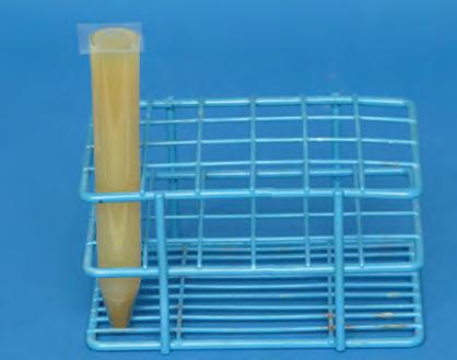
3. Pour the mixture into a 15-mL centrifuge tube. If the rotor on the centrifuge is not angled (i.e., if the tubes hang straight when not spinning), the centrifuge tube can be filled with flotation solution until a reverse meniscus is formed and a coverslip is added (Fig. 1.1). The tube is spun with the coverslip. The centrifugal force generated by the centrifuge will hold the coverslip in place. If the centrifuge has an angled rotor, fill the tube to approximately 10–12 mL (amount that will prevent spilling) and place in the centrifuge.
4. Spin the mixture in a benchtop centrifuge for about 5 minutes at approximately 500–650 × g (650 × g is 2500 rpm on a 4-in. rotor), regardless of whether the feces have been mixed with water or with the flotation solution. If a specific g force and speed setting cannot be determined, spinning the tube at the same speed used to separate serum from blood cells is sufficient.
If the initial spin is a water wash, the supernatant should be discarded, the sediment resuspended with flotation solution, and Steps 3 and 4 repeated.
Fig. 1.1 Centrifuge tube filled with flotation solution to the top and coverslip placed in contact with the fluid column.
5. Allow the centrifuge to stop without using the brake. The slight jerking that results from the use of the brake may dislodge parasites from the surface layer. If preferred, the tube can be allowed to sit for an additional 5–10 minutes to maximize recovery of parasite material that may not have completed flotation through the liquid column to the surface.
6. Following centrifugation, there are several ways to harvest the surface layer of fluid containing parasite eggs. If the tube has been spun with the coverslip in place, lift the coverslip off the tube and quickly place it on a microscope slide. When the tube is spun without the coverslip, remove the tube from the centrifuge after spinning and place in a test tube rack. Fill the tube with additional flotation solution to form a reverse meniscus. Place a coverslip on the tube and allow it to sit for an additional 5–10 minutes before removing the coverslip and placing it on a slide.
Alternatively, after the centrifuge comes to a stop, gently touch the surface of the fluid in the tube with a glass rod, microbiologic loop, or base of a small glass tube and then quickly touch the rod to a microscope slide to transfer the drop or two of adhering fluid. This procedure will be less efficient than allowing the tube to stand with a coverslip in place.
BENCHTOP (SIMPLE OR PASSIVE) FLOTATION PROCEDURE
When a centrifugal flotation procedure cannot be performed, sodium nitrate and saturated salt solutions can be used in a benchtop flotation test, although a number of studies have shown that this procedure will detect significantly fewer parasite infections than the centrifugal flotation. This technique is not recommended for 33% ZnSO4 or sugar flotation tests:
1. Mix several grams (a teaspoonful) of feces with the flotation solution in a cup.
2. Strain the mixture through cheesecloth or a tea strainer.
3. Pour into a test tube, pill vial, or container provided in a commercial kit. Add enough mixture or additional flotation fluid until there is a reverse meniscus on the top of the container. Place a coverslip on the fluid drop at the top.
4. Allow the flotation to stand for at least 10 minutes, remove the coverslip, place it on a slide, and examine. If the test is allowed to stand for too long, the salt may crystallize on the edges of the coverslip so that it will not lie flat on the slide.
FLOTATION SLIDES
Fecal flotation slides should be scanned using the 10× objective lens of the microscope (since most microscopes also have an eyepiece magnification of 10×, using the 10× objective gives a total magnification of 100×). Although most helminth eggs can be detected with the 4× objective, protozoan parasites are easily missed and this low power should not be used for scanning. The 40× lens should be used when there is uncertainty about the identity of structures on the slide and for scanning slides for Cryptosporidium oocysts. In some practices, to save expense, coverslips are not used. However, slides without coverslips dry out faster, do not have a flat plane of focus, and cannot be examined with the 40× lens, with the result that some parasites will be identified incorrectly or missed entirely.
Egg-counting techniques are also flotation tests and have several uses in food animals and horses. They can be used to assess the degree to which individual animals or groups are contributing to pasture contaminations with parasites, to assess efficacy of drug treatment (discussed in a later section in this chapter) and, in some cases, to determine relative levels of individual susceptibility to parasite infection.
Egg counts are of limited value in making judgments about the clinical condition of individual animals because many factors affect egg production, including parasite species, individual host immunity, and stage of infection. Also, counts performed on combined samples from a number of animals may not accurately reflect parasitism within that herd.
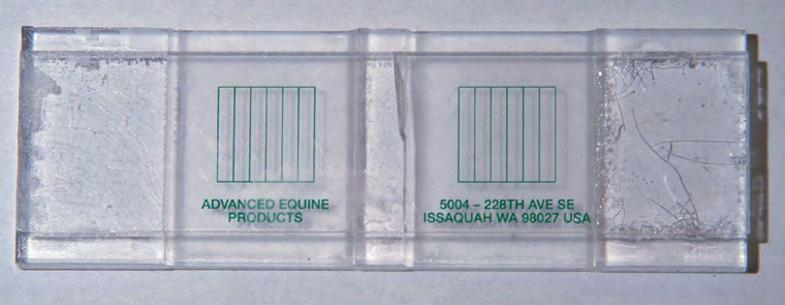
The easiest quantitative test to perform is the modified McMaster test. This test requires the use of special reusable slides (Fig. 1.2), which can be purchased from several suppliers (including Chalex Corporation, Wallowa, OR [www.vetslides.com]; Focal Point, www.mcmaster.co.za; JA Whitlock & Co., Eastwood, New South Wales, Australia [www.whitlock.com.au]). The capacity of the counting chamber and the number of chambers counted per sample affect the detection level of the test. Saturated salt solutions are usually used as the flotation solution in this test.
As it is most commonly used, the modified McMaster test has a detection level of 25 or 50 eggs per gram (EPG) of feces. This level is acceptable in many situations since parasite control programs do not usually require detection of lower egg numbers. However, the accuracy of the McMaster test is reduced when egg counts are at the lower limits of detection. When egg counts of less than 200 EPG are expected in a group of animals being evaluated (e.g., in adult cattle or camelids) or when it is important to detect low numbers of eggs more accurately (e.g., in fecal egg count reduction tests [FECRTs]), a modification of the test is appropriate. The sensitivity and precision of the McMaster test can be improved by using slides that allow examination of larger quantities of the egg/flotation solution mixture, or by counting additional aliquots of the sample.
Alternatively, other procedures with lower detection limits can be used including the Wisconsin sugar flotation test (double centrifugation procedure) or a modified Stoll egg-counting test. These procedures permit detection of fewer than 10 EPG of fecal material but are more time-consuming to perform. Another procedure, the mini-FLOTAC
1.2 McMaster slide used in the modified McMaster procedure for quantitative egg counts.
Fig.
test, has been developed in Europe. Several studies have demonstrated greater precision and accuracy of the mini-FLOTAC compared to McMaster and Wisconsin tests but at this time it has limited availability and has been used in the United States primarily in research.
Another commercially available test for determining equine fecal egg counts is Parasight (Lexington, KY [www.parasightsystem.com]). This system provides counts of equine strongyle and Parascaris spp. eggs. An equine sample is mixed with reagents and fluorescenceimaging is used with a software counting algorithm to produce a fecal egg count. Regardless of the procedure used to count parasites, the most important element is consistency. Each step of the procedure should be performed in the same way for every sample.
Modified McMaster test.
1. For ruminants, combine 4 g of fecal material with 56 mL of flotation solution to yield a total volume of 60 mL. The test can also be performed with 2 g of feces and 28 mL of flotation solution when only small amounts of feces are available. For horses, it is standard in the United States to use 4 g of feces and 26 mL of flotation solution. See Note 1 below for modified calculations. Portable electronic scales that weigh in 0.1-g increments are widely available and inexpensive. If a method of weighing feces is not available, adding manure to the measured flotation solution until the final desired volume is reached can also be used (e.g., add manure to 26 mL of fluid to a total volume of 30 mL). This method would be more accurate with horse or cattle manure compared to the pelleted manure of small ruminants or camelids.
2. Mix well and strain through cheesecloth or a tea strainer. The mixture does not have to be strained, but it will be much easier to read the slide if large pieces of debris are removed. An alternative to straining is to use a filter pipette to transfer material to the counting chamber (a pipette with 12 mesh/cm wire mesh at the end is available from JA Whitlock & Co. [www.whitlock.com.au]).
3. Immediately fill each chamber of the McMaster slide with the mixture using a pipette or syringe. The entire chamber must be filled, not just the area under the grid. If large air bubbles are present, remove the fluid and refill.
4. Allow the slide to sit for at least 5 minutes before examining to allow the flotation process to occur. There has been limited investigation of the maximum amount of time slides can be allowed to sit before reading and there is no standard recommendation. Allowing slides to sit for an hour does not seem to alter results.
5. Look at the slide with the 10× lens, focusing on the top layer, which contains the air bubbles. At this level, the lines of the grid will also be in focus. Count eggs, oocysts, and any other parasite stages, in each lane of both chambers. Each type of parasite should be counted separately. In some cases, eggs can be identified to genus or perhaps to species (e.g., Strongyloides, Trichuris, and Nematodirus), whereas others must be counted as a category of parasites (coccidia, strongylid eggs).
To determine the number of parasite EPG of feces, add the counts for both chambers for each parasite. The most commonly used McMaster slides in the United States are calibrated so that the number of eggs counted in a single chamber represents the number present in 0.15 mL of fecal mixture. If both chambers are counted and the results are added, the total represents the number of eggs present in 0.3 mL, which, for
example, is 1/200th of a total volume of 60 mL; therefore, the number of eggs counted must be multiplied by 200. However, if a total of 4 g of feces was used in the test, the result must be divided by 4 to yield EPG of feces. Multiplying by 200 and dividing by 4 is equivalent to multiplying the number of eggs counted by 50. Therefore, each egg observed represents 50 EPG in the final count. The same level of detection can be achieved by using 2 g of fecal material and 28 mL of flotation solution. The smaller amount of feces may be preferred when evaluating small lambs or kids.
Additional Notes
1. If a detection level of 25 EPG is desired, the McMaster test should be performed with 4 g of feces and 26 mL of flotation solution (results are then multiplied by 100 and divided by 4 as described earlier). Other combinations of manure and flotation solution can also be used with appropriate calculations using the formula
eggs gnoeggs counted /. //TV F
where T = total volume of feces/flotation solution mixture, V = volume of aliquot examined in slide, and F = grams of feces used.
2. If pelleted feces are very hard, 2–5 mL of water can be added first and left to soften manure for at least an hour. The flotation solution is then added to the softened manure (flotation solution volume reduced by the amount of water used to soften feces).
3. A variety of methods can be used to homogenize feces and liquid. As alternatives to mixing with a tongue blade or other utensil, some laboratories use a mechanical or handheld kitchen mixer to homogenize feces and water/flotation solution. Shaking manure and fluid in a jar with glass beads has also been used, but as previously stated, whatever method is used, it should be consistent across samples.
4. In another procedure for the McMaster test, feces is mixed initially with water, strained, and centrifuged, and the supernatant is discarded. Flotation solution is added to resuspend the sediment and mixed, and the mixture is used to fill the counting chambers. For example, 3 g of feces is mixed with 42 mL of water, strained and the fluid used to fill a 15-mL centrifuge tube, which is then centrifuged at 300–650 × g for 2 minutes. The supernatant is then discarded and flotation solution is added to partially fill the tube, which is either shaken or stirred to resuspend the sediment. Additional flotation solution is added to fill the tube, and the counting chamber is filled. If the eggs in a volume of 0.3 mL are counted, the number of eggs seen is multiplied by 50 to give the number of eggs per g. The EPG can also be calculated using the formula described in Note 1. This procedure is the most effective for reducing debris but increases the time required to perform each test.
Mini-FLOTAC
The Mini-FLOTAC device and the Fill-FLOTAC device, which are used together in the mini-FLOTAC procedure, are available in North America from the University of Georgia. The test should be performed as described in the brochure provided with the test device. This procedure is generally more time-consuming than the basic modified McMaster procedure but is more accurate and precise.