A ESTHESIA AND CO-EXISTING DISEASE
Rollerta L. Hines
Katheri11e .E. Marschall
Roberta L. Hines, MD
Nicholas M. Greene Professor and Chairman
Department of Anesthesiology
Yale University School of Medicine
Chief of Anesthesiology
Yale-New Haven Hospital
New Haven, Connecticut
Chapter 29: Psychiatric Disease, Substance Abuse, and Drug Overdose
Natalie F. Holt, MD, MPH
Assistant Professor of Anesthesiology
Yale University School of Medicine
New Haven, Connecticut
Associate Director of Anesthesiology
VA Connecticut Healthcare System
West Haven, Connecticut
Chapter 22: Renal Disease
Chapter 27: Diseases Related to Immune System Dysfunction
Chapter 28: Cancer
Viji Kurup, MBBS
Associate Professor of Anesthesiology
Yale University School of Medicine
New Haven, Connecticut
Chapter 2: Obstructive Respiratory Diseases
Chapter 3: Restrictive Respiratory Diseases and Lung Transplantation
William L. Lanier, Jr., MD
Professor of Anesthesiology
Mayo Clinic
Rochester, Minnesota
Chapter 13: Diseases Affecting the Brain
Chapter 14: Spinal Cord Disorders
Chapter 15: Diseases of the Autonomic and Peripheral Nervous Systems
Linda L. Maerz, MD
Associate Professor of Surgery and Anesthesiology
Yale University School of Medicine
New Haven, Connecticut
Chapter 4: Critical Illness
Adnan Malik, MD
Assistant Professor of Anesthesiology
Yale University School of Medicine
New Haven, Connecticut
Chapter 10: Heart Failure and Cardiomyopathies
Thomas J. Mancuso, MD
Senior Associate in Anesthesiology
Boston Children’s Hospital
Associate Professor of Anesthesiology
Harvard Medical School
Boston, Massachusetts
Chapter 30: Pediatric Diseases
Luiz Maracaja, MD
Assistant Professor of Anesthesiology
University of Texas Health Science Center in San Antonio
San Antonio, Texas
Chapter 11: Pericardial Disease and Cardiac Trauma
Katherine E. Marschall, MD, LLD (honoris causa)
Anesthesiologist–retired
New Haven, Connecticut
Chapter 25: Skin and Musculoskeletal Diseases
Chapter 29: Psychiatric Disease, Substance Abuse, and Drug
Overdose
Veronica Matei, MD
Assistant Professor of Anesthesiology
Yale University School of Medicine
New Haven, Connecticut
Chapter 20: Nutritional Diseases: Obesity and Malnutrition
Bryan G. Maxwell, MD, MPH
Anesthesiologist
Randall Children’s Hospital
Portland, Oregon
Chapter 7: Congenital Heart Disease
Raj K. Modak, MD
Associate Professor
Director of Cardiac Anesthesia
Henry Ford Medical Group
Henry Ford Hospital
Detroit, Michigan
Chapter 11: Pericardial Disease and Cardiac Trauma
Tori Myslajek, MD
Assistant Professor of Anesthesiology
Yale University School of Medicine
New Haven, Connecticut
Chapter 18: Diseases of the Gastrointestinal System
Adriana D. Oprea, MD
Assistant Professor of Anesthesiology
Yale University School of Medicine
New Haven, Connecticut
Chapter 24: Hematologic Disorders
Jeffrey J. Pasternak, MS, MD
Associate Professor of Anesthesiology
Mayo Clinic
Rochester, Minnesota
Chapter 13: Diseases Affecting the Brain
Chapter 14: Spinal Cord Disorders
Chapter 15: Diseases of the Autonomic and Peripheral Nervous Systems
Wanda M. Popescu, MD
Associate Professor of Anesthesiology
Yale University School of Medicine
New Haven, Connecticut
Chapter 10: Heart Failure and Cardiomyopathies
Chapter 20: Nutritional Diseases: Obesity and Malnutrition
Stanley H. Rosenbaum, MA, MD
Professor of Anesthesiology, Internal Medicine & Surgery
Yale University School of Medicine
New Haven, Connecticut
Chapter 4: Critical Illness
Robert B. Schonberger, MD, MA
Assistant Professor of Anesthesiology
Yale University School of Medicine
New Haven, Connecticut
Chapter 21: Fluid, Electrolyte, and Acid-Base Disorders
Denis Snegovskikh, MD
Attending Anesthesiologist
Anesthesia Associates of Willimantic, CT
Willimantic, Connecticut
Chapter 31: Pregnancy-Associated Diseases
Jochen Steppan, MD, DESA
Assistant Professor of Anesthesiology and Critical Care Medicine
Johns Hopkins University School of Medicine
Baltimore, Maryland
Chapter 7: Congenital Heart Disease
Hossam Tantawy, MBChB
Assistant Professor of Anesthesiology and Trauma Surgery
Yale University School of Medicine
New Haven, Connecticut
Chapter 18: Diseases of the Gastrointestinal System
Chapter 19: Inborn Errors of Metabolism
Jing Tao, MD
Assistant Professor of Anesthesiology
Yale University School of Medicine
New Haven, Connecticut
Chapter 2: Obstructive Respiratory Diseases
Chapter 19: Inborn Errors of Metabolism
Russell T. Wall, III, MD
Chair, Department of Anesthesiology and Perioperative Care
MedStar Georgetown University Hospital
Professor of Anesthesiology and Pharmacology
Georgetown University School of Medicine
Washington, DC
Chapter 23: Endocrine Disease
Zachary Walton, MD, PhD
Attending Anesthesiologist
Anesthesia Associates of Willimantic, CT
Willimantic, Connecticut
Chapter 31: Pregnancy-Associated Diseases
Kelley Teed Watson, MD
Anesthesiologist
Anesthesiology of Greenwood, Inc.
Greenwood, South Carolina
Chapter 8: Abnormalities of Cardiac Conduction and Cardiac Rhythm
Christopher A.J. Webb, MD
Assistant Professor of Anesthesiology
Columbia University Medical Center
New York, New York
Chapter 17: Diseases of the Liver and Biliary Tract
Paul David Weyker, MD
Assistant Professor of Anesthesiology
Columbia University Medical Center
New York, New York
Chapter 17: Diseases of the Liver and Biliary Tract
Jean G. Charchaflieh
Jing Tao, Viji Kurup
Restrictive Respiratory Diseases and Lung Transplantation 33
Ranjit Deshpande, Viji Kurup
Critical Illness 53
Linda L. Maerz, Stanley H. Rosenbaum 5 Ischemic Heart Disease 79
Shamsuddin Akhtar
Valvular Heart Disease 107
Adriana Herrera 7 Congenital Heart Disease 129
Jochen Steppan, Bryan G. Maxwell
Abnormalities of Cardiac Conduction and Cardiac Rhythm 151
Kelley Teed Watson
and Pulmonary Arterial Hypertension 183
Manuel Fontes, Paul M. Heerdt
Heart Failure and Cardiomyopathies 199
Wanda M. Popescu, Adnan Malik
Pericardial Disease and Cardiac Trauma 225
Raj K. Modak, Luiz Maracaja 12 Vascular Disease 237
Loreta Grecu, Nikhil Chawla
Diseases Affecting the Brain 265
Jeffrey J. Pasternak, William L. Lanier, Jr.
Cord Disorders 305
Jeffrey J. Pasternak, William L. Lanier, Jr.
Diseases of the Autonomic and Peripheral Nervous Systems 315
Jeffrey J. Pasternak, William L. Lanier, Jr. 16 Diseases of Aging 327
Shamsuddin Akhtar
Diseases of the Liver and Biliary Tract 345
Tricia Brentjens, Paul David Weyker, Christopher A.J. Webb
Diseases of the Gastrointestinal System 359
Hossam Tantawy, Tori Myslajek
Inborn Errors of Metabolism 377
Hossam Tantawy, Jing Tao
Nutritional Diseases: Obesity and Malnutrition 385
Veronica Matei, Wanda M. Popescu
Fluid, Electrolyte, and Acid-Base Disorders 407
Robert B. Schonberger
Renal Disease 425
Natalie F. Holt
Endocrine Disease 449
Russell T. Wall, III
Hematologic Disorders 477
Adriana D. Oprea
Skin and Musculoskeletal Diseases 507
Katherine E. Marschall
Infectious Diseases 539
Antonio Hernandez Conte
Diseases Related to Immune System Dysfunction 567
Natalie F. Holt
Cancer 585
Natalie F. Holt 29 Psychiatric Disease, Substance Abuse, and Drug Overdose 611
Katherine E. Marschall, Roberta L. Hines
Pediatric Diseases 635
Michelle W. Diu, Thomas J. Mancuso 31 Pregnancy-Associated Diseases 671
Zachary Walton, Denis Snegovskikh, Ferne Braveman Index 695
Sleep-Related Breathing Disorders
Physiology of Sleep
Sleep Stages
Physiologic Differences Between NREM and REM Sleep
Respiratory Control During Wakefulness and Sleep
Effects of Aging and Disease on Sleep
Cardiovascular System Physiology During NREM and REM Sleep
Cerebral Blood Flow, Spinal Cord Blood Flow, and Epileptogenicity During NREM and REM Sleep
Effects of Sleep on Energy Balance and Metabolism
Effects of Drugs on Sleep
Specific Sleep Disorders
Pathogenesis of Sleep-Related Breathing Disorders
Pathogenesis of Obstructive Sleep Apnea
Pathogenesis of Central Sleep Apnea
Pathogenesis of Sleep-Related Hypoventilation Disorders
Pathogenesis of Sleep-Related Hypoxemia Disorder
Pathophysiologic Consequences of Sleep-Related Breathing Disorders
Pathophysiologic Consequences of Obstructive Sleep Apnea
Pathophysiologic Consequences of Central Sleep Apnea
Pathophysiologic Consequences of Sleep-Related Hypoventilation Disorders
Prevalence of Sleep-Related Breathing Disorders
JEAN G. CHARCHAFLIEH
Prevalence of Obstructive Sleep Apnea
Prevalence of Central Sleep Apnea
Prevalence of Obesity Hypoventilation Syndrome
Diagnosis of Sleep-Related Breathing Disorders
Polysomnography
Morphometric Models
Questionnaires
Criteria for the Diagnosis of Obstructive Sleep Apnea in Adults
Criteria for the Diagnosis of Central Sleep Apnea
Criteria for the Diagnosis of Sleep-Related Hypoventilation Disorders
Criterion for the Diagnosis of Sleep-Related Hypoxemia Disorder
Treatment of Sleep-Related Breathing Disorders
Treatment of Obstructive Sleep Apnea
Treatment of Central Sleep Apnea
Treatment of Sleep-Related Hypoventilation Disorders
Perioperative Considerations in Patients With Sleep-Related Breathing Disorders
Practice Guidelines for Perioperative Management of Patients With Obstructive Sleep Apnea
Perioperative Opioid-Induced Respiratory Depression
Key Points
Scientific study of sleep in humans dates back only about a century, whereas the development of sleep medicine as a medical discipline dates back only about 50 years. Rapid eye movement (REM) sleep was first described in cats in 1957. The genetic mutation of narcolepsy was first described in dogs in 1999. The clock gene mutation was first described in mice in 2005, demonstrating that a mutation in the circadian system clock gene disturbed not only the sleep cycle but also energy balance, resulting in hyperphagia, hyperlipidemia, hyperglycemia, hypoinsulinemia, obesity, metabolic syndrome, and hepatic dysfunction. The term sleep apnea syndrome was first
introduced in 1975. Prior to that the term Pickwickian syndrome was used. In 1974 one of the first cases of what would be considered obstructive sleep apnea (OSA) was described as a case of periodic nocturnal upper airway obstruction in an obese patient with normal control of breathing, a positional increase in upper airway resistance, and associated dysrhythmias (bradycardia and asystole) that resolved with tracheostomy, which was the treatment of choice at that time. In 1981 the treatment of OSA was advanced by the understanding of its pathophysiology and by demonstrating the therapeutic efficacy of continuous positive airway pressure (CPAP) in a patient with
severe OSA who was scheduled for tracheostomy but refused the surgery and elected to undergo the “experimental” therapy with CPAP.
PHYSIOLOGY OF SLEEP
Our current understanding of the wake/sleep state maintains that wakefulness is accomplished by a brainstem neuronal pathway known as the ascending reticular activating system (ARAS), which involves several neurotransmitters including acetylcholine, dopamine, norepinephrine, histamine, and 5-hydroxytryptamine. Sleep is maintained by inhibition of the ARAS via a hypothalamic nucleus known as the ventrolateral preoptic (VLPO) nucleus. This involves two neurotransmitters: γ-aminobutyric acid (GABA) and galanin. There is reciprocal inhibition between the ARAS and the VLPO nucleus. The neurotransmitter adenosine promotes sleep by inhibiting cholinergic ARAS neurons and activating VLPO neurons. The timing and duration of sleep are influenced by three factors: (1) sleep homeostasis, which involves buildup of the inhibitory neurotransmitter adenosine during wakefulness, (2) circadian homeostasis, which is regulated by a hypothalamic nucleus that provides GABAergic input to the pineal gland, and (3) environmental zeitgebers (“timegivers”), which include light, temperature, eating, body position, and environmental stimulation. Light is the most important zeitgeber. It provides input to the hypothalamus to suppress release of melatonin from the pineal gland, whereas darkness stimulates the release of melatonin, also known as “the hormone of darkness.” In normal circadian rhythm, time of onset of release of melatonin under dim light conditions occurs about 2 hours before sleep onset. Temperature is another important zeitgeber. Falling core body temperature promotes falling to sleep, whereas rising body temperature promotes awakening. Caffeine inhibits sleep by blocking the effects of adenosine.
Sleep Stages
Electroencephalography (EEG) is an important method of studying wakefulness and sleep and defining sleep stages. The electrical activity of the brain can be categorized into three states: wakefulness, REM sleep, and non-REM (NREM) sleep. The latter can be further categorized into three stages: N1, N2, and N3, according to the progressive decrease in frequency and increase in amplitude of EEG waveforms. Muscle tone as measured by electromyography (EMG) is normal during wakefulness, decreased during NREM sleep, and abolished during REM sleep. In terms of vegetative functions and energy expenditures, REM sleep matches or exceeds that in awake levels and has been described as a state of an active brain in a paralyzed body.
Sleep occurs in all stages of human life, including in utero, but sleep duration and stage proportions differ according to age. Sleep stages are not equally distributed during the sleep period. Stage N3, also known as slow wave sleep, occurs during the first third of the night. REM sleep periods increase in duration and intensity as sleep progresses. REM sleep is defined by three electrical findings: (1) on EEG: low amplitude, mixed
frequency waves; (2) on electromyography: low or absent muscle tone (atonia); and (3) on electrooculogram (EOG): rapid eye movements. Tonic REM sleep refers to REM sleep–associated muscle atonia. Phasic REM sleep refers, in addition to atonia, to phasic bursts of rapid eye movements, muscle twitches, sympathetic activation, and dreaming that is likely to be recalled upon awakening, unlike NREM dreaming, which is less likely to be recalled.
Physiologic Differences Between NREM and REM Sleep
NREM sleep maintains homeostasis and autonomic stability at low energy levels—that is, with a low basic metabolic rate and a decreased heart rate, cardiac output, and blood pressure. Hormonal secretion is maintained.
REM is considered a more primitive state of sleep. It impairs homeostasis and disrupts autonomic stability. REM-induced autonomic instability manifests as irregularity in heart rate, cardiac output, blood pressure, and tidal volume and suppression of cardiac and respiratory chemoreceptor and baroreceptor reflexes. REM sleep is associated with skeletal muscle atonia affecting all skeletal muscles including upper airway dilator muscles and intercostal muscles but with significant sparing of the diaphragm.
Respiratory Control During Wakefulness and Sleep
The brainstem respiratory control center consists of two groups of neurons: a dorsal respiratory group that promotes inspiration and a ventral respiratory group that functions as the respiratory pacing center. The ventral group contains μ-opioid receptors that inhibit respiration when they are activated by endogenous or exogenous opioids. The respiratory control center sends output to the phrenic nerve and the hypoglossal nerve and receives input from three areas of the body: (1) electrical input from the forebrain regarding sleep/ wake state, sleep stage, and voluntary control of breathing; (2) chemical input from peripheral and central chemoreceptors regarding pH, Paco2, and Pao2; and (3) input via the vagus nerve from mechanoreceptors in the lungs and airway. REM sleep decreases all three aspects of breathing control to a greater extent than NREM sleep.
The transition from wakefulness to sleep can be associated with breathing irregularity, including periodic breathing and sleep-onset apnea. After this transition, sleep is usually associated with an increase in airway resistance and Paco2 (2–8 mm Hg) and a decrease in Pao2 (3–10 mm Hg), chemosensitivity, CO2 production (10%–15%), tidal volume, and minute ventilation.
Effects of Aging and Disease on Sleep
Aging decreases the percentage of sleep in its slow wave portion and in the REM portion and the total time in bed during
which one is asleep (also known as sleep efficiency). Aging increases the time it takes to fall asleep (also known as sleep latency) and the incidence of daytime napping.
Disease states can also disrupt sleep quality and quantity and produce vicious cycles in which sleep disruption and the disease state exacerbate each other until the cycle is broken by treating the disease or the sleep disruption or both. Both acute pain (including postoperative pain) and chronic pain disorders (e.g., fibromyalgia, chronic fatigue syndrome) also disrupt the quality and quantity of sleep. Clinically, fibromyalgia and chronic fatigue syndrome manifest with insomnia, nonrefreshing sleep, excessive daytime sleepiness, and fatigue.
Cardiovascular System Physiology During NREM and REM Sleep
NREM sleep increases vagal and baroreceptor control of the cardiovascular system and results in sinus dysrhythmia through the coupling of respiratory activity and cardiorespiratory centers in the brain. REM sleep–induced loss of homeostasis results in irregularity and periodic surges in heart rate, blood pressure, and cardiac output, which can present clinical risk in patients with cardiopulmonary disease or those with underdeveloped cardiorespiratory systems, such as infants (which increases the risk of sudden infant death syndrome). Phasic REM sleep is associated with phasic increases in sympathetic activity, resulting in heart rate and blood pressure surges without a corresponding increase in coronary blood flow. This can result in nocturnal angina and nocturnal myocardial infarction. Tonic REM sleep is associated with increased parasympathetic activity, resulting in abrupt decreases in heart rate, including pauses, which in patients with a congenital long QT syndrome or Brugada syndrome can trigger multifocal ventricular tachycardia or even sudden unexplained nocturnal death.
Cerebral Blood Flow, Spinal Cord Blood Flow, and Epileptogenicity During NREM and REM Sleep
NREM sleep is associated with a decrease in cerebral blood flow and spinal cord blood flow, with maintenance of autoregulation. REM sleep is associated with regional increases in cerebral blood flow and impaired autoregulation. Phasic REM sleep periods increase in intensity and duration toward early morning, with resulting early morning surges in blood pressure that can lead to an increased risk of stroke in the early morning hours. OSA is also associated with early morning surges in blood pressure, increased vascular reactivity to Pco2, and increased intracranial pressure that can result in additional risk of early morning stroke.
NREM sleep is more epileptogenic than both wakefulness and REM sleep because of increased thalamocortical synaptic synchrony and neuronal hyperpolarization, which promote seizure propagation. REM sleep is least epileptogenic because of decreases in thalamocortical synaptic synchrony
and interhemispheric neuronal connectivity and the presence of REM-induced muscle atonia.
Effects of Sleep on Energy Balance and Metabolism
Sleep and sleep deprivation are associated with hormonal changes that affect energy metabolism and other endocrine functions. Hormonal release can be regulated by sleep homeostasis, circadian rhythms, or both. There are sleep deprivation–related postprandial increases in both insulin and glucose to levels greater than would occur without sleep deprivation, which indicates insulin resistance. This might explain the association between sleep deprivation and insulin resistance and diabetes mellitus. Sleep deprivation–related thyroid stimulating hormone peak release indicates that sleep deprivation is a hypermetabolic state.
Effects of Drugs on Sleep
Drugs that affect the central nervous system, autonomic nervous system, or immune system may affect sleep architecture and cause sleep disorders. Many drugs are capable of these changes, and some are listed in Table 1.1. Alcohol, barbiturates, benzodiazepines, nonbenzodiazepine GABA receptor agonists such as zolpidem, opioids, acetylcholinesterase inhibitors such as donepezil (which is used to treat Alzheimer’s disease), antiepileptic drugs, adrenergic α1-agonists such as prazosin, adrenergic α2-agonists such as clonidine, β-blockers such as propranolol, β-agonists such as albuterol, nonsteroidal antiinflammatory drugs, corticosteroids, pseudoephedrine, theophylline, diphenhydramine, tricyclic antidepressants, monoamine oxidase inhibitors, selective serotonin reuptake inhibitors, serotonin and norepinephrine reuptake inhibitors, serotonin antagonist and reuptake inhibitors, dopamine and norepinephrine reuptake inhibitors, antimigraine drugs (triptans), and statins can all cause sleep disruption and sleep disorders.
SPECIFIC SLEEP DISORDERS
Specific sleep disorders are disorders that manifest predominantly but not exclusively with sleep manifestations. They include disorders that manifest primarily as: (1) decreased sleep (insomnia), which is the most common type of sleep disorder, (2) increased sleep (hypersomnias), (3) abnormal sleep behavior (parasomnias), (4) disruptions of circadian rhythm, and (5) sleep-induced exacerbations of certain pathophysiologic problems such as sleep-related movement disorders and sleep-related breathing disorders (SRBDs).
Narcolepsy represents the loss of boundaries between the three distinct states of wakefulness, NREM sleep, and REM sleep. Parasomnias represent admixtures of wakefulness with either NREM sleep or REM sleep. The admixture of wakefulness with NREM sleep results in NREM parasomnias that include confusional arousal, sleep terror, and sleep acting
Effects of Drugs on Sleep Architecture and Sleep Disorders
drug Effect on REM Sleep Effect on Slow Wave Sleep Effect on Sleep disorder
Alcohol
Barbiturates
Benzodiazepines
Zolpidem
opioids ↑ At high doses
prazosin
↑ Snoring and exacerbation of SRBd
↑ NREM parasomnia
↑ Hypoxia with oSA
Resolves nightmares
Clonidine Induces nightmares
β-Blockers
Corticosteroids
Caffeine
Amphetamine
tricyclic antidepressants
MAoIs
SSRIs
SNRIs
trazodone
to almost zero
↑ daytime sleepiness, Induce nightmares
Insomnia
Bizarre dreams
Bruxism
↑ periodic limb movements, restless legs syndrome (RLS)
↑ periodic limb movements, RLS
↑ REM sleep without atonia
↑ periodic limb movements, RLS
REM sleep behavior disorder
Mirtazapine ↑ periodic limb movements, RLS
Bupropion
Antipsychotics
Lithium
Statins
periodic limb movements
↑ periodic limb movements, RLS
Sleep walking
Insomnia
Sleep disruption
MAOIs, Monoamine oxidase inhibitors; NREM, non-REM sleep; OSA, obstructive sleep apnea; REM, rapid eye movement sleep; SNRIs, serotonin and norepinephrine reuptake inhibitors; SRBD, sleep-related breathing disorder; SSRIs, selective serotonin reuptake inhibitors.
(talking, walking, cooking, or eating). REM parasomnias include REM nightmares and REM sleep behavior disorder, which is REM sleep without the usual atonia, which allows physical enactment of dreams during REM sleep and can result in injury to self or others.
PATHOGENESIS OF SLEEP-RELATED BREATHING DISORDERS
Pathogenesis of Obstructive Sleep Apnea
The hallmark of OSA is sleep-induced and arousal-relieved upper airway obstruction. The pathogenesis of this airway obstruction is not fully understood. Comorbid conditions that are associated with increased prevalence rates for OSA include hypertension, coronary artery disease, myocardial infarction, congestive heart failure, atrial fibrillation, stroke, type 2 diabetes mellitus, nonalcoholic steatohepatitis (NASH), polycystic ovarian syndrome, Graves disease, hypothyroidism, and acromegaly. Predisposing factors include genetic inheritance, non-Caucasian race, upper airway narrowing, obesity, male gender, menopause, use of sedative drugs and alcohol, and cigarette smoking. Direct physiologic mechanisms involved in the pathogenesis of OSA include (1) anatomic and functional upper airway obstruction, (2) a decreased respiratory-related
arousal response, and (3) instability of the ventilatory response to chemical stimuli.
Narrowing of the Upper Airway
Airway obstruction can be due to anatomic narrowing or to functional collapse of the airway or to both factors. The most common sites of upper airway obstruction are the retropalatal and retroglossal regions of the oropharynx. Obstruction can be due to bony craniofacial abnormalities or, more commonly, excess soft tissue, such as thick parapharyngeal fat pads or enlarged tonsils. Children have many reasons for anatomic upper airway narrowing, including the very common enlargement of tonsils and adenoids, as well as the much less common congenital airway anomalies. The latter include Pierre-Robin syndrome, Down syndrome, achondroplasia, Prader-Willi syndrome, Klippel-Feil syndrome, Arnold-Chiari malformation type II, maxillary hypoplasia, micrognathia, retrognathia, tracheomalacia, and laryngomalacia.
In adults, acromegaly, thyroid enlargement, and hypothyroidism are additional causes of narrowing of the upper airway. Mallampati developed a clinical classification of oropharyngeal capacity to predict difficult tracheal intubation, and this was later found useful in predicting the risk of OSA as well. For every 1-point increase in the Mallampati score, the odds ratio for OSA is increased by 2.5.
Graves disease can cause OSA by extraluminal compression of the upper airway, and thyroid mass lesions can cause snoring, stridor, or sleep apnea. Toxic goiter may “burn out,” leading to hypothyroidism, which increases the risk of OSA by inducing obesity and macroglossia. Acromegaly increases the risk of OSA by maxillofacial skeletal changes, upper airway soft tissue enlargement (including tongue size), and obesity.
Functional collapse of the upper airway occurs when forces that can collapse the upper airway overcome the forces that can dilate the upper airway. Collapsing forces consist of intraluminal negative inspiratory pressure and extraluminal positive pressure. Dilating forces consist of pharyngeal dilating muscle tone and longitudinal traction on the upper airway by an increased lung volume, so-called tracheal tug. Excessive inspiratory efforts to help overcome upper airway obstruction can lead to even more upper airway collapse by generating excessive negative intraluminal pressure. The supine position enhances airway obstruction by increasing the effect of extraluminal positive pressure against the pharynx, which lacks any bony support. Sleep, particularly REM sleep, decreases muscle tone generally, including that of the upper airway, and decreases lung volume, which decreases the tracheal tug effect. Patients with OSA have a more collapsible upper airway with altered neuromuscular control. Their upper airway muscles have inflammatory infiltrates and denervation changes, which might decrease their ability to dilate the airway during sleep.
The respiratory-related arousal response is stimulated by (1) hypercapnia, (2) hypoxia, (3) upper airway obstruction, and (4) the work of breathing, which is the most reliable stimulator of arousal.
obesity
Obesity is a risk factor for OSA in all age groups. A 10% increase in body weight is associated with a 6-fold increase in the odds of having OSA and a 32% increase in the apneahypopnea index. A 10% weight loss is associated with a 26% decrease in the apnea-hypopnea index. Besides affecting the size of subcutaneous cervical fat, obesity could be associated with increased amounts of fat in the tongue and larger parapharyngeal fat pads.
Genetic Factors
Genes can affect the pathogenesis of OSA by influencing the regulation of sleep, breathing, energy metabolism, and craniofacial anatomy; certain alleles have been found to be associated with OSA. Heredity as a factor in OSA development is suggested by familial aggregation of cases of OSA.
Pathogenesis of Central Sleep Apnea
Central sleep apnea (CSA) refers to sleep apnea that is not associated with respiratory efforts during the apnea event. This absence of respiratory effort could be due to instability of neural control of respiration, weakness of respiratory muscles, or
both. Instability of respiratory control may include increased, decreased, or oscillating respiratory drive.
primary/Idiopathic
Central Sleep Apnea
Primary/idiopathic CSA has an unknown cause and manifests as periodic breathing with a cycle length composed of apnea plus the subsequent hyperpnea. There is then an oscillation between hyperventilation and apnea. Increased chemosensitivity to Pco2 predisposing to respiratory control system instability may be the underlying pathogenesis.
Secondary Central Sleep Apnea
The most common form of secondary CSA is narcoticinduced CSA, which is encountered in up to half of patients using opioids chronically. It can manifest either as periodic Biot’s breathing or irregular ataxic breathing. The latter is usually associated with significant hypoxia and prolonged apnea.
Central Sleep Apnea With Cheyne-Stokes
Breathing
CSA with Cheyne-Stokes breathing was the first form of a sleep-related breathing disorder to be described. In 1818 John Cheyne described the periodic nature of breathing in an obese patient who suffered from a stroke and heart failure. He described the patient as:
A.B., sixty years, of a sanguine temperament, circular chest, and full habit of body, for years had lived a very sedentary life, while he indulged habitually in the luxuries of the table .The patient suddenly developed palpitations and displayed signs of severe congestive heart failure. The only particularity in the last period of his illness, which lasted eight or nine days, was in the state of respiration. For several days his breathing was irregular; it would entirely cease for a quarter of a minute, then it would become perceptible, though very low, then by degrees it became heaving and quick, and then it would gradually cease again. This revolution in the state of his breathing occupied about a minute this symptom, as occurring in its highest degree, I have only seen during a few weeks previous to the death of the patient.
Congestive heart failure, stroke, and atrial fibrillation are the three most common conditions during which CSA with Cheyne-Stokes breathing is encountered. It is postulated that a significant decrease in ejection fraction and consequent increase in circulation time is at least partially responsible for this condition. The pathophysiology of this form of periodic breathing is described in terms of its four cyclical components: hypopnea, apnea, hypoxia, and hyperventilation (Fig. 1.1).
Pathogenesis of Sleep-Related Hypoventilation Disorders
Sleep-related hypoventilation disorders can be primary or due to a comorbid illness. Primary forms are rare and include the obesity hypoventilation syndrome (OHS/
Pickwickian syndrome) and central alveolar hypoventilation syndrome/Ondine’s curse. Comorbid forms are more common, since they are usually associated with (1) common respiratory diseases, such as chronic obstructive pulmonary disease (COPD) or the overlap syndrome (COPD plus OSA); (2) drug-induced respiratory depression; (3) neurologic disorders such as amyotrophic lateral sclerosis, spinal cord injury, or postpolio syndrome; (4) neuromuscular disorders; and (5) restrictive chest wall disorders such as kyphoscoliosis. The clinical features of OHS include: (1) marked obesity, (2) somnolence, (3) twitching, (4) cyanosis, (5) periodic respiration, (6) secondary polycythemia, (7) right ventricular hypertrophy, and (8) right ventricular failure/cor pulmonale. OHS is characterized by hypoventilation during wakefulness, which worsens in the supine position and during sleep.
Pathogenesis of Sleep-Related Hypoxemia Disorder
Sleep-related hypoxemia disorder is due to exacerbation of diurnal hypoxemia due to cardiopulmonary disease.
PATHOPHYSIOLOGIC CONSEQUENCES OF SLEEP-RELATED BREATHING DISORDERS
Pathophysiologic Consequences of Obstructive Sleep Apnea
Cardiovascular Consequences (table 1.2)
The pathophysiology of OSA is the result of three immediate events: apnea episodes, arousals, and increased respiratory effort. Direct and indirect effects of these events can interact and produce significant acute and chronic cardiac, neurologic, and metabolic morbidity and mortality.
Apneic and hypopneic episodes result in hypoxia, which can be prolonged and severe. OSA-induced hypoxia and reoxygenation cycles activate redox-sensitive genes, oxidative stress, inflammatory processes, the sympathetic nervous system, and the coagulation cascade, all of which can contribute to endothelial dysfunction and ultimately to systemic hypertension, pulmonary hypertension, atherosclerosis, right and left ventricular systolic and diastolic dysfunction, coronary artery disease, congestive heart failure, atrial fibrillation, stroke, and sudden cardiac death.
Depth of respiration
PCO2 of respiratory neurons
PCO2 of lung blood
Respiratory center excited
FIG. 1.1 Proposed underlying pathophysiology of Cheyne-Stokes breathing showing changing Pco2 in the pulmonary blood (red line) and delayed changes in the Pco2 of the fluids of the respiratory center (blue line). (From Hall JE. Regulation of respiration. In: Hall JE, ed. Guyton and Hall Textbook of Medical Physiology. 13th ed. Philadelphia: Elsevier; 2016:539.)
Arousal episodes lead to increased sympathetic system activity and decreased parasympathetic system activity, which results in increases in heart rate, left ventricular afterload, myocardial oxygen consumption, dysrhythmias, myocardial toxicity, and apoptosis. Arousal episodes lead to nonrestorative sleep and chronic sleep deprivation, which are also associated with increased sympathetic activity, inflammation, and a hypermetabolic state.
Increased inspiratory efforts can result in large swings in negative intrathoracic pressure, which are transmitted to the heart, lungs, and great vessels. The increase in transmural pressure in these structures can have multiple detrimental effects.
Swedish national data found that OSA is associated with an increased prevalence of coronary artery disease and that treatment of OSA reduces this risk. Untreated moderate to severe OSA is associated with an increased risk of repeat revascularization after percutaneous coronary intervention, and successful treatment of the OSA reduces this risk. OSA patients having coronary artery bypass surgery have an increased risk
Cardiovascular Consequences of Obstructive Sleep Apnea
Immediate results
Intermediate-term clinical consequences
Long-term clinical consequences
Hypoxemia, hypercarbia Arousal
decreased oxygen delivery oxidative stress
Inflammation
Hypercoagulability
pulmonary vascular constriction
Cardiac dysfunction
Endothelial dysfunction
Increased right ventricular afterload
Right ventricular hypertrophy
Sympathetic activation parasympathetic inactivation
tachycardia
Hypertension
Increased left ventricular afterload
Increased myocardial oxygen consumption
Myocardial toxicity
dysrhythmias
Reduced pleural pressure
Increased transmural pressure on heart and great vessels
Increased right and left ventricular afterload dysrhythmias, Aortic dilatation
Increased lung water
TABLE 1.2
of major adverse perioperative cardiac and cerebrovascular events. They also have a greater risk of significant dysrhythmias and atrial fibrillation in this setting.
Neurologic Consequences
The EEG changes of chronic sleep deprivation include overall slowing of the EEG, a decrease in deeper stages of sleep, and a compensatory increase in lighter stages of sleep. Psychomotor vigilance task testing demonstrates an increase in the number of lapses. OSA-induced disruption of sleep is associated with extensive daytime sleepiness, a decrease in cognition and performance (attention, memory, executive functioning), decreased quality of life, mood disorders, and increased rates of motor vehicle collisions. Caffeine consumption in OSA patients could be a behavioral compensatory mechanism to overcome their daytime sleepiness.
The mortality impact of OSA is evident in moderate to severe OSA. The economic impact is due to increased healthcare utilization, decreased productivity, and years of potential life lost. It is estimated that the yearly incidence of OSA-related motor vehicle accidents alone costs about $16 billion and 1400 lost lives. Treating all drivers with OSA with positive airway therapy (at a cost of ≈ $3 billion a year) would save about $11 billion and about 1000 lives.
Metabolic Consequences
With OSA, multiple mechanisms interact to produce metabolic derangements and disorders that can worsen OSA and produce a vicious cycle that must be broken by treating both of its elements. Pathophysiologic mechanisms of these metabolic derangements include hypoxic injury, systemic inflammation, increased sympathetic activity, alterations in hypothalamic-pituitary-adrenal function, and hormonal changes. The metabolic derangements include insulin resistance, glucose intolerance, and dyslipidemia. Metabolic disorders include type 2 diabetes mellitus, central obesity, and metabolic syndrome. OSA is encountered in 50% of patients with NASH and in 30%–50% of patients with polycystic ovarian syndrome.
Pathophysiologic Consequences of Central Sleep Apnea
Unlike OSA events, CSA respiratory events are not associated with increased respiratory effort and may terminate without arousal. Nevertheless, they are associated with hypoxia that can be severe and prolonged and can be associated with severe sleep disruption, including difficulty in establishing or maintaining a refreshing sleep state. The combination of sleep deprivation and hypoxia results in many associated cardiovascular, neurologic, and metabolic derangements.
Pathophysiologic Consequences of SleepRelated Hypoventilation Disorders
About 90% of patients with OHS also have some degree of OSA, exacerbating their degree of hypoxia and hypercarbia.
The major consequences of hypoxia and hypercarbia include pulmonary hypertension, cor pulmonale, and an increased risk of sudden unexplained nocturnal death. Patients with interstitial lung disease (e.g., interstitial pulmonary fibrosis) usually suffer from even more severe hypoxia and sleep disruption than those with COPD.
PREVALENCE OF SLEEP-RELATED BREATHING DISORDERS
Sleep-related breathing disorders are the second most common category of sleep disorders (after insomnia disorder) and are the most common sleep disorders encountered in sleep medicine clinics. OSA accounts for about 90% of sleep-related breathing disorders. Snoring is more common than OSA and is the most common reason for referral for a sleep study.
Prevalence of Obstructive Sleep Apnea
In 2014 the American Academy of Sleep Medicine (AASM) estimated that OSA affects at least 25 million adults in the United States. The proportion of OSA patients who are not clinically diagnosed is estimated to be roughly 80% among men and 90% among women. Patients with hypertension (including drug-resistant hypertension), type 2 diabetes mellitus, coronary artery disease, atrial fibrillation, permanent pacemakers, various forms of heart block, congestive heart failure, a history of stroke, and those coming for bariatric surgery have a much greater prevalence of OSA than the general population, and many of them are undiagnosed.
Prevalence of Central Sleep Apnea
CSA is not common. About 50% of cases of CSA are found in patients with congestive heart failure. Other common comorbidities include chronic renal failure, stroke, multiple sclerosis, neuromuscular disorders, chronic opioid use, and living at higher altitudes.
Prevalence of Obesity Hypoventilation Syndrome
OHS has an estimated prevalence of 0.15%–0.3% in the general population, with higher rates among women than men, probably owing to higher rates of obesity among women than men.
DIAGNOSIS OF SLEEP-RELATED BREATHING DISORDERS
The diagnosis of a sleep-related breathing disorder is based on criteria established by professional organizations, which also provide classifications of sleep disorders. The International Classification of Diseases, 10th edition (ICD-10), is developed by the World Health Organization and adopted by many government and billing organizations. The ICD-10 divides
TABLE 1.3
Physiologic Functions Studied During Polysomnography
Electroencephalogram to measure and evaluate sleep stages
Electrooculogram to measure eye movements
Chin electromyogram to measure muscle tone and the presence of REM sleep without atonia
Limb electromyogram to detect periodic limb movements and restless legs syndrome
Electrocardiogram to detect dysrhythmias
Upper airway sound recording to detect snoring
Nasal and oral airflow via a thermal sensor to detect apnea
Nasal airflow via a pressure sensor to detect hypopnea and arousals
thoracoabdominal inductance plethysmography to detect respiratory efforts
pulse oximeter to detect oxygen saturation/desaturation
Capnography to detect hypercarbia/hypoventilation
Body position sensor to note body position effects
Video recording or sleep technologist observation to detect parasomnias
sleep disorders into six categories: insomnias, hypersomnias, parasomnias, circadian rhythm sleep disorders, sleep-related movement disorders, and sleep-related breathing disorders. The latter are further divided into four categories: OSA, CSA, sleep-related hypoventilation disorders, and sleep-related hypoxemia disorder.
Polysomnography
Polysomnography (PSG) can be used to differentiate CSA from OSA; assess its severity; detect associated hypoventilation and hypoxia; detect associated EEG, ECG, and limb movement events; and, when indicated, titrate positive airway pressure (PAP) therapy and perform follow-up assessments of any implemented therapy for the sleep-related breathing disorder. Rules for performing and interpreting PSG are published in the AASM Manual for the Scoring of Sleep and Associated Events. The manual covers the performance and interpretation of polysomnographic studies and home sleep apnea testing. The impact of these rules extends beyond performing and scoring an individual sleep study. These rules also affect diagnosis rates, which then affect calculations in epidemiologic studies and the implementation of individual and public health therapeutic interventions.
Standard PSG consists of simultaneous recording of multiple (7–12) physiologic parameters during a full night of sleep in a sleep laboratory with a sleep technologist in attendance (Table 1.3). It should contain 6 or more hours of recordings. The recorded PSG study is divided into 30-second periods called epochs for scoring purposes. During scoring, each individual epoch must be scored for sleep stage and any respiratory events such as apnea or hypopnea with or without obstruction, cardiac or limb events, and associated arousal. Respiratory events are scored if they last 10 seconds or longer (Table 1.4).
Sleep apnea testing can be done in several ways, each with a decreasing degree of complexity: level 1 testing is PSG; level
1.4
Rules for Scoring Respiratory Events During Polysomnography in Adults
Respiratory Event Scoring Criteria
obstructive apnea Apnea for longer than 10 seconds with a ≥ 90% air flow reduction despite respiratory effort
Central apnea Apnea for longer than 10 seconds with a ≥ 90% air flow reduction without respiratory effort
Hypopnea A > 30% reduction in air flow for longer than 10 seconds associated with a ≥ 3% decline in oxygen saturation oR arousal
Hypoventilation A 10-minute period with a pco2 > 55 mm
Hg or a ≥ 10 mm Hg increase in pco2 to ≥ 50 mm Hg
periodic breathing ≥3 consecutive cycles of Cheyne-Stokes breathing with a cycle length ≥ 40 seconds or ≥ 5 episodes of Cheyne-Stokes breathing in 2 hours
2 testing is unattended PSG done at home (rarely done); level 3 testing is home apnea testing in combination with an actigraph (a device that keeps track of movements as an assessment of sleep state); and level 4 testing uses 1–2 channels to monitor pulse oximetry and airflow. Level 4 testing is inadequate for a diagnosis of OSA, since it lacks information about respiratory effort.
Overnight home oximetry is an example of a level 4 home sleep apnea test. Data derived from this monitoring include the hourly frequency of a drop in Sao2 by 3% or more and the T90, which is the total time spent with an oxygen saturation of less than 90%.
Morphometric Models
The association of anatomic risk factors with sleep apnea has been used to produce morphometric models to predict the likelihood of OSA. One morphometric model uses the triad of BMI, neck circumference, and oral cavity measurements and has a very high sensitivity and specificity. The oral cavity measurements include palatal height, maxillary intermolar distance, mandibular intermolar distance, and overjet (the horizontal distance between the edge of the upper incisors and the labial surface of the lower incisors). Note that overjet is not the same as overbite.
Questionnaires
Multiple tools in the form of questionnaires have been developed for screening populations for OSA. The Epworth Sleepiness Scale is used to assess excessive daytime sleepiness. The Berlin Questionnaire has three categories assessing snoring, sleepiness, and risk factors. The AASM developed a 10-item questionnaire to detect classic symptoms of OSA, and a 6-item checklist to identify patients who are at high risk for OSA. The American Society of Anesthesiologists (ASA) created an OSA
TABLE
checklist with three categories: predisposing physical characteristics, history of apparent airway obstruction during sleep, and somnolence. Chung et al. used an acronym of some of the clinical features and risk factors of OSA to develop the STOPBANG scoring model. The acronym STOP stands for Snoring, Tired (daytime sleepiness), Observed apnea, and high blood Pressure; and the acronym BANG stands for BMI 35 or greater, Age 50 years or older, Neck circumference 40 cm (17 inches) or larger, and male Gender. Ramachandran et al. developed the Perioperative Sleep Apnea Prediction (P-SAP) score based on logistic regression analysis of surgical patient data. It has nine elements: age, male gender, obesity, snoring, diabetes mellitus type 2, hypertension, thick neck, Mallampati class 3 or greater, and reduced thyromental distance. (These questionnaires are available as appendixes to this chapter in Expert Consult online.)
Compared to PSG, most questionnaires demonstrate a tradeoff between sensitivity and specificity, with a trend toward decreased specificity as the questionnaire score increases or the severity of OSA increases.
Criteria for the Diagnosis of Obstructive Sleep Apnea in Adults
Elements of the diagnosis of adult OSA include: (1) signs and symptoms such as extreme daytime sleepiness, fatigue, insomnia, snoring, subjective nocturnal respiratory disturbance, and observed apnea; (2) associated medical or psychiatric disorders such as hypertension, coronary artery disease, atrial fibrillation, congestive heart failure, stroke, diabetes mellitus, cognitive dysfunction, and mood disorders; and (3) predominantly obstructive respiratory events recorded during sleep center nocturnal PSG or during out-of-center sleep testing. The sum of apnea and hypopnea events per hour is defined as the apnea-hypopnea index (AHI). The sum of apnea, hypopnea, and arousal events is defined as the respiratory disturbance index (RDI).
Clinical findings of OSA in adults can be divided into three categories: (1) anatomic features; (2) nocturnal and diurnal signs and symptoms of OSA, including loud snoring, gasping, choking, breath-holding, breathing interruption, insomnia, restless sleep, nocturia, bruxism, morning headache, nonrefreshing sleep, fatigue, decreased cognitive and executive function, depression and irritability; and (3) commonly associated comorbidities.
Criteria for the Diagnosis of Central Sleep Apnea
Clinical findings of CSA can be divided into two categories: (1) nocturnal and diurnal signs and symptoms, including insomnia, frequent nocturnal awakenings with breath-holding, gasping or choking, mild snoring, breathing interruptions reported by bed partner, nonrestorative sleep, fatigue, and excessive daytime sleepiness; and (2) clinical findings of associated comorbidities, including neuromuscular diseases,
congestive heart failure, stroke, end-stage renal disease, and opioid use. PSG will show apneic periods without respiratory efforts.
Criteria for the Diagnosis of Sleep-Related Hypoventilation Disorders
Clinical findings in patients with sleep-related hypoventilation disorders can be divided into three categories: (1) specific signs and symptoms of diseases associated with an increased likelihood of a hypoventilation disorder, including neuromuscular diseases such as amyotrophic lateral sclerosis, postpolio syndrome, and facial muscle weakness in muscular dystrophy; (2) clinical findings due to chronic hypoxia (plethora) and hypercapnia; and (3) clinical findings due to systemic complications of chronic hypoxia and hypercapnia, including polycythemia, right heart failure, liver congestion, and peripheral edema. The BMI is typically over 30 kg/m2. PSG will demonstrate significant increases in Pco2 during both wakefulness and sleep.
Criterion for the Diagnosis of Sleep-Related Hypoxemia Disorder
The criterion for diagnosis of this disorder is 5 minutes of a sleep-related decrease in oxygen saturation to less than 88% with or without hypoventilation.
TREATMENT OF SLEEP-RELATED BREATHING DISORDERS
Treatment of Obstructive Sleep Apnea
Because of its high prevalence rate and a general lack of diagnosis, the first step in management of OSA is detection. In cases of suspected obstructed sleep apnea, objective testing should be performed to confirm the diagnosis and assess its severity using PSG. Testing should be followed by patient education, initiation of treatment, and long-term follow-up to assess the effect of therapy.
positive Airway pressure therapy
The PAP device is an air compressor that delivers air pressurized to specific levels. The device-patient interface can be a facemask, a nasal mask, or nasal pillows. PAP can be continuous (CPAP), bilevel (BiPAP) or autotitrating (APAP). The goal of PAP titration is to select the lowest airway pressure that would eliminate all respiratory events, including apneas, hypopneas, arousals, and snoring, so that the respiratory disturbance index decreases to less than 5 per hour, with acceptable oxygenation (Spo2 ≥ 90%) and an acceptable mask leak level. Suggested mechanisms for the efficacy of PAP therapy include (1) increasing the pharyngeal transmural pressure (pneumatic splint effect), (2) reducing pharyngeal wall thickness and airway edema, (3) increasing airway tone by mechanoreceptor stimulation, and (4) increasing end-expiratory lung volume and producing a tracheal tug effect.
CPAP consists of a single fixed PAP that is maintained during both inhalation and exhalation. BiPAP consists of two fixed airway pressures: a higher inspiratory pressure and a lower expiratory pressure. The transition from inspiratory to expiratory pressure is based on the machine’s detection of expiratory effort. BiPAP mode allows a lower expiratory pressure than what would be required with CPAP. BiPAP is an alternative therapy for OSA in patients requiring high levels of PAP who have difficulty exhaling against a fixed pressure, or who develop gastric distention from swallowing air while on CPAP, or who have co-existing central hypoventilation.
PAP therapy can be titrated either manually or automatically. Manual in-laboratory, PSG-guided, full night titration of fixed PAP is considered the norm. APAP titration consists of a single variable PAP that is maintained during both inhalation and exhalation, with variation from breath to breath according to the presence or absence of apnea, hypopnea, or snoring. APAP is an acceptable alternative for the treatment of uncomplicated moderate to severe OSA that is associated with snoring. APAP mode may improve patient adherence and may minimize the
average airway pressure by allowing higher PAP during periods of greater obstruction, such as the supine position and REM sleep, and lower PAP during periods of lesser obstruction. Expiratory positive airway pressure (EPAP) devices are disposable adhesive valves that direct exhaled airflow into small channels to increase resistance to exhalation and thereby create a degree of expiratory positive airway pressure.
oral Appliance therapy
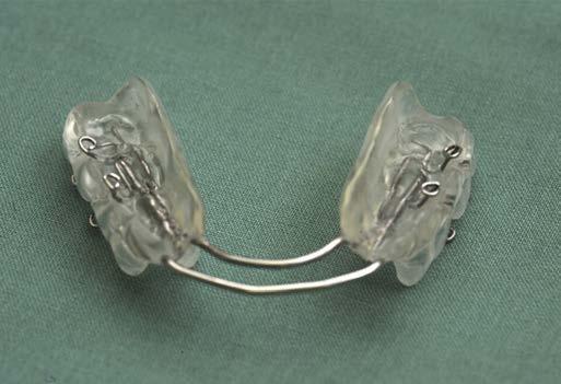
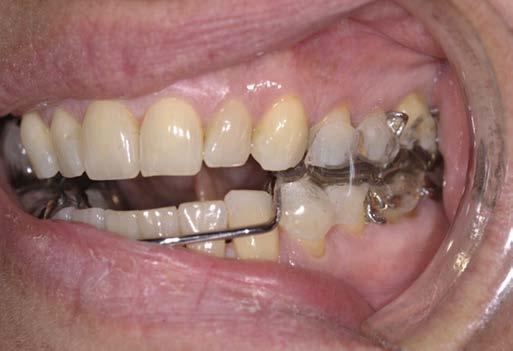
Oral appliance therapy is considered second-tier treatment in the management of OSA. The most common forms of oral appliances for OSA treatment include mandibular advancement devices and tongue retaining devices. Mandibular advancement devices are usually custom-made devices that are fitted to the teeth like a mouth guard and act to advance and stabilize the mandible to increase upper airway capacity (Fig. 1.2). Tongue retaining devices advance and retain the tongue in an anterior position by holding it in a suction cup placed over the front teeth. (See video at aveotsd.com.) Mandibular advancement devices are more costly but have greater efficacy and patient

FIG. 1.2 An oral appliance (mandibular advancement device) for use in obstructive sleep apnea. A, Device. B, Natural occlusion of this patient. C, Mandibular advancement device in position. Note the forward movement of the lower teeth/jaw with this device. (From Marcussen L, Henriksen JE, Thygesen T. Do mandibular advancement devices influence patients’ snoring and obstructive sleep apnea? A cone-beam computed tomography analysis of the upper airway volume. J Oral Maxillofacial Surg. 2015;73:1816-1826.)
A C B
compliance. Oral appliance therapy is indicated for the treatment of snoring, mild to moderate OSA, and selected cases of moderate to severe OSA, such as that due predominantly to the supine position or to a disproportionally large tongue relative to oral cavity capacity. This modality has been shown to be effective in reducing sleep interruption, daytime sleepiness, neurocognitive impairment, and cardiovascular complications. Side effects include excessive salivation, temporomandibular joint discomfort, and long-term occlusion changes.
Hypoglossal nerve stimulation uses a nerve stimulator that is implanted in the chest and has electronic sensing leads implanted between the internal and external intercostal muscles in the fourth intercostal space. These sensors detect breathing and signal the device to stimulate the hypoglossal nerve during inhalation, which results in enlargement of upper airway capacity. The system is turned on by the patient before going to sleep and turned off upon awakening.
Surgical therapy
Surgical treatment of the airway in the form of tracheostomy is the oldest form of therapy for OSA and has a very high rate of efficacy. However, its invasiveness is its major deterrent. In adults, in whom anatomic causes of OSA are relatively uncommon, airway surgery treatment for OSA is considered thirdtier therapy. These surgical procedures target soft tissue and bony tissue to enlarge airway capacity at the levels of the nose, palate, and/or tongue base and include maxillomandibular advancement, laser-assisted uvulopalatoplasty, uvulopalatopharyngoplasty, and palatal implants.
Bariatric surgery aims to restrict caloric intake or absorption or both. Bariatric surgery can be the sole therapy or an adjunctive treatment to PAP therapy in patients with morbid obesity associated with OSA or OHS. Screening for OSA should be performed in all patients undergoing bariatric surgery.
Medical therapy
Adjunctive medical therapy for OSA can be used in combination with any of the other forms of OSA therapy: PAP, oral appliances, or surgery. These adjuncts include diet, exercise, positional therapy, avoidance of alcohol and sedatives before sleep, supplemental oxygen, and pharmacologic therapy, such as with a stimulant drug like modafinil (Provigil). Positional therapy consists of devices that discourage or prevent the patient from sleeping in the supine position.
Comorbid conditions should be treated. Thyroid disorders should be treated surgically, medically, or both as indicated. Acromegaly should be treated surgically, medically, or both as indicated. Bromocriptine and somatostatin therapy can reduce the apnea-hypopnea index in patients with acromegaly by 50%–75%. However, continued PAP therapy is usually required owing to persistent skeletal changes.
Treatment of Central Sleep Apnea
In CSA related to congestive heart failure, first-tier therapy consists of CPAP therapy and nocturnal oxygen supplementation.
This can be augmented with BiPAP or drug therapy with acetazolamide and theophylline after medical optimization of congestive heart failure. Therapies for CSA associated with end-stage renal disease include CPAP, supplemental oxygen, use of bicarbonate during dialysis, and nocturnal dialysis.
Treatment of Sleep-Related Hypoventilation Disorders
Treatment of sleep-related hypoventilation disorders should enhance airway patency and ventilation, which is best achieved using noninvasive positive pressure ventilation (NIPPV) in one of three modes: (1) spontaneous mode, in which the patient cycles the device from inspiratory PAP to expiratory PAP; (2) spontaneous timed mode, in which a backup rate delivers PAP for a set inspiratory time if the patient does not trigger the device within a set period of time; and (3) timed mode, in which both the inspiratory time and respiratory rate are fixed. NIPPV is recommended for the treatment of hypoventilation due to any sleep-related breathing disorder.
PERIOPERATIVE CONSIDERATIONS IN PATIENTS WITH SLEEP-RELATED BREATHING DISORDERS
Management of sleep-related breathing disorders are a topic of special interest within the specialties of anesthesiology and sleep medicine. In 2011 this combined interest by the two specialties resulted in the establishment of the Society of Anesthesia and Sleep Medicine (SASM), which is an international society with a stated mission “to advance standards of care for clinical problems shared by Anesthesiology and Sleep Medicine, including perioperative management of sleep disordered breathing, and to promote interdisciplinary communication, education and research in matters common to anesthesia and sleep.”
The prevalence of OSA among surgical patients is higher than the overall prevalence of 2%–4% in the general population. The perioperative period can exacerbate sleep-related breathing disorders because of (1) sleep deprivation due to anxiety, pain, alterations in circadian rhythms, and nursing interventions; (2) REM sleep rebound, which worsens OSA; and (3) the suppressant effects of anesthetics, sedatives, and analgesics on airway patency, respiratory drive, and arousal. The effect of sleep-disordered breathing on perioperative outcomes has been the subject of many observational studies and systematic reviews, with conflicting findings based on study population, examined outcomes, and study design. The evidence is, however, mostly negative.
PRACTICE GUIDELINES FOR PERIOPERATIVE MANAGEMENT OF PATIENTS WITH OBSTRUCTIVE SLEEP APNEA
The AASM, the ASA, and the Society for Ambulatory Anesthesia (SAMBA) have provided practice parameters for the
perioperative management of OSA patients. Algorithms for the perioperative management of OSA patients have also been developed by individual groups.
In 2003 the AASM published a statement for the perioperative management of OSA in which it indicated that the literature is insufficient to develop standards-of-practice recommendations, and that the statement was based on a consensus of clinical experience and published peerreviewed medical evidence that, unfortunately, was scanty and of limited quality. The statement provided an introduction about OSA and listed the most common factors that contribute to increased perioperative risk in OSA patients, including: (1) increased risk of upper airway obstruction and respiratory depression due to effects of sedative, anesthetic, and narcotic medications; (2) decreased functional residual capacity (FRC) and decreased oxygen reserve due to obesity; and (3) the cardiopulmonary effects of OSA. It described the symptoms and signs of OSA, as well as a description of CPAP therapy, and provided a questionnaire and checklist for preoperative recognition of patients who are at high risk for OSA. The AASM also detailed recommendations for intraoperative and postoperative patient care, including transfer of care.
In 2006 the ASA developed comprehensive practice guidelines for the perioperative management of OSA patients and updated them in 2014. These guidelines provide a checklist for preoperative identification and assessment of OSA and detailed recommendations covering the areas of preoperative evaluation, considerations for inpatient versus outpatient surgery, preoperative preparation, intraoperative management, postoperative management, and criteria for discharge to unmonitored settings.
In 2012, SAMBA produced a consensus statement on preoperative selection of adult patients with OSA scheduled for ambulatory surgery, which concluded that patients with known OSA might be considered for ambulatory surgery if they were medically optimized and could use their CPAP postoperatively. Patients with presumed OSA could be considered for ambulatory surgery if they could be managed with nonopioid analgesia perioperatively.
The elements of the practice parameters for perioperative care of patients with OSA are noted in Table 1.5.
PERIOPERATIVE OPIOID-INDUCED RESPIRATORY DEPRESSION
The Anesthesia Patient Safety Foundation (APSF) made perioperative opioid-induced respiratory depression a top priority in 2006. In 2011 it held its second conference on this subject and focused on monitoring for this entity. The executive summary of this conference recommended that “all patients receiving postoperative opioid analgesia should have periodic assessment of level of consciousness and continuous monitoring of oxygenation by pulse oximetry,” and if supplemental oxygen is provided, “continuous monitoring of ventilation by capnography (PETCO2) or an equivalent method.”
In 2009 the ASA provided “Practice Guidelines for the Prevention, Detection, and Management of Respiratory Depression Associated with Neuraxial Opioid Administration.” These were updated in 2016. Like the APSF, the ASA recommended that all patients receiving neuraxial opioids be monitored for adequacy of ventilation, oxygenation, and level of consciousness, with increased monitoring for patients with high-risk conditions, including unstable medical conditions, obesity, OSA, concomitant administration of opioid analgesics or hypnotics by other routes, and extremes of age. They also recommended administering supplemental oxygen to patients with an altered level of consciousness, respiratory depression, or hypoxemia, and having resuscitative measures available as needed, including narcotic reversal drugs and NIPPV.
KEY POINTS
• Electroencephalography (EEG) is an important method of studying wakefulness and sleep and defining sleep stages. The electrical activity of the brain can be categorized into three states: wakefulness, rapid eye movement (REM) sleep, and non-REM (NREM) sleep. The latter can be further categorized into three stages: N1, N2, and N3, according to the progressive decrease in frequency and increase in amplitude of EEG waveforms. Muscle tone as measured by electromyography is normal during wakefulness, decreased during NREM sleep, and abolished during REM sleep.
• NREM sleep maintains homeostasis and autonomic stability at low energy levels—that is, with a low basic metabolic rate and a decreased heart rate, cardiac output, and blood pressure. Hormonal secretion is maintained.
• REM sleep impairs homeostasis and disrupts autonomic stability. REM-induced autonomic instability manifests as irregularity in the heart rate, cardiac output, blood pressure and tidal volume, and suppression of cardiac and respiratory chemoreceptor and baroreceptor reflexes. REM sleep is associated with skeletal muscle atonia affecting all skeletal muscles, including upper airway dilator muscles and intercostal muscles, but with significant sparing of the diaphragm.
• Specific sleep disorders are disorders that manifest predominantly but not exclusively with sleep manifestations. They include disorders that manifest primarily as: (1) decreased sleep (insomnia), which is the most common type of sleep disorder, (2) increased sleep (hypersomnias), (3) abnormal sleep behavior (parasomnias), (4) disruptions of circadian rhythm, and (5) sleep-induced exacerbations of certain pathophysiologic problems such as sleep-related movement disorders and sleep-related breathing disorders.
• The hallmark of obstructive sleep apnea (OSA) is sleepinduced and arousal-relieved upper airway obstruction.
• Functional collapse of the upper airway occurs when forces that can collapse the upper airway overcome the forces that can dilate the upper airway. Collapsing forces consist of intraluminal negative inspiratory pressure and extraluminal positive pressure. Dilating forces consist of pharyngeal
Perioperative Management of the Patient With Obstructive Sleep Apnea
potential Sources of perioperative Risk
Lack of institutional protocol for perioperative management of sleep apnea patients
patients with a known diagnosis of obstructive sleep apnea (oSA)
patients without a diagnosis of oSA
perioperative Risk Mitigation
develop and implement institutional protocol for perioperative management of sleep apnea patients.
Know sleep study results.
Know the therapy being used: oral appliance, positive airway pressure (pAp) with settings (mode, pressure level, supplemental oxygen if any).
Consult sleep medicine specialist as needed.
Use a screening tool to determine the likelihood of oSA: AASM questionnaire, ASA checklist, Berlin questionnaire, or Stop-BANG questionnaire.
Inpatient versus outpatient surgery decisions based on institutional protocol containing factors related to: (1) patient, (2) procedure, (3) facility, and (4) postdischarge setting
preoperative lack of optimization of therapy for oSA
preoperative sedative-induced airway compromise or respiratory depression
Intraoperative sedative/opioid/anestheticinduced upper airway compromise or respiratory depression during monitored anesthesia care (MAC)
At risk for oxygen desaturation
possible difficult mask ventilation or endotracheal intubation
Consult sleep medicine specialist to optimize therapy.
Use preoperative sedation only in a monitored setting.
Whenever possible, use topical, local, or regional anesthesia with minimal to no sedation. Continuous monitoring of ventilation adequacy
Use of the patient’s oSA therapy device during MAC with sedation
Consider general anesthesia with a secured airway vs. deep sedation with an unsecured airway.
Elevate head of bed to facilitate spontaneous ventilation/oxygenation.
preoxygenate sufficiently.
Maintain oxygen insufflation by nasal cannula during endotracheal intubation.
Apply ASA difficult Airway Algorithm, including the use of laryngeal mask airway, videolaryngoscope, fiberoptic bronchoscope, and transtracheal jet ventilation as indicated. optimize head/neck position for mask ventilation and endotracheal intubation. potential difficulty with noninvasive blood pressure monitoring and/or increased risk for cardiovascular complications
postextubation airway obstruction in the operating room or postanesthesia care unit with associated risk of negative pressure pulmonary edema
Consider intraarterial catheter for blood pressure monitoring and blood sampling for arterial blood gases.
Elevate the head of the bed.
Extubate only after patient clearly meets objective extubation criteria.
Maintain readiness for reintubation with the same device used during induction and expect that the difficulty of intubation will be greater than previously.
At risk for postoperative oxygen desaturation Supplemental oxygen therapy
Consider nasal airway.
Consider pAp therapy (this can be initiated de novo in the postoperative setting).
Communication failure during transfer of care
Identify the patient’s diagnosis of sleep apnea and its therapy. Alert staff about expected problems and their management. perioperative opioid-related respiratory depression due to opioids administered by neuraxial route, intravenous route with bolus injection, or via intravenous patientcontrolled analgesia (IV-pCA)
postdischarge opioid-induced respiratory depression and/or exacerbation of oSA
ASA,
Supplemental oxygen as needed
Continuous electronic monitoring of oxygenation and ventilation
Maintain patient’s oSA therapy whenever possible; use home settings as a guide. Avoid background mode with IV-pCA.
Consider opioid-sparing analgesic techniques (e.g., transcutaneous electrical nerve stimulation), and use nonopioid analgesics (e.g., NSAIds, acetaminophen, tramadol, ketamine, gabapentin) whenever possible.
Ensure companionship and a safe home environment for high-risk patients. Consult sleep medicine specialist to optimize sleep apnea therapy if needed.
dilating muscle tone and longitudinal traction on the upper airway by an increased lung volume, so-called tracheal tug.
• Central sleep apnea refers to sleep apnea that is not associated with respiratory efforts during the apnea event. This absence of respiratory effort could be due to instability of neural control of respiration, weakness of respiratory muscles, or both. Instability of respiratory control may include increased, decreased, or oscillating respiratory drive.
• Apneic and hypopneic episodes result in hypoxia, which can be prolonged and severe. OSA-induced hypoxia and
reoxygenation cycles activate redox-sensitive genes, oxidative stress, inflammatory processes, the sympathetic nervous system, and the coagulation cascade, all of which can contribute to endothelial dysfunction and ultimately to systemic hypertension, pulmonary hypertension, atherosclerosis, right and left ventricular systolic and diastolic dysfunction, coronary artery disease, congestive heart failure, atrial fibrillation, stroke, and sudden cardiac death.
• Polysomnography can be used to differentiate CSA from OSA, assess its severity, detect associated hypoventilation
American Society of Anesthesiologists; NSAIDs, Nonsteroidal antiinflammatory drugs.
and hypoxia, detect associated EEG, ECG, and limb movement events, and, when indicated, titrate positive airway pressure (PAP) therapy and perform follow-up assessment of any implemented therapy for the sleep-related breathing disorder.
• Because of its high prevalence rate and a general lack of diagnosis, the first step in management of OSA is detection.
• Suggested mechanisms for the efficacy of continuous PAP therapy include (1) increasing the pharyngeal transmural pressure (pneumatic splint effect), (2) reducing pharyngeal wall thickness and airway edema, (3) increasing airway tone by mechanoreceptor stimulation, and (4) increasing end-expiratory lung volume and producing a tracheal tug effect.
• The perioperative period can exacerbate sleep-related breathing disorders because of (1) sleep deprivation due to anxiety, pain, alterations in circadian rhythms, and nursing interventions; (2) REM sleep rebound, which worsens OSA; and (3) the suppressant effects of anesthetics, sedatives, and analgesics on airway patency, respiratory drive, and arousal.
• To avoid opioid-induced respiratory depression, all patients receiving opioids, including neuraxial opioids, should be monitored for adequacy of ventilation, oxygenation, and level of consciousness, with increased monitoring for patients with high-risk conditions, including unstable medical conditions, obesity, OSA, concomitant administration of opioid analgesics or hypnotics by other routes, and extremes of age.
RESOURCES
Aurora RN, Casey KR, Kristo D, et al. Practice parameters for the surgical modifications of the upper airway for obstructive sleep apnea in adults. Sleep. 2010;33:1408‐1413.
Aurora RN, Chowdhuri S, Ramar K, et al. The treatment of central sleep apnea syndromes in adults: practice parameters with an evidence-based literature review and meta-analyses. Sleep. 2012;35:17-40.
Biot MC. Contribution a l’ètude de phènomène respiratoire de Cheyne-Stokes. Lyon Mèd. 1876;23:517-528, 561-567.
Bradley TD, Logan AG, Kimoff RJ, et al. Continuous positive airway pressure for central sleep apnea and heart failure. N Engl J Med. 2005;353: 2025-2033.
Benumof JL. The elephant in the room is bigger than you think: finding obstructive sleep apnea patients dead in bed postoperatively. Anesth Analg 2015;120:491.
Bolden N, Smith CE, Auckley D. Avoiding adverse outcomes in patients with obstructive sleep apnea: development and implementation of a perioperative obstructive sleep apnea protocol. J Clin Anesth. 2009;21:286-293.
Chau EH, Lam D, Wong J, et al. Obesity hypoventilation syndrome: a review of epidemiology, pathophysiology, and perioperative considerations. Anesthesiology. 2012;117:188-205.
Chung F, Yegneswaran B, Liao P, et al. STOP questionnaire: a tool to screen patients for obstructive sleep apnea. Anesthesiology. 2008;108:812-821.
Chung F, Yegneswaran B, Liao P, et al. Validation of the Berlin questionnaire and American Society of Anesthesiologists checklist as screening tools for obstructive sleep apnea in surgical patients. Anesthesiology. 2008;108: 822-830.
Chung F, Liao P, Elsaid H, et al. Factors associated with postoperative exacerbation of sleep-disordered breathing. Anesthesiology. 2014;120:299-311.
Correa D, Farney RJ, Chung F, et al. Chronic opioid use and central sleep apnea: a review of the prevalence, mechanisms, and perioperative considerations. Anesth Analg. 2015;120:1273-1285.
Gay P, Weaver T, Loube D, et al. Positive Airway Pressure Task Force, Standards of Practice Committee, American Academy of Sleep Medicine. Evaluation of positive airway pressure treatment for sleep related breathing disorders in adults. Sleep. 2006;29:381-401.
Gross JB, Bachenberg KL, Benumof JL, et al. Practice guidelines for the perioperative management of patients with obstructive sleep apnea: a report by the American Society of Anesthesiologists Task Force on perioperative management of patients with obstructive sleep apnea. Anesthesiology 2006;104:1081-1093.
Johns MW. A new method for measuring daytime sleepiness: the Epworth sleepiness scale. Sleep. 1991;14:540-545.
Joshi GP, Ankichetty SP, Gan TJ, et al. Society for Ambulatory Anesthesia consensus statement on preoperative selection of adult patients with obstructive sleep apnea scheduled for ambulatory surgery. Anesth Analg 2012;15:1060-1068.
Kaw R, Pasupuleti V, Walker E, et al. Postoperative complications in patients with obstructive sleep apnea. Chest. 2012;141:436-441.
Kaneko Y, Floras JS, Usui K, et al. Cardiovascular effects of continuous positive airway pressure in patients with heart failure and obstructive sleep apnea. N Engl J Med. 2003;348:1233-1241.
Lockhart EM, Willingham MD, Abdallah AB, et al. Obstructive sleep apnea screening and postoperative mortality in a large surgical cohort. Sleep Med. 2013;14:407-415.
Marin JM, Soriano JB, Carrizo SJ, et al. Outcomes in patients with chronic obstructive pulmonary disease and obstructive sleep apnea: the overlap syndrome. Am J Respir Crit Care Med. 2010;182:325-331.
Memtsoudis SG, Stundner O, Rasul R, et al. The impact of sleep apnea on postoperative utilization of resources and adverse outcomes. Anesth Analg. 2014;118:407-418.
Meoli AL, Rosen CL, Kristo D, et al. Clinical Practice Review Committee, American Academy of Sleep Medicine. Upper airway management of the adult patient with obstructive sleep apnea in the perioperative period— avoiding complications. Sleep. 2003;26:1060-1065.
Mokhlesi B. Obesity hypoventilation syndrome: a state-of-the-art review. Respir Care. 2010;55:1347-1365.
Morgenthaler TI, Kapen S, Lee-Chiong T, et al. Standards of Practice Committee, American Academy of Sleep Medicine. Practice parameters for the medical therapy of obstructive sleep apnea. Sleep. 2006;29:1031-1035.
Netzer NC, Stoohs RA, Netzer CM, et al. Using the Berlin Questionnaire to identify patients at risk for the sleep apnea syndrome. Ann Intern Med 1999;131:485-491.
Peppard PE, Young T, Palta M, et al. Prospective study of the association between sleep-disordered breathing and hypertension. N Engl J Med 2000;342:1378-1384.
Practice guidelines for the prevention, detection, and management of respiratory depression associated with neuraxial opioid administration: an updated report by the American Society of Anesthesiologists Task Force on Neuraxial Opioids and the American Society of Regional Anesthesia and Pain Medicine. Anesthesiology. 2016;124:535-552.
Practice guidelines for the perioperative management of patients with obstructive sleep apnea: an updated report by the American Society of Anesthesiologists Task Force on Perioperative Management of Patients with Obstructive Sleep Apnea. Anesthesiology. 2014;120:268-286.
Ramar K, Dort LC, Katz SG, et al. Clinical practice guideline for the treatment of obstructive sleep apnea and snoring with oral appliance therapy: an update for 2015. J Clin Sleep Med. 2015;11:773-827.
<http://sasmhq.org/>. Accessed August 15, 2016.
Sateia MJ. International classification of sleep disorders-third edition: highlights and modifications. Chest. 2014;146:1387-1394.
Wedewardt J, Bitter T, Prinz C, et al. Cheyne-Stokes respiration in heart failure: cycle length is dependent on left ventricular ejection fraction. Sleep Med. 2010;11:137-142.