[Ebooks PDF] download Adams and stashak's lameness in horses seventh edition. edition gary m. baxter
Adams
And Stashak's Lameness In Horses Seventh Edition. Edition Gary M. Baxter
Visit to download the full and correct content document: https://ebookmass.com/product/adams-and-stashaks-lameness-in-horses-seventh-ed ition-edition-gary-m-baxter/
More products digital (pdf, epub, mobi) instant download maybe you interests ...
Voids in Materials: From Unavoidable Defects to Designed Cellular Materials 2nd Edition Gary M. Gladysz
https://ebookmass.com/product/voids-in-materials-fromunavoidable-defects-to-designed-cellular-materials-2nd-editiongary-m-gladysz/ Fundamental Immunology Seventh Edition
Blackwell Publishing was acquired by John Wiley & Sons in February 2007. Blackwell’s publishing program has been merged with Wiley’s global Scientific, Technical and Medical business to form Wiley‐Blackwell.
The right of insert Gary M. Baxter to be identified as the author of the editorial material in this work has been asserted in accordance with law.
Registered Office
John Wiley & Sons, Inc., 111 River Street, Hoboken, NJ 07030, USA
Editorial Office
111 River Street, Hoboken, NJ 07030, USA
For details of our global editorial offices, customer services, and more information about Wiley products visit us at www.wiley.com.
Wiley also publishes its books in a variety of electronic formats and by print‐on‐demand. Some content that appears in standard print versions of this book may not be available in other formats.
Limit of Liability/Disclaimer of Warranty
The contents of this work are intended to further general scientific research, understanding, and discussion only and are not intended and should not be relied upon as recommending or promoting scientific method, diagnosis, or treatment by physicians for any particular patient. In view of ongoing research, equipment modifications, changes in governmental regulations, and the constant flow of information relating to the use of medicines, equipment, and devices, the reader is urged to review and evaluate the information provided in the package insert or instructions for each medicine, equipment, or device for, among other things, any changes in the instructions or indication of usage and for added warnings and precautions. While the publisher and authors have used their best efforts in preparing this work, they make no representations or warranties with respect to the accuracy or completeness of the contents of this work and specifically disclaim all warranties, including without limitation any implied warranties of merchantability or fitness for a particular purpose. No warranty may be created or extended by sales representatives, written sales materials or promotional statements for this work. The fact that an organization, website, or product is referred to in this work as a citation and/or potential source of further information does not mean that the publisher and authors endorse the information or services the organization, website, or product may provide or recommendations it may make. This work is sold with the understanding that the publisher is not engaged in rendering professional services. The advice and strategies contained herein may not be suitable for your situation. You should consult with a specialist where appropriate. Further, readers should be aware that websites listed in this work may have changed or disappeared between when this work was written and when it is read. Neither the publisher nor authors shall be liable for any loss of profit or any other commercial damages, including but not limited to special, incidental, consequential, or other damages.
Library of Congress Cataloging‐in‐Publication Data
Names: Baxter, Gary M., editor.
Title: Adams and Stashak’s lameness in horses / [edited by] Gary M. Baxter.
Other titles: Lameness in horses
Description: Seventh edition. | Hoboken, NJ : Wiley-Blackwell, 2021. | Includes bibliographical references and index.
Identifiers: LCCN 2019046203 (print) | LCCN 2019046204 (ebook) | ISBN 9781119276685 (hardback) | ISBN 9781119276692 (adobe pdf) | ISBN 9781119276708 (epub)
Set in 10/10.5pt Sabon by SPi Global, Pondicherry, India
To my family for their understanding and encouragement and
To all of the faculty, residents, interns, students, staff, and referring veterinarians that I have worked with through the years at both Colorado State University and the University of Georgia
TABLE OF CONTENTS
List of Contributors xv
Preface to the Seventh Edition xix
Preface to the Sixth Edition xxi
Preface to the Fifth Edition xxiii
Preface to the Fourth Edition xxv
Acknowledgments xxvii
Common Terminologies and Abbreviations xxix
About the Companion Website xxxi
1 Functional Anatomy of the Equine Musculoskeletal System 1
AnnA Dee FAils
Anatomic Nomenclature and Usage 1
Thoracic Limb 1
Hindlimb 34
Axial Components 60
Acknowledgment 64
2 Examination for Lameness 67
History, Visual Exam, and Conformation 67
GAry M. BAxter AnD teD s. stAshAk
Introduction 67
Adaptive Strategies of Lame Horses 67
Classification of Lameness 68
Signalment and Use 71
Histor y (Anamnesis) 71
Visual Examination at Rest 71
Conformation 72
Conformation Components and Traits 74
Acknowledgment 91
Palpation and Manipulation 93
GAry M. BAxter AnD teD s. stAshAk
Foot 93
Pastern 96
Fetlock 97
Metacar pus/Metatarsus (MC/MT) 97
Suspensory Ligament 99
Flexor Tendons 100
Carpus 100
Forearm (Antebrachium) and Elbow 101
Shoulder and Scapula 102
Tarsus (Hock) 105
Tibia 107
Stifle 109
Femur 112
Hip 112
Pelvis 113
Back 113
Neck 115
Flexion Tests/Manipulation 115
Distal Limb/Phalangeal/Fetlock Flexion 115
Carpal Flexion 117
Elbow Flexion 117
Shoulder/Upper Forelimb Flexion 117
Tarsal/Hock Flexion 117
Stifle Flexion 119
Full‐Limb Forelimb and Hindlimb Flexion 119
Navicular Wedge Test 119
Direct or Local Pressure plus Movement 120
Subjective Assessment of Lameness 123
kevin G. keeGAn
Evaluation of the Horse for Lameness at the Walk 124
Evaluation of the Horse at the Trot 126
Evaluation of Vertical Movement of the Head for Forelimb Lameness 126
Withers Movement 129
Evaluation of Vertical Movement of the Pelvis for Hindlimb Lameness (the Vertical Pelvic Movement [VPM] Method) 129
Evaluation of Pelvic Rotation for Hindlimb Lameness (The Pelvic Rotation Method [PRM]) 131
Bilateral Lameness 132
Obser ving Movement of the Limbs 134
Joint Angle Measurements Associated with Lameness 134
Stride Timing and Length Variables Associated with Lameness 134
Hoof Trajectory Associated with Lameness 135
Lameness Evaluation During Ipsilateral Gaits 135
Evaluation of Lameness at the Lunge 136
Evaluation of Lameness Under Saddle 136
Subjective Scoring Methods 137
Objective Assessment of Lameness 139
kevin G. keeGAn
Measurement of Ground Reaction Forces (Kinetics) 139
Measurement of Movement (Kinematics) 140
Perineural and Intrasynovial Anesthesia 157
GAry M. BAxter
Types of Local Anesthetics 157
Skin Preparation and Restraint 157
Perineural Anesthesia 157
Intrasynovial Anesthesia 167
Acknowledgment 187
3 Diagnostic Imaging 189
Radiography
189
MyrA BArrett AnD elizABeth Acutt
Equipment 189
Digital Radiography Systems 191
Radiation Safety 194
Contrast Examinations 195
Principles of Radiographic Interpretation 198
Limitations of Radiography 210
Nor mal Radiographic Anatomy 211
Acknowledgments 299
Ultrasound
W. rich reDDinG
301
Introduction 301
Ultrasound to Evaluate Tendons and Ligaments 302
Patient Preparation and Scan Protocol 303
Ultrasonographic Assessment of Tendon/Ligament Pathology 316
Limitations of Ultrasonography 321
Ultrasound to Evaluate Joint Injury 323
Indications for Ultrasonography of Joints 324
Equipment and Technique 324
Ultrasonographic Appearance of Periarticular Structures 326
Ultrasonic Appearance of the Joint 327
Conclusions 331
Other Indications for Ultrasonography of the Musculoskeletal System 332
New Directions in the Use of Ultrasound 335
Nuclear Medicine/Scintigraphy 342
kurt selBerG, elizABeth Acutt, AnD AlejAnDro vAlDés‐MArtínez
Principles of Nuclear Medicine 342
Radiation Safety and Protection 343
Imaging Equipment 343
Method for a Scintigraphic Exam of the Musculoskeletal System 344
Indications for Nuclear Scintigraphy of the Musculoskeletal System in Horses 347
Normal Bone Scan 348
Scintigraphic Signs of Disease 356
Abnormal Conditions for Specific Anatomical Regions 361
Limitations of Nuclear Medicine 373
Computed Tomography 376
MAthieu spriet
Introduction 376
Equipment and Principles of CT 376
Clinical Use of CT in Equine Orthopedic Imaging 378
Contrast‐Enhanced CT 380
Positron Emission Tomography 383
Conclusion 383
Magnetic Resonance Imaging 387
MichAel schrAMMe AnD eMilie seGArD‐Weisse
Introduction 387
General Principles and Physics of MRI 387
Equipment, High‐ and Low‐Field Magnets, and RF Coils 387
Sequences and Protocols for Equine MRI 389
Artifacts of MRI 390
Inter pretation of Musculoskeletal MR Images 393
Contrast MRI Techniques 396
How to Read an Equine MRI Study 397
Indications, Case Selection, Advantages, and Disadvantages of MRI 397
Magnetic Resonance Imaging of the Foot and Pastern 397
Magnetic Resonance Imaging of the Fetlock Region 409
Magnetic Resonance Imaging of the Metacarpal and Metatarsal Regions 416
Magnetic Resonance Imaging of the Carpal Region 420
Magnetic Resonance Imaging of the Tarsal Region 421
Magnetic Resonance Imaging of the Stifle Region 424
Thermography 431
trAcy A. turner
Thermographic Instrumentation 431
Principles of Use 431
Specific Applications for Lameness Diagnostics 433
4 Lameness of the Distal Limb 439
Navicular Region/Palmar Foot 439
rAnDy B. eGGleston AnD GAry M. BAxter
Navicular Syndrome/Disease 439
Fractures of the Navicular (Distal Sesamoid) Bone 454
Soft Tissue Injuries in the Foot (DDFT and Podotrochlear Apparatus) 456
Acknowledgments 459
Coffin Joint and Distal Phalanx 463
GAry M. BAxter
Osteoarthritis (OA) of the Distal Interphalangeal (DIP) Joint 463
Fractures of the Distal Phalanx (P3, Coffin Bone) 465
Subchondral Cystic Lesions of the Distal Phalanx (P3) 471
Collateral Ligament Injuries of the Distal Phalanx 472
Ossification of the Collateral Cartilages of the Distal Phalanx (Sidebone) 474
Miscellaneous Conditions of the Foot 477
GAry M. BAxter
Sole Bruises, Corns, and Subsolar Abscesses 477
Septic Pedal Osteitis 479
Penetrating Injuries of the Foot 481
Keratoma 483
Avulsion Injuries of the Hoof 485
Acknowledgments 489
Laminitis 490
jAMes BelknAp, AnDy pArks, AnD kAty Dern
Overview 490
Relevant Anatomy 490
Pathogenesis: Pathophysiologic Considerations 491
Pathogenesis: Structural Considerations of the Equine Digit 492
The Clinical Presentation: The Horse at Risk of Laminitis 494
Clinical Signs: The Acute Onset of Laminitis 496
Hoof Wall Resections 509
Coronary Band Grooving and Resection 509
The Pastern 512
Ashlee e. WAtts AnD GAry M. BAxter
Osteoarthritis (OA) of the PIP Joint (High Ringbone) 512
Osteochondrosis (OC) of the PIP Joint 517
Luxation/Subluxation of the Proximal Interphalangeal (PIP) Joint 518
Fractures of the Middle (Second) Phalanx (P2) 522
Fractures of the Proximal (First) Phalanx (P1) 525
Desmitis of the Distal Sesamoidean Ligaments (DSLs) 532
Desmitis of Digital Annular Ligaments 535
SDFT and DDFT Abnormalities 536
The Fetlock 541
MAtt Brokken AnD AliciA Bertone
Osteochondral Fractures and Fragmentation of the Proximal Phalanx 541
Fractures of the Proximal Sesamoid Bones 543
Sesamoiditis 548
Axial Osteitis/Osteomyelitis of the Proximal Sesamoid Bones 549
Osteoar thritis of the Metacarpophalangeal/ Metatarsophalangeal Joint 550
Palmar/Plantar Osteochondral Disease 552
Fetlock Subchondral Cystic Lesions (SCLs) 553
Traumatic Rupture of the Suspensory Apparatus 554
Luxation of the Metacarpophalangeal/ Metatarsophalangeal Joint (Fetlock Luxation) 556
Constriction of or by the Fetlock Palmar/Plantar Annular Ligament 558
Acknowledgments 560
The Metacarpus and Metatarsus 563
kylA F. ortveD AnD AliciA l. Bertone
Bucked Shin Complex and Stress Fractures of the Dorsal Third Metacarpus 563
Fractures of the Condyles of the Third Metacarpal/ Metatarsal Bones (Condylar Fractures, Longitudinal Articular Fractures) 567
Fractures of the Third Metacarpal/Metatarsal (Cannon) Bone 571
Metacar pal/Metatarsal Exostosis (Splints) 572
Fractures of the Small Metacarpal and Metatarsal (Splint) Bones 576
Enostosis‐Like Lesions 580
Suspensory Ligament Desmitis 580
Superficial Digital Flexor (SDF) Tendinitis (Bowed Tendon) 585
Deep Digital Flexor Tendinitis 590
Desmitis of the Accessor y Ligament of the Deep Digital Flexor Tendon (Distal Check Ligament) 591
5 Lameness of the Proximal Limb 597
The Carpus 597
chris kAWcAk
Developmental Abnormalities of the Carpus 598
Miscellaneous Car pal Swellings 604
Dorsal Carpal Swelling 605
Intra‐ar ticular Fractures 607
Accessor y Carpal Bone Fracture 612
Carpal Luxations 613
Soft Tissue Damage to the Carpus 614
Osteoarthritis 615
Carpometacarpal Osteoarthritis 616
Osteochondrosis of the Carpus 617
Osteochondroma of the Distal Radius 617
Desmitis of the Accessory Ligament (Radial or Superior Check Ligament) of the Superficial Digital Flexor Tendon 619
The Antebrachium, Elbow, and Humerus 623
jereMy huBert
Fractures of the Radius 623
The Elbow 627
Fractures of the Humerus 633
Neoplastic Lesions of the Humerus 637
Paralysis of the Radial Nerve 637
Acknowledgment 638
The Shoulder and Scapula 641
jereMy huBert
Inflammation of the Intertubercular Bursa (Bicipital Bursitis) 641
Inflammation of the Infraspinatus Bursa 643
Osteochondrosis (OC) of the Scapulohumeral (SH) Joint or Shoulder 644
Osteoar thritis (OA) of the Scapulohumeral Joint 646
Luxation of the Scapulohumeral (Shoulder) Joint 648
Suprascapular Ner ve Injury (Sweeny) 650
Fractures of the Scapula 652
Fractures of the Supraglenoid Tubercle (Tuberosity) 653
Acknowledgment 655
The Tarsus 657
W. rich reDDinG
Introduction 657
Diagnosis of Tarsal Lameness 657
Articular Diseases of the Tarsus 662
Cunean Tenectomy 672
Fractures and Luxations of the Tarsus 680
Soft Tissue Injuries of the Tarsus 687
Periarticular Tarsal Cellulitis 697
Tibia and Crus 701
W. rich reDDinG
Introduction 701
Diagnostic Analgesia of the Crus 701
Imaging the Tibia/Crus 701
Clinical Conditions 701
Enostosis‐Like Lesions 702
Fractures of the Tibia 703
Incomplete Fractures 704
Proximal Physeal Fractures 705
Diaphyseal Fractures 706
Tibial Tuberosity Fractures 706
Gastrocnemius Disruption in Foals and Adults 707
The Stifle: Femoropatellar Region 709
GAry M. BAxter and Ken E. Sullins
Introduction 709
Femoropatellar Joint 711
Upward Fixation of the Patella (UFP) 717
Desmitis of the Patellar Ligaments 719
Patellar Luxation/Subluxation 721
Synovial Osteochondroma in the Hindlimb 722
The Stifle: Femorotibial Joint Region 725
chris kAWcAk
Subchondral Cystic Lesions (SCLs) of the Stifle 726 Fractures 729
Blood‐Derived Biologics: Platelet‐Rich Plasma, Autologous Conditioned Serum, Autologous Protein Solution, and Bone Marrow Aspirate 897
Cor ticosteroids 898
Miscellaneous 898
Oral/Nutritional 900
nicolAs s ernst AnD troy n truMBle
Nonsteroidal Anti‐Inflammatory Drugs 900
Nutraceuticals 903
Therapeutic Trimming and Shoeing 911
AnDreW h pArks
Introduction 911
Examination of the Limb for Therapeutic Shoeing 911
The Trim 912
Shoeing 912
Protection and Support 917
Clinical Applications of Principles 918
Summar y 920
Acupuncture Treatment of Limb
Lameness and Back Pain 921
kevin k hAussler
Introduction 921
Techniques of Stimulation 921
General Indications for Treatment 922
Lameness 922
Chronic Back Pain 923
Adverse Effects 924
Manual Therapy Techniques 925
kevin k. hAussler
Introduction 925
Therapeutic Touch 925
Massage Therapy 925
Passive Stretching Exercises 925
Mobilization 926
Joint Mobilization and Manipulation 926
Contraindications 927
Rehabilitation/Physical Therapy 929
MelissA kinG, kAtherine ellis, AnD nArelle c stuBBs
Introduction 929
Clinical and Client Demand for Rehabilitation/PT 929
Manual Therapy 930
Proprioceptive Facilitation/Neuromotor Control
Techniques 934
Physical Modalities 936
Conclusion 943
9 Occupational‐Related Lameness Conditions 949
The Thoroughbred Racehorse 949
ryAn cArpenter
Risk Factors for Injury 949
Diagnosis of Lameness 950
Common Conditions 951
The Standardbred Racehorse 964
kiMBerly johnston AnD FrAnk A. nickels
Description of the Sport 964
Lameness Exam 965
Specific Lameness Conditions 966
The Racing Quarter Horse 972
nAncy l. GooDMAn AnD lArry r. overly
Introduction 972
Conformation Relating to Lameness 972
Training the Racing Quarter Horse 973
Shoeing 973
Lameness Related to Track Surface 973
Lameness Examination 973
Specific Lameness Conditions 975
The Western Performance Horse 980
roBin M. DABAreiner
Introduction 980
Team Roping Horses 980
Tie‐Down and Breakaway Roping 981
Barrel Racing 982
Reining Horses 982
Cutting Horses 983
Jumping, Eventing, and Dressage
Horses 986
oMAr MAher
Introduction and Horses Used for the Disciplines 986
Structure of Training and Competition 987
Training Surfaces and Shoeing 989
Lameness Diagnosis 990
Common Lameness Problems 991
Acknowledgment 996
The Endurance Horse 998
toDD c holBrook
The Sport 998
Athletes and Exercise Conditions 998
Veterinary Control 998
The Lameness Examination 1000
Common Causes of Lameness 1002
The Western Pleasure Horse 1006
sherry A. johnson AnD DAviD D. FrisBie
Understanding the Sport 1006
Western Pleasure Terminology 1007
Training and Showing 1007
Commonly Encountered Musculoskeletal Issues in the Western Pleasure Athlete 1008
Foot Pain 1008
Fetlock Osteoarthritis 1010
Proximal Suspensory Desmopathy 1010
Distal Tarsal Disease 1012
Rehabilitation and Management of the Western Pleasure Athlete 1013
Conclusion 1013
Gaited Horses 1015
DAviD A. Wilson AnD kevin G. keeGAn
Evaluating Gaited Horses for Lameness 1015
What Is a Gaited Horse? 1015
Classification of Gaits 1015
Specific Gaits in “Gaited Horses” 1018
Lameness in the Gaited Horse 1020
The Draft Horse 1026
jAn F. hAWkins
Introduction 1026
Anamnesis 1026
Lameness Examination 1026
Common Causes of Lameness 1026
Diseases of Young Draft Horses 1030
10 Lameness in the Young Horse 1033
The
Physis/Physeal Fractures/ Physitis 1033
DAne M. tAtArniuk, troy n truMBle, AnD GAry M. BAxter
The Physis 1033
Classification and Treatment of Physeal Injuries/ Fractures 1037
Developmental Orthopedic Diseases 1041
Epiphysitis/Physitis/Physeal Dysplasia 1042
Etiology 1042
Clinical Signs 1043
Diagnosis 1043
Treatment 1046
Prognosis 1046
Angular Limb Deformities (ALDs)
and Cuboidal Bone Malformations 1048
nicolAs s. ernst, troy n. truMBle, AnD GAry M. BAxter
Angular Limb Deformities (ALDs) 1048
Etiology 1048
Clinical Signs 1048
Diagnosis 1049
Treatment 1051
Prognosis 1055
Cuboidal Bone Malformation/Incomplete
Ossification 1055
Etiology 1055
Clinical Signs 1055
Diagnosis 1056
Treatment 1056
Prognosis 1058
Flexural Deformities 1059
nicolAs s ernst, troy n truMBle, AnD GAry M. BAxter
Congenital Flexural Deformities 1059
Acquired Flexural Deformities 1062
Osteochondrosis 1071
c. WAyne McilWrAith
Introduction 1071
Osteochondritis Dissecans (OCD) 1071
Subchondral Cystic Lesions 1078
Lameness in Foals 1081
roBert j. hunt
Diagnosis 1081
Noninfectious Causes of Lameness 1082
Infectious Causes of Lameness 1089
11 Foot Care and Farriery 1091
Basic Foot Care 1091
stephen e o’GrADy
Introduction 1091
Evaluation of the Foot 1091
Principles of Trimming and Shoeing 1095
stephen e o’GrADy
Guidelines for Trimming 1095
Trimming the Foot 1098
Trimming the Barefoot Horse 110 0
The Horseshoe 1102
Placement and Application of the Shoe 1108
Non‐nail Alternatives in Farriery 1109
Acknowledgment 1111
Farriery for Common Hoof Problems 1112
stephen e o’GrADy
Conditions of the Foot That Respond to Farriery 1112
Clinical Conditions Affecting the Hoof 1121
Miscellaneous Conditions of the Foot 1129
Acknowledgment 1132
Natural Balance Trimming and Shoeing 1134
Gene ovnicek
Introduction 1134
Distortions of the Hoof 1134
Natural Balance Hoof Care Guidelines 1135
Natural Balance Evaluation, Exfoliation, and Mapping
Protocol 1135
Natural Balance Barefoot Trimming 1139
Natural Balance Shoeing 1139
Summary 1142
12 Miscellaneous Musculoskeletal Conditions 1143
Musculoskeletal Emergencies 1143
kAthryn A. seABAuGh
Introduction 1143
Fracture Management 1143
Musculoskeletal Wound Management 1148
Tendon and Ligament Lacerations 1149
Musculoskeletal Infections 1153
Specific Treatment Strategies for Musculoskeletal Infections 1158
The Poorly Performing Horse 1161
elizABeth j. DAviDson
Introduction 1161
Signalment and History 1161
Clinical Examination 1163
Diagnostics 1164
Treatment 1165
Evaluation of Proper Saddle Fit 1166
kevin k hAussler
Introduction 1166
Clinical Signs of Poor Saddle Fit 1166
Saddle Examination 1166
Static Examination of Saddle Fit 1166
Static Examination of Saddle Pads 1168
Dynamic Examination of Saddle Fit 1168
Prepurchase Examination 1170
rAnDy eGGleston
Structure of the Prepurchase Examination 1171
Summary 1174
Stance and Gait Anomalies Caused by Neurological Disease 1177
lutz s. GoehrinG
Neurological Gait and (Neuroanatomical)
Lesion Location 1177
Findings from a Neurological Exam That Can Help in Localizing a Lesion 1178
Pitfalls of Neurological Examination 1179
Select Neurological Syndromes Affecting the Gait 1180
Index 1183
LIST OF CONTRIBUTORS
ElizabEth V. acutt, bVsc
Department of Environmental and Radiological Health Sciences
Colorado State University
300 West Drake Rd Ft. Collins, CO 80523
Myra F. barrEtt, dVM, Ms, diploMatE acVr
Founding Member ACVR‐Equine Diagnostic Imaging
Associate Professor of Radiology College of Veterinary Medicine and Biomedical Sciences
Colorado State University Ft. Collins, CO 80523
Gary M. baxtEr, VMd, Ms, diploMatE acVs
Associate Dean for Clinical Services and Hospital Director
Veterinary Medical Center University of Georgia 2200 College Station Rd. Athens, GA 30602
JaMEs K. bElKnap, dVM, phd, diploMatE acVs
Professor Emeritus
Department of Veterinary Clinical Sciences College of Veterinary Medicine
The Ohio State University 601 Vernon Tharp St. Columbus, OH 43210
alicia l. bErtonE, dVM, phd, diploMatE acVs
Vice Provost for Graduate Studies and Dean of the Graduate School
Professor, Veterinary Clinical Sciences
Graduate School
Rm 118G, 1961 Tuttle Park Place Columbus, OH 43210
MatthEw t. broKKEn, dVM, diploMatE acVs & acVsMr
Department of Veterinary Clinical Sciences College of Veterinary Medicine
The Ohio State University 601 Vernon L. Tharp St. Columbus, OH 43220
ryan carpEntEr, dVM, Ms, diploMatE acVs
Equine Medical Center 10542 Walker Street Cypress, CA 90630
robin M. dabarEinEr, dVM, phd, diploMatE acVs
7893 CR 246 Caldwell, TX 77836
ElizabEth J. daVidson, dVM, diploMatE acVs & acVsMr
Department of Clinical Studies‐New Bolton Center School of Veterinary Medicine University of Pennsylvania 382 West Street Road Kennett Square, PA 19348
Kathryn V. dErn, dVM, Ms diploMatE acVs
Staff Surgeon Rood & Riddle Equine Hospital in Saratoga 63 Henning Rd. Saratoga Springs, NY 12866
randy b. EGGlEston, dVM, diploMatE acVs
Clinical Professor of Large Animal Surgery
Department of Large Animal Medicine College of Veterinary Medicine University of Georgia Athens, GA 30602
KathErinE Ellis, dVM
Resident, Equine Sports Medicine and Rehabilitation
Equine Orthopaedic Research Center
Colorado State University
300 West Drake Rd Ft. Collins, CO 80523
nicolas s. Ernst, dVM, Ms, diploMatE acVs
Associate Professor of Equine Surgery, Sports Medicine and Lameness
University of Minnesota, Leatherdale Equine Center 1801 Dudley Avenue St. Paul, MN 55108
anna dEE Fails, dVM, phd Department of Biomedical Sciences
Colorado State University Ft. Collins, CO 80523
daVid d. FrisbiE, dVM, phd, diploMatE acVs & acVsMr
Professor, College of Veterinary Medicine and Biomedical Sciences
Colorado State University Ft. Collins, CO 80523
lutz s. GoEhrinG, dVM, Ms, phd, diploMatE acViM/EcEiM
Professor of Equine Medicine and Reproduction Faculty of Veterinary Medicine
Ludwig‐Maximilians University Veterinärstr. 13 80539 Munich, Germany
nancy l. GoodMan, dVM
MG Equine Associates PC 6348 City Lights Lane Loveland, CO 80537
lauriE r. Goodrich, dVM, phd, diploMatE acVs
Professor, Equine Surgery and Lameness College of Veterinary Medicine and Biomedical Sciences
Colorado State University Ft. Collins, CO 80523
KEVin K. hausslEr, dVM, dc, phd, diploMatE acVsMr Associate Professor Orthopaedic Research Center College of Veterinary Medicine and Biomedical Sciences
Colorado State University
300 West Drake Rd Ft. Collins, CO 80523
Jan F. hawKins, dVM, diploMatE acVs
Professor of Large Animal Surgery
Section Chief Large Animal Surgery
Department of Veterinary Clinical Sciences
Purdue University
Lynn Hall of Veterinary Medicine
625 Harrison Street West Lafayette, IN 47907
todd c. holbrooK, dVM, diploMatE acViM, acVsMr
June Jacobs Endowed Chair and Professor of Equine Medicine
Department of Veterinary Clinical Sciences
College of Veterinary Medicine
Oklahoma State University Stillwater, OK 74078
JErEMy hubErt, bVsc, MrcVs, Ms, diploMatE acVs
Roberts and Stevenhage Veterinary Surgeons
136 Josiah Tongogara Street Bulawayo, Zimbabwe
robErt J. hunt, dVM, Ms, diploMatE acVs
Hagyard Equine Medical Institute
4250 Iron Works Pike Lexington, KY 40511
shErry a. Johnson, dVM, Ms, diploMatE acVsMr
PhD Candidate
Department of Clinical Sciences
Translational Medicine Institute
Colorado State University
2350 Gillette Drive
Ft. Collins, CO 80523
KiMbErly Johnston, VMd, diploMatE acVs
Veterinary Medicine and Surgery
Innovative BioTherapies
401 W. Morgan Road
Ann Arbor, MI 48108
chris KawcaK, dVM, phd, diploMatE acVs & acVsMr
Iron Rose Ranch Chair
Equine Orthopaedic Research Center
Colorado State University
300 West Drake Rd Ft. Collins, CO 80523
KEVin G. KEEGan, dVM, Ms, diploMatE acVs
Professor and Director, E. Paige Laurie Endowed Program in Equine Lameness, Department of Veterinary Medicine and Surgery
College of Veterinary Medicine
University of Missouri Columbia, MO 65211
MElissa KinG, dVM, phd, diploMatE acVsMr
Assistant Professor
Equine Orthopaedic Research Center College of Veterinary Medicine and Biomedical Sciences
Colorado State University
300 West Drake Rd Ft. Collins, CO 80523
drEw w. Koch, dVM
Resident, Equine Surgery
Department of Clinical Sciences
Colorado State University
300 West Drake Rd Ft. Collins, CO 80523
oMar MahEr, dV, diploMatE acVs, diploMatE acVsMr
Atlantic Equine Services 781 Citrus pl. Wellington, FL 33414
c. waynE Mcilwraith, bVsc, phd, dsc, FrcVs, diploMatE acVs & acVsMr
University Distinguished Professor in Orthopaedics
Barbara Cox Anthony University Chair in Orthopaedic Research
Colorado State University Ft. Collins, CO 80523
FranK a. nicKEls, dVM, Ms, diploMatE acVs
Professor, Large Animal Clinical Sciences
Veterinary Medical Center
Michigan State University 736 Wilson Rd East Lansing, MI 48824
stEphEn E. o’Grady, dVM, MrcVs
Virginia Therapeutic Farriery 833 Zion Hill Rd Keswick, VA 22947
Kyla F. ortVEd, dVM, phd, diploMatE acVs & acVsMr
Department of Clinical Studies, New Bolton Center School of Veterinary Medicine
University of Pennsylvania 382 West Street Road Kennett Square, PA 19348
larry r. oVErly, ii, dVM
Equine Sports Medicine 5550 Cerritos Ave., Ste. C Cypress, CA 90630
GEnE oVnicEK, rMF
Equine Digit Support System, Inc. 506 Hwy 115 Penrose, CO 81240
andrEw h. parKs, Ma, VEt Mb, MrcVs, diploMatE acVs
Professor of Large Animal Surgery
Department of Large Animal Medicine College of Veterinary Medicine University of Georgia Athens, GA 30622
w. rich rEddinG, dVM, Ms, diploMatE acVs & acVsMr
North Carolina State University College of Veterinary Medicine 1060 William Moore Drive Raleigh, NC 27607
hilary ricE, dVM
Littleton Equine Medical Center 8025 S Santa Fe Dr. Littleton, CO 80120
MichaEl schraMME, drMEdVEt, cErtEo, phd, diploMatE EcVs, acVs, EcVsMr, associatE EcVdi Professor of Equine Surgery Ecole Nationale Vétérinaire de Lyon VetAgro Sup 1, Avenue Bourgelat 69280 Marcy l’Etoile, France
Kathryn a. sEabauGh, dVM, Ms, diploMatE acVs & acVsMr
Assistant Professor Orthopaedic Research Center in the Translational Medicine Institute College of Veterinary Medicine and Biomedical Sciences
Colorado State University 300 West Drake Rd Ft. Collins, CO 80523
EMiliE sEGard‐wEissE, drVEt, diploMatE EcVdi
Clinical Associate Professor of Diagnostic Imaging Ecole Nationale Vétérinaire de Lyon VetAgro Sup 1, Avenue Bourgelat 69280 Marcy l’Etoile, France
Kurt sElbErG, Ms, dVM, Ms, diploMatE acVr, acVr‐Edi FoundinG MEMbEr College of Veterinary Medicine and Biomedical Sciences
Colorado State University
300 West Drake Rd Ft. Collins, CO 80523
laurEn E. sManiK, dVM
Resident, Equine Surgery Department of Clinical Sciences College of Veterinary Medicine and Biomedical Sciences
Veterinary Teaching Hospital
Colorado State University Ft. Collins, CO 80523
MathiEu spriEt, dVM, Ms, diploMatE acVr & EcVdi
Associate Professor of Diagnostic Imaging School of Veterinary Medicine University of California, Davis Davis, CA 95616
tEd s. stashaK, dVM, Ms diploMatE acVs
Professor Emeritus Surgery
Colorado State University 927 Los Alamos Road Santa Rosa, CA 95409
sara K.t. stEward, dVM
Equine Surgery Resident
Colorado State University
Veterinary Teaching Hospital
300 West Drake Rd Ft. Collins, CO 80523
narEllE c. stubbs, b.appsc(pt), M.aniM st(aniMal physiothErapy)
Consultant, Equine Sports Medicine and Rehabilitation West Palm Beach, FL 33412
KEnnEth E. sullins, dVM, Ms, diploMatE acVs
Professor and Chair
Dept. of Equine Medicine and Surgery College of Veterinary Medicine
Midwestern University 19555 N. 59th Ave. Glendale, AZ 85308
danE M. tatarniuK, dVM, Ms, diploMatE acVs
Clinical Assistant Professor, Equine Surgery & Sports Medicine
Department of Veterinary Clinical Sciences
Iowa State University – College of Veterinary Medicine 1600 S. 16th St. Ames, IA 50011
troy n. truMblE, dVM, phd, diploMatE acVs
Associate Professor College of Veterinary Medicine
Veterinary Medical Center
University of Minnesota 1365 Gortner Ave., 225 VMC St. Paul, MN 55108
tracy a. turnEr, dVM, Ms, diploMatE acVs & acVsMr, FEllow aMErican acadEMy thErMoloGy
Turner Equine Sports Medicine and Surgery 10777 110th St N Stillwater, MN 55082
stEphaniE J. ValbErG, dVM, phd, diploMatE acViM & acVsMr
Mary Anne McPhail Dressage Chair in Equine
Sports Medicine
Michigan State University
Department of Large Animal Clinical Sciences 736 Wilson Rd East Lansing, MI 48824
alEJandro Valdés‐MartínEz, MVz, diploMatE acVr
Veterinary Imaging Consultants, Inc Morrison, CO
rob Van wEssuM, dVM, Ms, diploMatE acVsMr, cErt pract KnMVd (Eq)
Equine All‐Sports Medicine Center PLLC 1820 Darling Road Mason, MI 48854
ashlEE E. watts, dVM, phd, diploMatE acVs
Associate Professor
Linda and Dennis H. Clark’68 Endowed Chair in Equine Studies
Director Comparative Orthopedics and Regenerative Medicine Laboratory
Texas A&M University College Station, TX 77843
daVid a. wilson, dVM, Ms, diploMatE acVs
Professor and Hospital Director College of Veterinary Medicine University of Missouri Columbia, MO 65211
PREFACE TO THE SEVENTH EDITION
Welcome to the seventh edition of Adams and Stashak’s Lameness in Horses. The seventh edition is a full‐color edition, and therefore a major effort was made to include new color images and illustrations. The goal was to have every image meet the standard of digital quality and clearly illustrate what the author had intended. Another major goal was to update existing material and add new information without expanding the size of the book. This required reorganization, consolidation, and addition and subtraction of material throughout the text. You will notice that the previous Chapters 2 and 3 were consolidated into a single chapter to permit the addition of more material on objective lameness diagnosis by Dr. Kevin Keegan. The large Chapter 5 in the sixth edition was condensed and divided into two individual chapters that focused on lameness problems of the distal vs. proximal aspects of the limbs. Additionally, the references throughout the book were grouped at the end of each major subheading instead of listed after each topic to prevent repetition of the references within chapters.
Also available to the reader are short “how to” video clips that demonstrate a variety of different physical
examination (palpation, hoof testing, flexion tests) and perineural and intrasynovial injection techniques. The clips will be available on a companion website intended to complement the text within the book. The goal is for the reader to use the video clips to better interpret and understand the text by being able to clearly see the procedure being performed on a live horse. The select perineural and intrasynovial injection video clips contain extensive anatomic details inserted directly into the live demonstrations to better illustrate the techniques. Important anatomic landmarks are clearly labeled on the videos for further clarity.
I wish to thank all of the authors that have contributed to the seventh edition as well as those that have contributed in the past and for the medical illustrators that helped create the new images and videos for the book. I would also like to thank all of the horses, clients, and veterinarians that have provided the case material, knowledge, and experiences that have been included within this text. I hope that the seventh edition continues to exemplify the excellent tradition that has been characteristic of the previous editions of Adams and Stashak’s Lameness in Horses.
PREFACE TO THE SIXTH EDITION
Welcome to the sixth edition of Adams and Stashak’s Lameness in Horses. When Dr. Stashak approached me about being an editor for the new edition, I failed to realize the complexity of the endeavor. However, I have tried to modify the book with the specific goal of providing the most current information as concisely as possible. You will notice that Dr. Stashak’s name has been added to the book title to reflect his numerous contributions to this text over the last few editions.
The primary objectives of the sixth edition were to update existing information and add new information without expanding the size of the book. This required reorganization, consolidation, and deletion of existing material in some cases. Expansive text on surgical procedures was condensed or eliminated in the sixth edition to focus on lameness and not surgery in horses.
You will notice that only Chapters 1–5 and 12 are similar in content to previous chapters in the fifth edition. However, Chapter 4 (Diagnostic Procedures) has been expanded considerably to reflect the advances that have been made in this important area over the last several years. Chapters 6–11 are new, although much of the information from the fifth edition has been reorganized into a different format within these chapters. Chapter 6 was added because of the growing importance of the axial skeleton in lameness and poor performance, especially in certain occupations. Chapters 7 and 8 focus on the principles of musculoskeletal diseases and treatments, respectively, and hopefully permit the reader to better understand these basic disease processes as well as the multitude of treatment options that are available for the numerous disease conditions covered elsewhere in the text. Chapter 9 contains a wealth of information from experienced equine veterinarians regarding lameness conditions unique to a horse’s specific sport. Knowing these specific occupation‐related conditions can be extremely helpful in lameness diagnosis. Chapter 10 is a “catch all” for many conditions and situations that do not fit neatly within another chapter yet are important aspects of the musculoskeletal system in the horse. Examples include prepurchase examina-
tions, saddle fit, headshaking, and assessment of the neurologic horse. Chapter 11 discusses the unique features of the musculoskeletal system in the growing horse and serves to remind us of the numerous differences between the immature and mature horse with respect to lameness.
A major effort was also made to include as many new color images and illustrations within the sixth edition as possible. Several older anatomical illustrations were converted to color, but many of the black and white illustrations were retained because they remain excellent examples. Conventional radiographs were replaced with digital images whenever possible due to their improved quality and reproducibility. The goal was to have every image clearly illustrate what the author had intended.
An instructional DVD titled The “How to” Guide for Equine Lameness Evaluation complements the sixth edition. Its primary purpose is to demonstrate physical examination procedures, manipulative tests, and other diagnostic techniques that are somewhat unique to the horse. Perineural and intrasynovial anesthetic techniques are illustrated both with still images and live demonstrations. Examples of lameness cases were included, so the observer could translate written text to the live horse regarding what to look for when evaluating a lame horse. Specific examples of uncommon musculoskeletal problems were also included with the idea that once you see one, you will never forget it. Finally, an example of how to evaluate lameness using objective data was included to make readers aware of the possible future of lameness diagnosis in the horse.
I wish to thank all who contributed to the text in any way, including the numerous horses, clients, and veterinarians who have provided me with the case material, knowledge, and experiences that have been included within this text. I hope that the sixth edition continues in the rich tradition of excellence that has been provided by previous editions of Adams’ Lameness in Horses. However, as the specialty of equine lameness continues to evolve, ideas to further improve the text are always welcomed. Thank you.
PREFACE TO THE FIFTH EDITION
First and foremost, I want to extend my sincere thanks to the veterinary profession, veterinary students, students in related equine science programs, paraprofessionals in the equine industry, and horse owners throughout the world for their wide acceptance of the fourth edition of Adams’ Lameness in Horses. The many favorable comments I received throughout the years have, to a large degree, provided me with the impetus to embark on the much‐needed revision of the fourth edition. That being said, it pleases me to provide the veterinary profession and persons in equine‐related fields with the extensively revised fifth edition of Adams’ Lameness in Horses. As with the fourth edition, the changes are substantial, including the addition of new authors, the reorganization of material, and the reduction in the number of Chapters from 14 to 9. As with the other editions, the fifth edition is designed to appeal to a wide audience in equine‐related fields.
Chapter 1 has been revised to provide the reader with an updated version of the functional anatomy of the equine locomotor system. The latest information regarding the dermal microcirculation of the foot and the anatomy of various joint capsules and their distribution has been added with detailed illustrations to support the discussion. As usual, Dr. Kainer’s attention to detail provides a complete reference for the various regions of the musculoskeletal system. I would like to thank Dr. Robert Bowker for his contributions to this chapter.
Chapter 2 has changed considerably and covers a discussion of conformation and locomotion. The part on conformation has been extensively revised and updated with as much reference material as possible in hopes of providing objective data from which to draw conclusions. Additionally, the discussion of normal movement, movement abnormalities, and factors that affect movement, which expands on the material from Chapter 13, “Natural and Artificial Gaits,” from the fourth edition, has also been included. Cherry Hill’s co‐authorship has provided much needed insight from a certified (carded by the U.S. breed associations) equine judge’s standpoint. Cherry’s background as a professional horse trainer and instructor has also added a
practical perspective that I believe will appeal to veterinarians and horsemen alike.
Chapter 3 is presented in the same format as in the previous edition, with the addition of new material to make it as current as possible. Most of the anecdotal material has been removed except where personal experience was interjected to provide another perspective. Many new illustrations have been added to facilitate the discussion.
Chapter 4, the imaging chapter, has been completely updated and includes two new parts, one on ultrasound and one on nuclear medicine. The discussion of these two imaging modalities, used extensively for lameness diagnosis, has greatly increased the amount of material presented. Chapter 4 is divided into three parts. Part I, authored by Dr. Richard Park, provides an updated discussion of radiography in the diagnosis of equine lameness. This is followed by Part II, a comprehensive discussion by Dr. Robert Wrigley on the usefulness of ultrasound in lameness diagnosis. This part’s many illustrations provide a useful and clear understanding of the anatomy being imaged. In Part III, Dr. Phillip Steyn provides a comprehensive discussion and presentation of illustrations on the value of nuclear medicine in the diagnosis of equine lameness. I would like to thank Dr. Richard Park for his leadership role in the development of this chapter.
Chapter 5 has also been completely updated with the addition of a new first author, Dr. Kate Savage, with Dr. Lewis acting as second author. This chapter provides the most current information regarding the role that nutrition plays in musculoskeletal development and disease.
Chapter 6 has also been completely revised and updated. With the departure of my colleague, Dr. Simon Turner, from the clinical arena to research, Dr. Gary Baxter has taken over as the first author of this chapter, with Dr. Turner serving as second author. A significant addition to this chapter is a comprehensive and practical discussion of the emergency (“first aid”) management of equine fracture patients for transport and/or treatment. Many illustrations have been added to support the discussion.
Chapter 7 has been extensively revised by Dr. Wayne McIlwraith. The addition of much research material to this chapter provides the reader with the most current information on the etiopathogenesis, diagnosis, and treatment of the various causes of joint disease and related structures. Many new illustrations have been added to augment the discussion of these various entities.
Chapter 8 has been extensively revised and greatly expanded, with the addition of new diseases. Dr. Alicia Bertone has updated discussion on the diseases associated with the fetlock region, including the metacarpus and carpus. Dr. Ken Sullins has updated discussion on the diseases of the hindlimb up to the coxofemeral joint. Dr. Dean Hendrickson has revised discussion on the diseases associated with the pelvis, back, and axial skeleton. The addition of these authors has greatly improved my ability to provide the reader with the most comprehensive and current discussion of the various diseases that cause lameness. As with the fourth edition, Chapter 8 concludes with discussion of “wobbler syndrome” and the various diseases of the spinal cord that can produce locomotor disorders that appear similar clinically. Dr. Alan Nixon has completely revised this section and, of note, has added a comprehensive discussion of the most current information on the diagnosis and treatment of equine protozoal myeloencephalitis (EPM).
Chapter 9 has been completely reorganized and updated and is presented in an entirely different format from that presented in the fourth edition. It incorporates information from Chapters 10 to 12 of the fourth edition. The addition of Cherry Hill, Richard Klimesh, and Gene Ovnicek as co‐authors has greatly improved the presentation of this material, which should make this chapter most useful to all who read it. (Chapter 14, “Methods of Therapy,” from the fourth edition has been eliminated, since most of this material is covered throughout the fifth edition for specific lesions or diseases and because many other texts cover the topic more completely than I possibly could in one chapter.)
With the expansion of the literature pertaining to lameness diagnosis and the recognition of new diseases, the reader will soon recognize that the reference lists have expanded in all portions of the text. In all cases the authors tried to include reference material from journals and text sources other than those of English‐speaking countries. This was difficult at times, since frequently only summaries and abstracts were written in English.
I am grateful and indebted to Mark Goldstein for his untiring efforts and the many tasks he performed to make the fifth edition possible. Mark scanned the entire
fourth edition onto computer. This unfortunately had to be done because the majority of the fourth edition text was lost in the archives of computer services. Following scanning, Mark proofread the material word for word, including checking superscripts and reference formatting. This had to be done, since the accuracy of the scanner at that time was only about 70%. Mark also did all the literature searches for the entire text and copied and organized the literature for distribution to contributing authors. Additionally, Mark combined new and old references for the fifth edition and added their numbered callouts in the text. Mark, thanks for your loyal and untiring effort; without you it would have been very difficult to complete the fifth edition.
The addition of numerous illustrations and photographs represents a tremendous time commitment and effort on behalf of the Computer‐Assisted Teaching Service laboratory at Colorado State University. For the majority of the new illustrations, I am deeply indebted to Jenger Smith for her skill and expertise in producing these fine illustrations for the fifth edition. Her desire to produce the best possible image and her untiring efforts are most appreciated. Additionally, I am grateful to Gale Mueller from Visible Productions for the excellent illustrations she made for Chapters 1, 3, and 7.
I am grateful to my colleagues, Drs. Baxter, Hendrickson, McIlwraith, and Trotter, including referring practitioners, for allowing me the courtesy of using some of their case material as examples. I also acknowledge the contribution of my colleagues and the surgical residents who have contributed to the care and treatment of some of the cases presented in this text. A special thanks is extended to the many practitioners who have referred cases that have been used in this text. Without their continued support, the accumulation of the case material would not have been possible. Additionally, I am grateful to the technicians who provided support in the care of these patients.
Dana Battaglia, managing editor, and the entire staff at Lippincott Williams & Wilkins have been most patient and helpful in the preparation of the fifth edition. I am grateful for their support and guidance. I also wish to thank Carroll Cann, former veterinary editor for Lippincott Williams & Wilkins, who provided early encouragement for this edition.
I hope the new fifth edition meets all the expectations and needs of those who read it. As always, I look forward to your cooperation in making corrections and suggested revisions for future editions.
Ft. Collins, CO
Ted S. Stashak
PREFACE TO THE FOURTH EDITION
When I was contacted by Mr. George Mundorff, executive editor for Lea & Febiger, regarding the possibility of revising the third edition of “Lameness in Horses” by Dr. O. R. Adams, I was excited but naive to the task at hand. Dr. Adams had, in his previous three editions, established the state of the art of lameness diagnosis and treatment, presenting it in a unique manner that appealed to veterinarians, horse owners and trainers, and farriers. Without a doubt, he defined and directly influenced the course of this subject more than any other individual during this time. I was truly fortunate to train under him during my internship and surgical residency at Colorado State University. His never‐ending thirst for knowledge, his humor, his friendship, and his love of the veterinary profession have inspired me throughout this endeavor. I only hope that I have served his memory well and that he would be proud of this fourth edition.
After considerable discussion with Lea & Febiger and the assurance of Mrs. Nancy Adams, Dr. Adams’ widow, I embarked on the revision with some basic changes in format in mind. These included the addition of new authors, changes in chapter sequence and presentation, the addition of new chapters and deletion of some old ones, and the transition from a monograph to a reference text. Because I wanted the fourth edition to represent the school where Dr. Adams attended and taught, I selected mostly authors from our faculty on the basis of their expertise and their ability to provide a broad base of opinion for the reader.
With the idea of approaching the discussion of lameness as one would approach a lameness examination itself, I changed the sequence of presentation. Using the newest accepted nomenclature, Chapter 1 deals with the functional anatomy of the equine locomotor system and represents a complete revision of Chapter 2 in the previous edition. Dr. Kainer starts with the forelimb, advancing from the foot up the limb, describing the regional anatomy of each site. The hindlimb is covered in similar fashion. The nomenclature may be confusing initially to older graduates of American veterinary schools, but recent graduates as well as foreign veterinarians will be well versed in this terminology. We felt it was time to make this transition since
the new nomenclature has been in use for at least 4 years. (Older terms are included parenthetically.)
Following a format similar to the previous edition, Chapter 2 deals with the relationship between conformation and lameness. I have eliminated “The Examination for Soundness,” which was Chapter 3 in the previous edition, because it discussed many topics unrelated to lameness and, simply, because the subject of soundness is so comprehensive it could be covered in a separate text. The present Chapter 3 deals with the diagnosis of lameness. After defining lameness and establishing how to determine which limb is lame, the description of the physical examination begins at the foot of the forelimb and proceeds upward. Emphasis is placed on recognition of problems peculiar to the region examined. Following this is a description and illustration of perineural and intrasynovial anesthesia.
The next logical step in the diagnosis of lameness is radiology, which is discussed in Chapter 4. This chapter is comprehensive; nothing like it has been published elsewhere. The format of the text and illustrations should answer any question the reader may have regarding the techniques for taking radiographs and interpreting them. The artwork beautifully illustrates the different structures seen on various radiographic views, and the illustrations are labeled so that anatomic sites are easily identified.
Chapters 5–7 are new. Discussing the role of nutrition in musculoskeletal development and disease, Chapter 5 illustrates a unique approach not used elsewhere. Dr. Lewis provides a comprehensive review of specific nutritional disorders, their causes, and their treatment for all phases of growth and development in the foal, during pregnancy and lactation in the mare, and during maintenance of the working horse. This information will benefit both the horseman and the veterinarian. Chapter 6, by Dr. Turner, starts with a brief review of endochondral ossification and then discusses the diseases associated with bones and muscles and their treatment. In Chapter 7, Dr. McIlwraith describes the developmental anatomy of joints and related structures, disease processes, clinical signs, and treatments. Both of
these chapters present in‐depth reviews, with major emphasis on the pathogenesis and pathobiology of the diseases. They are heavily referenced and will be of major interest to the veterinary profession.
Representing a complete revision of Chapter 8, “Lameness” updates the reader on new diseases as well as new findings and treatment for previously recognized entities. Unlike past editions, this material is heavily referenced. Information regarding the prevalence of the disease within various breeds according to sex and age introduces each subject. The format of the chapter has been changed to start with diseases relating to the foot region and then proceeding upward anatomically, consistent with the way most equine practitioners approach a systematic examination. Specific diseases of each region are discussed separately. This chapter, though referenced heavily and written technically, should be of interest to the horseman as well as the veterinary profession. I am particularly grateful to Dr. Alan Nixon for his thorough and comprehensive review of the diagnosis and treatment of the “wobbler’s syndrome” in horses. His presentation is clear and well illustrated, giving the reader the confidence to differentiate among the diseases that cause this syndrome.
Chapters 9–12 were written primarily for the horseman and farrier, though they will also be of interest to the veterinarian, particularly the equine practitioner. I have updated these chapters with new information, as well as listing what the horseman should look for when the horse is properly trimmed and shod. Chapter 13, “Natural and Artificial Gaits,” is essentially unchanged. Chapter 14, “Methods of Therapy,” has been updated and includes an extensive revision of different methods of external coaptation. This chapter is primarily directed toward the veterinary profession, though the horse owner will obtain insight into why different treatments are selected.
With the explosion of literature pertaining to musculoskeletal disease in the horse and the demands put on authors and editors alike, it became obvious that a transition from a monograph to a reference text was timely. To this end the authors have attempted to provide the latest information. As with any large text, however, authors and editors alike feel somewhat frustrated because at the time of publication some of this information will be out of date. With few exceptions, we stopped referencing material published in 1985. Occasionally publications in 1985 changed the presentation of the
materials so much that it could not be denied and therefore was included.
I am grateful to Dr. Robert Kainer, professor of anatomy and author of the first chapter, for taking the time to review and advise me on the nomenclature used in this book. A special thanks is extended to Dr. A. S. Turner for his review and comments on Chapter 8. The fine contributions of all the authors are sincerely appreciated. I want to thank Dr. Robert Perce (California) and Mr. Richard Klimesh (farrier, Colorado) for their advice on the chapters dealing with trimming and shoeing horses.
The addition of many new illustrations and photographs represents a tremendous time commitment and effort on behalf of the Office of Biomedical Media at Colorado State University. For the illustrations, I am indebted to Mr. Tom McCracken and Mr. John Dougherty for their expertise and the cooperation they have given me. For the photographs I am grateful to Mr. Al Kilminster and Mr. David Clack, for their expertise, cooperation, and commitment to excellence. For the design of the book cover, I thank Mr. Dave Carlson.
Most of the manuscript was typed by Mrs. Helen Acevedo. Her cooperation and patience with the many revisions necessary to complete this text are gratefully appreciated.
I am also grateful to my many colleagues who took the time to personally reveal their thoughts regarding certain topics. A special thanks is extended to the following: Dr. Joerg Auer (Texas), Dr. Peter Haynes (Louisiana), Dr. Larry Bramlage (Ohio), Dr. Joe Foerner (Illinois), Dr. Dallas Goble (Tennessee), Dr. Robert Baker (Southern California), Dr. Robert Copelan (Kentucky), and Dr. Scott Leith (deceased, Southern California).
Mr. Christian C. Febiger Spahr Jr., veterinary editor; Mr. George Mundorff, executive editor; Mr. Tom Colaiezzi, production manager; Ms. Constance Marino; Mrs. Dorothy Di Rienzi, manager of copy editors; and the entire staff at Lea & Febiger have been most helpful in the preparation of this book. I am grateful for their support and guidance.
I hope this book will be useful to all who read it. I hope to receive your cooperation in making corrections and suggested additions for further revisions.
Ft. Collins, CO
Ted S. Stashak
ACKNOWLEDGMENTS
A big thank you to the numerous people that have been involved with creating the seventh edition of Adams and Stashak’s Lameness in Horse. These include the staff at Wiley‐Blackwell—Erica Judisch, Susan Engelken, Melissa Hammer, and Purvi Patel to name a few—Dave Carlson for creating several new images, and Thel Melton at the Educational Resource Center (ERC) at the University of Georgia for creating the
perineural and intrasynovial injection video clips. Thel and others at the ERC spent numerous hours of detailed video editing to complete these clips, and for that I am truly grateful. And lastly, I would like to thank my colleagues at both the University of Georgia and Colorado State University for providing images, case material, and illustrations that may have been included in the new text.
COMMON TERMINOLOGIES AND ABBREVIATIONS
Terminology
Abbreviations
Distal or third phalanx P3; coffin bone
Middle or second phalanx P2
Proximal or first phalanx P1
Metacarpus/metatarsus
Second and fourth metacarpal/metatarsal bones
MC/MT or MC3/MT3; cannon bone
MC2, MC4, MT2, MT4; splint bones
Proximal sesamoid bones PSB
Distal sesamoidean ligaments DSL
Distal sesamoidean impar ligament DSIL
Collateral suspensory ligaments of navicular bone CSLs
Collateral ligaments CLs
Deep digital flexor tendon DDFT or DDF
Superficial digital flexor tendon SDFT or DDF
Accessory ligament of deep digital flexor tendon
ALDDFT, ICL or inferior check
Accessory ligament of superficial digital flexor tendon ALSDFT, SCL, or superior check
Digital flexor tendon sheath DFTS
Common digital extensor tendon CDET
Long digital extensor tendon LoDET
Lateral digital extensor tendon LDET
Lateral digital flexor tendon LDFT
Distal interphalangeal joint DIP joint or coffin joint
Proximal interphalangeal joint PIP joint or pastern joint
Extracorporeal shockwave treatment ESWT or shockwave
Intra‐articular IA
Intravenous IV
Intramuscular IM
Regional limb perfusion RLP
ABOUT THE COMPANION WEBSITE
This book is accompanied by a companion website:
www.wiley.com/go/baxter/lameness
The website includes: Short “how to” video clips that demonstrate a variety of different physical examination (palpation, hoof testing, flexion tests) and perineural and intrasynovial injection techniques. The goal is for the reader to clearly see these procedures being performed on live horses. The select perineural and intrasynovial injection video clips contain extensive anatomic detail inserted directly into the live demonstrations to better illustrate the techniques. Important anatomic landmarks are emphasized and clearly labeled within the videos.
CHAPTER
Functional Anatomy of the Equine Musculoskeletal System
AnnA Dee FAils
ANATOMIC NOMENCLATURE AND USAGE
Veterinary medical anatomists have been using the Nomina Anatomica Veterinaria, created by the International Committee on Veterinary Gross Anatomical Nomenclature since 1968 to standardize the names of anatomical structures.46 This chapter endeavors to use the most current, correct terms as outlined in that publication. Nonetheless, equine practitioners need to be equally fluent in older terminology, which is likely to be in wide usage among horse owners and equine professionals. This chapter will provide useful and common synonyms for many structures, along with their more technically correct terms.
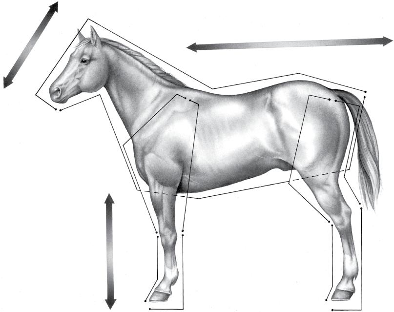
Figure 1.1 provides the directional terms for veterinary anatomy that will be used in this chapter. With the exception of the ocular and oral cavity structures, the terms anterior, posterior, superior, and inferior are not applicable to quadrupeds.
THORACIC LIMB
Digit and Fetlock
The digit is composed of distal (third), middle (second), and proximal (first) phalanges and associated structures (Figure 1.2). The fetlock consists of the metacarpophalangeal (fetlock) joint and the structures surrounding it. Because the digits and fetlocks of the thoracic limb and the pelvic limb are similar in most respects, the following descriptions pertain to both limbs unless otherwise indicated. When referring to structures of the forelimb, the term “palmar” is used; this will obviously be replaced with “plantar” when referring to the hindlimb. Likewise, such terms as metacarpophalangeal and metatarsophalangeal are counterparts in fore‐ and hindlimbs, respectively.
Foot
The foot consists of the hoof and all it encloses: the connective tissue corium (dermis), digital cushion, distal phalanx (coffin bone), most of the cartilages of the distal phalanx, distal interphalangeal (coffin) joint, distal part of the middle phalanx (short pastern bone), distal sesamoid (navicular) bone, podotrochlear bursa (navicular bursa), several ligaments, tendons of insertion of the common digital extensor and deep digital flexor muscles, blood vessels, and nerves. Skin between the heels is also part of the foot.
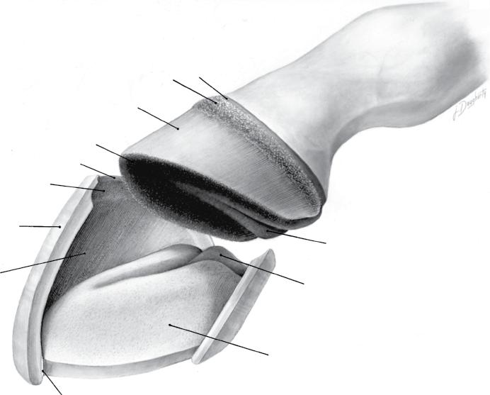
Hoof Wall, Sole, and frog
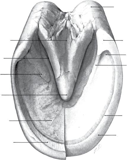
The hoof is continuous with the epidermis at the coronet, and the underlying corium of the hoof is likewise continuous with the dermis of the skin. The ground surface of the hoof comprises the sole, frog, heels, bars, and ground surface of the wall (Figure 1.3). The ground surface of the forefoot is normally larger and rounder than that of the hindfoot, reflecting the corresponding shape of the distal surface of the distal phalanx (coffin bone).
The hoof wall extends from the coronary band (also called the coronet), the transition between skin and hoof, distad to the ground. The surface of the wall is divided into the toe, medial and lateral quarters, and heels (Figures 1.3 and 1.4). From the toe, where it is thickest, the wall becomes progressively thinner and more elastic toward the heels, where it thickens again when it reflects to become the bars. Ranges for the angle of the toe between the dorsal surface of the hoof wall and the ground surface of the hoof vary widely.1,16 In the ideal digit, the dorsal surface of the hoof wall and the dorsal surface of the pastern should be parallel, reflecting the axial alignment of the phalanges.
The vascular and densely innervated collagenous connective tissue deep to the hoof is the corium. The corium
provides sensation, vascular supply, and attachment for the overlying stratified squamous epithelium that constitutes the hoof or ungual epidermis (L. ungula, hoof).
Regions of the corium are named according to the parts of the hoof under which they are located: perioplic corium, coronary corium, laminar corium, corium of the frog (cuneate corium), and solar corium. Histologically, coronary corium gives rise to elongated, distally directed papillae. Laminar corium forms a series of sheets that interdigitate with epidermal laminae of the stratum internum of the hoof wall. Shorter papillae extend from the perioplic, solar, and cuneate coria.
In the coronary region, the deepest layer (the stratum basale) of the ungual epidermis is a single layer of proliferating keratinocytes lying upon and between long dermal papillae. Cellular division here pushes cells distad into the stratum medium of the hoof wall, forming the epidermis that undergoes cornification.2 Nearly the entire hoof is composed of a thick layer of anucleate squamous keratinocytes.
For the most part, the keratinaceous tissues of the hoof are devoid of nerve endings; as a consequence it is the “insensitive” part of the foot. However, a few sensory
nerve endings from nerves in the corium penetrate between cells of the deepest layer of the epidermis.
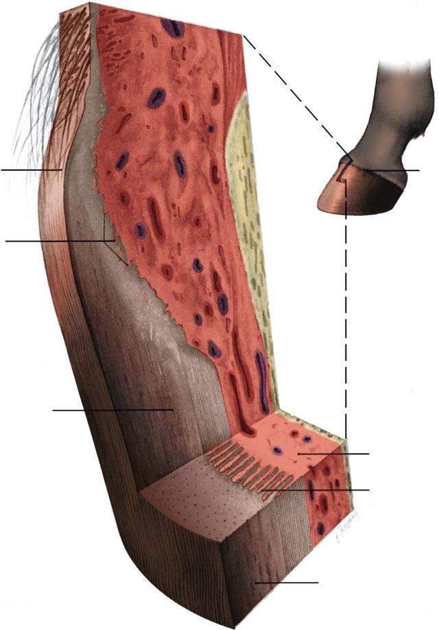
Three histological layers comprise the hoof wall: the stratum externum, stratum medium, and stratum internum (Figure 1.5). The superficial stratum externum, commonly called the periople, is a thin layer of horn extending distad from the coronet a variable distance; this thin, soft layer wears from the surface of the hoof wall so that it is present only on the bulbs of the heels and the proximal parts of the hoof wall. The bulk of the wall is the stratum medium consisting of cornified horn.2 The stratum internum comprises the epidermal laminae.
Distal to the coronary sulcus (Figure 1.4), about 600 primary epidermal laminae of the stratum internum interleave with the primary dermal laminae of the laminar corium (Figures 1.6 and 1.7). Approximately 100 microscopic secondary laminae branch at an angle from each primary lamina, further binding the hoof and corium together (Figures 1.3–1.6). The epidermal laminae are routinely referred to as “insensitive,” whereas the dermal laminae are called “sensitive.” In the strictest sense, though, only the keratinized parts of the primary epidermal laminae are insensitive; the deepest layer of
Caudal
Dorsal Cranial
Caudal
Dorsal
Dorsal
Rostral
Ventral
Proximal
Cranial
Dorsal
Distal
Dorsal
Plantar
Palmar
Caudal
Ventral
Cranial
Figure 1.1. Positional and directional terms.
Supraglenoid tubercle
Greater tubercle
Supraspinous fossa
Spine of the scapula
Deltoid tuberosity
Lateral epicondyle
Shaft of the radius
Intermediate carpal bone
Third carpal bone
Metacarpal tuberosity
Humeral head
Scapular car tilage
Infraspinous fossa
Tuber spinae
Shaft of the humerus
Olecranon fossa
Shaft of the ulna Olecranon tuber
Lateral styloid process
Accessory carpal bone
Ulnar carpal bone
Fourth carpal bone
Fourth metacarpal bone
Third metacarpal bone
Proximal phalanx
Middle phalanx
Distal phalanx
Lateral proximal sesamoid bone
Distal sesamoid bone
Figure 1.2. Bones of the left equine thoracic limb. Lateral view.
Central sulcus of the frog
Collateral sulcus
Crus of the sole
Apex of the frog
Body of the sole
Epidermal laminae
Bulb of the heel
Heel
Angle of the wall
Angle of the sole
Bar Quar ter
White line
Stratum medium of the hoof wall
Figure 1.3. Topography of the solar surface of the hoof. The right half has been trimmed to emphasize the region of the white line.
Perioplic corium Coronary corium
Laminar corium
Solar corium
Perioplic sulcus
Coronary sulcus
Stratum medium
Epidermal laminae of stratum internum
White line
Corium of the frog
Frog stay
Internal surface of the sole
Figure 1.4. Dissected view of relationships of the hoof to underlying regions of the corium (dermis).
Toe
Interdigitation of corial and epidermal laminae (stratum internum)
the epidermis, the stratum basale, including all of the secondary epidermal laminae, and the laminar corium are both innervated and therefore “sensitive.”42
Growth of the hoof wall is primarily from the coronary epidermis toward the ground. Trauma or inflammation stimulates greater production of horn. Ultrastructural studies indicate that during growth of the hoof, primary epidermal laminae move past the secondary epidermal
laminae by breaking and then reforming desmosomes between the two cell populations.23 The relationship between the epidermal and dermal laminae plus the blending of the laminar corium with the periosteum of the distal phalanx suspend and support the bone, aiding in the dissipation of concussion and the movement of blood. The growth of the wall progresses at the rate of approximately 6 mm per month, taking from 9 to
Periople Coronet
Papillae of coronary corium covered by coronary epidermis
Tubular and intertubular horn of the stratum medium of the horn wall
Stratum medium
Laminar corium
Figure 1.5. Three‐dimensional dissection of coronary region of the hoof wall.