
Visit to download the full and correct content document: https://ebookmass.com/product/examination-review-for-ultrasound-abdomen-and-obs tetrics-gynecology-2nd-edition-ebook-pdf/
More products digital (pdf, epub, mobi) instant download maybe you interests ...
Examination Review for Ultrasound: SPI: Sonographic Principles & Instrumentation 2nd Edition, (Ebook PDF)
https://ebookmass.com/product/examination-review-for-ultrasoundspi-sonographic-principles-instrumentation-2nd-edition-ebook-pdf/

Diagnostic Ultrasound Abdomen and Pelvis 2nd Edition
Aya Kamaya
https://ebookmass.com/product/diagnostic-ultrasound-abdomen-andpelvis-2nd-edition-aya-kamaya/

Obstetrics and Gynecology in Chinese Medicine 2nd Edition
https://ebookmass.com/product/obstetrics-and-gynecology-inchinese-medicine-2nd-edition/
Obstetrics & Gynecology PreTest Self-Assessment And Review 14th Edition Edition Shireen Madani Sims
https://ebookmass.com/product/obstetrics-gynecology-pretest-selfassessment-and-review-14th-edition-edition-shireen-madani-sims/

Obstetrics and Gynecology: Maintenance of Knowledge, An Issue of Obstetrics and Gynecology Clinics, 1e Janice
L. Bacon
https://ebookmass.com/product/obstetrics-and-gynecologymaintenance-of-knowledge-an-issue-of-obstetrics-and-gynecologyclinics-1e-janice-l-bacon/
Obstetrics & Gynecology (Diagnostic Medical Sonography Series) 4th Edition, (Ebook PDF)
https://ebookmass.com/product/obstetrics-gynecology-diagnosticmedical-sonography-series-4th-edition-ebook-pdf/
Obstetrics & Gynecology (Diagnostic Medical Sonography Series) Fourth Edition – Ebook PDF Version
https://ebookmass.com/product/obstetrics-gynecology-diagnosticmedical-sonography-series-fourth-edition-ebook-pdf-version/

Netter’s Obstetrics and Gynecology-Elsevier (2017) 3rd Edition Roger Smith
https://ebookmass.com/product/netters-obstetrics-and-gynecologyelsevier-2017-3rd-edition-roger-smith/

Case Files Obstetrics and Gynecology 5th Edition Eugene C. Toy
https://ebookmass.com/product/case-files-obstetrics-andgynecology-5th-edition-eugene-c-toy/

several revisions include an overview of gravidity and parity (Chapter 22), an improved review of the fetal heart (Chapter 27), and information concerning fetal alcohol syndrome (Chapter 32). Once more, these are only highlights, as there are countless improvements to this edition.
Additional Resources
As with the first edition, the review associated with this book does not need to end with its conclusion. There are several online resources for this book, including a mock registry examination that can be attempted by using the code at the beginning of this book and going to thepoint.lww.com/penny abobgyn2e. This exam simulator will provide the user with more intense “registry-like” questions, with topics that can be selected and answers that provide rationale. Instructors can use the faculty resources as well, which include an image bank and PowerPoint presentations.
Final Note
The material contained in this book is deliberately not exhaustive. With candid prayer for discernment, much care has been taken to explore each topic, and I have tried to select the most vital information for you as you study for these complicated examinations. Thus, this book, if used for its primary purpose of registry review, should emerge to the user as focused and purposeful. The exclusion of numerical statistics and extraneous facts from the text in most regards is intentional. However, in the pursuit of exhaustive comprehension, where data or information is critical to more complete patient care, the list of bibliographical references at the end of each chapter should provide you with essential aid.
In the classroom setting, many instructors have discovered that the use of the first edition of this book can certainly be beneficial. This edition will also provide the instructor with a topic-based review that can identify subject matter weaknesses. And instructors that offer registry review courses will find that this book and its resources will offer them a focused tool for preparing their students for the national certification exams.
For those who have moved beyond national certification, the manner in which this book has been constructed with clinical and sonographic findings highlighted certainly allows for its application during sonographic practice. Nevertheless, it is my greater hope that you exploit all relevant resources in your mission and obligation to provide each patient with the most favorable care you can afford.
I anticipate that this book will serve you, your students, and your patients well. I may never meet you face-to-face, but I would like to express gratitude for the choice that you have made to select this book. I wish you well as you care for our patients, and my hope is that you have a long-lasting and prosperous career as a registered diagnostic medical sonographer.
Steven M. Penny

I would first like to thank my acquisitions editor, Jay Campbell, for giving me the opportunity to pursue this second edition and for his confidence in me as an author. Thanks to all of the workers behind the scenes at Wolters Kluwer, including my editorial coordinator Lexi Pozonsky and marketing manager Leah Thompson, and the other members of the staff who make a project like this come together.
To my parents, Ted and Linda Penny, thank you for all that you have done for me and my family over the years. I hope that you know that I love you and that I am grateful for the love you have shown me throughout my life.
To my brother, Jeff Penny, and his family, Tammy, Nick, and Mackenzie thanks for your encouragement and for being in my life.
To my coworkers and the leadership at Johnston Community College, thank you for your companionship and direction over the years. To my past, current, and future students, thank you for demanding that I continue to learn, and thank you for allowing me to be your instructor.
To all of my former teachers and professors, thank you for your unwavering patience.

“By failing to prepare, you are preparing to fail.”
– Ben Franklin
The national certification examinations offered by the American Registry for Diagnostic Medical Sonography (ARDMS) and American Registry of Radiologic Technologist (ARRT) are not easy. These exams should be difficult because they have been created to test your knowledge of a vital imaging modality that can save people’s lives. Consequently, preparing for and challenging these exams should not be approached dispassionately. There are many resources on test taking that you can access at your local library and online. Below are three basic steps, which include tips for getting organized, studying, and preparing for these significant certification exams.
Step 1 Get Organized and Schedule the Exam
Both the ARDMS (ardms.org) and ARRT (arrt.org) provide content outlines for each of their certification examination. In fact, the content of this book is based on these content outlines, and accordingly this book includes pertinent information on each topic. However, you can use these content outlines as a guide for focused study as well. Keep your study materials which should include all of the resources you obtained throughout your sonography education organized by these content outlines. School lecture notes, note cards, quizzes, and tests should be organized well before you begin to study. Once you have organized your study materials, you should apply for the exam and then try to consider the best time to attempt it. When scheduling the examination, consider all of your other obligations (e.g., family responsibilities, vacations, job requirements) and allow for an ample amount of time to study so that you are thoroughly prepared on examination day. Do not postpone scheduling the exam. Scheduling the examination will provide you with a firm date, and it will hopefully help those of you who suffer from procrastination to focus on test preparation.
Step 2
Establish a Study Routine and Study Schedule
Next, since you have your deadline, find a quiet place to study and develop a study routine and schedule. Your study space should be quiet and free from distractions, like television and your cell phone (stay off of social media). The study schedule that you create for yourself should be realistic. That is to say, do not schedule two hours each night to study if you know that you will not be dedicated to that schedule. Instead, it may be best to schedule one solid hour each night. Also, studying in 45-minute increments with 15minute breaks may work best for some. You can create your own deadlines on your schedule and strive to meet them. Be sure to study at least for a few minutes every day to maintain momentum going into the exam.
The amount of time one requires to study will vary per individual. Only you know how much time you need to study, so if you struggle with certain topics, then allow for extra time to focus more attention on those topics. It may be best to review those topics you are most familiar with first, and as the exam approaches, review the topics that you struggle with just before your attempt, with the hopes of making the challenging information more readily accessible.
Most people know what manner they prepare for an exam best. Some test takers find flashcard useful, some create their own notes from reading, whereas others may simply read and choose to gradually answer registry review questions. A study group may be helpful for some as well. Nonetheless, the main concern of your studies should be learning the material and not just memorizing it to pass the exam. By learning the information, you will most likely be successful on the exam, with the added benefit of being able to apply your knowledge in your daily clinical practice as a registered sonographer.
Step 3
Confidently Attempt the Exam
Test anxiety is a challenge for many people. Some tips for reducing anxiety include eating well, getting plenty of rest and exercise, keeping a positive attitude, and taking practice tests. The ARDMS offers some tips for examday success, which include getting a good night’s sleep before exam day, knowing how long it takes to get to the testing center (traffic included), being early to the testing center, and being familiar with all of the test center requirements, like testing day registrant identification specifications. Currently, the ARDMS Sonographic Principles & Instrumentation (SPI) exam consists of 110 questions, whereas the specialty exams consist of 170 questions. Nearly all of the questions are in multiple choice format. The SPI exam may also include advanced item type question in the form of a semi-
interactive console, which requires the test taker to make adjustments to a simulated ultrasound machine console. The specialty exams may also include an advanced item type question referred to as hotspot questions. These questions require that the test taker use the cursor to mark the correct answer directly on the image.
Multiple choice questions consist of the stem which is the question and four possible answers to choose from. The correct answer must be included along with three other options referred to as distractors. You should read the question cautiously first, and try to answer the question before looking at the provided choices. If your given answer is provided, it will most likely be the correct choice to make. If you do not know the answer immediately, then try to eliminate the choices you know are incorrect. For these exams, you are allowed to mark questions and return to answer them later. You can also make changes before final submission. But be careful, it may be best to not change any of your answers. You should only make changes to questions that you feel confident you have answered incorrectly because for many of us our first impulse or guess is correct.
Bibliography
ARDMS.org. Review our tips for examination day success. Available at: http://www.ardms.org/get-certified/RDMS/Pages/Abdomen.aspx. Accessed March 16, 2017.
Fry R. Surefire Study Success: Surefire Tips to Improve Your Test-Taking Skills. New York: Rosen Publishing Group, 2016
Hill J. Test taking tips to give you an edge. Biomedical Instrumentation and Technology. 2009;43(3):223–224.
Mary KL. Test-taking strategies for CNOR certification. AORN Journal. 2007;85(2):315–332.
Medoff L. Stressed Out Students’ Guide to Dealing with Tests. New York: Kaplan Inc, 2008.

Abdominal Sonography Overview
The Liver
The Gallbladder
The Bile Ducts
The Pancreas
The Spleen
The Urinary Tract
The Adrenal Glands
Abdominal Vasculature
The Gastrointestinal Tract and Abdominal Wall
Noncardiac Chest and Retroperitoneum
The Face and Neck
The Male Pelvis
Musculoskeletal Imaging, Breast, and Superficial Structures SECTION
Gynecologic Sonography
Anatomy of the Female Pelvis
The Uterus and Vagina
The Ovaries and Fallopian Tubes
The Menstrual Cycle
SECTION III OBSTETRIC SONOGRAPHY REVIEW
Obstetric Sonography Overview
The First Trimester
The Fetal Head and Brain
The Fetal Face and Neck
The Fetal Spine and Musculoskeletal System
The Fetal Heart and Chest
The Fetal Gastrointestinal System
The Fetal Genitourinary System
Chromosomal Abnormalities
Multiple Gestations
Fetal Environment and Maternal Complications
Answers to

Introduction
A synopsis of abdominal sonography practice is provided in this chapter. Accordingly, the subsequent abdominal chapters will build on the foundation established in this chapter. It is important for the sonographer to obtain and recognize vital clinical information from patients, including laboratory results and other data that are obtained through patient inquiry. This chapter offers relevant laboratory findings, imaging artifacts, a brief overview of physics and instrumentation, infection control, and serves as a general abdominal imaging guide. Lastly, cross-referencing of potential information that may be encountered on the abdominal certification examination offered by the American Registry for Diagnostic Medical Sonography (www.ardms.org) and the abdominal portion of the examination offered by the American Registry of Radiologic Technologists (www.arrt.org)hasbeenperformedtoestablishthischapter.
Key Terms
anemia—a condition in which the red blood cell count or the hemoglobin is decreased anticoagulation therapy drug therapy in which anticoagulant medications are given to a patient to slow the rate at which the patient’s blood clots ascites—a collection of abdominal fluid within the peritoneal cavity chromaffin cells—the cells in the adrenal medulla that secrete epinephrine
and norepinephrine
clinical findings—the information gathered by obtaining a clinical history clinical history—a patient’s signs and symptoms, pertinent illnesses, past surgeries, laboratory findings, and the results of other diagnostic testing coagulopathies disorders that result from the body’s inability to coagulate or form blood clots also referred to as bleeding disorders
computed tomography—an imaging modality that uses X-ray to obtain cross-sectional images of the body in multiple planes; also referred to as CT or CAT scan
elastography—a sonographic technique employed to evaluate a mass based on its stiffness, ultimately providing a prediction as to whether a mass is more likely malignant or benign
endoscopy—a means of looking inside of the human body using an endoscope
exudate ascites a collection of abdominal fluid within the peritoneal cavity that may be associated with cancer
fluid-fluid level—a distinctive line seen within a cyst representing the layering of two different fluid densities
gastrin hormone produced by the stomach lining that is used to regulate the release of digestive acid
hematocrit a laboratory value that indicates the amount of red blood cells in the blood
homeostasis the body’s ability or tendency to maintain internal equilibrium by adjusting its physiologic processes
hyperthyroidism a condition that results from the overproduction of thyroid hormones
hypothyroidism a condition that results from the underproduction of thyroid hormones
intraluminal something located within the lumen or opening of an organ or structure
intraperitoneal located within the parietal peritoneum
Kaposi sarcoma—cancer that causes lesions to develop on the skin and other places; often associated with AIDS
leukocytosis an elevated white blood cell count
lymphadenopathy disease or enlargement of the lymph nodes
lymphedema build-up of lymph that is most likely caused by the obstruction of lymph drainage
mass effect—the displacement or alteration of normal anatomy that is
located adjacent to a tumor
Morrison pouch—the space between the liver and the right kidney; also referred to as the posterior right subhepatic space
multiloculated having many cavities
mural nodules small solid internal projections of tissue originating from the wall of cyst
nosocomial infections—hospital-acquired infections
nuclear medicine—a diagnostic imaging modality that utilizes the administration of radionuclides into the human body for an analysis of the function of organs or for the treatment of various abnormalities oncocytes—large cells of glandular origin
paracentesis a procedure that uses a needle to drain fluid from the abdominal cavity for diagnostic and/or therapeutic reasons
parietal peritoneum the portion of the peritoneum that lines the abdominal and pelvic cavities
pineal gland endocrine gland located in the brain that secretes melatonin
radiography a diagnostic imaging modality that uses ionizing radiation for imaging bones, joints, organs, and some other soft tissue structures
retroperitoneal posterior to the peritoneum
serosal fluid fluid that is secreted by the serous membranes to reduce friction in the peritoneal and other cavities of the body
signs an objective evidence of a disease such as abnormal laboratory findings and fever
sonographic findings—information gathered by performing a sonographic examination
space of Retzius the space between the urinary bladder and the pubic bone; also referred to as the retropubic space
standoff pad—a gel pad that is used to provide some distance between the transducer face and the skin surface, allowing superficial structures to be imaged more clearly
symptoms—any subjective evidence of a disease such as nausea, weakness, or numbness
thoracentesis a procedure that uses a needle to drain fluid from the pleural cavity for either diagnostic or therapeutic reasons
thymus gland—gland of the immune and lymphatic system located in the chest
transudate ascites a collection of abdominal fluid within the peritoneal cavity often associated with cirrhosis
tumor markers—substances produced by cancer cells or organs in response to cancer
unilocular having a single cavity
visceral peritoneum the portion of the peritoneum that is closely applied to each organ
voiding cystourethrogram—a radiographic examination used to evaluate the lower urinary tract, where a contrast agent is instilled into the urinary bladder by means of urethral catheterization
Wilson disease—a congenital disorder that causes a person to retain excess copper
SONOGRAPHIC TERMINOLOGY AND PRACTICE GUIDELINES
Before beginning your studies, you must have a fundamental appreciation of sonographic terminology and commonly used sonographic descriptive terms (Table 1-1) Abdominal sonograms may be requested for various reasons The American Institute of Ultrasound in Medicine (AIUM) publishes the practice guidelines for an abdominal sonogram on their website at www.aium.org(Table1-2).
SONOGRAPHIC DESCRIPTION OF ABNORMAL FINDINGS
The appreciation and recognition of sonographic pathology is vital for the sonographer. Not only should one be able to recognize the normal echogenicity of organs and structures, but one must also be capable of identifying abnormalities. The normal echogenicity of the abdominal organs from greatest (brightest) to least (darkest) is as follows: renal sinus → pancreas→spleen →liver→renalcortex→ renalpyramids→ gallbladder. Therefore,ifthe rightkidneyappears moreechogenicthan theliver,both the liver and the right kidney must be closely examined for a cause of this deviantsonographicfinding.

SOUND OFF
The normal echogenicity of the abdominal organs from greatest (brightest) to least (darkest) is as follows: renal sinus → pancreas → spleen → liver → renal cortex → renal pyramids → gallbladder.
TABLE 1-1
Sonographic descriptive terms
Sonographic Descriptive
Term Definition
Anechoic Without echoes
Complex
Having both cystic and solid components
Echogenic Structure that produces echoes
Heterogeneous Of differing composition
Homogeneous Of uniform composition
Hyperechoic
Hypoechoic
Isoechoic
Having many echoes
Having few echoes
Having the same echogenicity
Examples
Gallbladder
Simple renal cyst
Hemorrhagic cyst
Hepatic abscess
Fatty liver
Chronic renal disease
Graves disease
Diffuse liver metastasis
Normal liver
Normal testicle
Cavernous hemangioma
Angiomyolipoma
Hepatic adenoma
Thyroid adenoma
Focal nodular hyperplasia
TABLE 1-2 AIUM indications for abdomen and/or retroperitoneum
sonogram
AIUM Indications for an Ultrasound Examination of the Abdomen and/or Retroperitoneuma
Abdominal, flank, and/or back pain.
Signs or symptoms that may be referred from the abdominal and/or retroperitoneal regions such as jaundice or hematuria.
Palpable abnormalities such as an abdominal mass or organomegaly.
Abnormal laboratory values or abnormal findings on other imaging examinations.
Suggestive of abdominal and/or retroperitoneal pathology.
Follow-up of known or suspected abnormalities in the abdomen and/or retroperitoneum.
Search for metastatic disease or an occult primary neoplasm.
Evaluation of suspected congenital abnormalities.
Abdominal trauma.
Pretransplantation and posttransplantation evaluation
Planning for and guiding an invasive procedure.
•
•
• Searching for the presence of free or loculated peritoneal and/or retroperitoneal fluid.
Suspicion of hypertrophic pyloric stenosis or intussusceptions. Evaluation of a urinary tract infection.
An abdominal and/or retroperitoneal ultrasound examination should be performed when there is a valid medical reason. There are no absolute contraindications.
a This is a limited list of indications. Other indications exist. AIUM, American Institute of Ultrasound in Medicine.
Pathology is often described sonographically, relative to surrounding or adjacent tissue. For example, a hepatic mass may be described as hyperechoic compared to the surrounding echotexture of the normal liver. Solid tumors may be hyperechoic (occasionally described as echogenic), hypoechoic, homogeneous, heterogeneous, complex, isoechoic, cystic, or a combination of terms. For example, a renal mass may be described as a hypoechoicmasswithacentralareaofincreasedechogenicity.
Lastly, a cyst must fit certain criteria to be referred to as a simple cyst. Simple cysts have smooth walls or borders, demonstrate through transmission, are anechoic, and are round in shape (STAR criteria). Occasionally, with higher frequency transducers with superior resolution, a diminutive amount of internal debris may be noted within a simple cyst. However, cysts that have a large amount of internal debris, septations, mural nodules, have a fluid–fluid level, or other components may be described as complex cysts. Cysts may also be referred to as multiloculated or unilocular. Although simple cysts may not be worrisome, complex cysts may be followedcloselyorfurtheranalyzedwithanotherimagingmodality.
SOUND OFF
A simple cyst should have smooth walls, demonstrate through transmission, be completely anechoic, and be round in shape (STAR criteria).
PATIENT PREPARATION FOR AN ABDOMINAL SONOGRAM
Patients who are required to undergo an abdominal sonogram, and particularly patients who still have a gallbladder, need to fast for 8 hours, although some authors suggest that only a 6-hour fast may be warranted. However, diagnostic studies can be obtained in the nonfasting patient,
especially those requiring an emergency sonogram. The purpose of fasting is to ensure that the gallbladder is distended and to potentially reduce the amount of upper abdominal gas that may inhibit diagnostic accuracy. For renal sonograms, the patient is not typically required to fast, although some facilities may recommend that the patient be well hydrated. This is true especially if the urinary bladder needs to be assessed sonographically for intraluminal masses, irregularities, or urinary stones. Diabetic patients need to be scheduled early in the morning for studies that require them to fast to prevent hypoglycemic episodes. Abdominal sonography should also be performed prior to radiographic examinations that utilize barium contrast agents. For pediatric patients who must undergo renal sonography and urologic radiographic examinations, such as a voiding cystourethrogram, the renal sonogram is typically performed first. Small part sonography, such as scrotal and thyroid imaging, does not require patient preparation. The sonographicimagingprocessshouldbeclearlyexplainedtothepatient,anda thoroughclinicalhistoryobtainedpriortoperformingeachexamination.
GATHERING A CLINICAL HISTORY AND LABORATORY FINDINGS
Although the patient–sonographer interaction is exceedingly important, a review of prior examinations and relevant documents should be performed by the sonographer before any contact with the patient. Gathering a thorough clinical history includes a review of reports from previous sonograms, computed tomography (CT) scans, magnetic resonance imaging studies, nuclear medicine examinations, radiography procedures, endoscopy examinations, and any additional related laboratory and diagnostic reports available Moreover, sonographers must be capable of analyzing the clinical complaints of their patients. This practice will aid not only in clinical practice but also in answering complex certification examination questions By correlating clinical findings with sonographic findings, the sonographer can directly impact the patient’s outcome by providing the most targeted examination possible. Furthermore, when faced with a complicated, in-depth registry question, the test taker should be capable of eliminating information that is not applicable, to answer the question appropriately. Helpful information can be gathered from patient inquiry and from analyzing the registryreviewquestionathand.
Although not all patients visit the sonography department with laboratory results (labs) in hand, sonographers must be capable of analyzing labs when they are available. For most inpatients and emergency room patients, labs from blood work and/or a urinalysis will be available. Although an increase
in most labs reveals evidence of an abnormality, some lab levels decrease with certain abnormalities. For example, leukocytosis, or an elevation in white blood cell (WBC) count, in general, indicates the presence of an inflammatory response due to infection. Patients who have some form of “itis” (such as cholecystitis or pancreatitis), or possibly even an abscess, will most likely have an abnormal WBC count with existing infection. Conversely, a decrease in hematocrit indicates bleeding. Patients who have suffered recent trauma or have an active hemorrhage will most likely have a decreased hematocrit level. Keep these two labs in mind as you study. Other labs and specific associated pathologies will be included in organ-specific chapters. A summary of labs can be found in Table 1-3 for quick broad reference(Table1-3).
SOUND OFF
Patients who have some form of “itis” (such as cholecystitis or pancreatitis), or possibly even an abscess, will most likely have an abnormal WBC count with existing infection.
BASIC PATIENT CARE AND EMERGENCY SITUATIONS IN ABDOMINAL IMAGING
Sonographers must be capable of providing basic patient care for every patient equitably and in a timely manner Although we may spend a limited amount of time with each patient, we must also be prepared for emergency situations and know how to respond. Basic patient care includes the assessment of body temperature, pulse, respiration, and blood pressure if needed (Table 1-4). Sonographers should be competent at transferring patients safely from wheelchairs and stretchers to the examination stretcher, being mindful of intravenous therapy, postsurgical, and urinary catheter needs. For patients with intravenous therapy, the intravenous fluid bag should be continually elevated to prevent retrograde flow. For urinary catheter care, the urinary bag should be placed below the level of the urinary bladder to prevent retrograde urine flow that could result in a urinary tract infection. One of the most common causes of nosocomial infections, or hospital-acquiredinfections,istheurinarytractinfection.
TABLE 1-3 Basic overview of commonly encountered and relevant laboratory findings for abdominal imaging
Alanine aminotransferase
Albumin
Alkaline phosphatase
Aspartate aminotransferase
Bilirubin
λ-glutamyl transferase
PTT
PT
Urobilirubin (urine test)
Calcitonin
Thyroid-stimulating hormone
Thyroxine (T4) or free thyroxine
Triiodothyronine (T3)
BUN
Creatinine
Amylase
Biliary tree disease
Pancreatic disease
Hepatic disease
Liver damage
Biliary obstruction
Liver cancer
Pancreatic disease
Gallstones
Wilson disease
Liver damage
Pancreatic disease
Liver disease
Biliary obstruction
Other systemic disorders and syndromes
Liver disease
Biliary obstruction
Liver disease
Hereditary coagulopathies
Vitamin K (deficiency)
Anticoagulation therapy
Liver disease
Bleeding abnormalities
Anticoagulation therapy
Liver disease
Biliary obstruction
Thyroid cancer
Lung cancer
Anemia
Hypothyroidism
Hyperthyroidism
Hyperthyroidism
Hypothyroidism
Hyperthyroidism
Hypothyroidism
Renal disease
Renal obstruction
Dehydration
Gastrointestinal bleeding
Congestive heart failure
Renal damage
Renal infection
Renal obstruction
Pancreatic disorders
Gallbladder disease
Lipase
Serum calcium
Prostatic-specific antigen
Hematocrit
WBC
Biliary or pancreatic obstruction
Pancreatic disorders
Gallbladder disease
Biliary or pancreatic obstruction
Parathyroid abnormalities
Prostate abnormalities
Hemorrhage
Inflammatory disease/infection
BUN, blood urea nitrogen; PT, prothrombin time; PTT, partial thromboplastin time; WBC, white blood cell
TABLE 1-4 Normal numbers or ranges for basic patient care assessment
Basic Patient Assessment
Body temperature 98.6° (oral)
Adult pulse 60–100 beats/minute
Adult blood pressure <120/80
Adult respiration 12–20 breaths/minute
INFECTION CONTROL AND TRANSDUCER CARE
The cycle of infection may be depicted as a succession of steps (Fig. 1-1). Sonographers should continually employ standard precautions and good hygiene to prevent the spread of infection. Standard precautions are put into place to reduce the risk of microorganism transmission in the clinical setting. Standard precautions, formerly referred to as universal precautions, include (1) hand hygiene, (2) the use of personal protective equipment, (3) safe injection practices, (4) safe handling of potentially contaminated equipment and surfaces, and (5) respiratory hygiene and coughing etiquette. These precautions apply to blood, nonintact skin, mucous membranes, contaminatedequipment,andallotherbodyfluids,exceptforsweat.
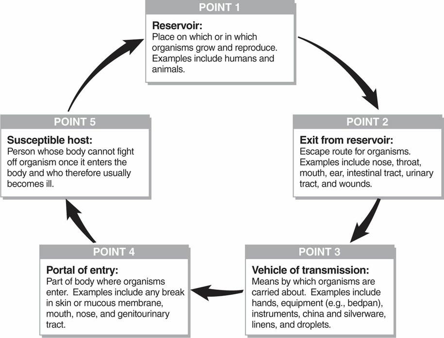
Figure 1-1 The cycle of infection.
The use of proper hand-washing and hand-cleaning techniques is one of the most effective means of preventing the spread of infection. The Center for Disease Control now recommends that health care workers employ an alcohol-based hand rub as the primary mode of hand hygiene in the clinical setting. Traditional hand-washing should be used as well when time permits, especially in situations when the hands are visibly soiled. Sonographers should also utilize personal protective equipment, such as gloves, gowns, face shields, and masks, when clinically applicable. Gloves, which are made of latex, nonlatex, or other synthetic material, should be worn during the sonographic examination and should be changed between patients. Be mindful of the potential for patient latex allergies and adapt accordingly by usinganotherformofsyntheticgloves.
Medical asepsis refers to the practices used to render an object or area free of pathogenic microorganisms. Although medical asepsis includes handwashing, it also includes the use of disinfectants in the clinical setting, as well as the use of transducer or probe covers. Probe covers should be used for endocavity examinations, such as endovaginal and endorectal imaging. The use of sterile or nonsterile probe covers for these examinations is recommended by the institution. Transducers used during invasive
procedures should be covered with a sterile probe cover, and sterile ultrasoundgelshouldbeutilized.
Following each examination, the transducer, ultrasound machine, stretcher, and any other equipment used during the examination should be thoroughly disinfected. The transducer should be cleaned with a disinfectant spray or wipe as recommended by the institution and manufacturer. Endocavity transducers should be sterilized in some manner after each examination. Lastly, to prevent the spread of infection, sonographers should maintaingoodpersonalhygieneandhealth.
INVASIVE AND STERILE PROCEDURES
Patient preparation for invasive procedures varies among clinical facilities. However, informed consent from the patient and laboratory results are universally obtained. Laboratory findings, including an analysis of prothrombin time (PT), partial thromboplastin time (PTT), international normalizing ratio, fibrinogen, and platelets, are used to evaluate the patient for coagulopathies. Sterile field preparation is performed prior to the procedure as well, and sterile asepsis, also referred to as surgical asepsis, mustbemaintained(Table1-5). Of course, sterile asepsis is always practiced inthesurgicalsuite.Someinvasiveproceduresthatarecommonlyperformed in the sonography department include thoracentesis, paracentesis, organ biopsies, mass biopsies, and abscess drainages. Biopsies can be performed using a freehand technique or under ultrasound guidance using a needle guide that attaches to the transducer. They may also be described as fine needle aspiration (FNA), which uses a thin needle and a syringe, or a core biopsy, which uses a much larger diameter needle to obtain a substantial tissue sample. An example of an FNA would be that of a thyroid biopsy, whereasanexampleofacorebiopsywouldbealiverbiopsy.
TABLE 1-5 Ten vital rules of surgical asepsis to remember
Always know which area and items are sterile and which are not.
If the sterility of an object is questionable, it is considered nonsterile.
If you recognize that an item has become nonsterile, act immediately.
A sterile field must never be left unmonitored. If a sterile field is left unattended, it is considered nonsterile.
A sterile person does not lean across a sterile field.
A sterile field ends at the level of the tabletop.
Cuffs of a sterile gown are not considered sterile.
The edges of a sterile wrapper are not considered sterile.
9. 10. If one sterile person must pass another, they must pass back-to-back. Coughing, sneezing, or excessive talking over a sterile field leads to contamination.
INSTRUMENTATION
Although a thorough physics review is beyond the scope of this book, there are several topics that must be addressed in preparation for the abdominal examination. Sonographic images are typically recorded and stored in a picture-archiving and communication system (PACS). PACS allows for both easy storage and comparison between sonographic findings and straightforwardcorrelationbetweenotherimagingmodalityfindings.
General abdominal imaging requires the use of a transducer that balances penetration with high-quality resolution. In general, the higher the frequency employed, the poorer the penetration abilities, but the better the resolution. Conversely, the lower frequencies provide better penetration with a sacrifice in resolution. Transducers that may be used for abdominal and small part sonographic imaging include linear array, matrix array, curved or convex array, phased array, or vector or sector array. Higher frequency linear array transducers (7.5 to 18 MHz or higher when appropriate) should be used for superficial structures, such as thyroid, scrotum, breast, musculoskeletal (MSK) imaging, and some gastrointestinal examinations (e.g., appendix and pylorus) A standoff pad or a mound of gel may be used for imaging some superficial structures, such as splinters or foreign objects just below the skin surface. Lower frequency curved array transducers (2.0 to 5.0 MHz) are employed for general abdominal imaging for the assessment of deeper or larger structures such as the liver, abdominal aorta, or pancreas. When applicable, sonographers should utilize technology such as power Doppler, color Doppler, pulsed Doppler, harmonics imaging, compound imaging, extended-field of view, elastography, and three-dimensional (3D) sonographytopromotediagnosticaccuracy.
SOUND OFF
↑Frequency = ↑Resolution = ↓Penetration
↓Frequency = ↓Resolution = ↑Penetration
IMAGING AND DOPPLER ARTIFACTS
Gray-scale, or brightness mode (B-mode), provides a two-dimensional (2D)
image of the human body in real time. Real-time imaging provides anatomy and motion, much like watching a live video of the internal structure being analyzed. Abdominal sonography involves careful analysis of vital structures. Often, artifacts will be observed during real-time imaging (Table 1-6).Also,thereareseveralDopplerimagingartifacts(Table1-7).
BODY SYSTEMS AND ABDOMINAL CAVITY
Overview of Body Systems
Throughout the following chapters, specific vital details will be provided for both normal and abnormal conditions that can be demonstrated with sonography. However, a fundamental understanding of several of the body systems is an important commission for the sonographer. The body consists of many mutually supporting body systems. These systems work together to preserve homeostasis, the body’s tendency to maintain internal equilibrium by adjusting internal processes. These systems include the cardiovascular system, endocrine system, lymphatic system, MSK system, nervous system, and reproductive system The excretory system includes the digestive system, urinary system, and respiratory system. Table 1-8 provides some insight into the basic functions of these systems and relevant topics to keep in mind as you study the various components that are applicable to abdominalimaging(Table1-8).
Artifact Explanation Example
Anisotropy Occurs when the sound beam strikes a structure in a nonperpendicular manner, resulting in a loss of the true echogenicity of the structure.
Seen when imaging tendons (Fig. 1-2).

Figure 1-2 Anisotropy. Left image is obtained perpendicular to the tendon (arrows), while the right image is obliqued.
Comet tail A type of
Seen with adenomyomatosis of the gallbladder
reverberation artifact caused by several small, highly reflective interfaces. (Fig. 1-3).
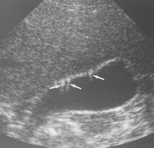
Figure 1-3 Comet tail artifact (arrows).
Dirty shadowing
Caused by air or bowel gas
Most often seen emanating from bowel. May be seen posterior to gas within an abscess (Fig 1-4)
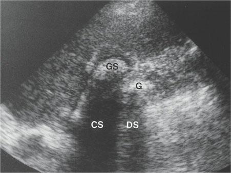
Figure 1-4 Dirty shadowing (DS) produced by gas (G). Clean shadow (CS) produced by gallstone (GS).
Edge shadowing
Reflective or refractive effect seen deep to the margins of a round structure that have a
Often seen arising from cystic structures and appears as narrow shadow lines originating at the edge of these structures (Fig. 1-5).
significantly different speed of sound compared to surrounding tissue. May be termed refractive shadowing
Mirror image Produced by a strong specular reflector and results in a copy of the anatomy being placed deeper than the correct location.
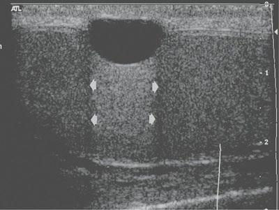
Figure 1-5 Edge artifact (arrowheads).
Seen posterior to the liver and diaphragm (Fig. 16).
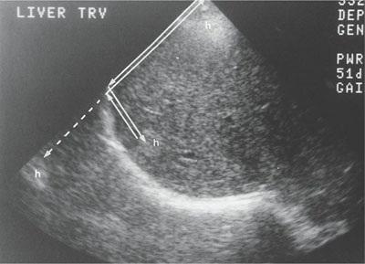
Figure 1-6 Mirror image artifact of a hemangioma (h) of the liver. The artifact is identified by the broken arrow outside of the liver.
Posterior (acoustic) enhancement or through transmission Produced when the sound beam is barely attenuated through a fluid or a fluid-filled structure
Seen posterior to fluid-filled structures such as the gallbladder and renal cysts, and with ascites (Fig. 1-7).
Refraction Caused by the bending of the ultrasound beam when it passes through an interface between two tissues with vastly dissimilar speeds of sound and the angle of the approach is not perpendicular

Figure 1-7 Through transmission (arrows) seen posterior to a liver cyst (Cy).
Seen when imaging through the rectus muscles of the abdominal wall (Fig. 1-8).
Reverberation artifact Caused by a large acoustic interface and subsequent production of false echoes.
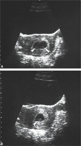
Figure 1-8 Refraction caused the appearance of two gestational sacs (image A arrowheads) when there was truly only one (image B arrowhead).
Seen as an echogenic region in the anterior aspect of the gallbladder or other fluid-filled structures (Fig. 1-9).
Ring-down artifact
Artifact that appears as a solid streak or a chain of parallel bands radiating away from a structure.

Figure 1-9 Reverberation. A. Reverberation in the anterior aspect of the urinary bladder (arrows) B Changing the scanning angle minimizes the artifact.
Seen emanating from gas bubbles within the abdomen. Can help to identify the presence of air in a structure, such as in the case of pneumobilia (Fig. 1-10).