Neuroanatomy Text and Atlas 4/E
Inkling Chapter (ENHANCED EBOOK) (NEUROANATOMY TEXT & ATLAS (MARTIN)) 4th Edition, (Ebook PDF)
Visit to download the full and correct content document: https://ebookmass.com/product/neuroanatomy-text-and-atlas-4-e-inkling-chapter-enh anced-ebook-neuroanatomy-text-atlas-martin-4th-edition-ebook-pdf/

More products digital (pdf, epub, mobi) instant download maybe you interests ...

Trapped: Brides of the Kindred Book 29 Faith Anderson
https://ebookmass.com/product/trapped-brides-of-the-kindredbook-29-faith-anderson/

Netter Atlas of Human Anatomy-Classic Regional Approach, 8e (Mar 29, 2022)_(0323793738)_(Elsevier) NOT TRUE PDF Frank H. Netter
https://ebookmass.com/product/netter-atlas-of-human-anatomyclassic-regional-approach-8e-mar-29-2022_0323793738_elsevier-nottrue-pdf-frank-h-netter/

Neuroanatomy: Text and Atlas 5th Edition Edition John D. Martin
https://ebookmass.com/product/neuroanatomy-text-and-atlas-5thedition-edition-john-d-martin/

Histology: A Text and Atlas 6th Edition, (Ebook PDF)
https://ebookmass.com/product/histology-a-text-and-atlas-6thedition-ebook-pdf/

Gifts, Glamping, & Glocks (A Camper & Criminals Cozy Mystery Series Book 29) Tonya Kappes
https://ebookmass.com/product/gifts-glamping-glocks-a-campercriminals-cozy-mystery-series-book-29-tonya-kappes/

Neuroscience for Neurosurgeons (Feb 29, 2024)_(110883146X)_(Cambridge University Press) 1st Edition Farhana Akter
https://ebookmass.com/product/neuroscience-for-neurosurgeonsfeb-29-2024_110883146x_cambridge-university-press-1st-editionfarhana-akter/

Cardiology-An Integrated Approach (Human Organ Systems) (Dec 29, 2017)_(007179154X)_(McGraw-Hill) 1st Edition Elmoselhi
https://ebookmass.com/product/cardiology-an-integrated-approachhuman-organ-systems-dec-29-2017_007179154x_mcgraw-hill-1stedition-elmoselhi/

Health Professions and Academia-How to Begin Your Career (Jun 29, 2022)_(3030942228)_(Springer).pdf John Paul (J.P) Sánchez
https://ebookmass.com/product/health-professions-and-academiahow-to-begin-your-career-jun-29-2022_3030942228_springer-pdfjohn-paul-j-p-sanchez/

978-1451176742 Text Atlas of Obstetric Dermatology
https://ebookmass.com/product/978-1451176742-text-atlas-ofobstetric-dermatology/

Contents
Preface
Acknowledgments
GuidetoUsingThisBook
SECTIONI|THECENTRALNERVOUSSYSTEM
1.OrganizationoftheCentralNervousSystem
NeuronsandGliaAretheTwoPrincipalCellularConstituentsoftheNervousSystem
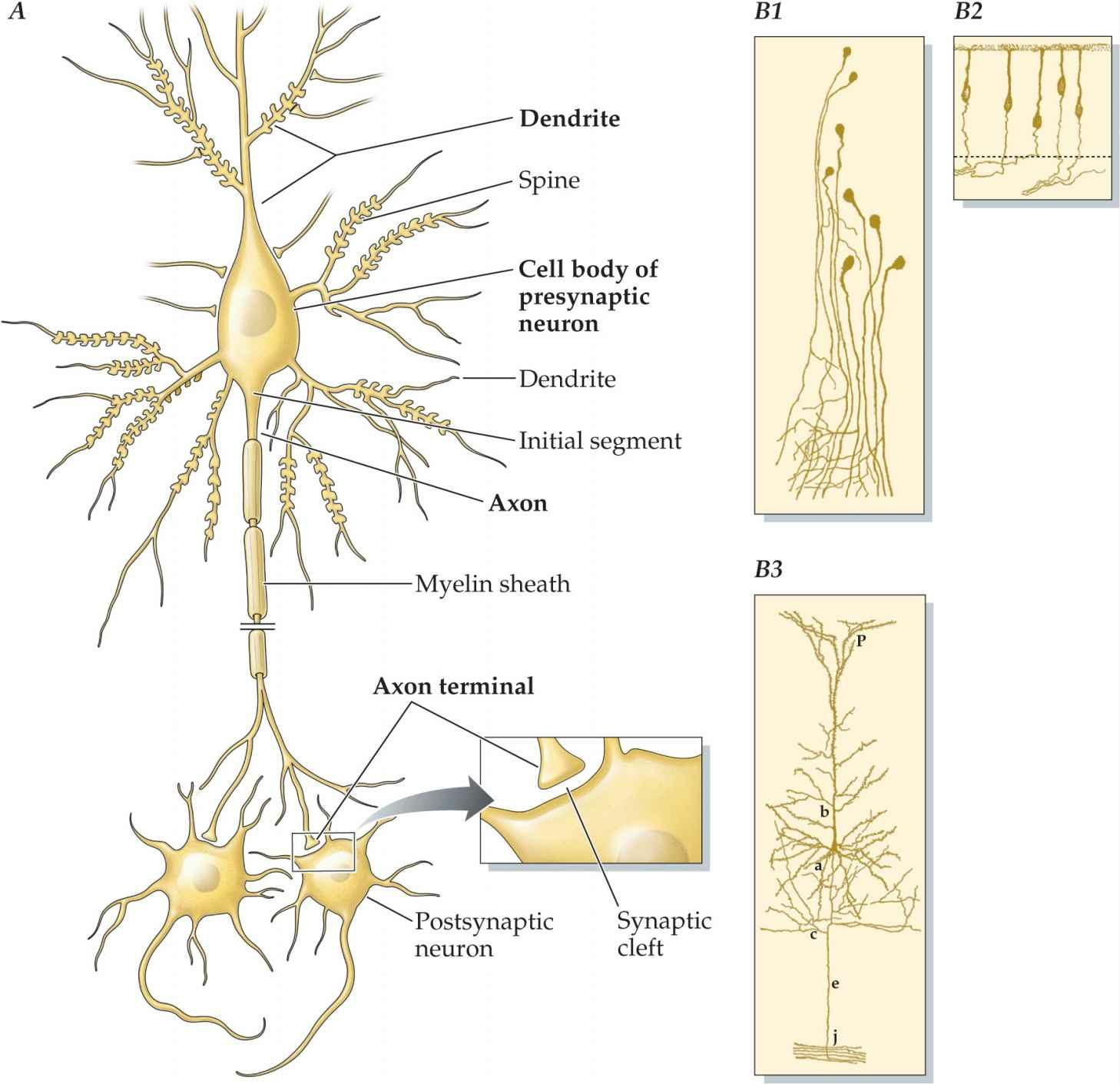
AllNeuronsHaveaCommonMorphologicalPlan
NeuronsCommunicateWithEachOtheratSynapses
GlialCellsProvideStructuralandMetabolicSupportforNeurons
TheNervousSystemConsistsofSeparatePeripheralandCentralComponents
TheSpinalCordDisplaystheSimplestOrganizationofAllSevenMajorDivisions
TheBrainStemandCerebellumRegulateBodyFunctionsandMovements
TheDiencephalonConsistsoftheThalamusandHypothalamus
TheCerebralHemispheresHavetheMostComplexShapeofAllCentralNervousSystemDivisions
The Subcortical Components of the Cerebral Hemispheres Mediate Diverse Motor, Cognitive, and EmotionalFunctions
TheFourLobesoftheCerebralCortexEachHaveDistinctFunctions
CavitiesWithintheCentralNervousSystemContainCerebrospinalFluid
TheCentralNervousSystemIsCoveredbyThreeMeningealLayers
AnIntroductiontoNeuroanatomicalTerms
2.StructuralandFunctionalOrganizationoftheCentralNervousSystem
TheDorsalColumn–MedialLemniscalSystemandCorticospinalTractHaveaComponentatEachLevel oftheNeuraxis
TheModulatorySystemsoftheBrainHaveDiffuseConnectionsandUseDifferentNeurotransmitters
NeuronsintheBasalForebrainandDiencephalonContainAcetylcholine
TheSubstantiaNigraandVentralTegmentalAreaContainDopaminergicNeurons
NeuronsintheLocusCeruleusGiveRisetoaNoradrenergicProjection
NeuronsoftheRapheNucleiUseSerotoninasTheirNeurotransmitter
GuidelinesforStudyingtheRegionalAnatomyandInterconnectionsoftheCentralNervousSystem
TheSpinalCordHasaCentralCellularRegionSurroundedbyaRegionThatContainsMyelinatedAxons
TheDirectionofInformationFlowhasItsOwnSetofTerms
SurfaceFeaturesoftheBrainStemMarkKeyInternalStructures
TheOrganizationoftheMedullaVariesFromCaudaltoRostral
ThePontineNucleiSurroundtheAxonsoftheCorticospinalTractintheBaseofthePons
TheDorsalSurfaceoftheMidbrainContainstheColliculi
TheThalamusTransmitsInformationFromSubcorticalStructurestotheCerebralCortex
TheInternalCapsuleContainsAscendingandDescendingAxons
CerebralCortexNeuronsAreOrganizedIntoLayers
TheCerebralCortexHasanInput-OutputOrganization
TheCytoarchitectonicMapoftheCerebralCortexIstheBasisforaMapofCorticalFunction
3.VasculatureoftheCentralNervousSystemandtheCerebrospinalFluid
NeuralTissueDependsonContinuousArterialBloodSupply
TheVertebralandCarotidArteriesSupplyBloodtotheCentralNervousSystem
TheSpinalandRadicularArteriesSupplyBloodtotheSpinalCord
TheVertebralandBasilarArteriesSupplyBloodtotheBrainStem
TheInternalCarotidArteryHasFourPrincipalPortions
TheAnteriorandPosteriorCirculationsSupplytheDiencephalonandCerebralHemispheres
CollateralCirculationCanRescueBrainRegionsDeprivedofBlood DeepBranchesoftheAnteriorandPosteriorCirculationsSupplySubcorticalStructures
DifferentFunctionalAreasoftheCerebralCortexAreSuppliedbyDifferentCerebralArteries
CerebralVeinsDrainIntotheDuralSinuses
TheBlood-BrainBarrierIsolatestheChemicalEnvironmentoftheCentralNervousSystemFromThatof theRestoftheBody
Cerebrospinal Fluid Serves Many Diverse Functions Most of the Cerebrospinal Fluid Is Produced by the ChoroidPlexus
CerebrospinalFluidCirculatesThroughouttheVentriclesandSubarachnoidSpace
CerebrospinalFluidIsDrawnFromtheLumbarCistern
TheDuralSinusesProvidetheReturnPathforCerebrospinalFluid
SECTIONII|SENSORYSYSTEMS
4.SomaticSensation:SpinalMechanosensorySystems
SomaticSensations
Functional Anatomy of the Spinal Mechanosensory System Mechanical Sensations Are Mediated by the DorsalColumn–MedialLemniscalSystem
Regional Anatomy of the Spinal Mechanosensory System The Peripheral Axon Terminals of Dorsal Root GanglionNeuronsContaintheSomaticSensoryReceptors
DermatomesHaveaSegmentalOrganization
TheSpinalCordGrayMatterHasaDorsoventralSensory-MotorOrganization MechanoreceptorAxonsTerminateinDeeperPortionsoftheSpinalGrayMatterandintheMedulla
TheAscendingBranchesofMechanoreceptiveSensoryFibersTravelinDorsalColumns
TheDorsalColumnNucleiAreSomatotopicallyOrganized
TheDecussationoftheDorsalColumn–MedialLemniscalSystemIsintheCaudalMedulla
MechanosensoryInformationIsProcessedintheVentralPosteriorNucleus
ThePrimarySomaticSensoryCortexHasaSomatotopicOrganization
ThePrimarySomaticSensoryCortexHasaColumnarOrganization
Higher-Order Somatic Sensory Cortical Areas Are Located in the Parietal Lobe, Parietal Operculum, andInsularCortex
5.SomaticSensation:SpinalSystemsforPain,Temperature,andItch
FunctionalAnatomyoftheSpinalProtectiveSystems
Pain,Temperature,andItchAreMediatedbytheAnterolateralSystem
VisceralPainIsMediatedbyDorsalHornNeuronsWhoseAxonsAscendintheDorsalColumns
RegionalAnatomyoftheSpinalProtectiveSystems
Small-DiameterSensoryFibersMediatePain,Temperature,andItch
Small-DiameterSensoryFibersTerminatePrimarilyintheSuperficialLaminaeoftheDorsalHorn AnterolateralSystemProjectionNeuronsAreLocatedintheDorsalHornandDecussateintheVentral Commissure
VascularLesionsoftheMedullaDifferentiallyAffectSomaticSensoryFunction
DescendingPainSuppressionPathwaysOriginateFromtheBrainStem ThreeSeparateNucleiintheThalamusProcessPain,Temperature,andItch
Limbic and Insular Areas Contain the Cortical Representations of Pain, Itch, and Temperature Sensations
6 SomaticSensation:TrigeminalandViscerosensorySystems
CranialNervesandNuclei
Important Differences Exist Between the Sensory and Motor Innervation of Cranial Structures and ThoseoftheLimbsandTrunk
ThereAreSevenFunctionalCategoriesofCranialNerves
CranialNerveNucleiAreOrganizedIntoDistinctiveColumns
FunctionalAnatomyoftheTrigeminalandViscerosensorySystems
SeparateTrigeminalPathwaysMediateTouchandPainandTemperatureSenses
TheViscerosensorySystemOriginatesFromtheCaudalSolitaryNucleus
RegionalAnatomyoftheTrigeminalandViscerosensorySystems
SeparateSensoryRootsInnervateDifferentPartsoftheFaceandMucousMembranesoftheHead
TheKeyComponentsoftheTrigeminalSystemArePresentatAllLevelsoftheBrainStem
TheCaudalSolitaryandParabrachialNucleiAreKeyBrainStemViscerosensoryIntegrativeCenters SomaticandVisceralSensationAreProcessedbySeparateThalamicNuclei
7.TheVisualSystem
FunctionalAnatomyoftheVisualSystem
AnatomicallySeparateVisualPathwaysMediatePerceptionandOcularReflexFunction
ThePathwaytothePrimaryVisualCortexIsImportantforPerceptionoftheForm,Color,andMotion ofVisualStimuli
ThePathwaytotheMidbrainIsImportantinVoluntaryandReflexiveControloftheEyes RegionalAnatomyoftheVisualSystem
TheVisualFieldofEachEyePartiallyOverlaps OpticalPropertiesoftheEyeTransformVisualStimuli
TheRetinaContainsThreeMajorCellLayers
EachOpticNerveContainsAlloftheAxonsofGanglionCellsintheIpsilateralRetina
TheSuperiorColliculusIsImportantinOcularMotorControlandOrientation
TheLateralGeniculateNucleusTransmitsRetinotopicInformationtothePrimaryVisualCortex
ThePrimaryVisualCortexHasaColumnarOrganization
The Magnocellular and Parvocellular Systems Have Differential Laminar Projections in the Primary VisualCortex
Higher-OrderVisualCorticalAreasAnalyzeDistinctAspectsofVisualStimuli
ObjectRecognitionIsTransmittedbytheVentralStreamandSpatialLocalizationandAction,bythe DorsalStream
TheVisualFieldChangesInCharacteristicWaysAfterDamagetotheVisualSystem
8.TheAuditorySystem
FunctionalAnatomyoftheAuditorySystem
ParallelAscendingAuditoryPathwaysAreInvolvedinDifferentAspectsofHearing RegionalAnatomyoftheAuditorySystem
TheAuditorySensoryOrgansAreLocatedWithintheMembranousLabyrinth
TheCochlearNucleiAretheFirstCentralNervousSystemRelaysforAuditoryInformation
TheSuperiorOlivaryComplexProcessesStimuliFromBothEarsforHorizontalSoundLocalization
TheOlivocochlearSystemRegulatesAuditorySensitivityinthePeriphery AuditoryBrainStemAxonsAscendintheLateralLemniscus
TheInferiorColliculusisLocatedintheMidbrainTectum
TheMedialGeniculateNucleusIstheThalamicAuditoryRelayNucleus
The Primary Auditory Cortex Comprises Several Tonotopically Organized Representations Within Heschl’sGyri
Caudal Secondary and Higher-Order Auditory Areas Give Rise to Projections for Distinguishing the LocationofSounds
Rostral Secondary and Higher-Order Auditory Areas Give Rise to Projections for Processing the LinguisticCharacteristicsofSounds
DamagetoFrontotemporalRegionsintheLeftHemisphereProducesAphasias
9.ChemicalSenses:TasteandSmell
TheGustatorySystem:Taste
TheAscendingGustatoryPathwayProjectstotheIpsilateralInsularCortex
RegionalAnatomyoftheGustatorySystem
Branches of the Facial, Glossopharyngeal, and Vagus Nerves Innervate Different Parts of the Oral Cavity
TheSolitaryNucleusIstheFirstCentralNervousSystemRelayforTaste
TheParvocellularPortionoftheVentralPosteriorMedialNucleusRelaysGustatoryInformationtothe
InsularCortexandOperculum
TheOlfactorySystem:Smell
TheOlfactoryProjectiontotheCerebralCortexDoesNotRelayThroughtheThalamus
RegionalAnatomyoftheOlfactorySystem
ThePrimaryOlfactoryNeuronsAreLocatedintheNasalMucosa
TheOlfactoryBulbIstheFirstCentralNervousSystemRelayforOlfactoryInput
TheOlfactoryBulbProjectstoStructuresontheVentralBrainSurfaceThroughtheOlfactoryTract
ThePrimaryOlfactoryCortexReceivesaDirectInputfromtheOlfactoryBulb
Olfactory and Gustatory Information Interacts in the Insular and Orbitofrontal Cortex for Sensing Flavors
SECTIONIII|MOTORSYSTEMS
10.DescendingMotorPathwaysandtheMotorFunctionoftheSpinalCord
FunctionalAnatomyoftheMotorSystemsforLimbControlandPosture
DiverseCentralNervousSystemStructuresComprisetheMotorSystems
ManyCorticalRegionsAreRecruitedIntoActionDuringVisuallyGuidedMovements
FunctionalAnatomyoftheDescendingMotorPathways
MultipleParallelMotorControlPathwaysOriginateFromtheCortexandBrainStem
ThreeRulesGoverntheLogicoftheOrganizationoftheDescendingMotorPathways
TwoLaterallyDescendingPathwaysControlLimbMuscles
FourMediallyDescendingPathwaysControlAxialandGirdleMusclestoRegulatePosture RegionalAnatomyoftheMotorSystemsandtheDescendingMotorPathways
TheCorticalMotorAreasAreLocatedintheFrontalLobe
The Projection From Cortical Motor Regions Passes Through the Internal Capsule En Route to the BrainStemandSpinalCord
TheCorticospinalTractCoursesintheBaseoftheMidbrain
ThePontineandMedullaryReticularFormationGivesRisetotheReticulospinalTracts
TheLateralCorticospinalTractDecussatesintheCaudalMedulla
The Intermediate Zone and Ventral Horn of the Spinal Cord Receive Input From the Descending Pathways
11.CranialNerveMotorNucleiandBrainStemMotorFunctions
OrganizationofCranialMotorNuclei
ThereAreThreeColumnsofCranialNerveMotorNuclei
NeuronsintheSomaticSkeletalMotorColumnInnervateTongueandExtraocularMuscles
The Branchiomeric Motor Column Innervates Skeletal Muscles That Develop From the Branchial Arches
TheAutonomicMotorColumnContainsParasympatheticPreganglionicNeurons
TheFunctionalOrganizationoftheCorticobulbarTract
TheCranialMotorNucleiAreControlledbytheCerebralCortexandDiencephalon
BilateralCorticobulbarTractProjectionsInnervatetheHypoglossalNucleus,TrigeminalNucleus,and NucleusAmbiguus
CorticalProjectionstotheFacialMotorNucleusHaveaComplexPattern
RegionalAnatomyofCranialMotorNucleiandCorticobulbarTract
LesionoftheGenuoftheInternalCapsuleInterruptstheCorticobulbarTract
TheTrigeminalMotorNucleusIsMedialtotheMainTrigeminalSensoryNucleus
TheFibersoftheFacialNerveHaveaComplexTrajectoryThroughthePons
TheGlossopharyngealNerveEntersandExitsFromtheRostralMedulla
ALevelThroughtheMid-MedullaRevealstheLocationsofSixCranialNerveNuclei
TheSpinalAccessoryNucleusIsLocatedattheJunctionoftheSpinalCordandMedulla
12 TheVestibularSystemandEyeMovements
FunctionalAnatomyoftheVestibularSystem
An Ascending Pathway From the Vestibular Nuclei to the Thalamus Is Important for Perception, Orientation,andPosture
TheVestibularSystemRegulatesBloodPressureinResponsetoChangesinBodyPostureandGravity
The Vestibular Nuclei Have Functionally Distinct Descending Spinal Projections for Axial Muscle Control
FunctionalAnatomyofEyeMovementControl
TheExtraocularMotorNeuronsAreLocatedinThreeCranialNerveMotorNuclei
TheVestibuloocularReflexMaintainsDirectionofGazeDuringHeadMovement VoluntaryEyeMovementsAreControlledbyNeuronsintheFrontalLobeandtheParietal-TemporalOccipitalAssociationCortex
RegionalOrganizationoftheVestibularandEyeMovementControlSystems
VestibularSensoryOrgansAreContainedWithintheMembranousLabyrinth
TheVestibularNucleiHaveFunctionallyDiverseProjections
TheExtraocularMotorNucleiAreLocatedAdjacenttotheMLFinthePonsandMidbrain ParasympatheticNeuronsintheMidbrainRegulatePupilSize
EyeMovementControlInvolvestheIntegratedFunctionsofManyBrainStemStructures
The Ventral Posterior Nucleus of the Thalamus Transmits Vestibular Information to the Parietal and InsularCorticalAreas
MultipleAreasoftheCerebralCortexFunctioninEyeMovementControl
13.TheCerebellum
GrossAnatomyoftheCerebellum
FunctionalAnatomyoftheCerebellum
TheCerebellumHasaBasicCircuit
AllThreeFunctionalDivisionsoftheCerebellumDisplayaSimilarInput-OutputOrganization
DamagetotheCerebellumProducesLimbMotorSignsontheSameSideastheLesion RegionalAnatomyoftheCerebellum
Spinal Cord and Medullary Sections Reveal Nuclei and Paths Transmitting Somatic Sensory InformationtotheCerebellum
TheInferiorOlivaryNucleusIstheOnlySourceofClimbingFibers
TheVestibulocerebellumReceivesInputFromPrimaryandSecondaryVestibularNeurons
ThePontineNucleiProvidetheMajorInputtotheCerebrocerebellum
TheIntrinsicCircuitryoftheCerebellarCortexIstheSamefortheDifferentFunctionalDivisions
TheDeepCerebellarNucleiAreLocatedWithintheWhiteMatter
The Ventrolateral Nucleus Relays Cerebellar Output to the Premotor and Primary Motor Cortical Areas
TheCerebellumisImportantforManyNonmotorFunctions
TheCorticopontineProjectionBringsInformationFromDiverseCorticalAreastotheCerebellumfor MotorControlandHigherBrainFunctions
14.TheBasalGanglia
OrganizationandDevelopmentoftheBasalGanglia
SeparateComponentsoftheBasalGangliaProcessIncomingInformationandMediatetheOutput
The Complex Shapes and Fractionation of Basal Ganglia Components Are Understood by How the BasalGangliaDevelop FunctionalAnatomyoftheBasalGanglia
Direct and Indirect Pathways Form Common Circuits Throughout All Functional Divisions of the BasalGanglia
KnowledgeofBasalGangliaConnectionsandNeurotransmittersProvidesInsightIntoTheirFunction InHealthandDisease
ParallelCircuitsCourseThroughtheBasalGanglia
IntegrationofInformationBetweentheBasalGangliaLoops RegionalAnatomyoftheBasalGanglia
The Anterior Limb of the Internal Capsule Separates the Head of the Caudate Nucleus from the Putamen
TheThreeComponentsoftheStriatumAreLocatedattheLeveloftheAnteriorHornoftheLateral Ventricle
The External Segment of the Globus Pallidus and the Ventral Pallidum are Separated by the Anterior Commissure
TheAnsaLenticularisandtheLenticularFasciculusAreOutputTractsoftheInternalSegmentofthe GlobusPallidus
LesionoftheSubthalamicNucleusProducesHemiballism
TheSubstantiaNigraContainsTwoAnatomicalDivisions
The Pedunculopontine Nucleus Is Part of a Parallel Path From the Basal Ganglia to Brain Stem LocomotorControlCenters
Stimulation-basedTreatmentsforMovementandNonmovementDisordersRelyonKnowledgeofthe RegionalAnatomyandCircuitryoftheBasalGanglia
TheVascularSupplyoftheBasalGangliaIsProvidedbytheMiddleCerebralArtery
SECTIONIV|INTEGRATIVESYSTEMS
15.TheHypothalamusandRegulationofBodilyFunctions
GrossAnatomyoftheHypothalamus
FunctionalAnatomyoftheHypothalamus
Separate Parvocellular and Magnocellular Neurosecretory Systems Regulate Hormone Release From theAnteriorandPosteriorLobesofthePituitary
The Parasympathetic and Sympathetic Divisions of the Autonomic Nervous System Originate From DifferentCentralNervousSystemLocations
HypothalamicNucleiCoordinateIntegratedVisceralResponsestoBodyandEnvironmentalStimuli
TheHypothalamusCoordinatesCircadianResponses,Sleep,andWakefulness RegionalAnatomyoftheHypothalamus
ThePreopticAreaInfluencesReleaseofReproductiveHormonesFromtheAnteriorPituitary SectionThroughtheMedianEminenceRevealsParvocellularandMagnocellularNuclei
ThePosteriorHypothalamusContainstheMammillaryBodies
Descending Autonomic Fibers Course in the Periaqueductal Gray Matter and in the Lateral Tegmentum
NucleiinthePonsAreImportantforBladderControl
DorsolateralBrainStemLesionsInterruptDescendingSympatheticFibers
PreganglionicNeuronsAreLocatedintheLateralIntermediateZoneoftheSpinalCord
16.TheLimbicSystemandCerebralCircuitsforReward,Emotions,andMemory AnatomicalandFunctionalOverviewofNeuralSystemsforReward,Emotions,andMemory
The Limbic Association Cortex Is Located on the Medial Surface of the Frontal, Parietal, and TemporalLobes
TheHippocampalFormationPlaysaRoleinConsolidatingExplicitMemories
The Amygdala Contains Three Major Functional Divisions for Emotions and Their Behavioral Expression
TheMesolimbicDopamineSystemandVentralStriatumAreImportantinReward ConnectionsExistBetweenComponentsoftheLimbicSystemandtheThreeEffectorSystems
AllMajorNeurotransmitterRegulatorySystemsHaveProjectionstotheLimbicSystem RegionalAnatomyofNeuralSystemsforEmotions,Learning,andMemory,andReward
TheNucleusAccumbensandOlfactoryTubercleComprisePartoftheBasalForebrain BasalForebrainCholinergicSystemsHaveDiffuseLimbicandNeocorticalProjections
TheCingulumCoursesBeneaththeCingulateandParahippocampalGyri
TheThreeNuclearDivisionsoftheAmygdalaAreRevealedinCoronalSection
TheHippocampalFormationIsLocatedintheFlooroftheInferiorHornoftheLateralVentricle ASagittalCutThroughtheMammillaryBodiesRevealstheFornixandMammillothalamicTract NucleiintheBrainStemLinkTelencephalicandDiencephalicLimbicStructuresWiththeAutonomic NervousSystemandtheSpinalCord
SECTIONV|ATLAS
AtlasI:SurfaceTopographyoftheCentralNervousSystem
AtlasII:Myelin-StainedSectionsThroughtheCentralNervousSystem
AnswerstoClinicalCases
AnswerstoStudyQuestions
Glossary
Index
Preface
Neuroanatomy plays a crucial role in the health science curriculum by preparing students to understand the anatomical basis of neurology and psychiatry Imaging the human brain, in both the clinical and research setting, helps us to identify its basic structure and connections. And when the brain becomes damaged by disease or trauma, imaging localizes the extent of the injury. Functional imaging helps to identify the parts of the brain that become active during our thoughts and actions, and reveals brain regions where drugs act to produce their neurological and psychiatric effects. Complementary experimental approaches in animals such as mapping neural connections, localizing particular neuroactive chemicals within different brain regions, and determining the effects of lesions provide the neuroscientist and clinician with the tools to study the biological substrates of disordered thought and behavior. To interpret this wealth of information requires a highlevelofneuroanatomicalcompetence.
Since the third edition of Neuroanatomy: Text and Atlas, clinical neuroscience has become even more dependent on localization of function for treatment of disease. Electrophysiological procedures, such as deep brain stimulation (DBS) for Parkinson disease, target small regions within the basal ganglia. DBS, as this is called, is routine in many major medical centers Interventional neuroradiology is a chosen approach for treating many vascular abnormalities, such as repair of arterial aneurysms. Surgery to resect small portions of the temporal lobe is the treatment of choice for many patients with epilepsy. Neurosurgeons routinely use high-resolution imaging tools to characterize the functions and even the connections of regions surrounding tumors, to resect the tumor safely and minimize risk of loss of speech or motor function Each of these innovative approaches clearly requires that the clinical team have a greater knowledge of functional neuroanatomy to design and carry out these tasks And this demand for knowledge of brain structure, function, and connectivity will only be more critical in the future as higher-resolution and more effective approachesaredevelopedtorepairthedamagedbrain.
Neuroanatomy helps to provide key insights into disease by providing a bridge between molecular and clinical neural science We are learning the genetic and molecular bases for many neurological and psychiatric diseases,suchasamyotrophiclateralsclerosisandschizophrenia.Localizingdefectivegenestoparticularbrain regions and neural circuits helps to further our understanding of how pathological changes in brain structure alterbrainfunction Andthisknowledge,inturn,willhopefullyleadtobreakthroughsintreatmentsandeven cures.
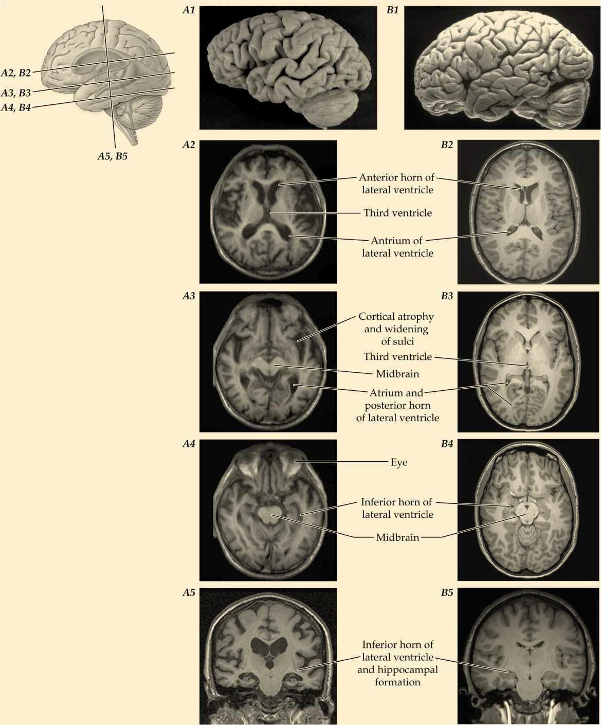
An important goal of Neuroanatomy:TextandAtlas is to prepare the reader for interpreting the new wealth of human brain images structural, functional, and connectivity by developing an understanding of the anatomical localization of brain function. To provide a workable focus, this book is largely restricted to the central nervous system It takes a traditional approach to gaining neuroanatomical competence: Because the basicimagingpictureisatwo-dimensionalslicethroughthebrain,thelocationsofstructuresareexaminedon two-dimensionalmyelin-stainedsectionsthroughthehumancentralnervoussystem.
What is new for the fourth edition of Neuroanatomy: Text and Atlas? All chapters have been revised to reflect advances in neural science since the last edition In addition to full color illustrations, there are many
newfeatures:
• Chapters begin with a clinical case to illustrate the connections and function of the key material. Some of thesecasesarespecializedandnotapttobeseeninroutinepractice.Theywerechosentoshowhowhuman behavior can change in remarkable ways following damage to a localized brain region; sometimes a very smallregion.
•Chaptersendwithaseriesofmultiplechoicereviewquestions.
• Material on central nervous system development is now included in the relevant individual chapters rather thanasingledevelopmentchapter.
•Thereareseparatechaptersontouchandpain.
Designed as a self-study guide and resource for information on the structure and function of the human central nervous system, this book can serve as both text and atlas for an introductory laboratory course in humanneuroanatomy.
For over 23 years, both at Columbia University’s College of Physicians and Surgeons and now at the City University of New York’s Medical School, we use this book in conjunction with a series of neuroanatomy laboratory exercises during the neuroscience teaching block in the curriculum. Rather than presenting the material in a traditional lecture format, we have successfully taught neuroanatomy in a dynamic small group learning environment Supplemented with use of brain models and specimens, neuroanatomy small group sessions complement neural science lecture material and round-out medical, graduate, and allied health sciencestudents’learningexperience
The organization of Neuroanatomy: Text and Atlas continues to parallel that of Principles of Neural Science, editedbyEricR.Kandel,JamesH.Schwartz,ThomasJessell,StevenA.Siegelbaum,andA.JamesHudspeth (McGraw-Hill) Like Principles of Neural Science, Neuroanatomy: Text and Atlas is aimed at medical students, and graduate students in neuroscience, biology, and psychology programs The content of many of the chapters is geared to dental students, such as a chapter focus on the trigeminal system, as well as physical therapyandoccupationaltherapystudentsbyconsideringthemotorsystemsindetail
JohnH.Martin