This work is subject to copyright. All rights are reserved by the Publisher, whether the whole or part of the material is concerned, specifically the rights of translation, reprinting, reuse of illustrations, recitation, broadcasting, reproduction on microfilms or in any other physical way, and transmission or information storage and retrieval, electronic adaptation, computer software, or by similar or dissimilar methodology now known or hereafter developed.
The use of general descriptive names, registered names, trademarks, service marks, etc. in this publication does not imply, even in the absence of a specific statement, that such names are exempt from the relevant protective laws and regulations and therefore free for general use.
The publisher, the authors and the editors are safe to assume that the advice and information in this book are believed to be true and accurate at the date of publication. Neither the publisher nor the authors or the editors give a warranty, express or implied, with respect to the material contained herein or for any errors or omissions that may have been made. The publisher remains neutral with regard to jurisdictional claims in published maps and institutional affiliations.
Printed on acid-free paper
This Springer imprint is published by Springer Nature
The registered company is Springer International Publishing AG
The registered company address is: Gewerbestrasse 11, 6330 Cham, Switzerland
I would like to thank my parents, wife (Abeer), and children (Maiar, Mirna, Amy) for their support and care throughout the journey of this textbook.
Alaa El-Ghobashy
I would like to thank my life partner, Andrea, for her unconditional support and for her acceptance of the time I dedicated to this project.
Javier Magrina
Preface
Surgical practice has undergone significant evolution over the past few decades from open access through to laparoscopy approach to most recently robotic techniques. Since the first description of robotic hysterectomy in 2005, the technique has gained popularity and its indications have broadened. Therefore, it was timely to offer a comprehensive review of the present status of robotic surgery in gynecology using the Da Vinci system.
This book is not only a compilation of the knowledge and experiences of the world renowned robotic surgeons, but it has also incorporated the recent advances and updates in gynecological surgery.
The textbook is aimed at practicing gynecologists, urogynecologists, and gynecological oncologists and is designed to provide a detailed guide to common robotic gynecologic procedures for the purpose of helping novice surgeons in their transition to robotic surgery and seasoned robotic surgeons to refine their surgical technique and expand their repertoire of robotic procedures.
The descriptive, step-by-step, text is complemented by figures, intraoperative photographs, and videos detailing the nuances of each procedure. Emphasis is placed on the operative setup, instrument and equipment needs, and surgical techniques for both the primary surgeon and the operative assistant.
This edition will provide unique insights into robotic gynecologic surgery and reduce the learning curve of accomplishing these increasingly popular procedures.
We would like to express our deepest thanks and gratitude to all the contributors, who so graciously have given their time and effort, and without whom this book would not have been born. There are many more people who have made this book possible specially Springer who supported this project since its inception. To all, thank you for the advice and help and for making this book a reality.
Alaa El-Ghobashy Javier Magrina
René H.M. Verheijen 3
Sorana
Shashank Agarwal, and Athula Ratnayake
Mete Gungor, M. Murat Naki, Ozguc Takmaz, and M. Faruk Kose
7 Robotic Simple Hysterectomy
Robotic-Assisted Video Laparoscopic Management of Genital and Extragenital Endometriosis
Sarika Gupta, Sarfraz Ahmad, and Robert W. Holloway 13 Robotic Radical Hysterectomy for Early-Stage Cervical
Alaa El-Ghobashy, San Soo Hoo, and Javier Magrina
Compartmental Theory in Uterine Cancer, Anatomical Considerations and Principles of Compartmental Cervical Cancer Surgery Step by Step
Rainer Kimmig
15 Peritoneal Mesometrial Resection (PMMR) with Therapeutic Lymphadenectomy (tLNE) in Endometrial Cancer
Rainer Kimmig
16 Pelvic Lymphadenectomy 127
Jordi Ponce, Marc Barahona, and M. Jesus Pla
17 Robotic Para-aortic Lymph Node Dissection 131
Brooke A. Schlappe and Mario M. Leitao Jr
18 Extraperitoneal Para-aortic Lymphadenectomy by Robot-Assisted Laparoscopy (S, SI, and XI Systems) 141
Fabrice Narducci, Lucie Bresson, Delphine Hudry, and Eric Leblanc
19 Robotic Debulking Surgery in Advanced Ovarian Cancer
Javier F. Magrina, Vanna Zanagnolo, Paul M. Magtibay III, and Paul M. Magtibay
Anna E. Wright, Sarvpreet Ubee, Kanagasabai Sahadevan, and Peter W. Cooke
21 Robotic Gastrointestinal (GI) Procedures in Gynecology .
John T. Kidwell and Nitin Mishra
153
163
177
22 Robotic-Assisted Total Pelvic Exenteration 185
Peter C. Lim and Elizabeth Y. Kang
23 Robot-Assisted Laparoscopic Fertility-Sparing Radical Trachelectomy 195 Jan Persson and Celine Lönnerfors
24 Research and Evidence-Based Robotic Practice 203 Rasiah Bharathan and Esther Moss
25 Complications of Robotic Surgery: Prevention and Management . .
. 211 Celine Lönnerfors and Jan Persson
26 The Surgical Assistant in Robotic-Assisted Laparoscopy ................... 235 Nita A. Desai, Ashley L. Gubbels, and Michael Hibner
27 Tips and Tricks for Robotic Surgery 239 O.E. O’Sullivan, B.A. O’Reilly, and M. Hewitt
Index
249
Contributors
Arnold P. Advincula, M.D., F.A.C.O.G., F.A.C.S. Division of Gynecologic Specialty Surgery, Department of OB/GYN, Columbia University Medical Center/NewYork-Presbyterian Hospital, New York, NY, USA
Shashank Agarwal Department of Anaesthesia, The Royal Wolverhampton Hospital, Wolverhampton, UK
Sarfraz Ahmad, Ph.D. Florida Hospital Gynecologic Oncology, Florida Hospital Cancer Institute and Global Robotics Institute, Orlando, FL, USA
Marc Barahona, M.D. University Hospital of Bellvitge (IDIBELL), University of Barcelona, Barcelona, Spain
Rasiah Bharathan, M.Sc., M.R.C.S., M.R.C.O.G. Department of Gynaecological Oncology, Royal Surrey County Hospital, Surrey, UK
Lucie Bresson Department of Gynecologic Oncology, Cancer Center Oscar Lambret, Lille Cedex, France
Peter W. Cooke Department of Urology, The Royal Wolverhampton NHS Trust, Wolverhampton, UK
Nita A. Desai, M.D. Division of Gynecologic Surgery, St. Joseph’s Hospital and Medical Center, Phoenix, AZ, USA
Alaa El-Ghobashy, M.D., M.R.C.O.G. Department of Gynaecological Oncology, The Royal Wolverhampton Hospitals NHS Trust, West Midlands, UK
Becca Falik, M.D. Center for Special Minimally Invasive and Robotic Surgery, Palo Alto, CA, USA
Stanford University Medical Center, Stanford, CA, USA
Ashley L. Gubbels, M.D. Division of Gynecologic Surgery, St. Joseph’s Hospital and Medical Center, Phoenix, AZ, USA
Mete Gungor Faculty of Medicine, Department of Obstetrics and Gynecology, Acıbadem Mehmet Ali Aydınlar University, Istanbul, Turkey
Sarika Gupta, M.D. Florida Hospital Gynecologic Oncology, Florida Hospital Cancer Institute and Global Robotics Institute, Orlando, FL, USA
Matt Hewitt Department of Robotic Surgery, Cork University Maternity Hospital, Cork, Ireland
Michael Hibner, M.D. Division of Gynecologic Surgery, St. Joseph’s Hospital and Medical Center, Phoenix, AZ, USA
Robert W. Holloway, M.D. Florida Hospital Gynecologic Oncology, Florida Hospital Cancer Institute and Global Robotics Institute, Orlando, FL, USA
San Soo Hoo Department of Gynaecological Oncology, The Royal Wolverhampton Hospitals NHS Trust, West Midlands, UK
Delphine Hudry Department of Gynecologic Oncology, Cancer Center Oscar Lambret, Lille Cedex, France
Elizabeth Y. Kang Center of Hope, University of Nevada School of Medicine, Reno, NV, USA
John T. Kidwell Department of Surgery, Mayo Clinic College of Medicine, Phoenix, AZ, USA
Sami Gokhan Kilic, M.D., F.A.C.O.G., F.A.C.S. Division of Minimally Invasive Gynecology and Research, Department of Obstetrics and Gynecology, The University of Texas Medical Branch, Galveston, TX, USA
Rainer Kimmig Department of Gynaecology and Obstetrics, West German Cancer Center, University Hospital Essen, Essen, Germany
M. Faruk Kose Faculty of Medicine, Department of Obstetrics and Gynecology, Acıbadem Mehmet Ali Aydınlar University, Istanbul, Turkey
Mertihan Kurdoglu, M.D. Division of Minimally Invasive Gynecology and Research, Department of Obstetrics and Gynecology, The University of Texas Medical Branch, Galveston, TX, USA
Sandra Madeuke Laveaux, M.D. Division of Gynecologic Specialty Surgery, Department of OB/GYN, Columbia University Medical Center/New York-Presbyterian Hospital, New York, NY, USA
Eric Leblanc Department of Gynecologic Oncology, Cancer Center Oscar Lambret, Lille Cedex, France
Mario M. Leitao Jr, M.D. Memorial Sloan Kettering Cancer Center, New York, NY, USA
Anjie Li, M.D. Center for Special Minimally Invasive and Robotic Surgery, Palo Alto, CA, USA
Stanford University Medical Center, Stanford, CA, USA
Peter C. Lim, M.D., F.A.C.O.G., F.A.C.S. Center of Hope, University of Nevada School of Medicine, Reno, NV, USA
Celine Lönnerfors, M.D., Ph.D. Department of Obstetrics and Gynecology, Skane University Hospital and Lund University, Lund, Sweden
Javier F. Magrina, M.D. Department of Medical and Surgical Gynecology, Mayo Clinic, Phoenix, AZ, USA
Paul M. Magtibay, M.D. Department of Medical and Surgical Gynecology, Mayo Clinic, Phoenix, AZ, USA
Paul M. Magtibay III, M.S. Department of Administration, Mayo Clinic, Phoenix, AZ, USA
Nitin Mishra, M.D. Department of Surgery, Mayo Clinic College of Medicine, Phoenix, AZ, USA
Esther Moss, M.R.C.O.G., M.Sc., Ph.D. Department of Gynaecological Oncology, University Hospitals of Leicester, Leicester, UK
Damian Murphy Department of Gynaecological Oncology, The Royal Wolverhampton Hospitals NHS Trust, West Midlands, UK
M. Murat Naki Faculty of Medicine, Department of Obstetrics and Gynecology, Acıbadem Mehmet Ali Aydınlar University, Istanbul, Turkey
Fabrice Narducci Department of Gynecologic Oncology, Cancer Center Oscar Lambret, Lille Cedex, France
Camran Nezhat, M.D., F.A.C.S., F.A.C.O.G. Center for Special Minimally Invasive and Robotic Surgery, Palo Alto, CA, USA
Stanford University Medical Center, Stanford, CA, USA
University of California San Francisco Medical Center, San Francisco, CA, USA
Marielle Nobbenhuis, M.D., Ph.D. Department of Gynaecological Oncology, The Royal Marsden NHS Foundation Trust, London, UK
B.A. O’Reilly Department of Robotic Surgery, Cork University Maternity Hospital, Cork, Ireland
O.E. O’Sullivan Department of Robotic Surgery, Cork University Maternity Hospital, Cork, Ireland
Jan Persson, M.D., Ph.D. Department of Obstetrics and Gynecology, Skane University Hospital and Lund University, Lund, Sweden
M. Jesus Pla, M.D., Ph.D. University Hospital of Bellvitge (IDIBELL), University of Barcelona, Barcelona, Spain
Jordi Ponce, M.D., Ph.D. University Hospital of Bellvitge (IDIBELL), University of Barcelona, Barcelona, Spain
Athula Ratnayake Department of Anaesthesia, The Royal Wolverhampton Hospital, Wolverhampton, UK
Kanagasabai Sahadevan City Hospitals Sunderland NHS Trust, Sunderland, UK
Brooke A. Schlappe, M.D. Memorial Sloan Kettering Cancer Center, New York, NY, USA
John H. Shepherd Department of Gynaecological Oncology, The Royal Marsden NHS Foundation Trust, London, UK
Ozguc Takmaz Faculty of Medicine, Department of Obstetrics and Gynecology, Acıbadem Mehmet Ali Aydınlar University, Istanbul, Turkey
Sarvpreet Ubee Department of Urology, The Royal Wolverhampton NHS Trust, Wolverhampton, UK
Bekir Serdar Unlu, M.D. Division of Minimally Invasive Gynecology and Research, Department of Obstetrics and Gynecology, The University of Texas Medical Branch, Galveston, TX, USA
René H.M. Verheijen Formerly University Medical Center Utrecht, Utrecht, Netherlands
Megan Wasson, D.O. Mayo Clinic, Phoenix, AZ, USA
Sorana White Department of Anaesthesia, The Royal Wolverhampton Hospital, Wolverhampton, UK
Anna E. Wright Department of Urology, The Royal Wolverhampton NHS Trust, Wolverhampton, UK
Johnny Yi, M.D. Mayo Clinic, Scottsdale, AZ, USA
Vanna Zanagnolo, M.D. Department of Gynecologic Oncology, European Institute of Oncology, Milan, Italy
The Development of Robotic Surgery: Evolution or Revolution?
John H. Shepherd and Marielle Nobbenhuis
A Historical Perspective
The history of mechanical automatons can be traced back to the ancient world with the development of the earliest mechanical machinery. During the fourth century BC, the Greek mathematician Archytas designed a mechanical bird, ‘the pigeon’ driven by steam. In 320 BC Aristotle postulated that automatons would replace human slavery. He quoted Greek mythology in which Hephaestus, the Greek god of craftsmen, created three-legged tables that could action under their own power.
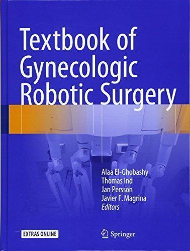
In the twelfth century Al-Jazari, a Muslim inventor designed automated machines that could play music and carry out simple duties. Villard de Honnecourt in the thirteenth century created similar machines. At the end of that century, Robert of Artouis designed and built a number of humanoid and animal robots displayed in his castle at Hesdin. It was some time later in 1495 that Leonardo da Vinci made several drawings of a mechanical knight in armour which was able to move its limbs and head (Fig. 1.1) [1].
This was based on his anatomical sketches and research described in the ‘Vitruvian Man’. There is no record as to whether the robot was in fact built. The following century Johannes Müller designed and built an automated eagle made of iron that did fly. Descartes, in his ‘Discourse on the Method’, 1657, postulated that automatons could be made by man but did not predict that one day they would be able to respond to human instruction [2].
A flurry of developments occurred in the early 1700s with mechanical toys created that could play music, fly, draw and even move as puppets. The most imaginative of these was ‘the Digesting Duck’ of Jacques de Vaucanson which had wings that flapped as well as a ‘digestive system’ which could swallow grain and defecate from a hidden storage chamber. Later that century in Japan, Hisashige Tanaka
J.H. Shepherd • M. Nobbenhuis (*)
Department
of Gynaecological Oncology, The Royal Marsden NHS Foundation Trust, London, UK
developed a number of complex mechanical toys that were able to fire arrows from a bow, serve Japanese tea and paint.
During the late nineteenth century, remotely controlled machinery was developed, mainly for usage during wartime as radio-controlled torpedoes and rockets.
Deep-sea robots followed in time (Fig. 1.2) as did the first remote-controlled robot to land and move on the surface of the moon followed in 1970.
The word robot is attributed to Joseph Kapak, derived from the Czech word ‘robota’ meaning service, in his 1921 play, ‘Universal Robots’. The film industry subsequently developed human machines as the forerunners of science fiction. A humanoid robot was exhibited in London at an exhibition of Model Engineers in 1928 designed by WH Richards with an aluminium body containing 11 electromagnets and a battery powered motor. This robot could move its hands and head by remote control. In 1939 Electro, a humanoid robot was exhibited at the world fair. The aluminium outer skin contained a motorised skeleton; it could respond to voice commands, smoke cigarettes, blow up balloons and move its head and arms.
The term robotics was coined by Asimov in his short story ‘Runaround 1942’ [3]. In this he described ‘three rules of robotics’ in which he postulated that (1) a robot should not injure a human being or through interaction allow one to come to harm; (2) a robot must obey all orders given to it from humans, except where such orders would contradict the previous Law; and (3) a robot must protect its own existence, except when to do so would contradict the previous two Laws. These rules remain a reasonable ethical framework upon which robot development may be applied to surgical care. Subsequently, in 1949 complex behavioural autonomous robots were created at the Burden Neurological Institute in Bristol by William Walter. He used analogue electronics to stimulate brain processes, whilst Alan Turing and John Von Neumann developed digital computation [4, 5]. Artificial intelligence was a short step away.
The first robotic arm was developed at the Rancho Los Amigos hospital in California and further modified at Stanford
A. El-Ghobashy et al. (eds.), Textbook of Gynecologic Robotic Surgery, https://doi.org/10.1007/978-3-319-63429-6_1
University in 1963. The following year the IBM system/360 was released and proved to be faster and more capable than previous machines. The Stanford Research Institute subsequently produced a mobile robot capable of reasoning with multiple sensory input in order to navigate. One of the first robotic applications came from the Stanford Artificial Intelligence Lab (SAIL) in 1969. They designed a robotic arm with six degrees of freedom all-electric mechanical manipulator exclusively for computer control. The Stanford Arm and SAIL helped to develop the knowledge base which has been applied in essentially all the industrial robots.
In the 1970s, the robots ‘Freddy’ and ‘Freddy II’ were built in the United Kingdom to assemble wooden blocks.
J.H. Shepherd and M. Nobbenhuis
The SCARA, Selective Compliance Assembly Robot Arm, created in 1978 was able to pick up parts and place them in various locations useful for assembly lines in factories. In 1986 Honda created a research programme capable of interacting successfully with humans.
It can be seen that with these exciting developments in technology, it was a short step to extending robotic usage into the operating theatre in order to aid and initiate already established laparoscopic and other instrumental techniques.
Surgical Developments
A major step forward in medicine was the invention by Dr. John Adler in 1994 of the CyberKnife, which was able to carry out stereotactic radiosurgery robotically for the treatment of the brain and subsequently other tumours [6]. With advances in microelectronics and computing robotic telecontrol technology with the use of robotic arms to assist in surgical procedures became a reality. Aesop (Computer Motion Inc., Goleta, California) utilised a voice-activated robotic arm. The same company developed Zeus, with remote control robotic arms. Intuitive Surgical Inc., Sunnyvale, California, produced the da Vinci robot controlled by a surgeon-operated console with foot and hand controls. Improvements in stereoscopic imaging gave a three-dimensional view far superior to previously available laparoscopic minimal access techniques although utilising similar optical equipment. Side carts with three and four robotic arms placed at the operating table side allowed further developments and an extension of numerous surgical techniques. In all surgical specialties, the use of fibre-optic technology has allowed diagnostic procedures to be extended to therapeutic and surgical procedures in a truly minimally invasive manner. Examples that can be given include: in urology, prostatectomy, cystectomy and nephrectomy; in colorectal surgery, anterior resection and hemicolectomy; in hepatobiliary and upper gastrointestinal surgery, liver resection, fundoplication and gastric banding, cholecystectomy, pancreatectomy and splenectomy; in cardiothoracic surgery, coronary artery bypass grafting and valve replacement; in otolaryngology, laryngectomy.
Whilst it may seem impractical and difficult to find a role for robotic assistance or minimal access surgery in the practice of obstetrics, in the field of gynaecology the possibilities are clearly endless. The pelvis lends itself anatomically to performing laparoscopy, and therefore robotic assistance will be applicable as has been shown with multiple procedures, when appropriate. The uterus is an obvious organ for such an approach when surgical intervention is necessary. Thus hysterectomy may be aided by robotic assistance and minimal access techniques. Similarly approaches to the pelvic sidewalls and retroperitoneum when dealing with endometriosis can be greatly facilitated with robotic assistance as may
Fig. 1.1 Model of Leonardo da Vinci’s mechanical knight with inner workings, as displayed in Berlin. Photo by Erik Möller
Fig. 1.2 Submersible, called ‘Alvin’, built for US Navy in 1964, operated by Woods Hale Oceanographic Institution
sacrocolpopexy and myomectomy. Magnification gained by the optics at the console can be a great aid to the surgeon as can the obliteration of any tremor with delicate procedures.
Oncological Surgery
Similarly it has been shown that pelvic oncological procedures including pelvic node dissection and radical hysterectomy may be greatly facilitated by the use of robotic assistance. With more flexibility using rotating arms, newly developed robots are able to access the pelvis and then the mid and upper abdomen without the necessity to de-dock. Thus more extensive procedures including pelvic exenteration and reconstruction as well as on occasions ovarian cancer surgery may be performed. The indications for these procedures will depend upon the particular circumstances present will be discussed in other sections of this textbook.
Surgical Training
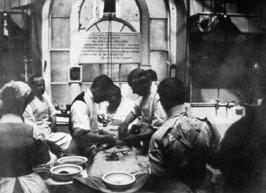
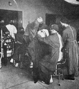
In the past surgical training has occurred in the operating theatre at the table side by observation, assisting and then carry out procedures under direct supervision (Figs. 1.3 and 1.4).
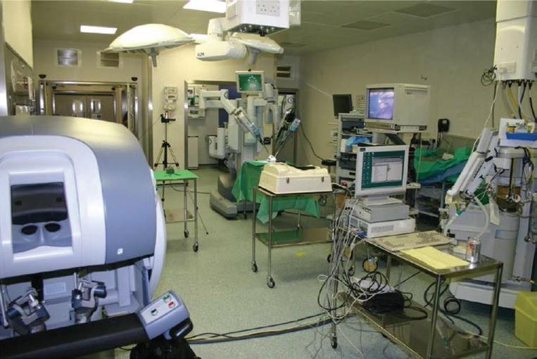
Whilst animal laboratories are not available in the United Kingdom, simulation of anatomical structures and pathology have now given way to computerised models in laboratories (Fig. 1.5).
Robotically assisted surgery may be ideally taught and learnt from such programmes and will have an increasing impact on the quality of training and therefore surgical practice. Just as airline pilots take refresher courses with tests in simulation chambers, so will the surgeons of the future be able to maintain their skills and test their ability. At the same
Fig. 1.3
St Bartholomews surgeons, London, in the 1900s. Archived photo from Medical Photography Department at St Bartholomews Hospital (from Professor John Shepherd’s personal collection)
Fig. 1.4
St Bartholomews surgeons in the 1940s. Archived photo from Medical Photography Department at St Bartholomews Hospital (from Professor John Shepherd’s personal collection)
Fig. 1.5 Set-up of robotic ‘lab’ at the Royal Marsden Hospital at time of introduction of robotic gynaecological programme in 2007 (With permission from Thomas Ind)
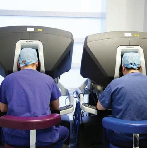
time the surgeon’s brain activity can be measured to assess fatigue and even stress levels. The impact on patient safety is quite clear. Newer models of robot equipment have dual controls which will allow tutoring and co-surgical techniques to be performed (Fig. 1.6).
Added Tools and Technology
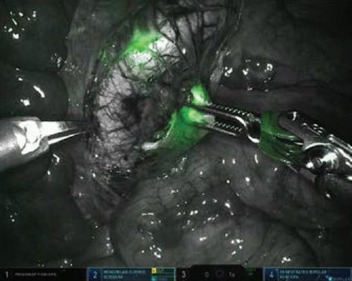
With further developments in imaging especially using MRI, three-dimensional images may be superimposed into the optics at the console of the robot to enable tumours and other anatomical structures to be visualised prior to a surgical procedure being carried out. This will be especially useful in cancer surgery for identifying tumours as well as other anatomical features, such as with the development and incorporation of fluorescent imaging identifying sentinel lymph nodes (Fig. 1.7).
Similarly, with developments in immunocytochemistry and microscopy in histology, in vivo identification of pathology becomes a realistic possibility allowing intelligent knives to excise malignant tissue with greater dexterity than the surgeons’ hand. With developments with haptic feedback, this will facilitate precision microsurgery. An alternative is the use of robotic endoscope holders providing an alternative to telesurgery systems by offering a third arm to the surgeon during an operation.
J.H. Shepherd and M. Nobbenhuis
The Future
The future is already here; we do not need to go back to it. Smaller robots with artificial intelligence are being developed with almost frightening possibilities for their use. Nanotechnology will supersede today’s machinery. Research will continue at an accelerating pace, and the place of new techniques and technologies will need to be carefully evaluated in a critical way as they become available. This will be at an inevitable cost, but this must be offset by an improvement in efficiency and success of treatments available. A reduction of morbidity and inevitable sequelae of treatment must be shown to be achieved with a reduction in hospitalisation and time away from home and work. Advances in medical care need to be supported and encouraged but their correct place carefully assessed. To quote Martin Luther King “Nothing in all the world is more dangerous than sincere ignorance and conscientious stupidity”. We just must accept anything is possible although not always practical.
References
1. Pasek A. Renaissance robotics: Leonardo da Vinci’s lost knight and enlivened materiality. Grad J Vis Mater Cult. 2014;7:1–25.
2. Descartes R.. Discours de la Méthode. Leiden; 1637.
3. Asimov I. The complete robot. Garden City: Doubleday; 1982.
4. Turing A. Computing machinery and intelligence. Mind. 1950;LIX(236):433–60.
5. Von Neumann J. The general and logical theory of automata. In: Jefferies LA, editor. Cerebral mechanisms in behaviour—the Hixon symposium. New York: Wiley; 1951. p. 1–31.
6. Adler John R Jr, et al. The Cyberknife: a frameless robotic system for radiosurgery. Stereotact Funct Neurosurg. 1997;69:124–8.
Fig. 1.6 Double console robotic surgery. The Royal Marsden Hospital (permission Press Office The Royal Marsden Hospital London)
Fig. 1.7 Sentinel lymph node detection external iliac artery using indocyanine green and Firefly filter (archive MA Nobbenhuis)
Training and Proctoring in Robotic Gynaecological Surgery
René H.M. Verheijen
Introduction
Although laparoscopic surgery had been introduced in the late 1960s, it lasted until this century for regulatory authorities and professionals to realize that medical training following a master-apprentice principle is insufficient to provide safe and adequate mastering and monitoring of competence and proficiency [1]. As a consequence, also the introduction of robotassisted surgery was viewed with scepticism and criticism on the way surgeons were trained [2]. This has rightfully led to a call for (a) more structured, (b) more validated and (c) more virtual training in specifically a field-like laparoscopic surgery where more and more technology is being introduced.
It has gradually been acknowledged that a long learning curve as well as the use of technical equipment put patients at risks during the apprenticeship. It was also recognized that these risks could easily be avoided by preparation through e-learning, followed by practicing first in dry and wet laboratory conditions, using virtual or physical models, and as a next step using animal or cadaver models to prepare for surgery in real patients.
Curricula have been developed that have been incorporated into specialist training for most of the surgical specialties. Also, some professional societies have set criteria within the specialty training programmes, which need to be met for a trainee to be allowed to start operating on a real patient as well as for established specialists continuing to do so.
Both the training methods as well as methods of assessment must be validated in order to objectively and accurately measure and monitor progress. E-learning modules have been developed to prepare for hands-on training. Virtual training modules have been developed for technical and procedural training. Box training for technical instruction as well as development of, e.g., eye-hand co-ordination has
R.H.M. Verheijen
Professor Emeritus of Gynaecological Oncology, Formerly University Medical Center Utrecht, Utrecht, Netherlands e-mail: rene.h.m.verheijen@gmail.com
Springer International Publishing AG 2018 A. El-Ghobashy et al. (eds.), Textbook of Gynecologic
equally been validated. In this way trainees become well prepared for surgery on life or cadaver models, which are more suitable for procedural training. Finally, performance during real-life operations can now equally objectively be evaluated using validated assessment tools, such as objective structured assessment of technical skills (OSATS) [3].
Although curricula and criteria for training in conventional laparoscopic surgery have now been well established in many parts of Europe, this is as yet not the case in robot-assisted surgery. No accredited training programmes or fellowships exist that might be used to certify specialists to perform robotassisted surgery. Nevertheless, already in 2007 the Society of American Gastrointestinal and Endoscopic Surgeons (SAGES) together with the Minimal Invasive Robotic Association (MIRA) drafted a position paper with formal guidelines for training and credentialing [4]. The European Board and College of Obstetricians and Gynaecologists (EBCOG) has also issued ‘Robotic Surgery Standards’ as part of their ‘Gynaecology Standards’ [5]. Although this latter document only describes training in broad terms, it does clearly define the learning curve of surgeons that should be ‘specifically trained’ for robot-assisted procedures, including sufficient systematic and validated system and procedural (didactic and skills) training, as well as proctor-assisted procedures.
Not surprisingly, urologists have been first to propose a curriculum for proper training. Although several groups (e.g. Florida Hospital Nicholson Center and Roswell Cancer Center) have developed surgical curricula, the curriculum developed by the EAU Robotic Urology Section (ERUS) is the only one that encompasses the whole learning path, from technical instruction to patient procedures [6].
From their experience gynaecologists could learn that modular training of procedures is more efficient than nonstructured training [7]. This seems a quite obvious conclusion, but in practice structured training is badly implemented. The Society of European Robotic Gynaecological Surgery (SERGS) is developing guidelines and a gynaecological curriculum for safe introduction in robot-assisted gynaecological surgery.
Baseline evaluation
E-learning Virtual training Console (observation)
Simulation based training course
Virtual reality Dry lab (model) Wet lab (animal)
Modular console training & structural assessment
Transition to full procedural training (video) Final evaluation
Modular Training
Specifically for training in complex procedures using sophisticated technology, the various aspects that are important to know and to master cannot be learned haphazardly. Modular training refers to both consecutive modules, each with an essential and defined part of the training, and to teaching the actual procedures in steps, rather than at once completely. This has been developed and validated by ERUS for the most common robot-assisted procedure, the radical prostatectomy [8].
Ideally, a curriculum is being built up from e-learning, through virtual and box training to artificial and animal model training (Fig. 2.1). Finally, full procedural training is done step-by-step. As each module contains essential information and teaches skills that are important for the next module, it is important that each module is followed and finished successfully, before embarking on the next module. Also each module is designed for specific types of information and/or skills.
Apart from other aspects, this modular training reflects also the three phases in which training of motoric skills is commonly divided, (a) a cognitive phase (knowledge), (b) an integrative or associative phase (skills) and (c) an autonomous phase (performance) (after Kopta [9]).
The e-learning module could, for example, contain basic information on technical features of the robot, clinical indications and regulatory issues. But in later stages of training and practice, e-learning also provides tools for permanent training by showing information provided by professionals themselves (e.g. WebSurg from IRCAD, websurg.com, and ESGO’s eAcademy, eacademy.esgo.org). Most e-learning tools are designed to teach cognitive and/or psychomotor skills. But it is difficult to compare their effectiveness in teaching surgical competencies with other educational interventions and curricula. Given these restrictions e-learning seems to perform at least as good as other educational tools [10].
Table 2.1
Virtual training systems for robot-assisted surgery
Name Manufacturer
dV-Trainer®
MIMIC Technologies
Da Vinci Skills Simulator® Intuitive Surgery
ProMIS®
Haptica
SEP® robot simulator SimSurgery
RoSS™ Trainer Simulated Surgical Systems
VR simulatora
aNot commercially available
University of Nebraska
Virtual training may teach technical skills in a simulated and therefore safe environment, at the same time providing tools for objective assessment of progress. Virtual systems are commercially available and offer exercises for specific skills and practice on virtual procedures or parts of them [11, 12] (Table 2.1). The exercises need to be validated before they can be used as a serious preparation for real-life surgery. Construct validation (whether the exercise is indeed discriminatory, i.e. really measures the ability or quality tested for) and face validation (to which extent the exercise resembles the real-life situation) need to have been carried out and have actually widely been published [13].
Model training may teach technical skills in a more realistic environment, be it by the addition of haptic feedback and working in a physical environment like a box or by providing a near to real-life environment as in animal or cadaver models.
E-learning modules and learning programmes are being developed. Manufacturers in particular are keen to develop training programmes, including e-learning, for safe and costeffective introduction of their equipment in the hospital. Although medical professionals and hospitals themselves are responsible for guidance and assessment, training programmes from within the profession are only slowly being developed and implemented and in all honesty lag behind or at best parallel manufacturers’ initiatives.
An important and final part of the training is procedural training, first virtually and/or on a model and finally in the patient. Life patient procedures should be performed in the presence of and guided by an experienced tutor. In the experience of ERUS a modular sequential introduction to complex procedures is the safest and most effective way to learn complex surgery. Rather than starting a procedure and finishing the whole procedure, with or without interference by the tutor, modular training takes the trainee step-by-step, through very well defined and structured steps which are not performed all in one session. Training in a specific procedure starts with first steps, after which the tutor should take over, adding further steps at each next procedure that the trainee is offered to perform. This step-by-step approach has the advantage that the trainee will have maximum attention for the essential steps that are being taught, without losing attention and concentration like in a procedure requiring a longer span of attention. In this way each step is learned more effec-
Fig. 2.1 Modular training programme as proposed by SERGS, based on a model developed by ERUS [8] R.H.
tively, and the procedure is done more safely than in a case where the whole procedure is performed at once.
Competency Based Assessment
After the successful introduction of competence-based training in general gynaecology and of structural assessment [14], these should also be the basis of advanced training in robot-assisted surgery. This provides a framework for trainees to assess regularly and systematically their progress. Thus necessary adjustments in the training and focus on specific needs can be made early on in the training.
The Royal College of Physicians and Surgeons of Canada were the first to recognize and use seven roles of a physician, each requiring specific competencies: professional, communicator, collaborator, leader, health advocate, scholar and medical expert as the central role [15] (Fig. 2.2). The performance in each of these roles determines the level of training in any field of medicine. Such evaluation of the various roles and the defined competencies is now an integrated part of assessment in general training in obstetrics and gynaecology, as reflected by compulsory national programmes such as in the United Kingdom and the Netherlands. It is important to realize that even in a technical field as robot-assisted surgery, these roles and competencies are essential for the future expert to develop and to assess. Robot-assisted surgery, e.g. requires good co-operation between the surgeon and the bedside team, including scrub nurses, surgical assistants and anaesthesiologists.
Name Abbreviation
Global evaluative assessment of robotic skillsa GEARS
Objective structured assessment of technical skills OSATS
aInstrument specifically designed for robot-assisted surgery
Assessment of each of the subsequent phases of training should therefore also include evaluation of these competencies in the different roles of the physician, and this should be and actually is integrated into the assessments (see further in structured assessment).
Structured Assessment
If anything has changed in surgical training, it is surely the systematic and structured way learning goals are being defined and assessed. The ‘see one, do one, teach one’ principle has long since been abandoned and assessment of surgical performance is no longer a matter of a short observation by a single tutor resulting in a brief and undocumented verdict. A regular, non-judgemental and objective evaluation of progress is essential for effective learning. Also, or particularly, training in robot-assisted surgery is not a matter of trial and error.
Modular set-up of the curriculum allows safe introduction of new skills and at the same time guarantees adequate preparation for each next step in the training. This should be monitored by assessments after each of the modules or parts thereof. This may be built in an e-learning module, but should be undertaken by a tutor in other parts. Following a structured assessment avoids forgetting important issues to assess and also forces the tutor to systematically review the various skills and competencies that need to be evaluated. Numerical scoring as in Global Evaluative Assessment of Robotic Skills (GEARS) and OSATS facilitates a quick evaluation, which allows also quick reference to earlier performance to measure progress. Various instruments have been developed and validated (Table 2.2). Such brief and standardized assessment should be followed by the identification of specific positive elements (‘what went well’) and issues that might need some more attention (‘what can be improved’). In this way the trainee is stimulated to set new goals for the next phase of the training.
GEARS is the only instrument specifically designed and validated for robot-assisted surgery [16, 17]. In order to integrate also non-technical competencies, a brief instrument, Non-
Fig. 2.2 CanMEDS roles describing a truly competent physician [15]
Table 2.2 Instruments for structured assessment in surgery
R.H. M. Verheijen
Table 2.3 Non-technical skills for surgeons (NOTSS) taxonomy
Category Elements
Situation awareness – Gathering information – Understanding information – Projecting and anticipating future state
Decision making – Considering options – Selecting and communicating options – Implementing and reviewing decisions
Communication and teamwork – Exchanging information – Establishing a shared understanding – Coordinating team activities
Leadership – Setting and maintaining standards – Supporting others – Coping with pressure
technical Skills for Surgeons (NOTSS), has been developed [18, 19] (Table 2.3). This provides a rating system that may be used within or in combination with instruments of objective assessment, such as GEARS and OSATS. The urologists have incorporated these instruments in their ERUS curriculum, and SERGS is developing this for the gynaecologists.
At the end of training assessment of a (full and unedited) video of a procedure performed by the trainee should be part of final evaluation. This also allows assessment by an independent assessor who will use tools like GEARS. Video assessment is now even offered commercially in order to monitor the performance of individual robotic surgeons [20].
Moments of structured assessment are not limited to the end of modules. In virtual training, every exercise will be individually and automatically scored, and exercises or (part of) procedures in models may each or at least regularly be followed by a brief assessment. In this way a portfolio is built up, which through the ratings of the subsequent exercises and procedures allows monitoring of progress of the trainee.
Conclusion
Training in robot-assisted surgery should be offered in a systematic and modular fashion with structured assessment. Tools are now available to objectively assess and monitor progress of trainees. These should be used, rather than the personal and unstructured opinion of tutors, in order for trainees to complete a portfolio that eventually may be used for certification. For urologists and gynaecologists, curricula have been developed which are basically divided into an introductory period of about 3 months of mainly e-learning and virtual learning and an intense 1 week course of simulation training in a dedicated training centre, followed by approximately 6 months procedural training (Fig. 2.2). This approach provides the professional community as well as patients a framework to safely develop and judge proficiency in robot-assisted surgery.
References
1. IGZ Netherlands. Risks of minimal invasive surgery underestimated (in Dutch). 2007. www.igz.nl
2. IGZ Netherlands. Unsatisfactory diligence at the introduction of surgical robots (in Dutch). 2010. www.igz.nl.
3. Faulkner H, Regehr G, Martin J, Reznick R. Validation of an objective structured assessment of technical skill for surgical residents. Acad Med. 1996;71:1363–5.
4. Herron DM, Marohn M, SAGES-MIRA Robotic Surgery Consensus Group. A consensus document on robotic surgery. Surg Endosc. 2008;22:313–25.
5. EBCOG. Standards of care for women’s health in Europe, Gynaecology Services, Standard 25. 2014. www.ebcog.eu.
6. Fisher RA, Dasgupta P, Mottrie A, Volpe A, Khan MS, Challacombe B, Ahmed K. An over-view of robot assisted surgery curricula and the status of their validation. Int J Surg. 2015;13:115–23.
7. Lovegrove C, Novarra G, Mottrie A, Guru KA, Brown M, Challacombe B, Popert R, Raza J, van der Poel H, Peabody J, Dasgupta P, Ahmed K. Structured and modular training pathways for robot-assisted radical prostatectomy (RARP): validation of the RARP assessment score and learning curve assessment. Eur Urol. 2016;69:626–35.
8. Volpe A, Ahmed K, Dasgupta P, Ficarra V, Novarra G, van der Poel H, Mottrie A. Pilot validation study of the European Association of Urology robotic training curriculum. Eur Urol. 2015;68:292–9.
9. Kopta JA. The development of motoric skills in orthopaedic education. Clin Orthop Relat Res. 1971;75:80–5.
10. Maertens H, Madani A, Landry T, Vermassen F, Van Herzeele I, Aggarwal R. Systematic review of e-learning for surgical training. BJS. 2016;103(11):1428–37. https://doi.org/10.1002/bjs.10236.
11. Abboudi H, Khan MS, Aboumarzouk O, Guru KA, Challacombe B, Dasgupta P, Ahmed K. Current status of validation for robotic surgery simulators—a systematic review. BJU Int. 2012;111:194–205.
12. Moglia A, Ferrari V, Morelli L, Ferrari M, Mosca F, Cuschieri A. A systematic review of virtual reality simulators for robot-assisted surgery. Eur Urol. 2016;69:1065–80.
13. Schreuder HW, Wolswijk R, Zweemer RP, Schijven MP, Verheijen RH. Training and learning robotic surgery, time for a more structured approach: a systematic review. BJOG. 2012;119:137–49.
14. Boerebach BCM, Arah OA, Heineman MJ, Lombarts KMJMH. Embracing the complexity of valid assessments of clinician’s performance: a call for in-depth examination of methodological and statistical contexts that affect the measurement of change. Acad Med. 2016;91:215–20.
15. The Royal College of Physicians and Surgeons of Canada. CanMEDS interactive. 2015. http://canmeds.royalcollege.ca
16. Goh A, Goldfarb DW, Sander JC, Miles BJ, Dunkin BJ. Global evaluative assessment of robotic skills: validation of a clinical assessment tool to measure robotic surgical skills. J Urol. 2012;1:247–52.
17. Sánchez R, Rodríguez O, Rosciano J, Vegas L, Bond V, Rojas A, Sanchez-Ismayel A. Robotic surgery training: construct validity of Global Evaluative Assessment of Robotic Skills (GEARS). J Robot Surg. 2016;10(3):227–31. https://doi.org/10.1007/ s11701-016-0572-1
18. Flin R, Yule S, Paterson-Brown S, Maran N, Rowley D, Youngson G. Experimental evaluation of a behavioural marker system for Surgeons’ Non-Technical Skills (NOTSS). Proc Hum Factors Ergon Soc Annu Meet. 2006;50:969–72.
19. Yule S, Flin R, Maran N, Rowley D, Youngson G, Paterson-Brown S. Surgeons’ non-technical skills in the operating room: reliability testing of the NOTSS behaviour rating system. World J Surg. 2008;32:548–56.
20. Lendvay TS, White L, Kowalewski T. Crowdsourcing to assess surgical skill. JAMA Surg. 2015;150:1086–7.
Anaesthesia for Robotic Gynaecological Surgery
Sorana White, Shashank Agarwal, and Athula Ratnayake
Introduction
The role of general anaesthesia is to produce a reversible and safe loss of consciousness, to maintain the patient’s physiological parameters within a normal range while blunting the sympathetic response to noxious stimuli and to facilitate optimum surgical conditions for the operation.
Anaesthesia for classical laparoscopic gynaecological surgery has been well described in many textbooks, but robotic gynaecological surgery is a new and evolving field, bringing different challenges in anaesthetic management. Principally a much steeper Trendelenburg position is required in order to improve access to the pelvic structures, usually in the order of 30°–45°. This, together with the CO2 pneumoperitoneum and increased length of surgery, has a marked effect on a patient’s physiology that can pose a significant challenge for the anaesthetist. Also, another major consideration is having very limited access to the patient once surgery is underway.
The patient’s journey starts with the initial diagnosis, counselling and consent followed by pre-assessment and optimisation for surgery. Once admitted to the hospital, the patient undergoes general anaesthesia and surgery followed by post-operative care. A sound understanding of the conduct of surgery and in particular the changes in physiology brought about by the steep Trendelenburg positioning and the CO2 pneumoperitoneum are paramount for ensuring patient safety during this journey.
Anaesthetic Management
General principles of preoperative assessment are followed, with particular attention to coexisting comorbidities. Patients are often relatively young and commonly anxious. Sedative
S. White • S. Agarwal • A. Ratnayake (*)
Royal Wolverhampton Hospital, Wolverhampton Road, Wolverhampton WV10 0QP, UK
premedication may be required. Caution should be exercised particularly if they are obese, as ventilation may be especially difficult.
Perioperative Management
Before inducing general anaesthesia, appropriate monitoring should be attached. This includes pulse oximetry, capnography, ECG and blood pressure monitoring (invasive if indicated). Endotracheal intubation provides a means for adequate ventilation, in addition to protection from aspiration. It is important to have intravenous lines secured, as they are usually inaccessible during the surgery. Further monitoring is also advised, e.g. temperature and neuromuscular monitoring.
At our institution the patient is anaesthetised on the operating table. They are supine on a non-slip mattress (although this is not universal practice). They are then placed in the lithotomy position with the arms fixed by their side. The perineum is positioned so that it is in alignment with the break in the table. Once the lower half of the table is removed, the surgeon will have good access.
The endotracheal tube is firmly fixed in position (ensuring ties are not so tight as to occlude venous drainage from above the neck), eyes are padded and the head is secured. Padded shoulder braces are attached and positioned away from the shoulders in the supine position. This is to avoid brachial plexus injuries in steep Trendelenburg position. We apply a heated blanket above the chest, before transferring the patient into the operating room. Subsequently drapes are applied, and surgery begins to site the trocars. Once this has been satisfactorily achieved, pneumoperitoneum is initiated followed by Trendelenburg position of 30°–45°. Additional ports are inserted so that the robotic arms (up to four) can be attached. Once the robot is positioned over the patient and the robotic arms docked, access to the airway, to any lines or monitoring is virtually impossible. It is important to note that moving the patient or performing CPR would require the robot to first be detached.
A. El-Ghobashy et al. (eds.), Textbook of Gynecologic Robotic Surgery, https://doi.org/10.1007/978-3-319-63429-6_3
Physiological Changes Caused by Steep
Trendelenburg
Airway
Increased intra-abdominal pressure secondary to CO2 pneumoperitoneum and the gravitational effects of the intraabdominal organs in Trendelenburg position result in a cephalad displacement of the diaphragm and consequently of mediastinal structures including the trachea. This can result in malpositioning of the endotracheal tube in the anaesthetised patient leading to endobronchial intubation [1].
Respiratory
Studies have shown that in procedures involving Trendelenburg position and pneumoperitoneum, lung compliance can be reduced by as much as 68% [2, 3].
The cephalad displacement of the diaphragm also results in collapse of the bases of the lungs (atelectasis) with reduced lung vital capacity and reduced functional residual capacity. Intra-abdominal pressures up to 15 mmHg are commonly used with the range being between 12 and 15 mmHg to allow enough operative space in the peritoneal cavity. When combined with Trendelenburg position, the European Association for Endoscopic Surgery recommends to avoid pressures higher than 12 mmHg because of decreased pulmonary compliance [10]. Following pneumoperitoneum, the increase in pulmonary blood volume further reduces lung compliance leading to higher airway peak and plateau pressures during mechanical ventilation and an increase in ventilation/perfusion mismatch. The need for higher airway ventilation pressures increases the risk of baro-/volutrauma to the lung. Higher intra-abdominal pneumoperitoneum pressures and pre-existing diaphragmatic defects have been associated with increased risk of post-operative pmeumothorax and pneumoperitoneum.
The amount of CO2 absorption into the blood from the pneumoperitoneum increases with the length of the operation [4]. With pre-existing lung disease such as emphysema and chronic bronchitis, gas exchange is impaired so the extent of hypercarbia may be exaggerated. Ultimately this results in a combination of hypoxia and hypercarbia.
Cardiovascular
The Trendelenburg position increases the return of blood from the legs causing an increase in preload and cardiac output. There is an increase in the central venous pressure (CVP), mean pulmonary artery pressure (MPAP) and pulmonary capillary wedge pressure. The increase in CVP and
MPAP has been shown to be up to threefold and twofold, respectively, in one study [5]. Mean arterial pressure increases to a greater extent than CVP, in part due to the increase in cardiac output and systemic vascular resistance during steep Trendelenburg and pneumoperitoneum. The main reason for this is compression of the intra-abdominal aorta resulting in an increase in the afterload as well as in humoral factors secondary to sympathetic stimulation [6].
Doppler studies have shown significant increases in stroke volume associated with this positioning with a compensatory decrease in heart rate and an increase in the time of isovolumetric relaxation of the heart [7].
These physiological principles are important as in patients with impaired left ventricular function the initial fluid redistribution (secondary to positioning) combined with increased afterload can precipitate heart failure. Furthermore, gas insufflation can result in traction on the peritoneum leading to vagal stimulation, causing bradycardia, and if severe it can lead to asystole. Finally, with increased duration of surgery, a combination of hypercarbia, acidosis and hypoxia can lead to arrhythmias and cardiovascular compromise [8].
Cerebrovascular
The steep Trendelenburg position and pneumoperitoneum are known to cause increased intracranial pressure (ICP). In patients with pre-existing raised ICP adopting this position can be catastrophic. Furthermore there can be a significant reduction in the cerebral tissue oxygen saturation in elderly patients.
Cerebral perfusion pressure (CPP) is calculated as the difference between the mean arterial pressure (MAP) and the highest of either intracranial pressure or CVP. As detailed above, MAP increases to a greater extent than CVP when a patient is positioned for robotically assisted surgery. Kalmar et al. showed using second-generation near-infrared spectrometry that the CPP and cerebral tissue oxygen saturation increased during surgery and were well above the level at which cerebral blood flow autoregulation would be affected or below which cerebral tissue hypoxia could occur.
The combination of altered respiratory physiology and CO2 pneumoperitoneum results in an increase in arterial partial pressure of CO2 which in turn leads not only to cerebral vasodilatation but also choroidal vasodilatation and an increase in intraocular pressure. Maintaining an acceptable end tidal CO2 as a surrogate marker of arterial partial pressure of CO2 and regularly monitoring the end tidal—arterial gradient is essential in minimising the risk of serious ocular consequences such as bilateral visual loss (Kalmar et al.).
Another factor to consider is cerebral oedema, which can occur due to a raised CVP, hypercarbia and cerebral vasodilatation. In order to minimise this appropriate ventilator
S. White et al.
strategies may be needed employed, such as the use of positive end expiratory pressure (PEEP). In addition intravenous fluid should be restricted, at least until the patient is levelled off near the end of surgery.
Ocular
Raised intraocular pressure and corneal abrasions are more likely, again due to patient position, and the potential reflux of gastric acid. Eyes should be taped shut and padded for extra precaution.
Haematological
Pelvic surgery is associated with deep venous thrombosis (DVT), and in lithotomy position this risk is even greater as the return of blood from the legs is impaired. Another complication of lithotomy position for a prolonged period is the potential for rhabdomyolysis [9].
Musculoskeletal
There is a risk of brachial plexus nerve injury with shoulder bolsters in place, but this needs to be balanced with the risk of patient sliding off the table in such a steep position. Normally shoulder bolsters are positioned 4–5 cm away from the patient’s shoulders when supine—once Trendelenburg position is established the anaesthetist needs to check the bolsters are not exerting traction onto the shoulders.
Another site for nerve injury is the common peroneal nerve, that can easily been compromised by the leg supports used for lithotomy position.
Monitoring of neuromuscular function must be in place as any coughing during surgery could be catastrophic once the robot is engaged.
Post-operative Management
Patients should be recovered by appropriately trained staff in a suitable environment. Those deemed high risk owing to their comorbidities or a turbulent perioperative phase should be managed in a high dependency environment.
The prolonged steep Trendelenburg position can result in complications in the recovery period that must be anticipated. Laryngeal oedema resulting in stridor and airway obstruction can occur, necessitating re-intubation. Postoperative confusion and delirium had also been reported, presumably secondary to cerebral oedema and inadequate
clearance of CO2, but studies suggesting this link have been underpowered due to the small numbers involved.
Post-operative pain relief is usually achieved through a multimodal analgesia technique. Intravenous or oral opiates, paracetamol and non-steroidal anti-inflammatory drugs are commonly used at our institution. The use of transverses abdominis plane (TAP) bocks and wound infiltration with local anaesthesia has also been described. Neuraxial blockade is generally not required for the post-operative pain relief and thus is rarely used.
Nausea and vomiting may persist in the post-operative, principally due to ileus, and anti-emetic medication should be given.
The Future
Minimally invasive robotic surgery has a future potential in providing cancer treatment to people who are unable to withstand the stress of a major laparotomy. As with other laparoscopic techniques, those that undergo surgery have an improved functional outcome, reduced length of hospital stay and faster recovery.
Due to the extreme positions involved and the effect on a patient’s physiology, innovative monitoring and safety devices will no doubt be developed to reduce risks of injury and aid anaesthetists in controlling physiological parameters. Also with the advent of remote site access (so that the operator might be in a different city), communication aids between team members will also be vital to the continued success of this type of surgery.
References
1. Chang CH, Lee HK, Nam SH. The displacement of the tracheal tube during robot-assisted radical prostatectomy. Eur J Anaesthesiol. 2010;27(5):478–80.
2. Danic MJ, Chow M, Gayload A, Bhandari A, Menon M, Brown M. Anesthesia consideration for robotic-assisted laparoscopic prostatectomy: a review of 1500 cases. J Robot Surg. 2007;1:119–23.
3. Suh MK, Seong KW, Jung SH, Kim SS. The effect of pneumoperitoneum and Trendelenburg position on respiratory mechanics during pelviscopic surgery. Korean J Anesthesiol. 2010;59(5):329–34. Epub 2010 Nov 25.
4. Murdock CM, Wolff AJ, Van Geem T. Risk factors for hypercarbia, subcutaneous emphysema, pneumothorax, and pneumomediastinum during laparoscopy. Obstet Gynecol. 2000;95(5):704–9.
5. Lestar M, Gunnarsson L, Lagerstrand L, Wiklund P, OdebergWernerman S. Hemodynamic perturbations during robot-assisted laparoscopic radical prostatectomy in 45° Trendelenburg position. Anesth Analg. 2011;113(5):1069–75. DOI 10.1213/ ANE.0b013e3182075d1f. Epub 2011 Jan 13.
6. Falabella A, Moore-Jeffries E, Sullivan MJ, Nelson R, Lew M. Cardiac function during steep Trendelenburg position and CO2 pneumoperitoneum for robotic-assisted prostatectomy: a transoesophageal Doppler probe study. Int J Med Robot. 2007;3:312–5.
7. Meininger D, Westphal K, Bremerich DH, Runkel H, Probst M, Zwissler B, Byhahn C. Effects of posture and prolonged pneumoperitoneum on hemodynamic parameters during laparoscopy. World J Surg. 2008;32(7):1400–5. DOI 10.1007/s00268-007-9424-5
8. Kikuno N, Urakami S, Shigeno K, Kishi H, Shiina H, Igawa M. Traumatic rhabdomyolysis resulting from continuous compression in the exaggerated lithotomy position for radical perineal prostatectomy. Int J Urol. 2002;9(9):521–4.
9. Biswas S, Gnanasekaran I, Ivatury RR, Simon R, Patel AN. Exaggerated lithotomy position-related rhabdomyolysis. Am Surg. 1997;63(4):361–4.
10. Neudecker J, Sauerland S, Neugebauer E, Bergamaschi R, Bonjer HJ, Cuschieri A, et al. The European Association for Endoscopic Surgery clinical practice guideline on pneumoperitoneum for laparoscopic surgery. Surg Endosc. 2002;16:1121–43.
S. White et al.
Robotic Machine and Instruments
Alaa El-Ghobashy
and
Damian Murphy
Introduction
The da Vinci Robotic System, manufactured by Intuitive Surgical, USA, was approved by the Food and Drug Administration (FDA) in 2000. It gained popularity worldwide as it facilitates the minimal access completion of complex surgical procedures. There are five known models of the da Vinci system (Standard, S, Si, X and Xi).
Earlier systems had one camera and two instrument arms. A fourth arm was subsequently added to assist the surgeon in handling and retracting without the need for an assistant. Further development included the high-definition 3D vision, motorised and dual console facilities. The latest robotic version, the da Vinci Xi Model, came to the market in 2014. It offers upgraded and better movement of the mechanical arms with an overhead alignment.
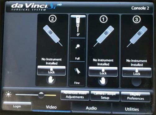
In principle, the da Vinci surgical system consists of three main components: the surgeon’s console, the patient’s surgical cart and the vision cart. Moreover, there are other accessories that are used with the da Vinci robot, namely metal trocars and EndoWrist instruments. The system translates the operator’s hand, wrist and finger movements into delicate real-time precise corresponding/matching movements of the surgical instruments. In this chapter, we will describe the widely available da Vinci Si model in details.
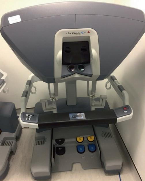
Surgeon Console
The console is the workstation where the surgeon can sit comfortably and control the da Vinci system away from the sterile surgical field. The part of the system features the following elements: stereoviewer, master controllers, footswitch panel, arm rest bar with left/right side pods and touchpad for preference/feature selections.
A. El-Ghobashy, M.D., M.R.C.O.G.
(*) •
D. Murphy
Department of Gynaecological Oncology, The Royal Wolverhampton Hospitals NHS Trust, West Midlands, UK
e-mail: alaaelghobashy@nhs.net
The surgeon views a 3D, real-time and high resolution image of the surgical field that is approximately magnified ×10 through the stereoviewer (Fig. 4.1). The system status icons and messages can also be seen while the surgeon operates. This allows maximum control of the system and warns the surgeon of any faults without having to move the head away from the stereoviewer. There are two infrared sensors on both sides of the stereoviewer that deactivate the robotic arms when the surgeon’s head is moved away. Images can be seen either in full screen mode or in TiloPro™ mode (3D image and up to two auxiliary images). There is also an adjustable two-way audio communication with microphones
Fig. 4.1 Surgeon console with stereoviewer
and speakers to allow the surgeon to exchange information with the rest of the operating team [1].
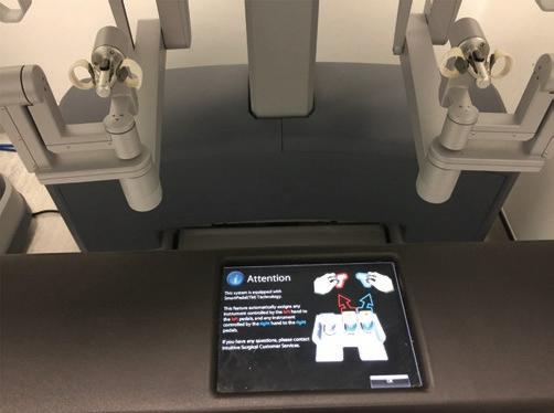
The master controllers are manoeuvred by the surgeon after inserting two fingers (index and thumb) in an adjustable Velcro straps to control the movement of the EndoWrist instruments and the camera (Fig. 4.2). The movements are created by opening and closing the controllers and by bringing them towards or away from the surgeon. The movements are precise, dextrous, scaled (fine 3:1 or normal 2:1) and filtered by the computer to avoid the transmission of any tremors to the instruments. The controllers in the Si model contain grey buttons (finger clutches) which when pressed disengage the controllers from the robotic arms to allow repositioning of the masters to a comfortable location without any change of the instruments’ sites. It is generally recommended to adjust the working space of the masters when the surgeon’s arms start to lift off from the armrest bar. The controllers can also adjust the camera focus when pressed and rotated clockwise and anticlockwise.
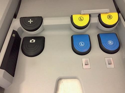
Located on the floor beneath the console is the footswitch panel. It contains three pedals to the left side (camera control, main clutch and the control of arms swap). There are other pedals to the right side (coagulation and cutting diathermy pedals) which are connected at the back of the console through coloured cables to the electrosurgical generator (Fig. 4.3).
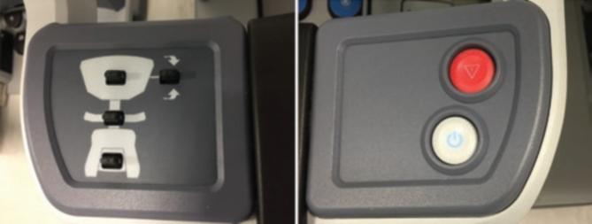
In the armrest bar, there are left side pods which allow the ergonomic adjustment according to the surgeon’s seating preferences (Fig. 4.4a). This avoids strains and discomfort during lengthy operations. Emergency stop and power buttons are located to the right side (right pods, Fig. 4.4b). In the middle of the armrest, there is a touchpad (integrated control interface) that offers adjustment of the audio-video settings as well as system control. Surgeons can save their preferred console settings in the users’ profile for automatic recall in future cases (Fig. 4.5).
Patient Cart
This is the surgical part of the system that is connected to the patient (Fig. 4.6). It is composed of motor-driven base with a main column attached to instruments and camera arms. The motor-driven patient cart facilitates the fast and controlled docking of the system to the patient. This part includes the steering