Download ebooks file Urological care for patients with progressive neurological conditions 1st ed. 2
ed. 2020 Edition John T. Stoffel
Visit to download the full and correct content document: https://ebookmass.com/product/urological-care-for-patients-with-progressive-neurolog ical-conditions-1st-ed-2020-edition-john-t-stoffel/
More products digital (pdf, epub, mobi) instant download maybe you interests ...
Physical Management for Neurological Conditions 4th Edition Geert Verheyden
This work is subject to copyright. All rights are reserved by the Publisher, whether the whole or part of the material is concerned, specifically the rights of translation, reprinting, reuse of illustrations, recitation, broadcasting, reproduction on microfilms or in any other physical way, and transmission or information storage and retrieval, electronic adaptation, computer software, or by similar or dissimilar methodology now known or hereafter developed.
The use of general descriptive names, registered names, trademarks, service marks, etc. in this publication does not imply, even in the absence of a specific statement, that such names are exempt from the relevant protective laws and regulations and therefore free for general use.
The publisher, the authors, and the editors are safe to assume that the advice and information in this book are believed to be true and accurate at the date of publication. Neither the publisher nor the authors or the editors give a warranty, expressed or implied, with respect to the material contained herein or for any errors or omissions that may have been made. The publisher remains neutral with regard to jurisdictional claims in published maps and institutional affiliations.
This Springer imprint is published by the registered company Springer Nature Switzerland AG The registered company address is: Gewerbestrasse 11, 6330 Cham, Switzerland
Acknowledgments
Editing a textbook about a topic that you care deeply about becomes a labor of love. I relied on several people for feedback and advice during this process. The idea of this book came from my experience training with Dr. Edward McGuire who taught me to look at neurogenic bladder patients by disease process, rather than by end organ. I would also like to thank Dr. David Bloom at the University of Michigan who always gave sound advice and wisdom about persistence and the need to advocate for uncommon diseases affecting patients and families. Finally, I would like to thank my wife and family for support, ad hoc editing, and for always reminding me about what is important.
Thanks also to Dr. Elizabeth V. Dray, my co-editor, for her continued dedication and effort on this project even after finishing fellowship.
John T. Stoffel
Contributors
Kelly Bree University of California – San Diego Health, La Jolla, CA, USA
Benjamin M. Brucker Department of Urology, New York University, Langone Health, New York, NY, USA
Anne P. Cameron Department of Urology, University of Michigan, Ann Arbor, MI, USA
Christopher Chermansky Department of Urology, University of Pittsburgh School of Medicine, Pittsburgh, PA, USA
Catherine Dowling Department of Neurology, University of Michigan Medical School, Ann Arbor, MI, USA
Elizabeth V. Dray Division of Urology, Department of Surgery, Greenville Health System, Greenville, SC, USA
Christopher S. Elliott Stanford University Medical Center, Department of Urology, Stanford, CA, USA
Santa Clara Valley Medical Center, Division of Urology, San Jose, CA, USA
Sean P. Elliott Department of Urology, University of Minnesota, Minneapolis, MN, USA
Jairam R. Eswara, MD Urology Services Department, St. Elizabeth’s Medical Center, Brighton, MA, USA
Department of Urology, Tufts Medical Center and Tufts Medical School, Boston, MA, USA
David Ginsberg Department of Urology, Keck USC Institute of Urology, Los Angeles, CA, USA
Michael Harper Department of Medicine, Division of Geriatrics, University of California, San Francisco, CA, USA
Natalia Hernandez Department of Urology, Houston Methodist Hospital, Houston, TX, USA
Rose Khavari Department of Urology, Houston Methodist Hospital, Houston, TX, USA
Giulia Lane Department of Urology, University of Michigan, Ann Arbor, MI, USA
Yang Mao-Draayer Department of Neurology, University of Michigan Medical School, Ann Arbor, MI, USA
Graduate Program in Immunology, Program in Biomedical Sciences, University of Michigan Medical School, Ann Arbor, MI, USA
Lisa Irene Mathias Division of Neurourology & Pelvic Reconstructive Surgery, University of Michigan, Ann Arbor, MI, USA
Jeremy B. Myers Genitourinary Injury and Reconstructive Urology, University of Utah Department of Surgery, Salt Lake City, UT, USA
University of Utah Division of Urology, Salt Lake City, UT, USA
Jalesh N. Panicker Department of Uro-Neurology, The National Hospital for Neurology and Neurosurgery, Queen Square, London, UK
Joseph J. Pariser Department of Urology, University of Minnesota, Minneapolis, MN, USA
Benoit Peyronnet Department of Urology, New York University, Langone Health, New York, NY, USA
Venkat M. Ramakrishnan, MD, PhD Division of Urology, Brigham and Women’s Hospital, Harvard Medical School, Boston, MA, USA
Paholo Barboglio Romo Department of Urology, University of Michigan, Ann Arbor, MI, USA
Yahir Santiago-Lastra University of California – San Diego Health, La Jolla, CA, USA
Claudia Sevilla Department of Urology, Keck USC Institute of Urology, Los Angeles, CA, USA
Katherine Shapiro Department of Urology, University of Pittsburgh School of Medicine, Pittsburgh, PA, USA
Kazuko Shem Santa Clara Valley Medical Center, Department of Physical Medicine and Rehabilitation, San Jose, CA, USA
Mini Singh Department of Neurology, University of Michigan Medical School, Ann Arbor, MI, USA
John T. Stoffel Division of Neurourology and Pelvic Reconstruction, Department of Urology, University of Michigan Medical School, Ann Arbor, MI, USA
Anne M. Suskind Department of Urology, Obstetrics, Gynecology and Reproductive Sciences, University of California, San Francisco, CA, USA
Gregory Vurture Department of Urology, New York University, Langone Health, New York, NY, USA
Blayne Welk Department of Surgery (Urology) and Epidemiology & Biostatistics, Western University, London, ON, Canada
Part I
Fundamentals of Neuro-urology
Chapter 1 Introduction
John T. Stoffel
and Elizabeth V. Dray
Introduction
Urologic care for patients with progressive neurologic conditions is both similar and different than care for other neurogenic bladder patients. Although the goals of safe urine storage and evacuation remain consistent among all neurogenic bladder patients, there are considerably more variables to consider when treating patients with progressive neurologic conditions. First, progressive disease may cause ongoing changes in bladder physiology. For example, overactive bladder symptoms may transition into an underactive bladder or new incontinence may develop as higher brain function is lost. Second, continuing cognitive changes may mean that self-care options that were previously effective may become unsuitable for patients over time. This coincides with a loss of independence as patients are unable to remember to take medications or follow a timed schedule. Third, changes to muscle strength and dexterity may force adjustments in bladder management strategies. The most obvious change includes switching from voiding or intermittent catheterization to an indwelling catheter, but other factors such as inability to ambulate, talk, or breathe weigh heavily on urologic treatment strategies. Given these changes over time, it is clear that care for progressive neurologic patients is many times more reactive and less scheduled compared to other neurogenic bladder neurogenic bladder patients.
If properly addressed, most neurogenic bladder patients, including those with a progressive condition, can expect reasonable urinary-specific quality of life and protection against morbidity such as urinary tract infections and renal failure.
J. T. Stoffel (*)
Division of Neurourology and Pelvic Reconstruction, Department of Urology, University of Michigan Medical School, Ann Arbor, MI, USA
e-mail: jstoffel@med.umich.edu
E. V. Dray
Division of Urology, Department of Surgery, Greenville Health System, Greenville, SC, USA
J. T. Stoffel, E. V. Dray (eds.), Urological Care for Patients with Progressive Neurological Conditions, https://doi.org/10.1007/978-3-030-23277-1_1
However, many practitioners have limited understanding on how to best treat patients with neurogenic bladder problems related to a progressive neurologic condition. Some conditions such as multiple sclerosis are more common and the urologic needs are a better understood among the urologic community, but other conditions such as cerebral palsy or Huntington’s Chorea are rarely encountered by urologists. In fact, there is little guidance supported by data on how to best establish urologic care for most progressive neurologic conditions. Barriers to consistent urologic care in this population include lack of knowledge regarding disease pathophysiology, lack of knowledge on how to best evaluate these patients, and a lack of knowledge regarding risk/benefit stratification for available treatments (Fig. 1.1).
Lack of knowledge regarding disease state is the first barrier to overcome. By definition, a neurogenic bladder is a condition in which urinary symptoms are related to an underlying neurologic condition. It is our feeling that great attention should be focused on understanding the relationship between neurologic condition and neurogenic bladder symptoms. Looking at Fig. 1.2, it is easy to see how care for patient with a progressive brain involvement such as Huntington’s chorea can be very different than a patient with a process that affects neuromuscular junctions such as Eaton Lambert just based on the different involved regions of the nervous system. Interpreting symptoms in this light leads to a more holistic approach that appreciates the different needs of patient groups.
The other barriers to good urologic care are less easy to overcome. Urologic testing strategies regarding neurogenic bladder patients, in general, are likewise not always clear. It is understood that urodynamics are helpful in understanding neurogenic bladder physiology, but there are no standardized, disease-specific urodynamic protocols that address the unique pathophysiology or limitations of patients with progressive conditions. Similarly, there are no guidelines for imaging, quality of life assessment, or home health care for these patients. Lack of knowledge of regarding risk stratification for treatments can also limit options for these patients. Since many of the patients fall outside standard procedural and preoperative assessment protocols, it frequently falls on the urologists to determine if an intervention is worth the risk to the patient. Consequently, this lack of knowledge regarding risk/
Fig. 1.1 Barriers to urologic care for the patient with a progressive neurologic condition
J. T. Stoffel and E. V. Dray
Parasympathetic (S2-S4)
benefit can unfortunately lead to under treatment in some of the patients and over treatment in others.
The purpose of this textbook, Urologic Care for the Patient with a Progressive Neurologic Condition, is to address these knowledge gaps and provide readily available information for the reader to use in the care of these patients. Our goals are to create a resource where providers can quickly access a summary of a specific disease pathophysiology, see the timeline of symptom progression, and understand unique characteristics about the disease which can impact urologic care. In short, the book should be a practical reference for the learner to quickly review and ensure appropriate urologic care is provided for this patient.
In Part 1, Chaps. 2, 3, 4, 5, 6, 7, and 8, we emphasize areas of knowledge that can be applied to any patients with progressive neurologic conditions, such as basic bladder physiology, neuro-anatomy, fundamentals of a neurologic exam, urodynamic testing, imaging modalities and limitations, quality of life assessments, and generalized neurogenic bladder care strategies. Special note is made in these chapters on how to apply these tests to patients with progressive neurologic conditions, if applicable.
In Part 2, Chaps. 9, 10, 11, 12, 13, 14, 15, 16, 17, and 18, we focus on providing a high level summary regarding the urologic care for patients with Parkinson’s disease, Alzheimer’s/dementia, ALS, cerebral palsy, Huntington’s chorea, multiple sclerosis, Friedrich’s ataxia, Guillain Barre, Eaton Lambert, and diabetes. Each progressive disease is reviewed for disease pathophysiology (mechanism of action) and the organs that are affected. Key tests for diagnosing the disease are discussed and timeline of disease progression is reviewed. Special attention is focused on
Bladder
Sympathetic (T10-L2)
Sacral Spinal Cord
Bifida
Fig. 1.2 Some neurologic conditions causing neurogenic bladder
J. T. Stoffel and E. V. Dray
common urologic symptoms that are disease-specific, and if applicable, unique surgical risks for each disease state are highlighted.
In Part 3, Chaps. 19 and 20, we discuss how individual differences in disease states can be used to develop a home health care plan and prevent additional urologic morbidity.
It is our sincere hope that this textbook will raise awareness around the urologic care of this sometimes-marginalized population. It is important to remember that safe bladder and a stable urinary quality of life can be achieved for these people. Ultimately, we hope that this textbook can add to the health and happiness for patients with progressive neurologic diseases.
Chapter 2 Basic Bladder Physiology and Anatomy
Venkat M. Ramakrishnan and Jairam R. Eswara
Introduction
The urinary bladder is a critical organ at a key juncture in the urological outflow tract. Though it primarily serves as a storage reservoir for urine, the bladder’s mechanical, contractile, and neurological properties allow humans and animals alike to adapt to a variety of scenarios, such as urinating at opportune or socially acceptable times, or holding urine during times of immense sympathetic stress such as the classic “fight-or-flight” response. Such examples clearly paint the bladder as an organ of convenience. These are scenarios that many take for granted, but those with bladder dysfunction (via bladder cancer, injury, neuropathy, or otherwise) are acutely aware of the sequelae that affect other organ structures (of particular interest to urologists are the upper urological tracts and kidneys) and overall quality of life. Many patients with a significant bladder-related component of their disease(s) often contend with a life of urinary frequency, loss of control, leakage, unpredictability, and – in several cases – significant social and psychological impairment [1]. To best understand the body of bladder dysfunction in the setting of degenerative neurologic conditions and treatment strategies presented in this book, we provide a brief overview of bladder structure, function, and physiology.
V. M. Ramakrishnan
Division of Urology, Brigham and Women’s Hospital, Harvard Medical School, Boston, MA, USA
e-mail: vramakrishnan@bwh.harvard.ed
J. R. Eswara (*)
Urology Services Department, St. Elizabeth’s Medical Center, Brighton, MA, USA
Department of Urology, Tufts Medical Center and Tufts Medical School, Boston, MA, USA
J. T. Stoffel, E. V. Dray (eds.), Urological Care for Patients with Progressive Neurological Conditions, https://doi.org/10.1007/978-3-030-23277-1_2
Basic Urinary Bladder and Detrusor Function
The urinary bladder is a hollow subperitoneal organ positioned beneath the abdominal viscera and housed deep within the pelvis. It is situated superior to the prostate gland (the space of Retzius lies anteriorly and is often accessed during prostatectomy) and directly anterior to the rectum in males (bordered posteriorly by a space known as the rectovesical excavation, the base of which is comprised of Denonvillier’s fascia). In females, the bladder lies directly anterior to the vagina (the conceptual space known as the vesicouterine pouch of Meiring separates the two). Given that the bladder originates from the urogenital sinus and was, at one point, continuous with the allantois, it remains loosely associated with the anterior abdominal wall via the obliterated urachus and related median umbilical ligament
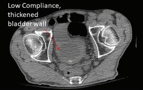
The body of the bladder itself is comprised of smooth muscle (the detrusor muscle) as well as up to 50% collagen (particularly types I, III, and IV) and 2–3% elastin [2]. The detrusor is primarily responsible for changes to compliance (defined as the change in volume per unit of pressure) and capacity. This is evidenced by the fact that bladder injury yields a dramatic increase in the amount of reparative collagen deposition (namely type III collagen) and a concurrent decrease in compliance. Moreover, increased age decreases the muscle-to-collagen ratio and the collagen, which cross-links over time, also decreases the overall bladder compliance. Anatomically, the low compliance bladder wall appears thickened on crosssectional imaging (Fig. 2.1), partly as a result of these extracellular matrix changes.
The internal structure of the bladder and proximal urethra also contain notable features. Posteromedially, the ureters enter the bladder at their corresponding ureteric orifices, forming two corners of a triangle known as the bladder trigone that is positioned to guide urine down the bladder neck and into the proximal prostatic urethra. The ureteric orifices possess one-way mucosal flaps that prevent the reflux of urine back into the ureters (vesicoureteral reflux); this anti-reflux mechanism can be defeated in instance of high intravesical pressure or defects to the native anatomy. In males, the smooth internal urethral sphincter (derived from the detrusor muscle) encircles the bladder neck just above the prostate gland. The striated external ure-
Fig. 2.1 CT image of a thickened, low-compliance bladder representing alterations in extracellular matrix
V. M. Ramakrishnan and J. R. Eswara
2 Basic Bladder Physiology and Anatomy
thral sphincter is positioned just inferior to the gland and medially to the bulbourethral (Cowper’s) glands, and is contiguous with the urogenital diaphragm. In females, the sphincter complexes are positioned adjacent to one another. Compared to males, the female striated sphincter runs more longitudinally. The sphincter urethra wraps around the urethra, similar to males. The distal sphincter complex function is augmented by a urethrovaginal muscle and compressor urethrae muscle group which helps promote continence in the absence of urethral length (Fig. 2.2).
Arterial blood supply to the bladder comes from the branches of the iliac vessels, and specifically the internal iliacs, which give off the superior vesical and umbilical arteries as well as the inferior vesical artery with its prostatic branches. Venous drainage occurs via corresponding veins that feed into the internal iliac veins, with the exception of a prostatic venous plexus that envelops the inferior aspect of the bladder as well as the entire prostate. Posterior, lymph drainage of the bladder courses directly to the external iliac nodes and, from there, to the common iliac nodes. Anterior drainage is housed in the prevesical plexus that also joins the external iliac nodes.
Fig. 2.2 Schematics depicting male (a) and female (b) continence
Anatomy of male continence a
neck
urethra
sphincter
Anatomy of Female Continence
Bladder Neck continuing into internal sphincter
Compressor urethrae
Striated Sphincter (external sphincter)
Urethral Meatus
V. M. Ramakrishnan and J. R. Eswara
Urinary Bladder Neurological Control and Innervation
Neurological control of urinary bladder function is incredibly intricate (Fig. 2.3). Globally, there are three input sources that drive micturition. The bladder contains afferent fibers (myelinated A and unmyelinated C fibers) with mechanoreceptors that interpret pressure/stretch (A), irritation (C) in the bladder wall [3]. This conveys characteristics such as awareness, fullness, urgency, pain, and temperature. Also mentioned earlier was that the bladder is an organ of convenience. Cognitive interpretation of various situations and the “decision” to urinate is carried out by higher cortical centers. Lastly, emotional behaviors and stress responses are controlled by the limbic system. These three sources – bladder afferents, the cerebral cortex, and the limbic system – all provide information to the medial frontal cortex of the forebrain within the periaqueductal gray matter. From here, stimuli are sent to the pons, which houses the brain’s micturition (medial pons) and continence centers (lateral pons). These are oppositional roles – that is, one function supports the contraction of the detrusor and subsequent expulsion or urine (i.e., micturition), while the other relaxes the detrusor and simultaneously stimulates the striated external urethral sphincter, thereby promoting the voluntary storage of urine (i.e., continence). The nerves that ultimately execute these functions originate from the spinal cord.
Anatomically, the innervation of the urinary bladder is an equally complex affair [2]. Multimodal control is obtained via sympathetic, parasympathetic, somatic efferent, and afferent nerve fibers. Interestingly, the framework for this level of control is laid during embryonic development, as the detrusor is of combined mesodermal and neural crest origin. At the lumbar spinal cord levels L1 and L2, sympathetic preganglionic fibers originating in the intermediate gray matter exit via the ventral root of the spinal cord, coursing through white rami communicantes and into the sympathetic trunk. At L1 and L2, these fibers can leave the sympathetic trunk and run along the aorta anteriorly and laterally via the intermesenteric (aortic) plexus
Fig. 2.3 Overview concept of bladder and CNS signaling
Efferent ner ves facilitate relaxation (storage) or contraction (empty) of bladder
Afferent ner ves carry pressure stretch, pain, temperature infor mation from bladder
Cor tex, limbic system, processes input
Bladder
Brain
and the inferior mesenteric ganglion. Fibers can also exit the sympathetic trunk and join the sacral splanchnic nerves and inferior hypogastric (pelvic) plexus. All of these fibers and tracts ultimately coalesce with a complex web of postganglionic fibers to innervate the bladder wall, ureters, prostate gland, and the external genitalia. Parasympathetic preganglionic fibers exit the spinal cord at the levels of S2–4, briefly entering the sacral plexus before joining the pelvic splanchnic nerves prior to innervating the bladder wall. Afferent fibers (mechanoreceptors – that is, “stretch” or distensibility receptors) sense bladder fullness and travel back via the sacral and inferior hypogastric plexuses to the sacral spinal cord [3]. Of note, these fibers are also in close relation to (and in the case of the parasympathetic supply, the same as) those that innervate the descending and sigmoid colon as well as the rectum.
Neurological control of voiding is critically dependent on the activity of various neurotransmitters, all of which are released based on direction from the pons [4, 5]. Also vital to this process is coordinated communication between the bladder and the urethral sphincters. To start, one is stimulated to urinate when the bladder fills to a capacity of approximately 200–500 cc of urine, though maximal capacities can be significantly higher. The act of filling stretches the aforementioned mechanoreceptors and conveys to the pons a need to induce urination [2]. The pontine micturition center then signals for detrusor contraction, mediated by acetylcholine (ACh) via M2, M3, and M5 muscarinic cholinergic receptors on detrusor myocytes via a G-protein–calcium channel mechanism. M2 receptors are the most numerous (with up to 75% prevalence) as opposed to M3 and M5 (approximately 25%) [6]. When the detrusor is stimulated to contract, concurrent relaxation of the internal urethral sphincter primes the bladder to release urine when convenient. The pontine micturition center inhibits the spinal guarding reflexes which act to inhibit involuntary bladder emptying and relaxes the external urethral sphincter via Onuf’s nucleus such that urethral pressure decreases. Combined with the concurrent increase in bladder pressure, urine flows down the pressure gradient and out the body. The act of voluntarily releasing urine via this process is what defines urinary continence
Cessation of voiding is highlighted by detrusor relaxation, which is achieved via activation of the pontine continence center and the downstream sympathetic action of norepinephrine on β-2 and β-3 adrenergic receptors. This elicits a G-protein–potassium efflux-dependent mechanism that relaxes the detrusor and prevents contraction (this is also one of the mechanisms capitalized on by pharmaceutical β-3 agonists, which are often used to treat bladder overactivity [7]). Concurrent involuntary contraction of the smooth internal urethral sphincter is achieved sympathetically, with norepinephrine acting on α-1 adrenergic receptors that are G-protein–calcium channel-mediated. The external urethral sphincter can also be voluntarily stimulated to contract (thereby promoting continence) under the auspices of somatic parasympathetic control, via the pudendal nerve, with ACh binding to nicotinic inotropic receptors. Figure 2.4 summarizes the bladder receptor physiology.
With the basic overview of the interplay between the nervous system, detrusor, and urethral sphincter complete, several additional points must be made. First, sympathetic control of voiding can override parasympathetic inputs. The norepinephrine required to relax the detrusor can also act on α-2 adrenergic receptors in
Muscarinic Receptors:
- Stimulate Bladder contraction
- M2 is dominant type (75%)
-Acetylcholinc mediated
Beta Adrenergic Receptors:
- Stimulate Bladder relaxation
- Norepinephrine mediated
Alpha Adrenergic Receptors:
- Stimulate Sphincter contraction
- Norepinephrine mediated
the pelvic ganglia to block the transmission of parasympathetic signaling. Second, with the aforementioned framework, it is easy to understand why disruption of this highly coordinated control system – via multiple sclerosis lesions, for instance –often results in detrusor-sphincter dyssynergia (DSD), a condition in which the detrusor contracts against a closed urethral sphincter. Third, this intricate system is also active during sexual activity. Sympathetic overdrive during ejaculation forces the internal urethral sphincter to contact and creates a natural one-way valve that promotes the antegrade flow of semen out the penis [8]. Fourth, it is important to recognize that blood flow to the muscle and mucosa are directly affected by intramural tension [9]. Continued distension can lead to ischemia as the supplying vessels are stretched and resistance is increased. In diseases of severe distension – most notably in those with spinal cord injury or other neurological conditions – bladder sensation is diminished or completely absent. This results in a sequence of overdistension, ischemia, consequent injury and/or death of once-healthy mucosa and muscle, preferential remodeling of the affected areas with collagen over muscle, and ultimately the permanent alteration of bladder compliance.
Thus far, we have outlined a complex system of higher cortical control for voiding and storing urine. Concurrently, there also exist primitive autonomic reflexes within the spinal cord and extra-neurological urothelial factors that affect these processes. Regarding the latter, factors such as obesity and metabolic disease (in particular, diabetes and associated neuropathies), fibrosis, ischemia, inflammation, and even various foods all play a role in establishing long-term lower urinary tract dysfunction. Many of these factors are well integrated with one another.
Bladder Neck
Bladder
Urethra
Prostate
Fig. 2.4 Contraction and relaxation is mediated by receptors in bladder
V. M. Ramakrishnan and J. R. Eswara
Tools for Evaluating Function of Bladder Anatomy
Bladder Diaries
There exist a whole host of methods for assessing bladder (and specifically, detrusor) function, the simplest of which is to have patients complete a bladder diary. Factors such as time of day, fluids and food (type and quantity), frequency and volume of urination, and in-depth analysis of accidents (such as leakage, urge to urinate, and surrounding activities (for e.g., sneezing, exercising, running, etc.)) are all systematically measured over a period of time. This provides clinicians and patients with useful information regarding possible intrinsic and extrinsic factors that contribute to a patient’s symptoms. Recent studies have demonstrated that 3 days’ worth of data collection are sufficient enough to assess lower urinary tract symptoms in adults [10, 11] and 2 days in children [11], though shorter durations are expectedly associated with a higher false-negative rate. The diary remains a good evaluation to determine how bladder physiology is impacting daily functioning.
Bladder Scanning and PVR
An additional tool for evaluating bladder physiology is to have the patient attempt to void and measuring a post-void residual (PVR) with a bladder-scanning ultrasound can provide immediate numerical data and, depending on the scanner, correlative imaging. PVRs of less than 10% of the voided volume are considered insignificant. It is important to recognize that false-positives for urinary retention can result in patients with excessive adiposity, gynecological disease [12], severe cardiovascular disease [12], and oncologic burden, for instance. In conjunction with measuring PVR, evaluation of the patient’s electrolytes (particularly, the renal function) and renal ultrasound can provide evidence of possible upper tract involvement.
Urodynamics
Urodynamics (UDS) is a critical tool for bladder and voiding evaluation and is part of the armamentarium of every urologist. Urodynamic testing is covered in more detail in chapter 8. Broadly, UDS is comprised of a group of metrics that interrogate bladder physiology, define abnormalities of the lower urinary tract, and elucidates issues with the transport, storage, and evacuation of urine in the context of the patient’s urological symptoms. The first metric, cystometry, can be further subdivided into filling and voiding cystometry and is the key test that evaluates detrusor function. Filling cystometry establishes a pressure–volume curve for bladder filling and can easily assess bladder sensation, capacity, compliance, and detrusor activity. This helps the practitioner determine if the bladder physiology is normal or has been
V. M. Ramakrishnan and J. R. Eswara
potentially impacted by a neurologic condition. Voiding cystometry does the same, but for bladder emptying. Critical to both of these tests is an awareness of intravesical pressure (Pves) as well as the abdominal pressure (Pabd) surrounding the bladder, which can be estimated via probes inserted into the rectum, vagina, extraperitoneal space, or ostomy. From these pressures, the true detrusor pressure (Pdet = Pves – Pabd) can be determined. The resultant graph is primarily comprised of two key phases –the filling/storage phase and the voiding phase. In short, urodynamics can summarize how the bladder anatomy described above functions in real time.
Uroflowmetry is also part of the suite of UDS metrics and simply measures the flow rate of urine (in mL/seconds) over a defined time period (seconds). The emphasis is made on identifying the maximum flow rate, the time to maximal flow, the voided volume, and the total flow time from start to finish. As mentioned earlier, voiding is critically dependent on the carefully orchestrated interplay between the bladder and urethral sphincters. Uroflowmetry provides data that is a direct combined reflection of the effectiveness of detrusor contraction, degree of urethral sphincter relaxation, and patency of the urethra via urethral pressure measurement [13].
Two final subsets of UDS worth emphasizing are Video-UDS and electromyography. Video-UDS combines the analytics of UDS with real-time fluoroscopic imagery of the lower urinary tract and is particularly useful for differentiating bladder neck obstruction from dysfunctional voiding. The modality can also be used to visualize vesicoureteral reflux, evaluate neurogenic bladder disease, and has the added benefit of potentially identifying malignancies/anatomic abnormalities along the urinary tract including the bladder mucosa by visualizing filling defects or outpouching during the cystogram. Electromyography, on the other hand, relies on electrodes placed in or near pelvic floor muscles of interest. This facilitates the quantitative analysis of muscle depolarization and expedites the evaluation of underlying neurological abnormalities affecting urethral sphincter and pelvic floor muscle function.
Concluding Remarks
The urinary bladder plays a critical role in the storage and expulsion of metabolic waste products. The detrusor is, quite literally, central to the action, though its function and tone depend on how a complex web of neural inputs interact with end organ receptors in the bladder and adjacent sphincters. Neurologic diseases can impact signaling to the bladder and cause changes in storage and emptying to ultimately result in the wide spectrum of symptoms and signs associated with a neurogenic bladder.
References
1. Lai HH, Rawal A, Shen B, Vetter J. The relationship between anxiety and overactive bladder or urinary incontinence symptoms in the clinical population. Urology. 2016;98:50–7.
2. Sam P, LaGrange CA. Anatomy, abdomen and pelvis, bladder detrusor muscle. Treasure Island: StatPearls; 2019.
3. Umans BD, Liberles SD. Neural sensing of organ volume. Trends Neurosci. 2018;41:911–24.
4. de Groat WC, Griffiths D, Yoshimura N. Neural control of the lower urinary tract. Compr Physiol. 2015;5:327–96.
5. Fowler CJ, Griffiths D, de Groat WC. The neural control of micturition. Nat Rev Neurosci. 2008;9:453–66.
6. Mansfield KJ, et al. Muscarinic receptor subtypes in human bladder detrusor and mucosa, studied by radioligand binding and quantitative competitive RT-PCR: changes in ageing. Br J Pharmacol. 2005;144:1089–99.
7. Andersson KE. On the site and mechanism of action of beta3-adrenoceptor agonists in the bladder. Int Neurourol J. 2017;21:6–11.
8. Raz S. Adrenergic influence on the internal urinary sphincter. Isr J Med Sci. 1974;10:608–11.
9. Kozlowski R, Siroky MB, Krane RJ, Azadzoi KM. Regulation of blood flow and microcirculation resistance in rabbit bladder. J Urol. 2002;168:1608–14.
10. Jimenez-Cidre MA, et al. The 3-day bladder diary is a feasible, reliable and valid tool to evaluate the lower urinary tract symptoms in women. Neurourol Urodyn. 2015;34:128–32.
11. Konstantinidis C, Kratiras Z, Samarinas M, Skriapas K. Optimal bladder diary duration for patients with suprapontine neurogenic lower urinary tract dysfunction. Int Braz J Urol. 2016;42:766–72.
12. Kim TH, et al. Falsely elevated postvoid residual urine volume in uterine myoma. Ann Rehabil Med. 2017;41:332–6.
13. Corona LE, Cameron AP, Clemens JQ, Qin Y, Stoffel JT. Urethral pressure measurement as a tool for the urodynamic diagnosis of detrusor sphincter dyssynergia. Int Neurourol J. 2018;22:268–74.
Chapter 3 Neuroanatomy: Overview of Functional Signaling Pathways
Blayne Welk and Jalesh N. Panicker
Introduction
The lower urinary tract (LUT) has two essential functions: storage and voiding of urine. During the storage mode, urine is allowed to passively fill the bladder: the kidneys must be able to drain freely into the bladder, the bladder must have a normal capacity, the walls of the bladder must be compliant and able to accommodate the increasing volume of urine, a normal sensory signal must be relayed to the central nervous system (CNS) for conscious perception of fullness, and the urinary sphincter must be competent and be able to prevent leakage. During the voiding phase, urine is allowed to fully exit the bladder: a conscious decision is made to urinate at a socially acceptable time, the urinary sphincters and other pelvic floor muscles relax, and there is a coordinated contraction of the bladder which is then able to fully empty through an unobstructed urethra. The neurologic control of storage and voiding of urine is dictated by several key structures and pathways in the central and peripheral nervous system that control and coordinate the bladder’s detrusor muscle (urinary reservoir) and the urethral sphincter (urinary outlet).
B. Welk (*)
Department of Surgery (Urology) and Epidemiology & Biostatistics, Western University, London, ON, Canada
J. N. Panicker
Department of Uro-Neurology, The National Hospital for Neurology and Neurosurgery, Queen Square, London, UK
J. T. Stoffel, E. V. Dray (eds.), Urological Care for Patients with Progressive Neurological Conditions, https://doi.org/10.1007/978-3-030-23277-1_3
B. Welk and J. N. Panicker
General Neuroanatomy Review
The CNS consists of the brain and spinal cord. The peripheral nervous system (PNS) consists of the afferents (sensory fibers that travel toward the CNS to relay sensations from the body or environment), and efferents (motor fibers that travel away from the CNS and signal organs or muscles in the body). Sensory afferents from the bladder sense fullness and enter the CNS through A-delta fibers via the dorsal root ganglia of the spinal cord. C-fibers are another afferent; however, they remain dormant in the healthy bladder. In pathological situations, such as following spinal cord injury or exposure to noxious stimuli, these become active and also respond to bladder filling, leading to abnormal bladder sensations or pain. Motor efferents exit the CNS via the ventral roots of the spinal cord.
Within the PNS, there is a somatic nervous system (which allows voluntary control of structures such as the urinary sphincter), and the autonomic nervous system (which provides unconscious control over visceral and endocrine function). The only somatic control of the LUT is through the pudendal nerve, which is derived from the cell bodies in an area of the ventral gray matter of the sacral spinal cord (S1–S3 segments) that is called Onuf’s nucleus.
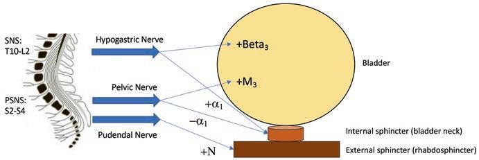
The autonomic nervous system has both parasympathetic (cranio-sacral) and sympathetic divisions (thoraco-lumbar) based on their anatomic relationship to the CNS (Fig. 3.1). The parasympathetic nervous system pathways relevant to urination arise from a different region of the gray matter of the sacral spinal cord and innervate the LUT through the pelvic nerves; the parasympathetic preganglionic motor efferents are long, and their ganglion are located near the bladder in the pelvic plexus. The sympathetic nervous system pathways relevant to urination arise from the T10-L2 spinal cord segments and innervate the LUT through the hypogastric nerve; the sympathetic preganglionic motor efferents have varying lengths. The pudendal, pelvic, and hypogastric nerves have both efferent and afferent fibers, which regulate motor and sensory functions of the LUT. There are several voiding
Fig. 3.1 Schematic of the sympathetic and parasympathetic pathways leading to the bladder. The hypogastric nerve stimulates beta3 receptors in the bladder (to relax the bladder), and alpha1 receptors (to contract the sphincter) during storage of urine. The pelvic nerve stimulates muscarinic3 receptors (to contract the bladder) and inhibits alpha1 receptors (relaxing the sphincter) during voiding of urine. The pudendal nerve contracts the external sphincter by activating nicotinic receptors
and storage spinal reflexes mediated by interneurons in the T10-L2 and S2–S4 spinal cord segments.
The bladder itself is made of smooth muscle, extracellular matrix, and urothelium. The smooth muscle component is referred to as the detrusor. There are two urinary sphincters: the internal (or bladder neck) sphincter and the external (or rhabdosphincter) sphincter. Conceptually, these are often considered as separate structures; however, there is considerable overlap and intermingling between the smooth muscles of the internal sphincter and the striated muscles of the external sphincter [1, 2].
Central Control of the LUT in Health
Functional brain imaging has greatly improved our understanding of the role of different regions of the brain in urine storage and voiding [3, 4]. The pontine micturition center (PMC, previously referred to as Barrington’s nucleus in animal studies) and the periaqueductal gray (PAG) are key centers in the brainstem and midbrain that are involved in urinary control. During the storage of urine, sensations of bladder fullness are conveyed from the LUT, and the first point of relay is the periaqueductal gray (PAG) in the midbrain. The insula, hypothalamus, thalamus, and dorsal anterior cingulate cortex are important regions involved in the conscious perception of bladder fullness. The medial prefrontal cortex is the “checkpoint” where the conscious decision to void occurs. Until this decision is made, the medial prefrontal cortex inhibits the PAG, which in turn inhibits the PMC. As the sensory afferents are increasingly activated during storage, this leads to increasing activation of the bladder’s sympathetic efferent innervation of the bladder and the internal urethral sphincter to promote further storage of urine. The sympathetic efferents release the neurotransmitter norepinephrine which activates beta-3 receptors in the bladder, resulting in relaxation of the detrusor, and alpha-1 receptors in the internal urethral sphincter that results in contraction of the urinary outlet. At the same time, the sympathetic nervous system inhibits contraction of the bladder by inhibiting the parasympathetic ganglia. Pudendal nerve efferents are activated and through the neurotransmitter acetylcholine activates nicotinic receptors, thereby increasing the tone of the striated external urethral sphincter [5–7].
At an appropriate time and place to void, inhibition of the PAG from different brain regions including the medial prefrontal cortex and the hypothalamus ceases; this in turns removes the PAG-mediated inhibition of the PMC and facilitates voiding. Inhibition of pudendal nerve functions results in relaxation of the external urethral sphincter. Sympathetic-mediated activity is inhibited, and the parasympathetic innervation mediates detrusor contractions. The neurotransmitter acetylcholine activates muscarinic receptors in the bladder wall, and inhibits the internal urethral sphincter with the release of nitrous oxide, leading to further relaxation of the urinary outlet [5–7].
The guarding reflex is a spinal reflex relevant to many neurologic diseases [8]. As the bladder fills, there is an unconscious and involuntary contraction of the external urethral sphincter-mediated through the bladder sensory afferents (in the pelvic
B. Welk and J. N. Panicker
nerve), sacral spinal cord, and pudendal nerve. When the sensory input from a full bladder penetrates consciousness, the reflex is further augmented by the somatic efferents in the pudendal nerve that further contract the external urethral sphincter and is associated with awareness of this action. As the guarding reflex is activated, pudendal sensory afferents inhibit parasympathetic innervation of the bladder through interneurons in S2–S4 of the sacral spinal cord, thus allowing further bladder filling. Input from the PMC is required to regulate the guarding reflex. For example, in complete suprasacral spinal cord injuries, there is often incomplete bladder emptying due to the failure of the PMC to switch off the guarding reflex during voiding. Other pathways can activate the guarding reflex, such as the ventrolateral medulla (nucleus retroambiguus), which activates the Onuf’s nucleus following the anticipation of a cough or sneeze to prevent stress incontinence.
LUT Dysfunction Following Progressive Neurologic Disease
The neural control of LUT functions is affected following neurological disease, and from an understanding of neuroanatomy, it is possible to infer the pattern of LUT dysfunction. However, the constellation of storage and voiding symptoms that a patient may experience can be influenced by different variables such as severity of disease, coexisting non-neurogenic urological complications (such as stress incontinence, or benign prostatic enlargement) and functional status. Diseases that affect signaling or function of the cerebral cortex (such as dementia, or Huntington’s chorea) are often associated with urgency and urgency incontinence due to inconsistent control of the PMC. Diseases that affect both the brain and spinal cord (such as multiple sclerosis) can lead to a variety of urinary symptoms such as urgency and urgency incontinence (due to damage to the pathways controlling the PMC), and urinary retention (due to damage to the nerves in the spinal cord that control bladder emptying), or detrusor sphincter dyssynergia resulting from a neurological disconnect between the PMC and the guarding reflex. Other diseases (such as GuillainBarre syndrome, or diabetes) affect the peripheral nervous system, and therefore damage to the pelvic, hypogastric, and pudendal nerves can lead to urinary retention and an areflexic bladder. In most cases, as disease severity increases, the urologic symptoms also get worse.
References
1. Yucel S, Baskin LS. An anatomical description of the male and female urethral sphincter complex. J Urol. 2004;171(5):1890–7.
2. Koraitim MM. The male urethral sphincter complex revisited: an anatomical concept and its physiological correlate. J Urol. 2008;179(5):1683–9.
3 Neuroanatomy: Overview of Functional Signaling Pathways
3. Fowler CJ, Griffiths DJ. A decade of functional brain imaging applied to bladder control. Neurourol Urodyn. 2010;29(1):49–55.
4. Drake MJ, Fowler CJ, Griffiths D, Mayer E, Paton JFR, Birder L. Neural control of the lower urinary and gastrointestinal tracts: supraspinal CNS mechanisms. Neurourol Urodyn. 2010;29(1):119–27.
5. Clemens JQ. Basic bladder neurophysiology. Urol Clin North Am. 2010;37(4):487–94.
6. Fowler CJ, Griffiths D, de Groat WC. The neural control of micturition. Nat Rev Neurosci. 2008;9(6):453–66.
7. Benarroch EE. Neural control of the bladder: recent advances and neurologic implications. Neurology. Wolters Kluwer Health, Inc. on behalf of the American Academy of Neurology. 2010;75(20):1839–46.
8. Park JM, Bloom DA, McGuire EJ. The guarding reflex revisited. Br J Urol. 1997;80(6):940–5.
Chapter 4 Measuring Urologic Quality of Life in People with Progressive Neurologic Conditions
John T. Stoffel
Introduction
When caring for patients with progressive neurologic diseases, it is important to remember that almost all medical decisions are influenced by two factors: safety and quality of life. Patient safety issues center around avoiding morbidity and/or mortality. Urologic patient safety examples include preventing and treating urinary tract infections, managing bladder pressures to reduce risk of hydronephrosis, and treating urinary incontinence to avoid progression of sacral decubitus ulcers. Quality of life, however, is a more complicated variable to measure among neurogenic bladder patients with progressive neurologic conditions. Rate of disease progression, physical impairment, and cognitive decline will all impact how a person “measures” his or her quality of life. An example of differences in QOL perception is seen among multiple sclerosis patients at different stages of disease progression. In one study, relapsing/remitting patients reported more severe urinary symptoms than secondary progressive patients despite having very similar urodynamic findings and voiding diaries [1]. This chapter will review some common health-related quality of life measures (HRQOL) and patient-reported outcome measures (PROM) relevant to neurogenic bladder care among patients with progressive neurologic diseases. By better understanding the domains and target populations of these instruments, it is hoped that practitioners will begin to use questionnaires to assess quality of life more frequently for these patients.
J. T. Stoffel (*)
Division of Neurourology and Pelvic Reconstruction, Department of Urology, University of Michigan Medical School, Ann Arbor, MI, USA