Lippincott Illustrated Reviews: Biochemistry (Lippincott Illustrated Reviews Series) 7th Edition, (Ebook PDF)
Visit to download the full and correct content document: https://ebookmass.com/product/lippincott-illustrated-reviews-biochemistry-lippincott-ill ustrated-reviews-series-7th-edition-ebook-pdf/
More products digital (pdf, epub, mobi) instant download maybe you interests ...
Lippincott Illustrated Reviews: Pharmacology (Lippincott Illustrated Reviews Series) 7th Edition, (Ebook PDF)
https://ebookmass.com/product/lippincott-illustrated-reviewspharmacology-lippincott-illustrated-reviews-series-7th-editionebook-pdf/
Lippincott Illustrated Reviews Flash Cards: Biochemistry (Lippincott Illustrated Reviews Series) 1st Edition, (Ebook PDF)
https://ebookmass.com/product/lippincott-illustrated-reviewsflash-cards-biochemistry-lippincott-illustrated-reviewsseries-1st-edition-ebook-pdf/
Lippincott Illustrated Reviews: Cell and Molecular Biology (Lippincott Illustrated Reviews Series) 2nd Edition, (Ebook PDF)
https://ebookmass.com/product/lippincott-illustrated-reviewscell-and-molecular-biology-lippincott-illustrated-reviewsseries-2nd-edition-ebook-pdf/
Harperu2019s Illustrated Biochemistry 31/e 31st Edition, (Ebook PDF)
https://ebookmass.com/product/harpers-illustratedbiochemistry-31-e-31st-edition-ebook-pdf/
Pearson Reviews & Rationales: Comprehensive Review for NCLEX RN (Hogan, Pearson Reviews & Rationales Series) 3rd Edition, (Ebook PDF)
https://ebookmass.com/product/pearson-reviews-rationalescomprehensive-review-for-nclex-rn-hogan-pearson-reviewsrationales-series-3rd-edition-ebook-pdf/
Harper’s Illustrated Biochemistry 31st Edition Victor W. Rodwell
https://ebookmass.com/product/harpers-illustratedbiochemistry-31st-edition-victor-w-rodwell-2/
Harper’s Illustrated Biochemistry, 31st Edition Victor W. Rodwell
https://ebookmass.com/product/harpers-illustratedbiochemistry-31st-edition-victor-w-rodwell/
Harper's Illustrated Biochemistry, 32th ed 32nd Edition
Peter J. Kennelly
https://ebookmass.com/product/harpers-illustratedbiochemistry-32th-ed-32nd-edition-peter-j-kennelly-2/
Harper's
Illustrated Biochemistry,
Peter J. Kennelly
32th ed 32nd Edition
https://ebookmass.com/product/harpers-illustratedbiochemistry-32th-ed-32nd-edition-peter-j-kennelly/
Dedication
This book is dedicated to my grandchildren, Charlie and Isabella, with the promise that I will not write another, and to my students, past and present, with deep gratitude for 25 years of opportunities to teach and learn.
Contents
UNIT I: Protein Structure and Function
Chapter 1:Amino Acids
Chapter 2:Protein Structure
Chapter 3:Globular Proteins
Chapter 4:Fibrous Proteins
Chapter 5:Enzymes
UNIT II: Bioenergetics and Carbohydrate Metabolism
Chapter 6:Bioenergetics and Oxidative Phosphorylation
Chapter 7:Introduction to Carbohydrates
Chapter 8:Introduction to Metabolism and Glycolysis
Chapter 9:Tricarboxylic Acid Cycle and Pyruvate Dehydrogenase Complex
Chapter 10:Gluconeogenesis
Chapter 11:Glycogen Metabolism
Chapter 12:Monosaccharide and Disaccharide Metabolism
Chapter 13:Pentose Phosphate Pathway and Nicotinamide Adenine
Dinucleotide Phosphate
Chapter 14:Glycosaminoglycans, Proteoglycans, and Glycoproteins
UNIT III: Lipid Metabolism
Chapter 15:Dietary Lipid Metabolism
Chapter 16:Fatty Acid, Triacylglycerol, and Ketone Body Metabolism
Chapter 17:Phospholipid, Glycosphingolipid, and Eicosanoid Metabolism
Chapter 18:Cholesterol, Lipoprotein, and Steroid Metabolism
UNIT IV: Nitrogen Metabolism
Chapter 19:Amino Acids: Nitrogen Disposal
Chapter 20:Amino Acids: Degradation and Synthesis
Chapter 21:Amino Acids: Conversion to Specialized Products
Chapter 22:Nucleotide Metabolism
UNIT V: Integration of Metabolism
Chapter 23:Metabolic Effects of Insulin and Glucagon
Chapter 24:The Feed–Fast Cycle
Chapter 25:Diabetes Mellitus
Chapter 26:Obesity
UNIT VI: Medical Nutrition
Chapter 27:Nutrition: Overview and Macronutrients
Chapter 28:Micronutrients: Vitamins
Chapter 29:Micronutrients: Minerals
UNIT VII: Storage and Expression of Genetic Information
Chapter 30:DNA Structure, Replication, and Repair
Chapter 31:RNA Structure, Synthesis, and Processing
Chapter 32:Protein Synthesis
Chapter 33:Regulation of Gene Expression
Chapter 34:Biotechnology and Human Disease
Appendix Index
Figure Sources
Bonus chapter online! Chapter 35: Blood Clotting (Use your scratch-off code provided in the front of this book for access to this and other free online resources on .)
Amino Acids 1
For additional ancillary materials related to this chapter, please visit thePoint.
I. OVERVIEW
Proteins are the most abundant and functionally diverse molecules in living systems. Virtually every life process depends on this class of macromolecules. For example, enzymes and polypeptide hormones direct and regulate metabolism in the body, whereas contractile proteins in muscle permit movement. In bone, the protein collagen forms a framework for the deposition of calcium phosphate crystals, acting like the steel cables in reinforced concrete. In the bloodstream, proteins, such as hemoglobin and albumin, transport molecules essential to life, whereas immunoglobulins fight infectious bacteria and viruses. In short, proteins display an incredible diversity of functions, yet all share the common structural feature of being linear polymers of amino acids. This chapter describes the properties of amino acids. Chapter 2 explores how these simple building blocks are joined to form proteins that have unique three-dimensional structures, making them capable of performing specific biologic functions.
II. STRUCTURE
Although >300 different amino acids have been described in nature, only 20 are commonly found as constituents of mammalian proteins. [Note: These standard amino acids are the only amino acids that are encoded by DNA, the genetic material in the cell (see p. 411). Nonstandard amino acids are produced by chemical modification of standard amino acids (see p. 45).] Each amino acid has a carboxyl group, a primary amino group (except for proline, which has a secondary amino group), and a distinctive side chain (R group) bonded to the α-
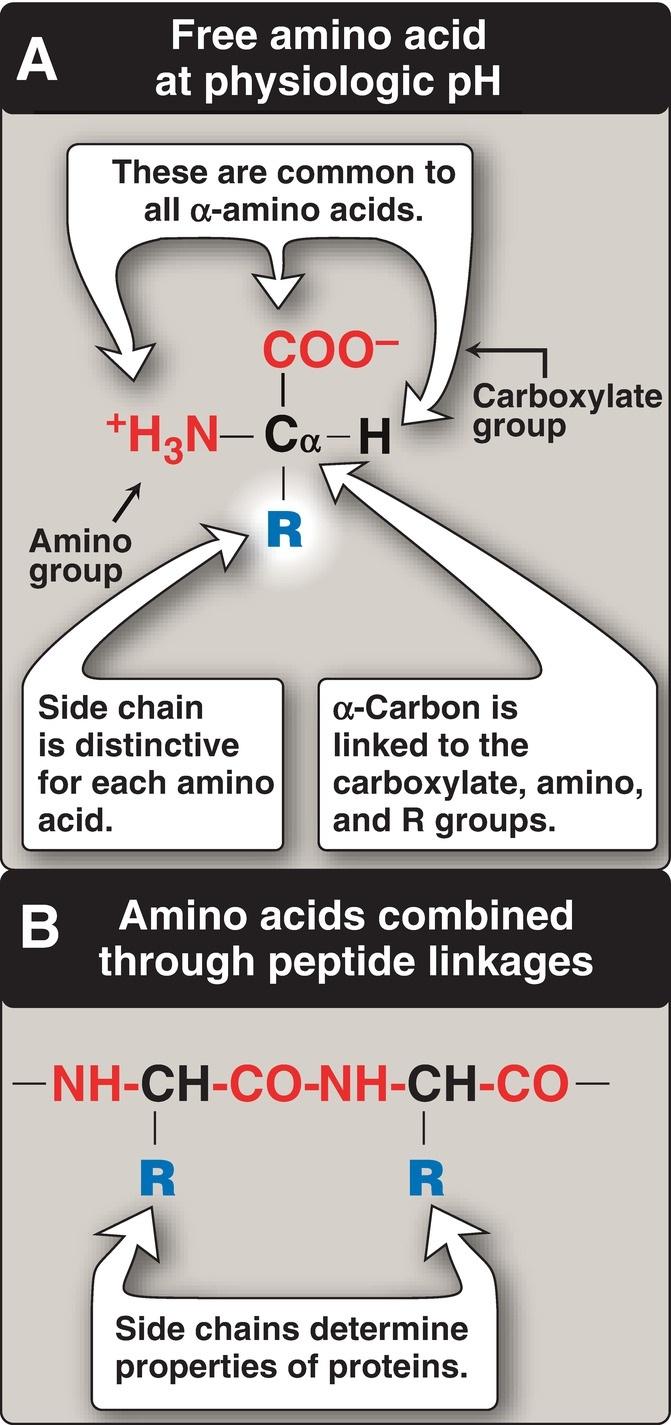
carbon atom. At physiologic pH (~7.4), the carboxyl group is dissociated, forming the negatively charged carboxylate ion ( COO ), and the amino group is protonated ( NH3+) (Fig. 1.1A). In proteins, almost all of these carboxyl and amino groups are combined through peptide linkage and, in general, are not available for chemical reaction except for hydrogen bond formation (Fig. 1.1B). Thus, it is the nature of the side chains that ultimately dictates the role an amino acid plays in a protein. Therefore, it is useful to classify the amino acids according to the properties of their side chains, that is, whether they are nonpolar (have an even distribution of electrons) or polar (have an uneven distribution of electrons, such as acids and bases) as shown in Figures 1.2 and 1.3.
Figure 1.1 A, B. Structural features of amino acids.
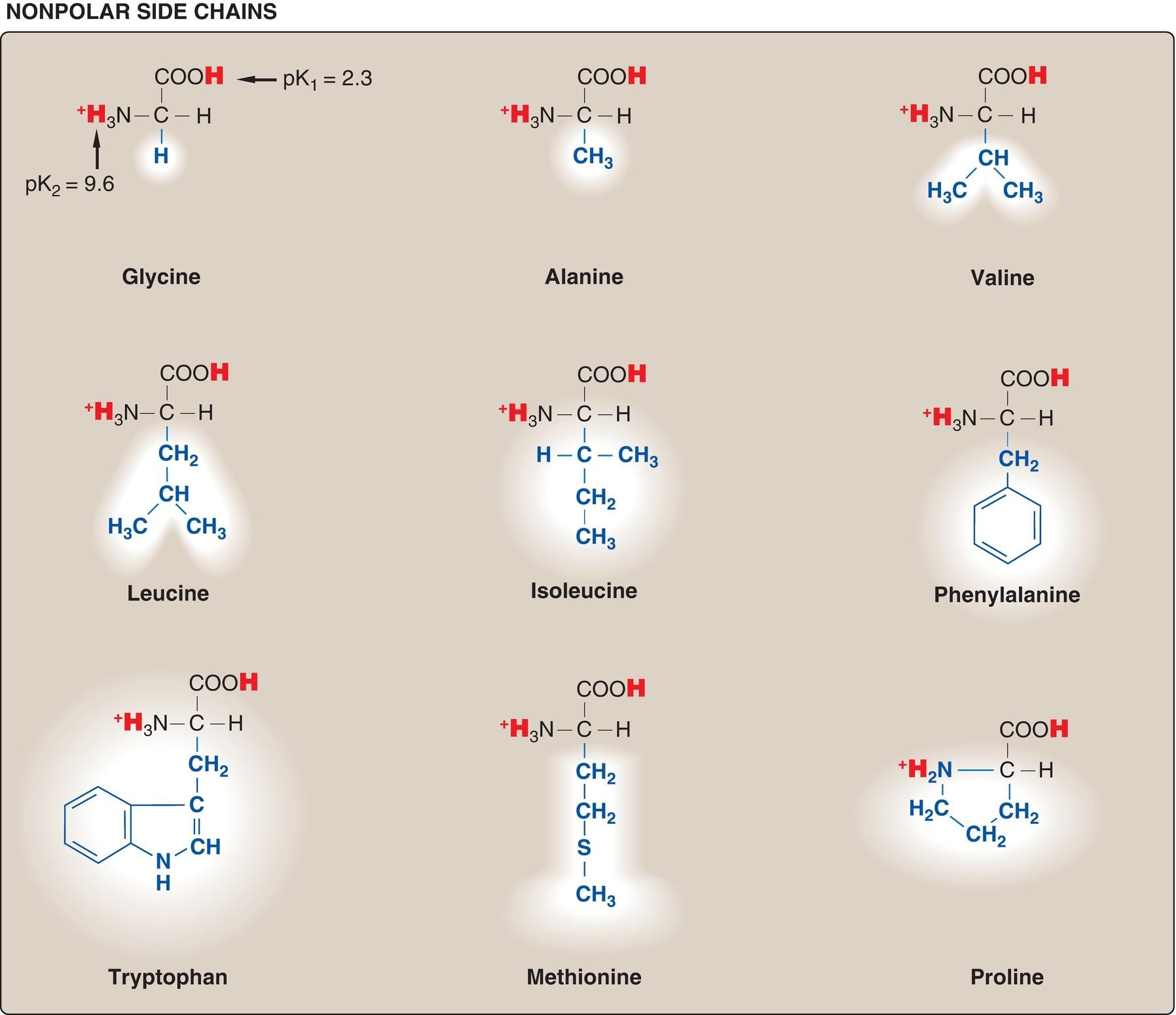
Figure 1.2 Classification of the 20 standard amino acids, according to the charge and polarity of their side chains at acidic pH, is shown here and continues in Figure 1.3. Each amino acid is shown in its fully protonated form, with dissociable hydrogen ions represented in red. The pK values for the α-carboxyl and α-amino groups of the nonpolar amino acids are similar to those shown for glycine.
Figure 1.3 Classification of the 20 standard amino acids, according to the charge
and polarity of their side chains at acidic pH (continued from Fig. 1.2). [Note: At physiologic pH (7.35 to 7.45), the α-carboxyl groups, the acidic side chains, and the side chain of free histidine are deprotonated.]
A. Amino acids with nonpolar side chains
Each of these amino acids has a nonpolar side chain that does not gain or lose protons or participate in hydrogen or ionic bonds (see Fig. 1.2). The side chains of these amino acids can be thought of as “oily” or lipid-like, a property that promotes hydrophobic interactions (see Fig. 2.10, p. 19).
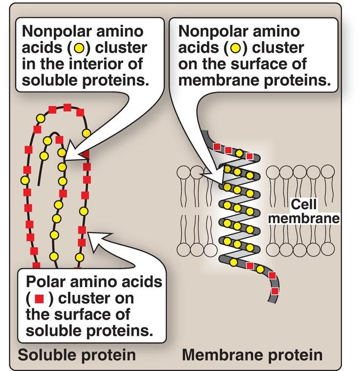
1. Location in proteins: In proteins found in aqueous solutions (a polar environment), the side chains of the nonpolar amino acids tend to cluster together in the interior of the protein (Fig. 1.4). This phenomenon, known as the hydrophobic effect, is the result of the hydrophobicity of the nonpolar R groups, which act much like droplets of oil that coalesce in an aqueous environment. By filling up the interior of the folded protein, these nonpolar R groups help give the protein its threedimensional shape. However, for proteins that are located in a hydrophobic environment, such as a membrane, the nonpolar R groups are found on the outside surface of the protein, interacting with the lipid environment (see Fig. 1.4). The importance of these hydrophobic interactions in stabilizing protein structure is discussed on p. 19.
Figure 1.4 Location of nonpolar amino acids in soluble and membrane proteins.
Sickle cell anemia, a disease of red blood cells that causes them to become sickle shaped rather than disc shaped, results from the replacement of polar glutamate with nonpolar valine at the sixth position in the β subunit of hemoglobin A (see p. 36).
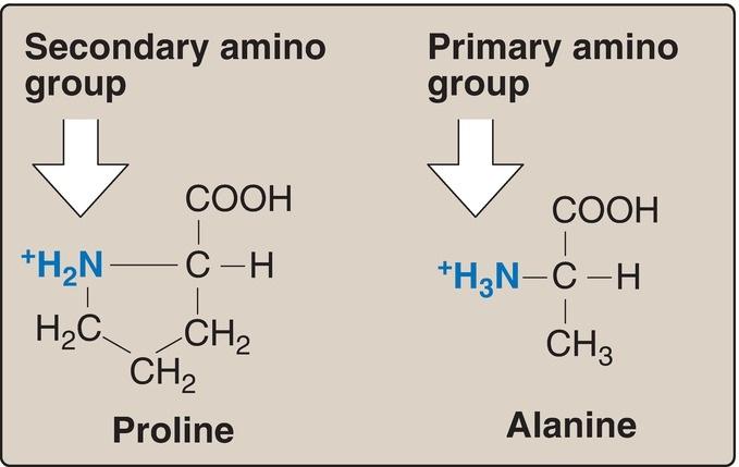
2. Proline: Proline differs from other amino acids in that its side chain and α-amino nitrogen form a rigid, five-membered ring structure (Fig. 1.5).
Proline, then, has a secondary (rather than a primary) amino group. It is frequently referred to as an “imino acid.” The unique geometry of proline contributes to the formation of the fibrous structure of collagen (see p. 45), but it interrupts the α-helices found in globular proteins (see p. 16).
Figure 1.5 Comparison of the secondary amino group found in proline with the primary amino group found in other amino acids such as alanine.
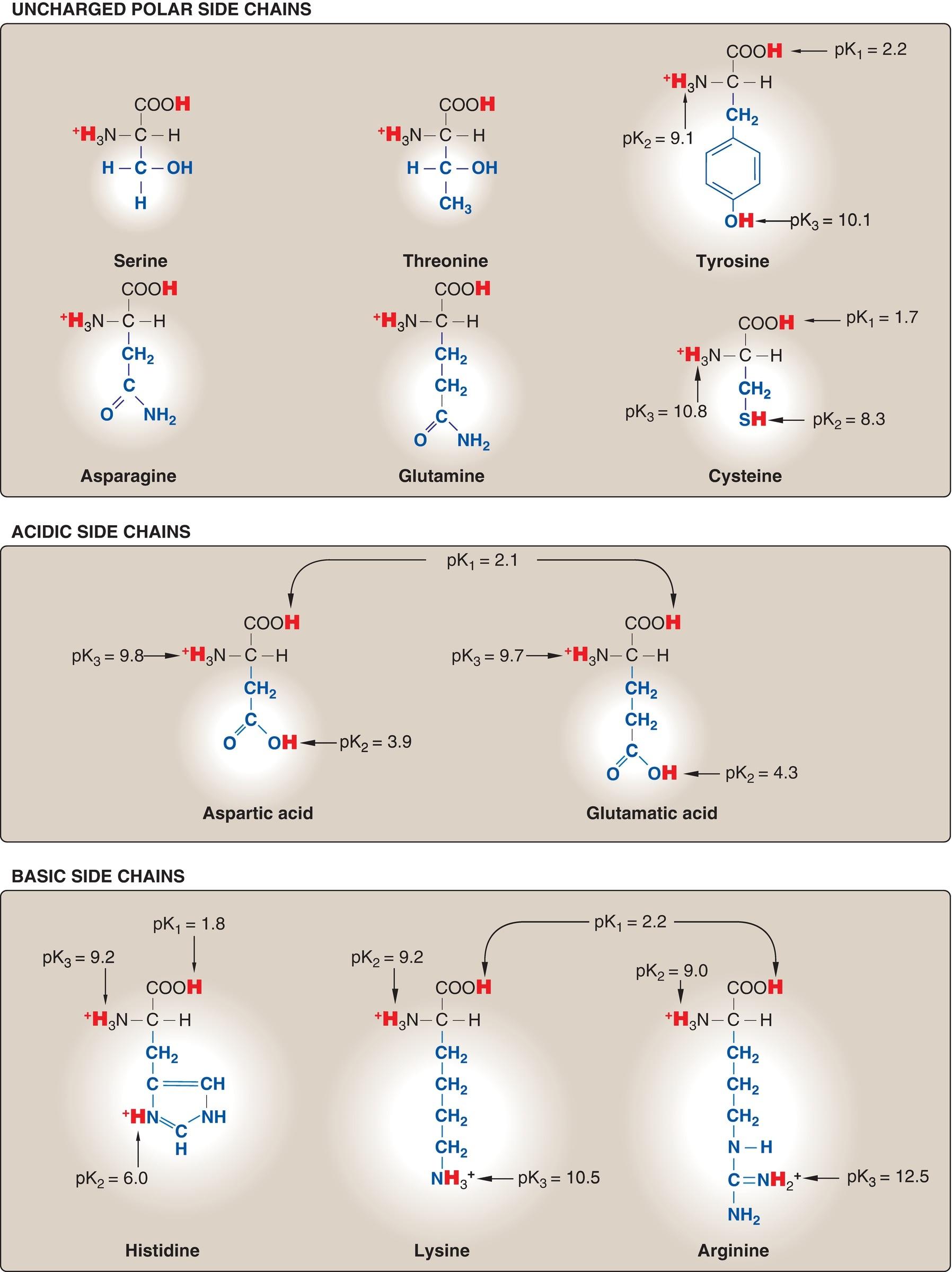
B. Amino acids with uncharged polar side chains
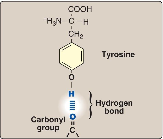
These amino acids have zero net charge at physiologic pH, although the side chains of cysteine and tyrosine can lose a proton at an alkaline pH (see Fig. 1.3). Serine, threonine, and tyrosine each contain a polar hydroxyl group that can participate in hydrogen bond formation (Fig. 1.6). The side chains of asparagine and glutamine each contain a carbonyl group and an amide group, both of which can also participate in hydrogen bonds.
Figure 1.6 Hydrogen bond between the phenolic hydroxyl group of tyrosine and another molecule containing a carbonyl group.
1. Disulfide bond: The side chain of cysteine contains a sulfhydryl (thiol) group ( SH), which is an important component of the active site of many enzymes. In proteins, the –SH groups of two cysteines can be oxidized to form a covalent cross-link called a disulfide bond ( S–S–). Two disulfide-linked cysteines are referred to as cystine. (See p. 19 for a further discussion of disulfide bond formation.)
Many extracellular proteins are stabilized by disulfide bonds. Albumin, a blood protein that functions as a transporter for a variety of molecules, is an example.
2. Side chains as attachment sites for other compounds: The polar hydroxyl group of serine, threonine, and (rarely) tyrosine can serve as a site of attachment for structures such as a phosphate group. In addition, the amide group of asparagine, as well as the hydroxyl group of serine or threonine, can serve as a site of attachment for oligosaccharide chains in glycoproteins (see p. 165).
C. Amino acids with acidic side chains
The amino acids aspartic acid and glutamic acid are proton donors. At physiologic pH, the side chains of these amino acids are fully ionized, containing a negatively charged carboxylate group ( COO ). The fully ionized forms are called aspartate and glutamate.
D. Amino acids with basic side chains
The side chains of the basic amino acids accept protons (see Fig. 1.3). At physiologic pH, the R groups of lysine and arginine are fully ionized and positively charged. In contrast, the free amino acid histidine is weakly basic and largely uncharged at physiologic pH. However, when histidine is incorporated into a protein, its R group can be either positively charged (protonated) or neutral, depending on the ionic environment provided by the protein. This important property of histidine contributes to the buffering role it plays in the functioning of such proteins as hemoglobin (see p. 30). [Note: Histidine is the only amino acid with a side chain that can ionize within the physiologic pH range.]
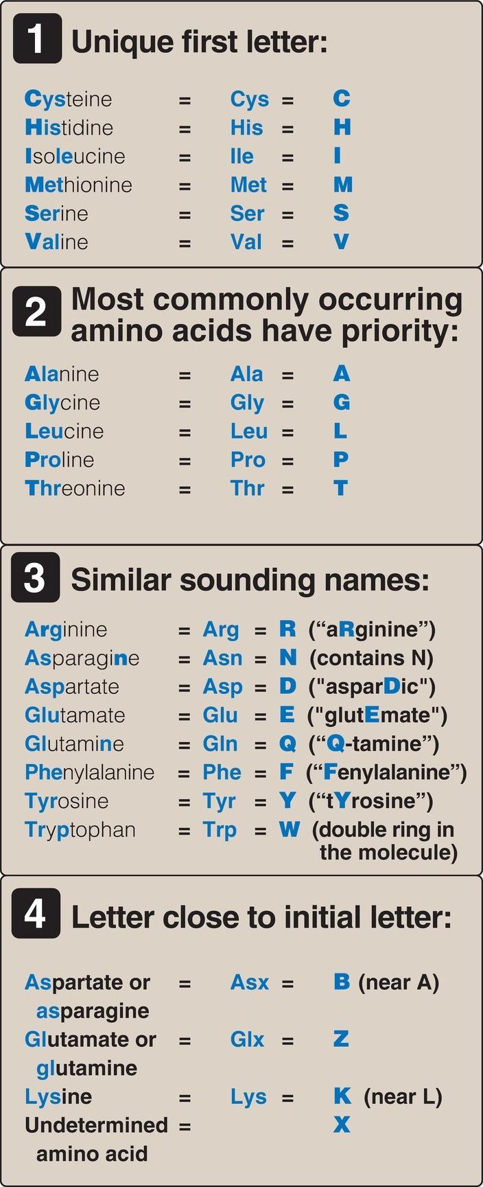
E. Abbreviations and symbols for commonly occurring amino acids
Each amino acid name has an associated three-letter abbreviation and a oneletter symbol (Fig. 1.7). The one-letter codes are determined by the following rules.
Figure 1.7 Abbreviations and symbols for the standard amino acids.
1. Unique first letter: If only one amino acid begins with a given letter, then that letter is used as its symbol. For example, V = valine.
2. Most commonly occurring amino acids have priority: If more than one amino acid begins with a particular letter, the most common of these amino acids receives this letter as its symbol. For example, glycine is more common than glutamate, so G = glycine.
3. Similar sounding names: Some one-letter symbols sound like the amino acid they represent. For example, F = phenylalanine, or W = tryptophan (“twyptophan” as Elmer Fudd would say).
4. Letter close to initial letter: For the remaining amino acids, a one-letter symbol is assigned that is as close in the alphabet as possible to the initial letter of the amino acid, for example, K = lysine. Furthermore, B is assigned to Asx, signifying either aspartic acid or asparagine; Z is assigned to Glx, signifying either glutamic acid or glutamine; and X is assigned to an unidentified amino acid.
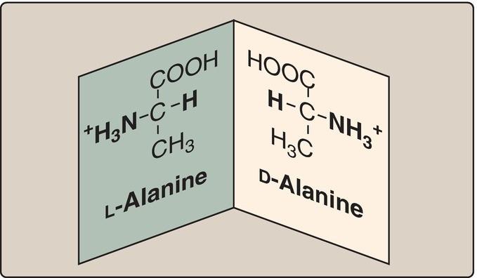
F. Amino acid isomers
Because the α-carbon of an amino acid is attached to four different chemical groups, it is an asymmetric (chiral) atom. Glycine is the exception because its α-carbon has two hydrogen substituents. Amino acids with a chiral αcarbon exist in two different isomeric forms, designated D and L, which are enantiomers, or mirror images (Fig. 1.8). [Note: Enantiomers are optically active. If an isomer, either D or L, causes the plane of polarized light to rotate clockwise, it is designated the (+) form.] All amino acids found in mammalian proteins are of the L configuration. However, D-amino acids are found in some antibiotics and in bacterial cell walls (see p. 252). [Note: Racemases enzymatically interconvert the D- and L-isomers of free amino acids.]
Figure 1.8 D and L forms of alanine are mirror images (enantiomers).
III. ACIDIC AND BASIC PROPERTIES
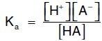
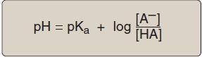
Amino acids in aqueous solution contain weakly acidic α-carboxyl groups and weakly basic α-amino groups. In addition, each of the acidic and basic amino acids contains an ionizable group in its side chain. Thus, both free amino acids and some amino acids combined in peptide linkages can act as buffers. Acids may be defined as proton donors and bases as proton acceptors. Acids (or bases) described as weak ionize to only a limited extent. The concentration of protons ([H+]) in aqueous solution is expressed as pH, where pH = log 1/[H+] or –log [H+]. The quantitative relationship between the pH of the solution and concentration of a weak acid (HA) and its conjugate base (A ) is described by the Henderson-Hasselbalch equation.
A. Equation derivation
Consider the release of a proton by a weak acid represented by HA:
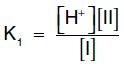
The salt or conjugate base, A , is the ionized form of a weak acid. By definition, the dissociation constant of the acid, Ka, is:
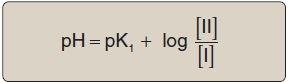
[Note: The larger the Ka, the stronger the acid, because most of the HA has dissociated into H+ and A . Conversely, the smaller the Ka, the less acid has dissociated and, therefore, the weaker the acid.] By solving for the [H+] in the above equation, taking the logarithm of both sides of the equation, multiplying both sides of the equation by 1, and substituting pH = log [H+] and pKa = log Ka, we obtain the Henderson-Hasselbalch equation:
B. Buffers
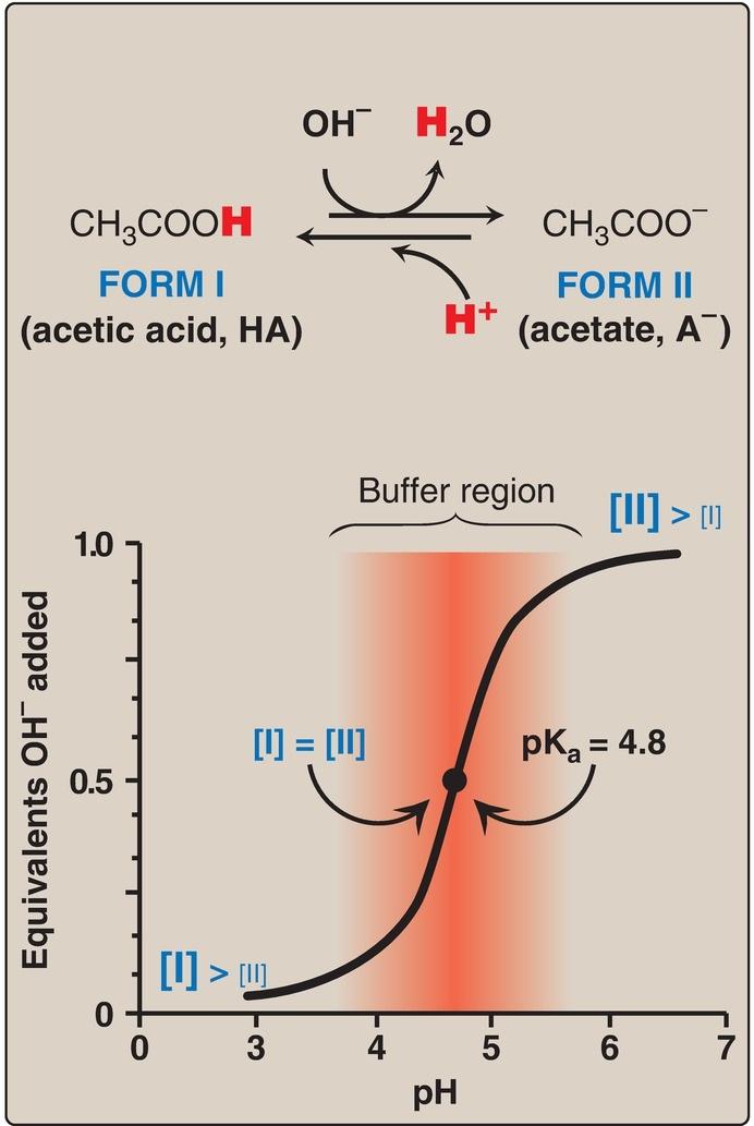
A buffer is a solution that resists change in pH following the addition of an acid or base. A buffer can be created by mixing a weak acid (HA) with its conjugate base (A ). If an acid such as HCl is added to a buffer, A can neutralize it, being converted to HA in the process. If a base is added, HA can likewise neutralize it, being converted to A in the process. Maximum buffering capacity occurs at a pH equal to the pKa, but a conjugate acidbase pair can still serve as an effective buffer when the pH of a solution is within approximately ±1 pH unit of the pKa. If the amounts of HA and A are equal, the pH is equal to the pKa. As shown in Figure 1.9, a solution containing acetic acid (HA = CH3 – COOH) and acetate (A = CH3 –COO ) with a pKa of 4.8 resists a change in pH from pH 3.8 to 5.8, with maximum buffering at pH 4.8. At pH values less than the pKa, the protonated acid form (CH3 – COOH) is the predominant species in solution.
At pH values greater than the pKa, the deprotonated base form (CH3 –COO ) is the predominant species.
Figure 1.9 Titration curve of acetic acid.
C. Amino acid titration
The titration curve of an amino acid can be analyzed in the same way as described for acetic acid.
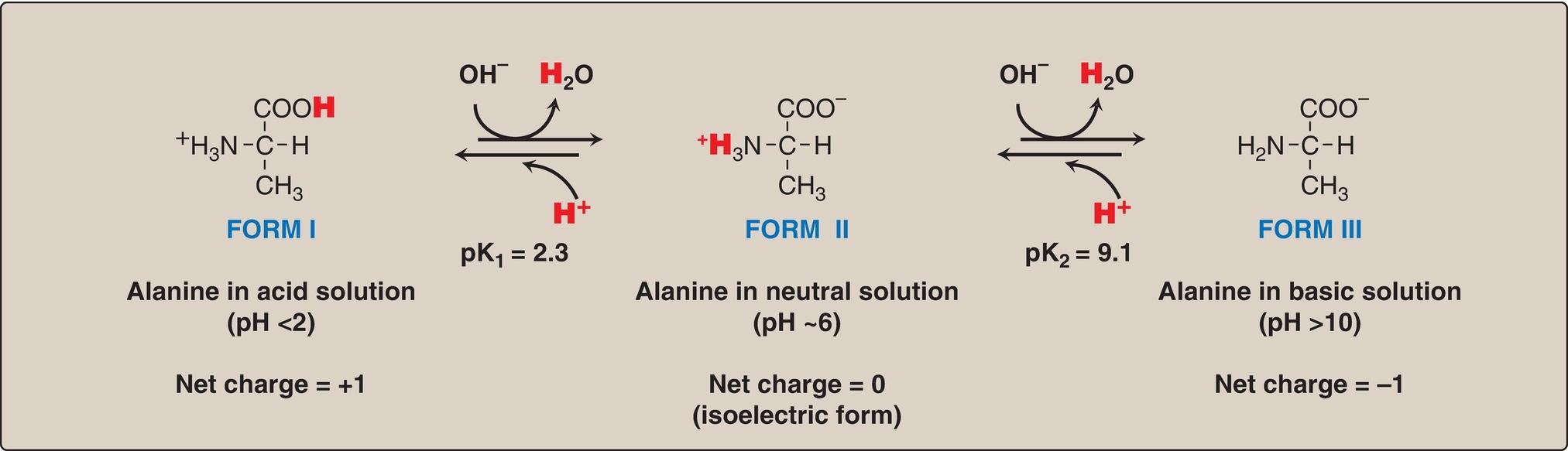
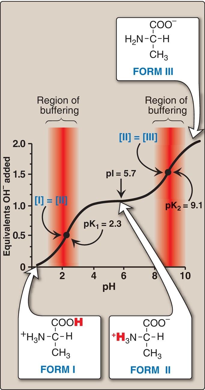
1. Carboxyl group dissociation: Consider alanine, for example, which contains an ionizable α-carboxyl and α-amino group. [Note: Its –CH3 R group is nonionizable.] At a low (acidic) pH, both of these groups are protonated (Fig. 1.10). As the pH of the solution is raised, the COOH group of form I can dissociate by donating a H+ to the medium. The release of a H+ results in the formation of the carboxylate group, COO . This structure is shown as form II, which is the dipolar form of the molecule (see Fig. 1.10). This form, also called a zwitterion (from the German word for “hybrid”), is the isoelectric form of alanine, that is, it has an overall (net) charge of zero.
Figure 1.10 Ionic forms of alanine in acidic, neutral, and basic solutions.
2. Application of the Henderson-Hasselbalch equation: The dissociation constant of the carboxyl group of an amino acid is called K1, rather than Ka, because the molecule contains a second titratable group. The Henderson-Hasselbalch equation can be used to analyze the dissociation of the carboxyl group of alanine in the same way as described for acetic acid:
where I is the fully protonated form of alanine and II is the isoelectric form of alanine (see Fig. 1.10). This equation can be rearranged and converted to its logarithmic form to yield:
3. Amino group dissociation: The second titratable group of alanine is the amino ( NH3+) group shown in Figure 1.10. Because this is a much weaker acid than the –COOH group, it has a much smaller dissociation constant, K2. [Note: Its pKa is, therefore, larger.] Release of a H+ from the protonated amino group of form II results in the fully deprotonated form of alanine, form III (see Fig. 1.10).
4. Alanine pKs: The sequential dissociation of H+ from the carboxyl and amino groups of alanine is summarized in Figure 1.10. Each titratable group has a pKa that is numerically equal to the pH at which exactly one half of the H+ have been removed from that group. The pKa for the most acidic group ( COOH) is pK1, whereas the pKa for the next most acidic group ( NH3+) is pK2. [Note: The pKa of the α-carboxyl group of amino acids is ~2, whereas that of the α-amino group is ~9.]
5. Alanine titration curve: By applying the Henderson-Hasselbalch equation to each dissociable acidic group, it is possible to calculate the complete titration curve of a weak acid. Figure 1.11 shows the change in pH that occurs during the addition of base to the fully protonated form of alanine (I) to produce the completely deprotonated form (III). Note the following:
Figure 1.11 The titration curve of alanine.
a. Buffer pairs: The –COOH/–COO pair can serve as a buffer in the pH region around pK1, and the –NH3+/–NH2 pair can buffer in the region around pK2.
b. When pH = pK: When the pH is equal to pK1 (2.3), equal amounts of forms I and II of alanine exist in solution. When the pH is equal to pK2 (9.1), equal amounts of forms II and III are present in solution.
c. Isoelectric point: At neutral pH, alanine exists predominantly as the dipolar form II in which the amino and carboxyl groups are ionized, but the net charge is zero. The isoelectric point (pI) is the pH at which an amino acid is electrically neutral, that is, in which the sum of the positive charges equals the sum of the negative charges. For an amino acid, such as alanine, that has only two dissociable hydrogens (one from the α-carboxyl and one from the α-amino group), the pI is the average of pK1 and pK2 (pI = [2.3 + 9.1]/2 = 5.7) as shown in Figure 1.11. The pI is, thus, midway between pK1 (2.3) and pK2 (9.1). pI corresponds to the pH at which the form II (with a net charge of zero) predominates and at which there are also equal amounts of forms I (net charge of +1) and III (net charge of 1).
Separation of plasma proteins by charge typically is done at a pH above the pI of the major proteins. Therefore, the charge on the proteins is negative. In an electric field, the proteins will move toward the positive electrode at a rate determined by their net negative charge. Variations in the mobility pattern are suggestive of certain diseases.
6. Net charge at neutral pH: At physiologic pH, amino acids have a negatively charged group ( COO ) and a positively charged group ( NH3+), both attached to the α-carbon. [Note: Glutamate, aspartate, histidine, arginine, and lysine have additional potentially charged groups in their side chains.] Substances such as amino acids that can act either as an acid or a base are defined as amphoteric and are referred to as ampholytes (amphoteric electrolytes).