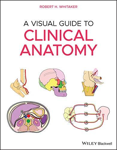
A Visual Guide to Clinical Anatomy
Robert H. Whitaker, MA, MD, MCHIR, FRCS
University of Cambridge Cambridge, UK
This edition first published 2021 © 2021 Robert H. Whitaker. Published 2021 by John Wiley & Sons Ltd.
All rights reserved. No part of this publication may be reproduced, stored in a retrieval system, or transmitted, in any form or by any means, electronic, mechanical, photocopying, recording or otherwise, except as permitted by law. Advice on how to obtain permission to reuse material from this title is available at http://www.wiley.com/go/permissions.
The right of Robert H. Whitaker to be identified as the author of this work has been asserted in accordance with law.
Registered Office(s)
John Wiley & Sons, Inc., 111 River Street, Hoboken, NJ 07030, USA
John Wiley & Sons Ltd, The Atrium, Southern Gate, Chichester, West Sussex, PO19 8SQ, UK
Editorial Office
9600 Garsington Road, Oxford, OX4 2DQ, UK
For details of our global editorial offices, customer services, and more information about Wiley products visit us at www.wiley.com.
Wiley also publishes its books in a variety of electronic formats and by print‐on‐demand. Some content that appears in standard print versions of this book may not be available in other formats.
Limit of Liability/Disclaimer of Warranty
The contents of this work are intended to further general scientific research, understanding, and discussion only and are not intended and should not be relied upon as recommending or promoting scientific method, diagnosis, or treatment by physicians for any particular patient. In view of ongoing research, equipment modifications, changes in governmental regulations, and the constant flow of information relating to the use of medicines, equipment, and devices, the reader is urged to review and evaluate the information provided in the package insert or instructions for each medicine, equipment, or device for, among other things, any changes in the instructions or indication of usage and for added warnings and precautions. While the publisher and authors have used their best efforts in preparing this work, they make no representations or warranties with respect to the accuracy or completeness of the contents of this work and specifically disclaim all warranties, including without limitation any implied warranties of merchantability or fitness for a particular purpose. No warranty may be created or extended by sales representatives, written sales materials or promotional statements for this work. The fact that an organization, website, or product is referred to in this work as a citation and/or potential source of further information does not mean that the publisher and authors endorse the information or services the organization, website, or product may provide or recommendations it may make. This work is sold with the understanding that the publisher is not engaged in rendering professional services. The advice and strategies contained herein may not be suitable for your situation. You should consult with a specialist where appropriate. Further, readers should be aware that websites listed in this work may have changed or disappeared between when this work was written and when it is read. Neither the publisher nor authors shall be liable for any loss of profit or any other commercial damages, including but not limited to special, incidental, consequential, or other damages.
Library of Congress Cataloging‐in‐Publication Data
Names: Whitaker, R. H. (Robert H.), author, illustrator.
Title: A visual guide to clinical anatomy / Robert H. Whitaker.
Description: Hoboken, NJ : Wiley-Blackwell, 2021.
Identifiers: LCCN 2020027865 (print) | LCCN 2020027866 (ebook) | ISBN 9781119708100 (paperback) | ISBN 9781119708162 (adobe pdf) | ISBN 9781119708148 (epub)
Subjects: MESH: Body Regions–anatomy & histology | Clinical Medicine | Pictorial Work
Classification: LCC QM25 (print) | LCC QM25 (ebook) | NLM QS 17 | DDC 611.0022/2–dc23
LC record available at https://lccn.loc.gov/2020027865
LC ebook record available at https://lccn.loc.gov/2020027866
Cover Design: Wiley
Cover Image: Courtesy of Robert H. Whitaker
Set in 10/12pt Sabon by SPi Global, Pondicherry, India
10 9 8 7 6 5 4 3 2 1
Preface, vii
Robert H. Whitaker
Foreword, ix
By Professor Harold Ellis
Foreword, xi
By Professor Sir Roy Calne
About the Author, xiii
SECTION 1: UPPER LIMB, 1
1.1 General Anatomy, 2
1.2 Shoulder and Arm, 13
1.3 Axilla, Brachial Plexus and Nerve Lesions, 31
1.4 Elbow and Forearm, 52
1.5 Wrist and Hand, 62
SECTION 2: LOWER LIMB, 81
2.1 Nerves, Vessels and Lymphatics, 82
2.2 Gluteal Region, Hip and Thigh, 99
2.3 Knee and Popliteal Fossa, 116
2.4 Lower Leg, 125
2.5 Ankle and Foot, 132
SECTION 3: THORAX, 147
3.1 Surface Anatomy and Breast, 148
3.2 Mediastinum, Thoracic Inlet, Diaphragm and Lymphatics, 159
3.3 Heart and Pericardium, 166
3.4 Trachea, Lungs and Oesophagus, 187
3.5 Vessels and Nerves, 200
3.6 Cross Sections, 207
SECTION 4: ABDOMEN AND PELVIS, 215
4.1 Surface Anatomy and Hernia, 216
4.2 Peritoneum, Vessels and Nerves, 230
4.3 General Bowel Anatomy and Foregut, 239
4.4 Midgut and Hindgut, 251
4.5 Biliary System, Portal System, Pancreas and Spleen, 265
4.6 Female and Male Genitourinary and Renal Tracts, 277
4.7 Pelvis and Perineum, 298
4.8 Referred Pain and Autonomics, 312
SECTION 5: HEAD AND NECK, 325
5.1 Skull Bones, Air Sinuses and Scalp, 326
5.2 Brain, Spine, Spinal Cord and Vessels, 335
5.3 Intracranial Haemorrhages, 350
5.4 Cranial Nerves in General and Skull Foramina, 354
5.5 Cranial Nerves I, V, VII, 364
5.6 Cranial Nerves VIII, IX, X, XI, XII, 372
5.7 Rules and Nerve Supply of Muscles, 379
5.8 Eye Visual Pathways, CN III, IV, VI, Autonomics and Oculomotor Versus Horner’s, 385
5.9 Eye Lids, Orbit, Lacrimal Gland, Retina and Optic Nerve, 391
5.10 Eye Light and Near Reactions, Eye Movements and Pupil Control, 398
5.11 Outer, Middle and Inner Ear, 406
5.12 Mouth and Mandible, 413
5.13 Nose and Face, 424
5.14 Neck Fascia, Vessels and General Anatomy, 430
5.15 Neck Triangles, Muscles, Thyroid and Parathyroid Glands, 440
5.16 Parotid and Submandibular Glands, 448
5.17 Larynx and Pharynx, 452
5.18 Autonomic Nervous System, in General, Sympathetic and Parasympathetic, 464
5.19 Temporal, Infratemporal and Pterygopalatine Fossae, 481
5.20 Cavernous Sinus and other Venous Sinuses, 489
Upper Limb
1.1 General Anatomy, 2
1.2 Shoulder and Arm, 13
1.3 Axilla, Brachial Plexus and Nerve Lesions, 31
1.4 Elbow and Forearm, 52
1.5 Wrist and Hand, 62
A Visual Guide to Clinical Anatomy, First Edition. Robert H. Whitaker.
1.1 General Anatomy
SURFACE ANATOMY
Function of any bone:
To give form
For muscle attachments
Movement
Protection of internal organs
Metabolic
Calcium, phosphorus Haemopoiesis
CLASSIFICATION OF JOINTS 1
FIBROUSCARTILAGINOUS
Skull sutures
Interosseous membranes
Inferior tibio bular
11th, 12th costotransverse
Primary
Costochondral
1st sternochondral
Spheno-occipital
Secondary
Midline symphyses
Intervertebral
Hyaline cartilage
SYNOVIAL
ATYPICAL SYNOVIALTYPICAL SYNOVIAL
Articular surface covered with brocartilage
Tempormandibular
Sternoclavicular
Acromioclavicular
2-7 Sternochondral
Articular surface covered with hyaline cartilage
All other synovial joints
PLANE
Tarsus and carpus
Gliding
CLASSIFICATION OF JOINTS 2
Flexion Extension HINGE Interphalangeal
SADDLE CONDYLOID 1st Carpometacarpal
Flexion Extension Adduction Abduction Circumduction “Controlled” rotation = opposition
Head of humerus
MODIFIED HINGE
Flexion Extension Rotation with flexed knee Knee
MOVEMENTS IN SYNOVIAL JOINTS
PIVOT
Both ends of radius Atlanto-axial
Rotation (Uni-axial)
LANDMARKS AROUND SHOULDER
Acromion
Cephalic vein in deltopectoral groove Deltoid
CONDYLOID Metacarpophalangeal
Flexion Extension Adduction Abduction Circumduction
BALL AND SOCKET Hip Shoulder (Sternoclavicular and Talocalcaneonavicular many features of one)
Flexion
Extension Adduction
Abduction
Circumduction “Free” rotation
Coracoid process of scapula
Clavicle
Pectoralis major
Palpable around the shoulder are:
The acromion
The head of the humerus
The corocoid process
The clavicle
Upper trunk over rst rib
Median over brachial artery
Ulnar behind medial epicondyle
Supraclavicular
Median between palmaris longus and exor carpi radialis
Ulnar lateral to pisiform
Extensor pollicis longus
Extensor pollicis brevis
Abductor pollicis longus
POSITION OF RELATIVELY SUPERFICIAL NERVES IN UPPER LIMB
Super cial branch of the radial nerve palpable over tendon of extensor pollicis longus
Cephalic vein
PALPABLE STRUCTURES IN THE UPPER LIMB
Both superior to deltopectoral groove
Coracoid process Lesser tuberosity
Acromioclavicular joint
Medial and lateral epicondyles
Olecranon
Head of radius
Anconeus (posterior to olecranon)
Radial and ulnar styloid processes
Dorsal (Lister’s) tubercle of radius
Hook of hamate
Biceps tendon and aponeurosis
Brachial, radial and ulnar pulses
VULNERABLE NERVES IN THE ARM
Acromion
Axillary nerve
Posterior axilla
Radial nerve
Lateral epicondyle
RADIAL NERVE
2/3 1/3
Passes from where the posterior axilla meets the arm to a point 2/3 down a line from acromion to the lateral epicondyle then it passes anterior to the lateral epicondyle
LEFT SCAPULA
T7
Rib 8
Covers half the ribs 2-7 8th rib is rst below Upper border at T2 Medial spine at T3 Lower border at T7
T2
T3
DERMATOMES IN THE OUTSTRETCHED UPPER LIMB (Anterior view)
UPPER LIMB DERMATOMES
ANTERIORPOSTERIOR
Note the axial lines that separate non-consecutive dermatomes
C4
C5
C6
T2
T1
C8
C7
C4
C5
C6
C7 C 8 T1
T2T3
C4
C5
C6
C7 C 8 T1 T2 T3
UPPER LIMB DERMATOMES
Supraclavicular nerves (cervical plexus)
Upper lateral cutaneous nerve of arm (axillary)
Lower lateral cutaneous nerve of arm (radial)
Lateral cutaneous nerve of forearm (musculocutaneous)
ANTERIOR
Intercostobrachial
Medial cutaneous nerve of arm (medial cord)
Medial cutaneous nerve of forearm (medial cord)
Note: The axial lines that separate non-consecutive dermatomes
CUTANEOUS NERVES OF THE UPPER LIMB
Upper lateral cutaneous nerve of arm (axillary C5,6)
Lower lateral cutaneous nerve of arm (radial C5,6)
Lateral cutaneous nerve of forearm (musculocutaneous C5,6)
Median (C6,7,8)
POSTERIORANTERIOR
Supraclavicular (C3,4)
Intercostobrachial (T2)
Medial cutaneous nerve of arm (C8,T1)
Post cutaneous nerve of arm and forearm (radial C5,6,7,8)
Medial cutaneous nerve of forearm (radial C5,6,7,8)
Ulnar (C8,T1)
Upper lateral cutaneous nerve of arm (axillary C5,6)
Lower lateral cutaneous nerve of arm (radial C5,6)
Lateral cutaneous nerve of forearm (musculocutaneous C5,6)
Radial (C6,7,8)
Lateral cordMedial cordPosterior cord
C4
Supraclavicular nerves (cervical plexus)
C5
Upper lateral cutaneous nerve of arm (axillary)
C5
Lower lateral cutaneous nerve of arm (radial) C6
Lateral cutaneous nerve of forearm (musculocutaneous)
C6,7 Radial nerve
C7
C8
T1
APPROXIMATE DERMATOMES OF UPPER LIMB
Medial cutaneous nerve of forearm T1 Medial cutaneous nerve of arm
Ulnar nerve and medial cutaneous nerve of forearm
Palmar and digital branches of median and ulnar nerves
Above the elbow the anterior dermatomes reach round posteriorly to meet an axial line. Supply posteriorly is by the posterior cutaneous nerve of arm. Below the elbow supply is by the posterior cutaneous nerve of forearm (C6,7)
SEGMENTAL NERVE SUPPLY IN UPPER LIMB
SHOULDER:
Flexion/abduction/lateral rotation C5 Extension/adduction/medial rotation C6,7,8
ELBOW:
Flexion (biceps re ex)
C5,6
Extension (triceps re ex) C6,7,8
FOREARM:
Pronation
Supination
WRIST:
Flexion/extension
FINGERS/THUMB (LONG TENDONS):
Flexion/extension
HAND (SMALL MUSCLES):
All movements
C7,8
C6
C7,8
C7,8
T1
Extension
Adduction
Medial rotation
C6,7,8
LIMB TENDON REFLEXES
Flexion
Abduction
Lateral rotation
Pronation
UPPER LIMB MYOTOMES
Small muscles of hand:
1, 2 (S)
4 (L)
6 (C)
7, 8 (C)
C7,8C5,6
C7,8
Anterior and posterior circumflex humeral
Profunda brachii
Radial
Posterior interosseous
ARTERIAL SUPPLY OF UPPER LIMB
Subclavian (to outer border of 1st rib)
Axillary (to lower border of teres major)
Pectoralis minor
Teres major
Lateral thoracic
Brachial
Ulnar
Common interosseous
Anterior interosseous
Ulnar
Deep palmar arch
Superficial palmar arch
ARTERIAL ANASTOMOSIS AROUND RIGHT ELBOW
Median nerve
Anastomotic branch
Profunda brachii
Lateral intermuscular septum
Interosseous recurrent Radial recurrent
Radial
Posterior interosseous (passing above interosseous membrane)
Ulnar nerve
Lower border of teres major
Superior ulnar collateral
Inferior ulnar collateral
Medial intermuscular septum
Anterior ulnar recurrent
Posterior ulnar recurrent
Ulnar
Anterior interosseous
Axilla
Median nerve
Lateral Medial brachial artery
Cubital fossa
Usual arrangment but in 15% the nerve passes behind artery
Mnemonic for anastomotic vessels:
Pus - Profunda
Spells - Superior ulnar collateral
Nasty - Nutrient
Infection - Inferior ulnar collateral
Deltoid
Cephalic vein
Pierces the clavipectoral fascia at upper end of the deltopectoral groove to enter axillary vein
Radial artery
Pulse. Lateral to the tendon of radialis
exor carpi
SUPERFICIAL VEINS AND PULSES IN UPPER LIMB
Pectoralis major
Brachial artery
Pulse. In the cubital fossa, lateral to the median nerve
Medial epicondyle
Basilic vein
Commencement of the basilic vein (medial side)
Pierces the fascia in the medial mid arm to join the venae commitantes which together, at the inferior border of teres major, become the axillary vein
Dorsal venous arch
Commencement of the cephalic vein (lateral side)
POSTERIORANTERIOR
lymphatics
LYMPHATIC DRAINAGE IN UPPER LIMB Super cial
lymphatics
Lymphatics from palm of hand and anterior forearm follow basilic vein to lymph nodes in the cubital fossa and then pass deep, to follow the deep veins to the lateral axillary and then central nodes
Lymphatics from dorsum of hand, posterior forearm and posterior arm follow cephalic vein to supraclavicular or infraclavicular lymph nodes
AXILLARY LYMPH NODES
Apical
Infraclavicular
Posterior
Supratrochlear
75% of lymphatics from the breast drain to axillary nodes. Others to internal thoracic, abdominal nodes or to other breast
Subclavian lymph trunk
Mnemonic for axillary lymph nodes:
A - Anterior
P - Posterior
I - Infraclavicular
C - Central
A - Apical
L - Lateral
1.2 Shoulder and Arm
SHOULDER JOINT
(GLENOHUMERAL)
Blood:
Circum ex humeral arteries
Nerves: Subscapular, suprascapular, axillary (Hilton's law)
Bursae: Subscapular, subacromial, infraspinatus, supraspinatus
Stability: Bones (poor), Capsule (relatively poor), Muscles +++, ligaments +++
Support: Rotator cu (subscapularis, supraspinatus, infraspinatus, teres minor), long head biceps, triceps in abduction, muscles from chest to arm
SHOULDER JOINT
(GLENOHUMERAL)
LIGAMENTS: Anterior
1,2,3: Glenohumeral
Anterior: superior, middle, inferior (weak thickenings of capsule)
4: Subscapularis
5: Teres major
6: Supraspinatus
7: Short head biceps
8: Pectoralis minor
Opening of subscapular bursa
Coraco-acromial (strong ++)
Subacromial bursa (large)
Coracohumeral (strong)
Transverse humeral (intertubercular)
Shallow glenoid fossa - deepened by glenoid labrum. Synovial, Ball and socket. Humeral head is 1/3 hemisphere
allow wide abduction, exion and extension.
Capsule: Strong and taut superiorly (anti-sag), inferiorly lax and inserted lower to Synovium: Envelops biceps tendon, communicates with bursae anteriorly and posteriorly
Long head of biceps
SHOULDER JOINT AND ROTATOR CUFF MUSCLES
Capsule
Glenoid labrum
Lax capsule inferiorly
Synovial sheath of biceps
Rotator Cu Muscles:
Subscapularis (anterior)
Infraspinatus (posterior)
Teres minor (posterior)
Supraspinatus (superior)
All blend with capsule of shoulder joint
Lax capsule inferiorly allows dislocation of head of humerus inferiorly and usually anteriorly
The tendon of the long head of biceps lies within the capsule but not within the synovial membrane. It attaches to the supraglenoid tubercle
SUBACROMIAL BURSA AND PAINFUL ARC SYNDROME
Subacromial bursa
Rotator cu (Tendon of supraspinatus)
During abduction the greater tuberosity impinges on the supraspinatus tendon at its point of least blood supply beneath the acromion
Abduction
Acromion
Greater tuberosity
Inferior capsule
Coracoacromial ligament (arch)
ACROMIOCLAVICULAR JOINT
Acromioclavicular ligament
Conoid
Coracoclavicular ligament
Trapezoid
Synovial
Atypical
Thick superior capsule (acromioclavicular ligament)
Incomplete fibrocartilaginous disc in upper joint
Strong coracoclavicular ligament
Nerve: Lateral supraclavicular (C4)
Movements: Gliding (passive) and 20° of rotation of scapula
ACROMIOCLAVICULAR JOINT AND CORACOCLAVICULAR LIGAMENTS
Coracoclavicular ligaments
ConoidTrapezoid
Acromioclavicular ligament
Costoclavicular ligament
ACROMIOCLAVICULAR JOINT
Synovial, Atypical, Plane (passive gliding)
Thick superior capsule (acromioclavicular ligament)
Incomplete fibrocartilaginous disc in upper joint
Strong coracoclavicular ligament
Nerve: Lateral supraclavicular (C4)
The acromioclavicular joint can dislocate but forces usually pass through coracoclavicular ligament to the clavicle then via the costoclavicular ligament to the manubrium and sternum. Excessive force will fracture the clavicle
Movements: gliding (passive) and 20o of rotation of scapula
Upper Limb: 1.2 Shoulder and Arm
STERNOCLAVICULAR JOINT
Capsule
Costoclavicular ligament (anterior and posterior bres)
Synovial
Interclavicular ligament
Atypical ( brocartilage on joint surfaces)
Fibrocartilaginous disc dividing it into 2 cavities
Manubrial surface is concave
All the features of a ball and socket joint
Disc attached to capsule, acts as shock absorber
Capsule thick above and posteriorly
Fulcrum at costoclavicular ligament
Clavicle rotates 40 degrees
Nerves: supraclavicular (C3,4)
Clavicle elevated At distal end, medial end depresses
At rest
Clavicle depressed at distal end, medial end elevates
Disc
1st costal cartilage with primary cartilaginous joint at either end
Ligaments:
Thickening of capsule (above and posteriorly) = anterior and posterior sternoclavicular ligaments
Interclavicular
Costoclavicular (strong)
MOVEMENTS AT THE STERNOCLAVICULAR JOINT
Distal clavicle is pushed forwards, medial end retracts
Distal clavicle pushed posteriorly, medial end protrudes
In view of all the movements described here and the ability to pivot on the costoclavicular ligament, the sternoclavicular joint can be regarded has having many the features of a ball and socket joint
Distal and medial clavicle can rotate together
Upper Limb: 1.2 Shoulder and Arm
Subscapularis
Internal rotation only
Crosses anteriorly at level of joint
Infraspinatus
Teres minor
External rotation only
Crosses posteriorly at level of joint
Teres major
Latissimus dorsi
Pectoralis major
Internal rotation and adduction
Crosses both anteriorly and inferior to level of joint
Combinations of several muscles
PRINCIPLES OF MOVEMENTS AT JOINTS - SHOULDER
Supraspinatus
Deltoid
Abduction only
Crosses superorly at level of joint
External rotation and adduction
Crosses both posteriorly and inferior to level of joint
SUPRASPINATUS
Origin: 3/4 supraspinous fossa and upper spine of scapula
Insertion: Superior facet of greater tuberosity of humerus and joint capsule
Action: Abducts and stabilises shoulder
Nerve supply: Suprascapular (C5,6 upper trunk)
Supraspinatus
Infraspinatus
INFRASPINATUS
Origin: Medial 3/4 infraspinous fossa and intermuscular septum
Insertion: Medial facet of greater tuberosity of humerus and joint capsule
Action: Lateral rotation and stabilisation of shoulder
Nerve supply: Suprascapular (C5,6 upper trunk)
Upper Limb: 1.2 Shoulder and Arm
DELTOID
Origin: Lateral third clavicle, acromion, spine of scapula as far as deltoid tubercle of scapula
Insertion: Deltoid tubercle of humerus
Action: Abducts arm. Anterior bres ex and medially rotate. Posterior bres extend and laterally rotate
Nerve supply: Axillary (C5,6 posterior cord)
Anterior bres
Posterior bres
Special note: There is little doubt that the lateral bres of deltoid help supraspinatus to initiate abduction of the shoulder but from the images above it is obvious that the anterior and posterior bres cross the joint more e ectively as soon as abduction begins and thus give more power to abduction
RHOMBOID MAJOR
Origin: Spines of T2-5 and supraspinous ligaments
Insertion: Lower half posterior medial scapula
Action: Retracts and rotates scapula to rest position
Nerve supply: Dorsal scapular (C5 from root)
RHOMBOID MINOR
Origin: Spines C7 and T1 and lower ligamentum nuchae
Insertion: Posteromedial scapula level with spine
Action: Retracts and rotates scapula to rest
Nerve supply: Dorsal scapular (C5 from root)
LEVATOR SCAPULAE
Origin: Posterior tubercles of transverse processes C1-4
Insertion: Upper medial border of scapula
Action: Raises medial end of scapula
Nerve supply: Dorsal scapular (C5 root)
Levator scapulae
Rhomboid minor
Rhomboid major
Upper Limb: 1.2 Shoulder and Arm
SUBSCAPULARIS
Origin: Medial 2/3 subscapular fossa
Insertion: Lesser tuberosity of humerus, half medial lip of bicipital groove, shoulder joint capsule
Action: Medial rotation stabilisation of shoulder
Nerve supply: Upper and lower subscapular (posterior cord C6,7)
Clavicular head
PECTORALIS MAJOR
Origin: Medial half of clavicle
Insertion: Anterior lamina (of trilaminar insertion) and lateral lip of bicipital groove, deep fascia, anterior lip of deltoid tuberosity
Sternocostal head
Origin: Anterior and lateral manubrium, body of sternum, aponeurosis of external oblique, upper 7 costal cartilages (not always 1st or 7th)
Insertion: Manubrial bres to intermediate lamina. Sternocostal bres to posterior lamina with highest bres into capsule of shoulder
Action: Flexion, adduction, internal rotation
Nerve supply: lateral and medial pectoral nerves Anterior
Upper Limb: 1.2 Shoulder and Arm
PECTORALIS MINOR
Origin: Ribs 3,4,5
Insertion: Coracoid process
Action: Protracts scapula with serratus anterior
Nerve supply: Medial and lateral pectoral nerves
TERES MAJOR
Origin: Oval area on lower third lateral side of inferior angle of scapula
Insertion: Medial lip of bicipital groove
Action: Medial rotation, adduction, stabilisation of shoulder
Nerve supply: Lower subscapular (posterior cord C5,6)
TERES MINOR
Origin: Middle third lateral border of scapula
Insertion: Inferior facet of greater tuberosity and joint capsule
Action: Lateral rotation and stabilisation of shoulder
Nerve supply: Axillary nerve (C5,6 (posterior cord)
Posterior
Anterior