Foot Surgery Viewed Through the Prism of Comparative Anatomy
From Normal to Useful
Cyrille Cazeau
First published 2020 in Great Britain and the United States by ISTE Ltd and John Wiley & Sons, Inc.
Apart from any fair dealing for the purposes of research or private study, or criticism or review, as permitted under the Copyright, Designs and Patents Act 1988, this publication may only be reproduced, stored or transmitted, in any form or by any means, with the prior permission in writing of the publishers, or in the case of reprographic reproduction in accordance with the terms and licenses issued by the CLA. Enquiries concerning reproduction outside these terms should be sent to the publishers at the undermentioned address:
ISTE Ltd
John Wiley & Sons, Inc.
27-37 St George’s Road 111 River Street London SW19 4EU Hoboken, NJ 07030
UK USA
www.iste.co.uk
www.wiley.com
© ISTE Ltd 2020
The rights of Cyrille Cazeau to be identified as the author of this work have been asserted by him in accordance with the Copyright, Designs and Patents Act 1988.
Library of Congress Control Number: 2020940426
British Library Cataloguing-in-Publication Data
A CIP record for this book is available from the British Library
ISBN 978-1-78630-604-3
1.4.
1.5.
1.6.
Chapter 2. The Aim of Surgery: Restore
2.1.
2.3.
2.3.1.
2.4.
2.4.1.
2.5.
2.5.2.
2.5.3.
2.5.4.
2.5.5.
2.5.6.
2.6.
2.7.
2.7.1.
2.7.2.
2.7.3.
2.7.4.
2.7.5.
2.7.6.
2.8.
2.8.1.
2.8.2.
2.9.
2.9.1.
2.9.2.
2.9.3.
2.9.4.
2.9.5.
Chapter 3. Rapid Healing Through Immediate Use
3.1.
3.2.
3.3.
3.3.1.
3.3.2.
3.3.3.
3.3.4.
3.3.5.
3.3.7.
3.3.8.
3.3.9.
3.3.10.
3.3.11.
3.4.
3.5.
3.6.
3.6.1.
3.6.2.
3.6.3.
3.6.4.
3.6.5.
3.6.6.
3.7.
Foreword
The fact that an orthopedic surgeon, a foot specialist, integrates comparative anatomy into his or her surgical procedures to repair an impaired function is not self-evident. For any paleoanthropologist accustomed to analyzing the bone fragments of our ancestors and their locomotor system, comparative anatomy is common practice; it is an essential point of view from which we base our functional interpretations and evolutionary hypotheses. It is therefore with immense pleasure, a certain pride and not without trepidation that I accepted Dr Cyrille Cazeau’s invitation to introduce his work, by evoking some convergences between the comparative approach and the object of study. While it is indeed comparative anatomy and locomotor function that brought us together, comparative anatomy – which Cyrille Cazeau has been working with for many years – gives it a central place in his teachings and workshops, questions it and nourishes it with encounters, such as the one with Pau Golano, an exceptional anatomist whom Cyrille Cazeau introduced me to and I got to know better, to the point of integrating it into his practice of percutaneous foot surgery. For the surgeon, comparing is to understand the human specificity, to distinguish what is essential to the function – useful – from what is simply accessory and, if necessary, can be sacrificed. My approach to the fossil hominid foot, which is unique because it is often fragmented and atypical in shape, owes much in its construction to my encounter with Cyrille Cazeau. This normal and useful element that he questions in his practice has
indeed become the central notion in my practice as a paleoanthropologist when it comes to functionally interpreting locomotor anatomy that is often very partial, whereas the only well-known and usable bipedal reference system was the human functional anatomy model, which is so particular. It is in response to this questioning that we sought to study other bipedal reference frames, more particularly non-human primates. So for us, comparing is also understanding non-human specificities. In our approaches to a paleontological object, we now see that bipedalism is not the proper domain of man: it is carried out in a varied manner in our closest cousins, catarrhine primates, whose anatomy seems to be “made” for other mechanically different locomotor modes. This is also how we understand that other efficient (and not transitional) bipeds could have existed in our distant ancestors, relying on the numerous paleoanthropological discoveries of the last two decades and the unexpected anatomical variation that they have revealed in hominids over the last 7 million years. At the time of publication, the discovery of a new primate species, Danuvius guggenmosi, in the Hammerschmiede deposit in present-day Bavaria, provides evidence of a potentially much older form of bipedalism. Its age – 11.6 million years –before the supposed differentiation date of the first hominids (one of the commonly accepted features of which was the practice of a habitual bipedalism), drives us to continue this comparative work to better understand human and non-human specificities. Beyond the paleontological object, the functional interpretation that is proposed and its evolutionary implications, it is essential that our comparative and paleoanthropological work be useful. The fact that this usefulness manifests itself in the daily life of each person is most often a very distant perspective, rarely palpable. With Foot Surgery Viewed Through the Prism of Comparative Anatomy, Cyrille Cazeau shows us that this perspective is not so distant and, in so doing, invites those working in fundamental and applied research, anatomists, zoologists, physiologists, biomechanists, orthopedists, surgeons, paleoanthropologists, etc. to engage in more discussion to achieve something useful. Finally, Cyrille Cazeau brings together multiple scales, from macroscopic anatomical structures to the bone’s microstructure, from millions of years of evolution to the “instantaneous” surgical procedure, towards an application that aims to improve an impaired function. This book, which questions the notion of an
Foreword xi
anatomical standard, to give full scope to the useful and functional, is therefore not a normal book. It is a book that will be useful to everyone in his or her practice.
Gilles BERILLON Paleoanthropologist Director of Research
at the CNRS
Laboratoire Histoire naturelle de l’Homme Préhistorique
Musée de l’Homme, Paris
July 2020
Introduction
Orthopedic surgery aims to improve the function of the musculoskeletal system. It aims to repair, rebuild or replace damaged anatomical elements with a prosthesis.
Physical actions in orthopedic surgery always lead to anatomical sacrifices, the benefits of which must outweigh the disadvantages.
To do this, we must differentiate between the anatomical elements that constitute our original anatomical normality and the elements that are truly useful for carrying out a given function, such as permanent terrestrial human bipedalism.
To help differentiate between “normal” and “useful” elements, it is useful to compare similar functional anatomy models, whose differences highlight the specificity of each.
To illustrate this approach, we take the study of the foot as an example. The human model is compared to those with the closest phylogeny: non-human primates and fossil hominids.
Thus, studying anatomical homologies allows us to rank the different anatomical players according to their usefulness for a given function. Some considered as “normal” define the anatomical normality acquired during ontogenesis, while others, essential to achieving a function, are described as “useful”.
The surgical consequences are direct in the choices made to correct a forefoot deformity such as hallux valgus. A choice means determination, and determination is negation. We thus implicitly deny everything that does not fall within its definition. Surgery requires that we make a determination that we decide on a procedure, an action by rejecting other possible ones. This is where the quest for the most efficient, the most correct procedure to achieve the best possible result lies, in this interaction between determination and negation.
Classical surgery mainly focuses on restoring ad integrum defining original normality. The study of comparative functional anatomy helps to define surgery that will restore the specific useful function, even if it is accompanied by anatomical sacrifices. Therefore, the preliminary work consists of dividing and then ranking the usefulness of the anatomical players for a given species, in this case humans.
The purpose of the surgical act, which always constitutes a physical aggression, a transgression, is to propose in return a result leading, according to Aristotle, the patient as close as possible to recovery. To do this, the injured organ must be put back into its usual working context as soon as possible. We propose a concept – that of percutaneous surgery – composed of linked and inseparable elements. It is advisable to correct the deformations in the three planes of space through minimal surgical approaches, to offer reliable mechanical stability before any biological consolidation allowing immediate complete support and to use an anesthesia technique that avoids the painful postoperative storm which follows traditional techniques.
The benefit, therefore, is faster functional recovery, which is vital for a better outcome.
Comparison: The Key to Understanding Human Specificity
1.1. Comparison, a source of clarity
It is difficult to understand the specificity of an object or a function, if we do not compare it with a model that resembles it. Highlighting differences will allow us to grasp their distinctiveness. The smaller the difference, the more difficult it is to detect if it is isolated from a similar environment, but the more it characterizes the specificity of the object being studied.
This easy-to-understand approach when comparing color, from blue to green via emerald, for example, is more complex when it comes to functional anatomy models. Our understanding of specificity cannot be dissociated from the effort of classification and the establishment of phylogeny.
Indeed, by carefully establishing derived and ancestral characteristics, it is possible to establish differences and then classify according to the principle of parsimony. According to Aristotle, let us “search for relations between apparently independent things and search for similarities between things that are dissimilar in the eyes of the common man”. Knowing things makes it possible to see them acutely; verbalizing them makes it possible to think about them.
1.2. Different models of the foot and ankle throughout evolution
Locomotion involves a variety of movements adapted to different environments and lifestyles.
Identical morphological organization and processes common to all vertebrates are implemented, but thanks to original anatomical elements, animals can move with highly sophisticated means and respond, each in their own way, to the constraints of their environment.
Among the many elements involved in the musculoskeletal system’s function, the foot and ankle (crurotarsal or talocrural joint) were chosen because they undergo the most spectacular and elaborate transformations during adaptation to different types of locomotion: running, trotting, tree climbing, etc. Mammals have anatomical adaptations that reflect their specific mode of locomotion.
1.2.1. Evolution of the chiridial limb
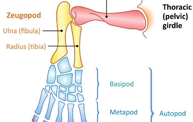
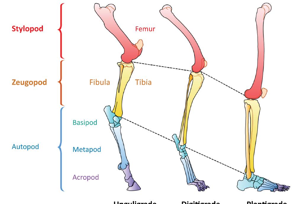
The chiridial limb represents the general limb structure of tetrapods. There are significant differences between mammalian classes, but the general pattern is identical, as shown in Figure 1.1.
The foot represents the autopod (Figure 1.1); succeeding the leg sector which constitutes the zeugopod. The autopod consists of three parts: the tarsus (basipod), the metatarsus (metapod) and the phalanxes (acropod).
Figure 1.1. The chiridial limb: general structure of a tetrapod’s limb. For a color version of the figure, see www.iste.co.uk/cazeau/foot.zip
The typical shape is pentadactyl and almost always has a locomotor function.
When our gait is increased or semi-increased, the foot is plantigrade, with five complete segments numbered from 1 to 5, from the tibial (medial) side to the fibular (lateral) side. In quadrupedal gait, the hands and feet have similar functions. The number of segments is reduced, four in the digitigrade of carnivores and even less in unguligrades.
Let us quickly put the tarsus and the zeugo-autopodal joint into context.
The appearance of the chiridial limb in the Late Devonian period (370 million years ago) was a fundamental acquisition that enabled the transition from aquatic to terrestrial life. Anatomically speaking, it was the transition from an aquatic fin to a limb that enabled the conquest of continents. Seventy million years after its alleged disappearance, the famous missing link, the coelacanth fish, was caught in 1938 in the Comoros. Its fin seemed to have the anatomical shape of a section with the chiridial limb.
The structure is as follows (Figure 1.1): the proximal segment or stylopod is represented by the arm (humerus) or thigh (femur), the middle segment or zeugopod is the forearm (radius and ulna) or leg (tibia and fibula) and the distal segment or autopod, consisting of three parts, is the hand or foot. The basipod corresponds to the tarsus (and the carpus), the metapod to the metatarsus (metacarpus) and the acropod to the phalanxes.
Here, we are interested in the ankle (zeugo-autopodial joint). This limb has maintained its general structure but has evolved differently according to lineages.
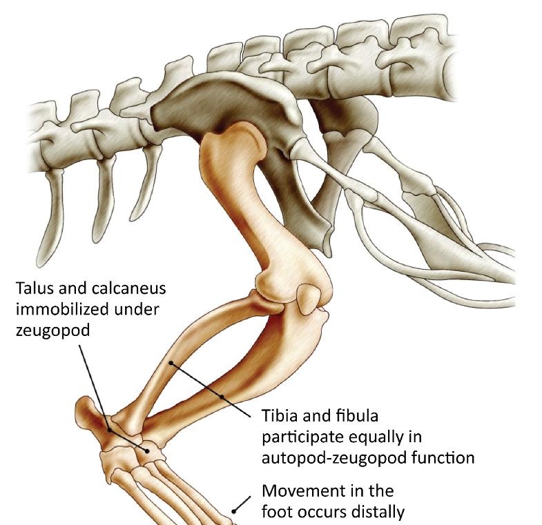
The ancestors of the first amphibians (labyrinthodontia) were known since the Devonian period. They had an autopod made of multiple parts whose main center of mobility was the joint spaces between the basipod and the metapod (Chopart).
During evolution, numerous fusions took place; a talus and a calcaneus were observed in the whole group at the origin of the mammals, turtles, reptiles and birds (amniotes).
Then, two main developments occurred; the first group came to be represented by turtles (chelonians), most reptiles and birds, and the second by mammals.
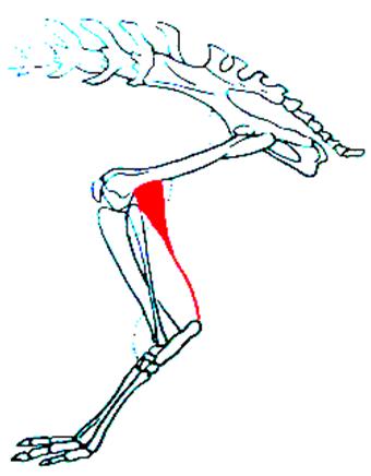
In the first, the talus-calcaneus pair is immobilized on the zeugopod, the fibula participates as much as the tibia in the function and the foot’s movement is more distal in the tarsus (Figure 1.2).
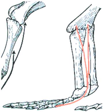
The gastrocnemius muscle remains inserted opposite the plantar fascia (Figure 1.3).
Figure 1.2. Lower limb of a reptile (crocodile)
Figure 1.3. Insertion of gastrocnemius on plantar fascia below mediotarsal joint. For a color version of the figure, see www.iste.co.uk/cazeau/foot.zip
In mammals, the functional joint is located between the zeugopod and the basipod. The talus is superimposed on the calcaneus which develops the sustentaculum tali, the fibula participates much less in the joint and regresses and the insertion of the gastrocnemius muscle is transferred to the calcaneal tuberosity, effectively increasing its strength (Figure 1.4).
Figure 1.4. Insertion of gastrocnemius on calcaneus. For a color version of the figure, see www.iste.co.uk/cazeau/foot.zip
Anatomical adaptations are then made according to the type of locomotion. These concern the entire limb, but the autopod undergoes the most significant transformations (Figure 1.5).
Figure 1.5. Transformation of lower limb and, in particular, the autopod to match the type of locomotion. For a color version of the figure, see www.iste.co.uk/cazeau/foot.zip
Adaptation to quadruped running was achieved through three types of simultaneous modifications: lengthening of the limbs (especially at the expense of the metapod), raising of the autopod and progressive reduction in the number of fingers. The same applies to the adaptations for jumping, tree climbing, flying or when returning to aquatic life.
1.2.2. The human model
Following this general description, we will focus on the human model from now on.
1.2.2.1.
Bone factors for transverse ankle stabilization
The transverse stabilizing factors of the ankle concern the bones and are related to the following:
– shape of the talus: extended very transversely, the grooves are wide and the pulley (its anatomical shape) is well developed from front to back, giving a wide stance in an erect position whatever the degree of flexion/extension. The tarsus is wide and robust, adapted to the upright position, and the contact surface between the talus and the tibia is proportionally the most important. The bearing surface is estimated at 4.5 to 9 cm2;
– narrow mortise and tenon fitting (Figures 1.6 and 1.7): this results from the bimalleolar clamp, the two malleoli descending very low in relation to the talus;
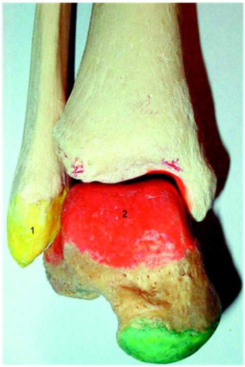
Figure 1.6. Human crurotarsal joint (front view). 1) Lateral malleolus; 2) tibial articular surface of the talus. For a color version of the figure, see www.iste.co.uk/cazeau/foot.zip
Figure 1.7. Proximal side of a human crurotarsal joint (view from below). For a color version of the figure, see www.iste.co.uk/cazeau/foot.zip
– permanent adaptation of the intermalleolar gap; it varies due to the physiology of the distal tibiofibular joint; in fact, as the talus pulley is narrower backwards than forwards, the intermalleolar gap varies by about 5 mm to maintain perfect transverse stability. Cohesion is ensured by the anterior and posterior tibiofibular ligaments at the level of the syndesmosis. Contrary to animals, the distal tibiofibular joint does not closely connect its two constituent bones, providing stability during flexion/extension; – ligament factors are also involved in stability; the powerful collateral ligament systems oppose rotational movements in the longitudinal axis. The system, which is already robust in humans and limited to short components, thickens and strengthens from carnivores to ruminants and then to equids.
1.2.2.2. Principles of anteroposterior stability and factors limiting flexion/extension
The amplitude of flexion/extension primarily depends on the extent of the proximal articular surface of the talus and the tibial surface. Other factors are bone abutments, ligament tension and, to a lesser extent, the muscle and tendon elements.
1.2.2.2.1. Factors limiting flexion
These are as follows: – bone factors: in extreme flexion, the upper surface of the talus neck rests against the anterior edge of the tibial pilon;
Lateral
– capsule and ligament factors: the posterior elements become strained, i.e. the posterior talofibular component, the deep plane of the medial collateral ligament and the posterior capsule, which is flexible.
1.2.2.2.2. Factors limiting extension
These are as follows:
– bone factors: the posterior tubercles of the talus rest against the posterior margin of the tibia, which descends lower than the anterior margin;
– capsule and ligament factors: the anterior elements become strained; anterior talofibular joint, anterior part of the deltoid ligament, anterior capsule, which is flexible;
– muscular factors: in men, the terminal tendons of the muscles in the leg area are short; their tonus is inadvertently significant, limiting joint amplitude, and the possibilities of storing some elastic energy. The muscle mass is low, favoring the erect position but not the lower limb’s swing speed. The amplitude of extension is important, as are all the articular segments of the hind limb.
1.2.3. Comparative functional anatomy
In the human model, the anatomy of the tibiotarsal joint is very different from that of other mammals, but the result is identical: exclusive flexion/extension.
Transverse stability is ensured by the dynamic quality of the bimalleolar tibiofibular syndesmosis and the short collateral ligaments.
The absence of long components, associated with the shallow tibiotalar joint grooves, has the disadvantage of creating a weakness in this joint, which is partly responsible for the high frequency of ankle sprains.
On the other hand, the absence of these long components has the advantage of not limiting the movements of the midtarsal joint, allowing the inversion of the foot, a condition of the arboreal life of primates, in particular those whose weight is carried by their hind limbs (lemurs and tarsiers).
1.3. Different functional anatomy models of bipedalism, from turkeys to humans and great apes
1.3.1. Challenges in terms of phylogeny
Bipedalism is a functional anatomy entity that includes a wide variety of biomechanical mechanisms.
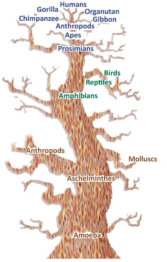
Figure 1.8. Haeckel’s phylogenetic tree placing humans at the top. For a color version of the figure, see www.iste.co.uk/cazeau/foot.zip
This concept is highly controversial and has been overvalued over the centuries, out of ignorance, belief in a divine creation or to feed the various theories of evolution, with an inseparable anthropocentric feeling of superiority, with humans at the top (Figure 1.8). It is still considered today as one of the answers to the hominization process. This may have led us to
forcibly unite in a direct and vertical phylogenetic relationship between apes and humans and to automatically place Australopithecus as an intermediate form.
Yet the human species moves according to a highly specialized, unique mode of bipedalism, very different even from that adopted by animals known phylogenetically to be the closest, i.e. the great apes, of which the bonobo is one.
The choice of a resemblance criterion is as crucial as it is arbitrary, having a direct influence on classification and phylogeny. Thus, the overrepresentation of a morphological or functional feature relative to the complete characteristics of an individual leads to erroneous conclusions.
The “bipedalism” criterion, historically overvalued, thus groups together various entities. Humans do not have exclusivity; the types vary greatly within the different animal classes – present and extinct – and notable differences can be observed among fossil hominoidea, primates and even within the genus Homo.
1.3.2. Definition of bipedalism
Different types of terrestrial progressions exist: quadrupedalism, bipedalism and crawling aptodes. What does “bipedalism” mean? According to the Latin etymology, it means walking on two feet. According to Sabine Renous, bipedal progression is characterized by the following elements: the vertebrate’s body rests entirely on repetitive synchronous or alternating movements of the hind limbs. The forelimbs, freed from being required for terrestrial locomotion, are entirely available for new activities, such as flight in birds or the non-specialized use of the hand in humans.
In the animal world, “bipedalism” generally characterizes movement on the extremities of the lower limbs. This does not presume the support, which can be terrestrial or arboreal only, or mixed. Moreover, this bipedalism can be exclusive as in humans or be integrated into a locomotor repertoire more or less varied in terms of quadrupedalism or the arboreal substrate.
Humans do not therefore have the exclusivity of bipedalism based on these definitions, and anatomy differs according to zoological groups: dinosaurs, birds, primates, bears, etc.
1.3.3. Different bipeds
1.3.3.1.
Dinosaurs
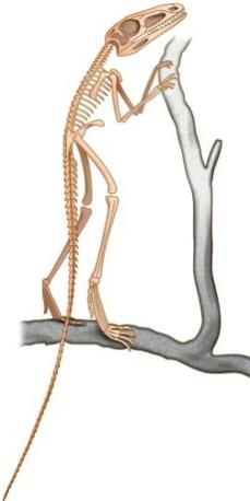
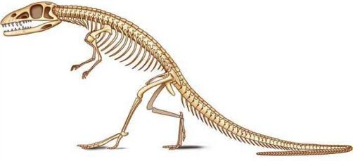
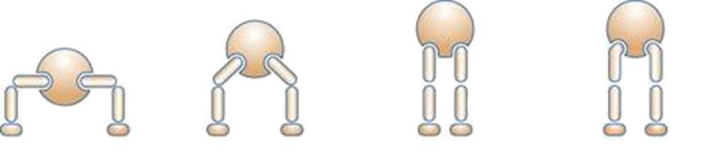
The first known forms to adopt bipedalism were some archosaur dinosaurs, which include most of the reptilian forms of the secondary era (250 to 64 million years ago) (Figure 1.9a and 1.9b), especially during the Jurassic and Cretaceous periods. Authors emphasize the anatomical particularities of the locomotor system. The classical arrangement of the reptilian limbs was then the transverse limb, still observed today in crocodiles (Figure 1.10), barely allowing the body to be lifted. The disadvantage is that considerable forces were required on the limbs to hold the trunk and belly above the ground, causing, among other things, breathing problems due to crushing of the rib cage.
Figure 1.9. Bipedal dinosaurs of the secondary era
Figure 1.10. Transition from transverse to parasagittal limb, various types of erect positions
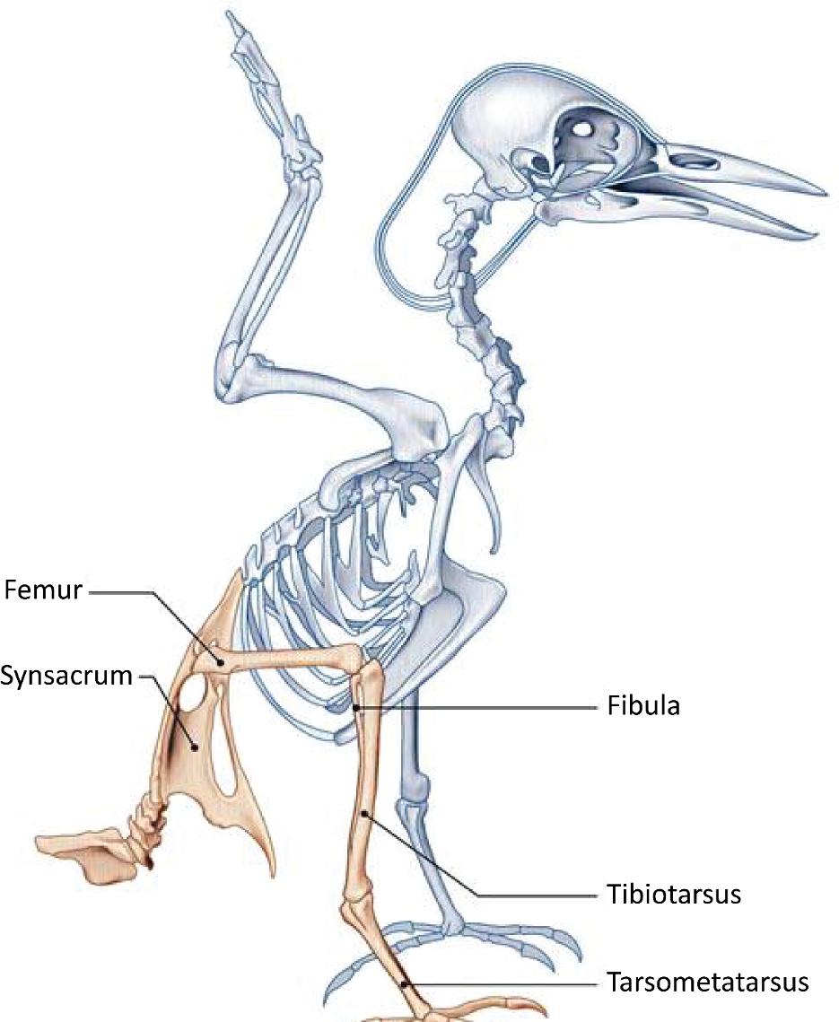
Based on this model, many archosaurs adopted a semi-vertical bipedal position, with tail development and straightening from the hind limbs. In the middle of the Triassic period, archosaurs experienced a great evolutionary form of disappearance. The evolution was achieved by adopting certain lineages of an erect posture, necessary for bipedalism. In animals that have an erect position, the lower limbs were positioned vertically under the body, the weight transmitted directly to the ground via the limbs, without exerting any leverage (minimum muscular and articular moments). In order for this change to occur, many bone and joint changes happened. Different mechanical models existed by zoologic groups and periods. At the hips, some animals (Euparkeria) were in a “semi-erect” position (Figure 1.10), with the acetabulum looking down and out, articulating on a femur that did not have a cervical portion. Others (Saurosuchus) were in the “erect on pillars” position, with the lower limbs upright and the femur without a cervical portion fitting into a downward-facing acetabulum. This likely limited the stride range and decreased the force on the hip abductor muscles. Finally, other groups adopted the “classic erect” position, with the acetabulum facing down and out, articulating with a vertical femur but off-center due to the presence of a cervical portion, resembling the current quadruped model. Anatomically, the pelvis, which supports the weight of the abdomen, developed and took on a triradiate shape. The ilium in particular developed and joined – in addition to the sacral vertebrae – with the presacral, lumbar and caudal postsacral vertebrae to form a synsacrum as in birds (Figure 1.11). The femoral heads became stronger and formed a bearing surface, with four bony processes on the femur providing numerous muscle insertions between the pelvis and the femur, as well as and especially between the tail and the femur. The tibia became thicker and the fibula atrophied.
As far as the ankle and foot are concerned, bipedalism was possible in these different groups based on different mechanical models. It may have been a “primitive mesotarsal” ankle with the characteristics of the reptilian intratarsal joint that later came to be found in birds; the distal part of the tibia merging with the posterior tarsus, the anterior tarsus with the metatarsals (Figure 1.12) and the talocalcaneal block being fused to the tibia. Mobility was present in the Chopart and Lisfranc joints. In other groups of bipedal dinosaurs, the mobile hinge was located between the talus and calcaneus (an
evolved mesotarsal ankle). In others, it was located in the tibiotalar joint, identical to that of mammals.
Figure 1.11. Bird skeleton