Flexible Bronchoscopy
Fourth Edition
Edited
by Ko-Pen Wang, MD
Division of Pulmonary and Critical Care Medicine
Johns Hopkins Bayview Medical Center
Johns Hopkins University School of Medicine
Baltimore, MD, USA
Atul C. Mehta, MD, FACP, FCCP
Lerner College of Medicine
Buoncore Family Endowed Chair in Lung Transplantation
Department of Pulmonary Medicine
Respiratory Institute
Cleveland Clinic
Cleveland, OH, USA
J. Francis Turner, Jr., MD, FACP, FCCP, FCCM
Division of Pulmonary and Critical Care Medicine
University of Tennessee Graduate School of Medicine
Knoxville, TN, USA;
National Supercomputing Institute
University of Nevada, NV, USA
This edition first published 2020 © 2020 by John Wiley & Sons Ltd.
Edition History [3e, 2012]
All rights reserved. No part of this publication may be reproduced, stored in a retrieval system, or transmitted, in any form or by any means, electronic, mechanical, photocopying, recording or otherwise, except as permitted by law. Advice on how to obtain permission to reuse material from this title is available at http://www.wiley.com/go/permissions.
The right of Ko‐Pen Wang, Atul C. Mehta, and J. Francis Turner, Jr. be identified as the authors of editorial work has been asserted in accordance with law.
Registered Office(s)
John Wiley & Sons, Inc., 111 River Street, Hoboken, NJ 07030, USA
John Wiley & Sons Ltd, The Atrium, Southern Gate, Chichester, West Sussex, PO19 8SQ, UK
Editorial Office
9600 Garsington Road, Oxford, OX4 2DQ, UK
For details of our global editorial offices, customer services, and more information about Wiley products visit us at www.wiley.com.
Wiley also publishes its books in a variety of electronic formats and by print‐on‐demand. Some content that appears in standard print versions of this book may not be available in other formats.
Limit of Liability/Disclaimer of Warranty
The contents of this work are intended to further general scientific research, understanding, and discussion only and are not intended and should not be relied upon as recommending or promoting scientific method, diagnosis, or treatment by physicians for any particular patient. In view of ongoing research, equipment modifications, changes in governmental regulations, and the constant flow of information relating to the use of medicines, equipment, and devices, the reader is urged to review and evaluate the information provided in the package insert or instructions for each medicine, equipment, or device for, among other things, any changes in the instructions or indication of usage and for added warnings and precautions. While the publisher and authors have used their best efforts in preparing this work, they make no representations or warranties with respect to the accuracy or completeness of the contents of this work and specifically disclaim all warranties, including without limitation any implied warranties of merchantability or fitness for a particular purpose. No warranty may be created or extended by sales representatives, written sales materials or promotional statements for this work. The fact that an organization, website, or product is referred to in this work as a citation and/or potential source of further information does not mean that the publisher and authors endorse the information or services the organization, website, or product may provide or recommendations it may make. This work is sold with the understanding that the publisher is not engaged in rendering professional services. The advice and strategies contained herein may not be suitable for your situation. You should consult with a specialist where appropriate. Further, readers should be aware that websites listed in this work may have changed or disappeared between when this work was written and when it is read. Neither the publisher nor authors shall be liable for any loss of profit or any other commercial damages, including but not limited to special, incidental, consequential, or other damages.
Library of Congress Cataloging‐in‐Publication Data
Names: Wang, Ko Pen, editor. | Mehta, Atul C., editor. | Turner, J. Francis, Jr., editor.
Title: Flexible bronchoscopy / edited by Ko-Pen Wang, Atul C. Mehta, J. Francis Turner, Jr.
Description: Fourth edition. | Hoboken, NJ : John Wiley & Sons, 2020. | Includes bibliographical references and index.
Identifiers: LCCN 2019058267 (print) | LCCN 2019058268 (ebook) | ISBN 9781119389057 (hardback) | ISBN 9781119389224 (adobe pdf) | ISBN 9781119389217 (epub)
Subjects: MESH: Bronchoscopy | Bronchial Diseases–diagnosis | Bronchial Diseases–therapy
Classification: LCC RC734.B7 (print) | LCC RC734.B7 (ebook) | NLM WF 500 | DDC 616.2/307545–dc23
LC record available at https://lccn.loc.gov/2019058267
LC ebook record available at https://lccn.loc.gov/2019058268
Cover Design: Wiley
Cover Images: Courtesy of Professors Kurimoto and Miyazawa, Courtesy of J. Francis Turner, Courtesy of Yang Xia, Courtesy of Eric Carlson and Tom Schlieve, © yodiyim/Getty Images
Set in 9.5/12.5pt STIXTwoText by SPi Global, Pondicherry, India 10
Contents
List of Contributors ix
Preface xiii
About the Companion Website xv
1 A Short History of Flexible Bronchoscopy: From Fiberoptics to Robotics 1
Heinrich D. Becker
2 Professor Ikeda’s Genius: The Flexible Bronchoscope and Flexible Bronchoscopy Training 21
Jason Akulian and David Feller-Kopman
3 Applied Anatomy of the Airways 27
Mani S. Kavuru, Atul C. Mehta, and J. Francis Turner, Jr.
4 Infection Control and Radiation Safety in the Bronchoscopy Suite 35
Prasoon Jain, Sarah Hadique, and Atul C. Mehta
5 Anesthetic Management for Diagnostic and Therapeutic Bronchoscopy 63
Blake A. Moore, J. Francis Turner, Jr., and Ko-Pen Wang
6 Indications and Contraindications in Flexible Bronchoscopy 81
Robert F. Browning Jr., J. Francis Turner, Jr., and Ko-Pen Wang
7 Radial-Probe Ultrasonography in Flexible Bronchoscopy 103
Noriaki Kurimoto, Takeshi Isobe, Takeo Inoue, and Teruomi Miyazawa
8 Convex-Probe Ultrasonography in Flexible Bronchoscopy 111
Andrew Pattison and Kazuhiro Yasufuku
9 Early Diagnosis of Lung Cancer: Autofluorescence Bronchoscopy, Narrow-Band Imaging, Optical Coherence Tomography, Raman Spectroscopy 127
Renelle Myers and Stephen C.T. Lam
10 Electromagnetic Navigation Bronchoscopy 137
Carlos Aravena Leon and Thomas R. Gildea
11 Virtual Bronchoscopic Navigation 155
Swati Baveja, Wolfgang Hohenforst-Schmidt, J. Francis Turner, Jr., and Ko-Pen Wang
12 Indirect Laryngoscopy: Anatomy and Use of the Flexible Bronchoscope 161
Eric R. Carlson and Thomas Schlieve
13 Bronchoscopy for Airway Lesions: Washing, Brushing, and Endobronchial Biopsy 179
Heinrich D. Becker
14 Bronchoalveolar Lavage 185
Wei Zhang, Yi Huang, and Richard Helmers
15 Bronchoscopic Lung Biopsy 207
Sean McKay, Robert F. Browning Jr., J. Francis Turner, Jr., and Ko-Pen Wang
16 Transbronchial Needle Aspiration for Cytology and Histology Specimens 221
Yang Xia, J. Francis Turner, Jr., Atul C. Mehta, and Ko-Pen Wang
17 Staging of Bronchogenic Carcinoma: Our Path from 1994 to the Present 251
J. Francis Turner, Jr. and Ko-Pen Wang
18 The Future of Interventional Pulmonology 263
Ala Eddin Sagar and David E. Ost
19 Application of Laser, Electrocautery, Argon Plasma Coagulation, and Cryotherapy in Flexible Bronchoscopy 271
Danai Khemasuwan and Semra Bilaceroglu
20 Flexible Bronchoscopy and the Application of Endobronchial Brachytherapy, Fiducial Placement, Radiofrequency Ablation, and Microwave Ablation 285
Michael A. Jantz
21 Foreign Body Aspiration and Flexible Bronchoscopy 319
Erik E. Folch, Alexander S. Rabin, and Atul C. Mehta
22 The Role of Bronchoscopy in Hemoptysis 333
Guo-wu Zhou, Hai-dong Huang, Chong Bai, and Sunit R. Patel
23 Endobronchial Stents 351
Septimiu D. Murgu, Jonathan S. Kurman, J. Francis Turner, Jr., Atul C. Mehta, and Ko-Pen Wang
24 Balloon Bronchoplasty 379
Ben Bevill, Luca Paoletti, and Nicholas J. Pastis Jr.
25 Rigid Bronchoscopy 387
J. Francis Turner, Jr. and Ko-Pen Wang
26 Pediatric Flexible Bronchoscopy 397
Xicheng Liu, Chen Meng, Jing Ma, and Shunying Zhao
27 Bronchoscopy in the Intensive Care Unit 411
Ali Sadoughi and Alexander Chen
28 Bronchial Thermoplasty Management of Asthma 421
Ara A. Chrissian, Samir Makani, and Michael J. Simoff
29 Endoscopic Management of Emphysema 433
Stefano Gasparini and Martina Bonifazi
30 Endoscopic Management of Bronchopleural Fistulas 445
Ming-yao Ke, Hai-dong Huang, and J. Francis Turner, Jr.
31 Clinical Management of Benign Airway Stenosis and Tracheobronchomalacia 455
Qiang Li and Yang Xia
Index 471
List of Contributors
Jason Akulian, MD, MPH
Section of Interventional Pulmonology
Division of Pulmonary and Critical Care Medicine
University of North Carolina at Chapel Hill
Chapel Hill, NC, USA
Chong Bai, MD, PhD
Respiratory and Critical Care Medicine
Changhai Hospital
Second Military Hospital Shanghai, China
Swati Baveja, MBBS
Division of Pulmonary and Critical Care Medicine
University of Tennessee Graduate School of Medicine Knoxville, TN, USA
Heinrich D. Becker, MD
Formerly Department of Interdisiplinary Endoscopy
Thoraxclinic at Heidelberg University Heidelberg, Germany
Ben Bevill, MD
Division of Pulmonary and Critical Care Medicine
University of Tennessee Graduate School of Medicine Knoxville, TN, USA
Semra Bilaceroglu, MD
Izmir Dr Suat Seren Training and Research Hospital for Thoracic Medicine and Surgery
Health Sciences University Izmir, Turkey
Martina Bonifazi, MD
Department of Biomedical Sciences and Public Health
Università Politecnica delle Marche, Ancona; Pulmonary Diseases Unit
Department of Internal Medicine
Azienda Ospedaliero-Universitaria ‘Ospedali Riuniti’ Ancona, Italy
Robert F. Browning Jr., MD, FACP, FCCP
Interventional Pulmonology
Walter Reed National Military Medical Center, Bethesda; Uniformed Services University of Health Sciences Bethesda, MD, USA
Eric R. Carlson, DMD, MD, FACS Department of Oral and Maxillofacial Surgery University of Tennessee Medical Center University of Tennessee Cancer Institute Knoxville, TN, USA
Alexander Chen, MD
Division of Pulmonary and Critical Care Washington University School of Medicine St Louis, MO, USA
Ara A. Chrissian, MD
Pulmonary and Critical Care
Loma Linda University Loma Linda, CA, USA
David Feller-Kopman, MD
Interventional Pulmonology
Division of Pulmonary and Critical Care Medicine Johns Hopkins University School of Medicine Baltimore, MD, USA
Erik E. Folch, MD, MSc Division of Pulmonary and Critical Care Medicine Massachusetts General Hospital Boston, MA, USA
Stefano Gasparini, MD, FCCP Department of Biomedical Sciences and Public Health Universita Politecnica delle Marche Ancona; and Pulmonary Diseases Unit
Department of Internal Medicine Azienda Ospedaliero‐Universitaria ‘spedali Riuniti’ Ancona, Italy
Thomas R. Gildea, MD, MS, FCCP Department of Pulmonary Allergy and Critical Care Medicine Respiratory Institute Cleveland Clinic Cleveland, OH, USA
Sarah Hadique, MD
Department of Pulmonary and Critical Care Medicine
West Virginia University
Morgantown, WV, USA
Richard Helmers, MD
Mayo Clinic Health System
Eau Claire, WI, USA
Wolfgang Hohenforst-Schmidt, MD
Sana Clinic Group Franken Department of Cardiology/Pulmonology/Intensive Care/ Nephrology
“Hof” Clinics
University of Erlangen Hof, Germany
Hai-dong Huang, MD
Respiratory and Critical Care Medicine
Second Military Medical University
Shanghai;
Interventional Pulmonology Center of SMMU
Respiratory and Critical Care Medicine
Changhai Hospital
Second Military Medical University (SMMU) Shanghai; International Training Center for Interventional Pulmonology
Henry Ford Hospital Shanghai, China
Yi Huang, MD
Shanghai Changhai Hospital Shanghai, China
Takeo Inoue, MD
Division of Respiratory Medicine
Department of Internal Medicine
St Marianna University School of Medicine Kawasaki, Japan
Takeshi Isobe, MD
Division of Medical Oncology and Respiratory Medicine Department of Internal Medicine
Shimane University Hospital Izumo, Japan
Prasoon Jain, MD, FCCP
Louis A. Johnson VA Medical Center Clarksburg, WV, USA
Michael A. Jantz, MD
Division of Pulmonary, Critical Care, and Sleep Medicine
University of Florida Gainesville, FL, USA
Mani S. Kavuru, MD
Division of Pulmonary and Critical Care Medicine
Jefferson Center for Critical Care
Thomas Jefferson University and Hospital Philadelphia, PA, USA
Ming-yao Ke, MD
Department of the Respiratory Centre
Second Affiliated Hospital of XiaMen Medical College Xiamen, Fujian, China
Danai Khemasuwan, MD, MBA
Tufts University School of Medicine and St Elizabeth’s Medical Center Boston, MA, USA
Noriaki Kurimoto, MD
Division of Medical Oncology and Respiratory Medicine
Department of Internal Medicine
Shimane University Hospital Izumo, Japan
Jonathan S. Kurman, MD
Division of Pulmonary and Critical Care Department of Medicine
Medical College of Wisconsin Milwaukee, WI, USA
Stephen C.T. Lam, MD, FCCP
Department of Integrative Oncology
British Columbia Cancer Agency Vancouver, British Columbia; Cancer Imaging Department and Department of Medicine
University of British Columbia Vancouver, British Columbia, Canada
Carlos Aravena Leon, MD
Department of Pulmonary
Allergy and Critical Care Medicine
Respiratory Institute Cleveland Clinic Cleveland, OH, USA; Department of Respiratory Diseases
Faculty of Medicine
Pontificia Universidad Catolica de Chile Santiago, Chile
Qiang Li, MD
Department of Respiratory and Critical Care Medicine
Shanghai East Hospital
Tongji University School of Medicine Shanghai, China
Xicheng Liu, MD
Department of Interventional Pulmonology
Beijing Children’s Hospital
Capital University of Medicine
Beijing, China
Jing Ma, MD
Department of Interventional Pulmonology
Qilu Children’s Hospital of Shandong University Jinan, China
Samir Makani, MD
Division of Pulmonary and Critical Care Medicine
Henry Ford Hospital Detroit, MI, USA
Sean McKay, MD
Interventional Pulmonology
Walter Reed National Military Medical Center
Uniformed Services University of Health Sciences
Bethesda, MD, USA
Atul C. Mehta, MD, FACP, FCCP
Professor of Medicine
Lerner College of Medicine
Buoncore Family Endowed Chair in Lung Transplantation
Department of Pulmonary Medicine
Respiratory Institute
Cleveland Clinic
Cleveland, OH, USA
Chen Meng, MD
Department of Interventional Pulmonology
Qilu Children’s Hospital of Shandong University Jinan, China
Teruomi Miyazawa, MD, PhD, FCCP
Division of Respiratory Medicine
Department of Internal Medicine
St Marianna University School of Medicine
Kawasaki, Japan
Blake A. Moore, MD
Section on Cardiothoracic Anesthesia
University of Tennessee Graduate School of Medicine
Knoxville, TN, USA
Septimiu D. Murgu, MD
Department of Medicine
Section of Pulmonary and Critical Care
University of Chicago Chicago, IL, USA
Renelle Myers, MD, FRCPC
Department of Integrative Oncology
British Columbia Cancer Agency
Vancouver, British Columbia; Department of Medicine
University of British Columbia Vancouver, British Columbia, Canada
David E. Ost, MD, MPH, FACP
Division of Internal Medicine
Department of Pulmonary Medicine
MD Anderson Cancer Center
University of Texas Houston, TX, USA
Luca Paoletti, MD
Division of Pulmonary and Critical Care Medicine
Medical University of South Carolina Charleston, SC, USA
Nicholas J. Pastis Jr., MD, FCCP
Division of Pulmonary and Critical Care Medicine
Medical University of South Carolina Charleston, SC, USA
Sunit R. Patel, MD
Medical Group
Trulock, CA, USA
Andrew Pattison, MD
Division of Thoracic Surgery
Toronto General Hospital
University Health Network University of Toronto Toronto, Ontario, Canada
Alexander S. Rabin, MD
Department of Pulmonary and Critical Care Medicine
Massachusetts General Hospital Boston, MA, USA
Ali Sadoughi, MD
Division of Pulmonary and Critical Care
Albert Einstein College of Medicine/Montefiore Medical Center
New York, USA
Ala Eddin Sagar, MD
Banner MD Anderson Cancer Center Phoenix, AZ, USA
Thomas Schlieve, DDS, MD
Division of Oral and Maxillofacial Surgery
University of Texas Southwestern Medical School
Parkland Memorial Hospital Dallas, TX, USA
Michael J. Simoff, MD, FCCP
Division of Pulmonary and Critical Care Medicine
Henry Ford Hospital Detroit, MI, USA
J. Francis Turner, Jr., MD, FACP, FCCP, FCCM
Division of Pulmonary and Critical Care Medicine
University of Tennessee Graduate School of Medicine Knoxville, TN; National Supercomputing Institute
University of Nevada Las Vegas, NV, USA
Ko-Pen Wang, MD
Division of Pulmonary and Critical Care Medicine
Johns Hopkins Bayview Medical Center
Johns Hopkins University School of Medicine Baltimore, MD, USA
Wei Zhang, MD
Shanghai Changhai Hospital Shanghai, China
Yang Xia, MD
Division of Pulmonary and Critical Care Medicine
Second Affiliated Hospital of Zhejiang University School of Medicine
Hangzhou, China
Kazuhiro Yasufuku, MD, PhD
Division of Thoracic Surgery
Toronto General Hospital University Health Network
University of Toronto Toronto, Ontario, Canada
Shunying Zhao, MD
Department of Interventional Pulmonology
Beijing Children’s Hospital
Capital University of Medicine Beijing, China
Guo-wu Zhou, MD, PhD
Respiratory and Critical Care Medicine
Changhai Hospital
Second Military Hospital Shanghai;
Respiratory and Critical Care Medicine
China-Japan Friendship Hospital Beijing, China
Preface
We are honored to be the editors of the fourth edition of Flexible Bronchoscopy. In this role, we wish to recognize that this book is the collaborative efforts of many experts in the art and science of bronchoscopy, as reflected by the contributions of our eminent authors.
The art of bronchoscopy is now recognized as a critical skill not only in the technical performance of obtaining necessary tissue and a variety of therapeutics, but also to ensuring the best application of our rapidly expanding technology.
Modern bronchoscopy was born over 100 years ago when Professor Gustav Killian began utilizing the rigid bronchoscope in 1897. This was followed by the genius of Professor Shigeto Ikeda in developing the flexible bronchoscope in 1964. Since these early years, technology has enhanced our capabilities at a steady pace with intermittent brilliant leaps forward with the insight of modern thought leaders and inventors since the 1970s. These developments are well known to the field of bronchoscopy and interventional pulmonology. They include the development of the flexible transbronchial needle for nodal aspiration and bronchoscopic lung cancer staging, followed by Professors Heinrich Becker, Teruomi Miyazawa, Kazuhiro Yasufuku, and others shepherding the application of ultrasound in the tracheobronchial tree. Also, we acknowledge the seminal work of Professor François Dumon and the development of silicone stents in 1990, and other advances as detailed by the authors in this textbook.
In this book, our authors delineate the bedrock techniques of bronchoscopy, such as anesthesia for bronchoscopy, rigid bronchoscopy, and bronchoscopic lung biopsy. Also covered are the history and applications of the more recent developments of linear and radial‐probe ultrasound, navigational bronchoscopy for localization and sampling of peripheral pulmonary nodules, as well as authoritative information on therapeutic procedures such as ablative therapy utilizing “hot” instruments with laser, electrocautery, and argon plasma coagulation or “cold” interventions utilizing spray cryotherapy.
As bronchoscopy and interventional pulmonology mature as a specialty, the challenge will be to explore how we might best apply this burgeoning technology in an effective, safe, and economically prudent manner. Optimal application of many standard techniques still yields substantial benefit in the diagnosis and staging of benign and malignant disease while new techniques need to be carefully judged against the established ones.
This is an exciting time for those of us privileged to be helping patients with diseases of the chest through our interest in the art and science of bronchoscopy. We hope that the wisdom and enthusiasm shared by our authors will allow you to excel in your chosen profession and improve the welfare of our patients.
Ko‐Pen Wang
Atul C. Mehta J. Francis Turner, Jr.
About the Companion Website
Don’t forget to visit the companion website for this book: www.wiley.com/go/wang4e
There you will find valuable video material designed to enhance your learning. Scan this QR code to visit the companion website.
A Short History of Flexible Bronchoscopy
From Fiberoptics to Robotics
Heinrich D. Becker
Department of Interdisiplinary Endoscopy, Thoraxclinic at Heidelberg University, Heidelberg, Germany
It is already 70 years since the beginning of bronchoscopic examination and the appearance of the flexible bronchofiberscope represents the opening of a new page in bronchoscopic examination. Future bronchoscopic examinations should make further progress on this milestone of the flexible bronchofiberscope.
(Shigeto Ikeda [1])
1.1 Introduction
There is ample literature about the history of bronchoscopy in general. In this chapter, I will describe the steps that led to the development of the first flexible bronchoscope from prototype to the final device and the crucial steps of further evolution from fiberscopes to videoscopes, endobronchial ultrasound (EBUS) scopes, and the latest robotic flexible bronchoscope. The introduction of adjuvant technologies created a wide range of diagnostic and therapeutic applications for flexible bronchoscopy that has made it the central indispensible tool in pulmonary medicine today. I will describe how, driven by changing concepts, planned search for technical solutions or chance detection, new technologies were added to existing ones, leading to new concepts and strategies in a logical pattern. The examples given are early and advanced lung cancer, central airway obstruction, solitary pulmonary nodules (SPN), diseases of lung tissue, emphysema, and asthma. And finally, based on current developments I will take a look at the future of flexible bronchoscopy.
1.2 Shigeto Ikeda and the Invention of the Flexible Bronchoscope
From its introduction by Gustav Killian in 1897, the rigid bronchoscope remained the standard instrument for inspection of the airways during the following 70 years. Due to the comparatively complicated procedure, requiring special skills and in many cases additional general anesthesia, application of rigid bronchsocopy was mainly restricted to ENT departments, thoracic surgery, and specialized pulmonology centers. Only after Shigeto Ikeda introduced the flexible bronchoscope in 1967 did the art of bronchoscopy spread to many medical disciplines worldwide.
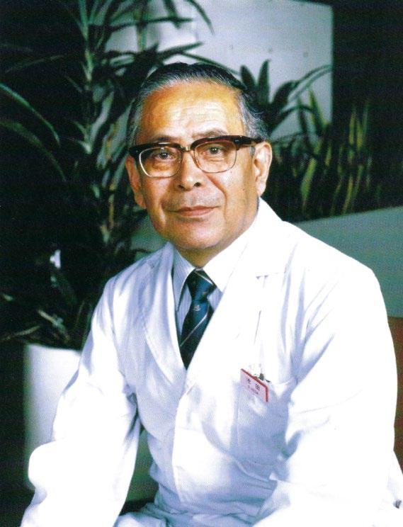
Ikeda was born in 1925 (Figure 1.1). After graduating from high school, he began studying medicine at Keio University in 1944. However, he had to interrupt his studies for one year as he suffered from specific pleuritis and underwent thoracoplasty. After recovery, he graduated in 1952 but in the same year, he had to have lung resection for a tuberculous mass during his internship in the Division of Tuberculous Surgery. Here he began studies on bronchial anatomy, including bronchography and motion pictures. As he found illumination by electric bulbs at the tip of rigid telescopes unsatisfactory, in 1962 he designed a telescope with “cold light.” A glass fiber bundle, connected to a 500 W xenon light source, was attached to the telescope and provided sufficient illumination for obtaining photographs and taking movies, for which he constructed special cameras.
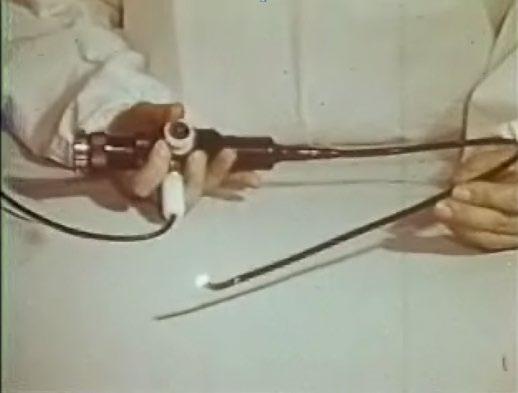
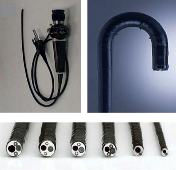
However, visualization of the bronchi in both upper lobes was often difficult due to the anatomical structures. Thus the need for a flexible bronchoscope, based on the concept of the gastrointestinal fiberscope presented by Basil Hirshowitz in 1961, was apparent [2,3]. As Machida Co. and Olympus Optical Co. had produced the first gastrofiberscopes in Japan from 1962, Ikeda approached Machida in 1964 and Olympus in 1965 for the construction of prototypes for bronchoscopy. He formulated specific requirements regarding diameter, more and smaller optical fibers, flexible light guide, fixed tip <1 cm, length 1 m, fixed focus 0.5–3 cm, visual angle 80°, and tip flexion of 60°. For ease of introduction, a special semiflexible orotracheal tube was constructed, that could be straightened in case specimens had to be obtained by a rigid forceps (Figure 1.2).
In 1966, when Ikeda presented the first prototype at the 9th International Congress on Diseases of the Chest in Copenhagen, Denmark, he created huge excitement and the story was even published in the New York Times. Further improvements were made on the following prototypes: control mechanisms for lengthwise rotation and bending of the tip were built into the control section, improved imaging was achieved by regular arrangement of smaller glass fibers, and a lens was mounted on the tip (Figure 1.3, movie). Finally, in the seventh prototype a channel was integrated into the scope for the introduction of sampling devices; Ikeda was confident that the instrument was ready for

demonstrating the first bronchoscope at my first visit to Japan. (Note: he was left handed, which is why the line to the light source and the suction of flexible bronchoscopes are running to the left so that the control section of the scope rests easily in your left hand and the lines are not pulling.) On the left is the first scope with the special orotracheal tube that could be straightened for taking rigid biopsies.
Figure 1.1 Shigeto Ikeda, 1925–2001.
Figure 1.2 Ikeda
commercialization and introduced and popularized flexible fiberbronchoscopy throughout the world.
In the following years, more and more experience was gained in clinical application and by 1980 flexible fiber bronchoscopy had become a routine procedure and spread worldwide. In 1980, after visiting Dumon in Marseille, Ikeda’s group began Nd:YAG laser treatment and photodynamic therapy (PDT) of malignant lesions. For better image resolution and processing, together with Asahi Pentax Co. he introduced charge-coupled device (CCD) chip technology to build the first videoscope which was
later adopted by Machida-Toshiba and Olympus. On his mission to promote the art of flexible fiberbronchoscopy, Ikeda traveled extensively all over the world and in 1978 founded the World Association for Bronchology (WAB) within which nowadays, as the World Association for Bronchology and Interventional Pulmonology (WABIP), national and continental assocations are united.
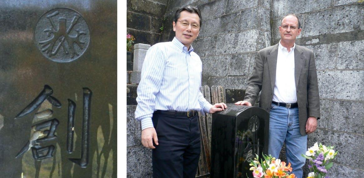
In recognition of his outstanding achievements, Ikeda received many national and international awards. After resignation from his clinical and academic positions in 1991, he preserved a keen interest in the development of bronchoscopy and stayed closely connected to the scientifc societies. Despite his fragile health due to several strokes and heart attacks, according to his lifelong motto “Never give up,” he attended all the meetings until the year 2000 before he died in 2001 [1,4–8] (Figure 1.4).
1.3 Further Development of Flexible Endoscopes
In the beginning, Machida and Olympus were the only manufacturers of flexible fiberbronchoscopes but soon other companies in Japan, the US and Europe began entering the market and today there are around 20 in business. As Olympus had the widest distribution network, it remains the main player in the market. With growing experience in fiberbrochoscopy, there was an increasing demand for different types of bronchoscopes for special
Figure 1.3 Movie clip of the first flexible fiberscope (see video on the website: function with flexion and rotation).
Source: Courtesy of T. Shirakawa.
Figure 1.4 With Teruomi Miyazawa at Ikeda’s tomb. The inscription on the gravestone beneath the emblem of the WAB reads: “Invention.”
diagnostic and therapeutic applications and also for other disciplines in medicine that realized the value of the new technology, such as anesthesia, pediatrics, and ICU medicine.
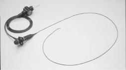
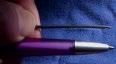
The development of new imaging and therapeutic technologies that could be applied via flexible bronchoscopes drove new advances. Improved fiberbronchoscopes were brought to the market in comparatively quick succession. Different diameters from 6 mm down to 2.2 mm for peripheral airways and pediatric use became available (Figure 1.5). Recently, miniaturized bronchoscopes with an outer diameter of less than 1 mm have been constructed for laboratory research in small animals. A variety of bronchoscopes with wide channels of 3 mm for interventional procedures and 1.2 mm for peripheral use are now available. Improvement of fiberoptics is allowing better analysis of small structures and photodocumentation. For observation by trainees, a teaching attachment (lecture scope) can be mounted onto the control section of the bronchoscope. Fiberscopes with battery illumination and integrated monitor are useful for
the ICU, sleep laboratory, on wards and in the outpatient department.
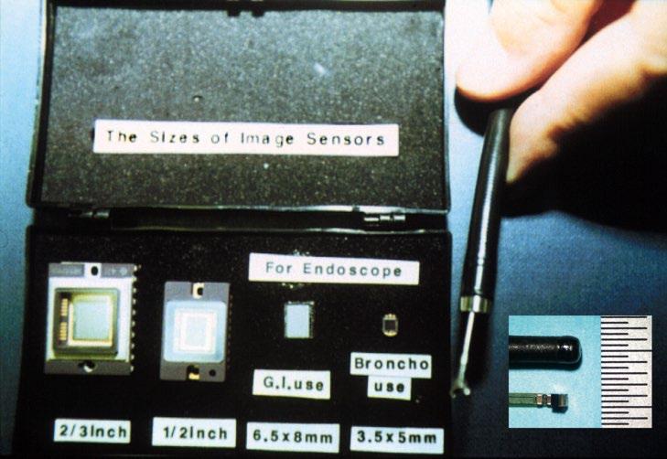
The next big step was the introduction of videobronchoscopy by Pentax in 1987, which became possible due to miniaturization of CCD chips, that could be integrated into the tip of flexible bronchoscopes instead of optical fibers [9] (Figure 1.6). Via a video processor, moving images can be followed on a monitor, stored on film and data banks, and printed. They can be directly integrated into the report or transmitted online to other departments. The biggest advantage is that images can be processed according to colors, contour enhancement, magnification, and different light sources. The CCDs have become so efficient that today videbronchoscopes with high-definition television (HDTV) with superior image quality are available. In very small endoscopes, the CCD is integrated into the control section and transmits the fiberoptic image to the processor (hybrid scope).
The next technological revolution was the introduction of EBUS in 1996, adding a new dimension to flexible
(a)
(c)
(b)
Figure 1.5 Modern flexible scopes of different diameters and sizes of biopsy channels.
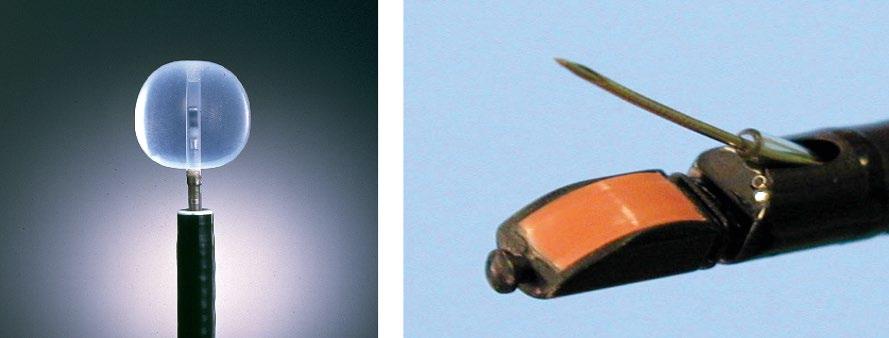
bronchoscopy by widening the view of the bronchoscopist beyond the airway. In 1999, the miniaturized radial probe was launched, that could be introduced via the biopsy channel of the flexible bronchoscope [10]. As there was an increasing demand for real-time transbronchial needle aspiration of mediastinal lymph nodes (TBNA), a dedicated ultrasonic endoscope was added in 2002 [11] (Figure 1.7) Today, EBUS-TBNA has become the standard for mediastinal staging and the radial probe for approach to peripheral lesions. “Endobronchial ultrasound (EBUS) is the single most useful pulmonary procedure in decades and should be available to all patients with thoracic adenopathy requiring evaluation” (Kevin L. Kovitz).
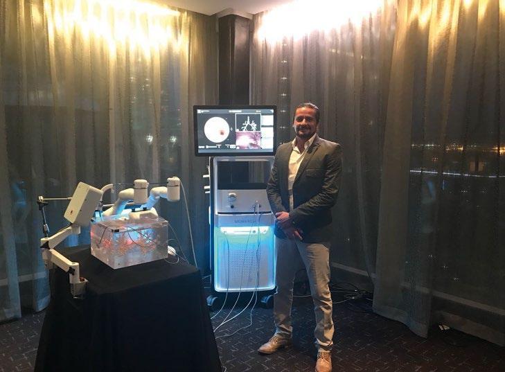
The latest revolution in flexible bronchoscopy is the introduction of robotic bronchoscopy. In the attempt to
become more independent from the bronchoscopist’s individual skills in handling and accuracy, robotic surgery has been developed. It is expected to improve access to peripheral airways by flexibility in all directions in order to place tools with precision consistently in the desired location and maintain stability in position under vision and by active remote robotic control. In 2018, a paper on clinical application of the first robotic bronchoscope (Monarch®, Auris Surgical Robotics Inc.) was published by my former coworker Jose Rojas-Solano [12] (Figure 1.8) A second upcoming robotic platform is the Ion™ endoluminal system by Intuitive Surgical, Inc., which currently is waiting for 510(k) clearance.
A general problem with flexible bronchoscopes is disinfection because of the small channels with risk of
Figure 1.6 The exponential downsizing of CCD chips for endoscopes.
Figure 1.7 Radial miniature EBUS probe inserted via the channel of a flexible bronchoscope and ultrasonic bronchoscope, so-called convex probe (cP) bronchoscope.
cross-contamination [13]. As few optical systems can withstand the autoclaving procedure, disposable videobronchoscopes have been developed [14]. The costs of reusable bronchosopes have been assessed for application in the ICU which, albeit with many imponderables, seemed favorable. However, due to the many different diagnostic and therapeutic applications of flexible bronchoscopy as illustrated below, it is highly improbable that disposable flexible bronchoscopes will be taking over more than the current approximately 30% of the market. So far, the only practical solution is high-level disinfection of instruments and strict observation of hygienic guidelines.
With all these technical improvements, bronchoscopy has become an essential part of pulmonary medicine. The bronchoscope market is driven by increasing prevalence of pulmonary diseases that can be diagnosed and treated by minimal invasive procedures with the bronchoscope, improved reimbursement, and technological advancements. The global market was valued at US$15.4 billion in 2017 and is expected to have increased 8.3% annually by the year 2025. Thus it can be expected that this will be an important incentive for manufacturers to research and invest in innovations [15].
1.4 Further Developments in Flexible Bronchoscopy
The ease of application and access beyond the central airways opened vast opportunities for the introduction of new optical, diagnostic, and therapeutic techniques [16], starting with transbronchial lung biopsy (TBLB),
performed by Anderson and Zavala after Ikeda’s visit to the United States [17].
In 1974, Reynolds published first experiences with bronchoalveolar lavage (BAL) [18]. Cortese demonstrated the potential of early lung cancer detection by fluorescence, induced by injection of hematoporphyrin derivates (HPD) in 1978 [19]. In the same year, Wang began TBNA of mediastinal lymph nodes via the flexible bronchoscope, which by then was only rarely applied via rigid instruments [20]. Also, the first endobronchial application of the Nd:YAG laser was performed by Toty [21] and in 1980 Dumon published his results on photoablation of stenoses by ND:YAG laser treatment, which for some time was the most applied technique [22,23]. In parallel, Hayata and Kato applied PDT after sensitization by HPD for treatment of centrally located early lung cancer [24]. In 1979 Hilarss used radioactive probes for treatment of central airway cancer for endoscopic high-dose radiation therapy (HDR) [25].
With rapid development and miniaturization of electronic imaging by charge CCDs, the first videobronchoscope was developed by Ikeda in cooperation with Pentax Co. in 1987. In 1990, the first dedicated silicone stent for the airways was presented by Dumon [26]. As implanting silicone stents by flexible bronchoscopy is difficult and intravascular metallic stents proved unsuitable for the airways, we introduced the Ultraflex® Nitinol® stent in 1992 [27]. In 1991 Lam reported on autofluorescence bronchoscopy (AFB) without HPD for early detection of lung cancer [28]. The need for local staging of these lesions was met by the introduction of radial EBUS in 1999 [10], which in addition opened a wide range of applications for diagnosis within the mediastinum and lung. Currently, in connection with
Figure 1.8 J. Rojas-Solano with the Monarch robotic bronchoscope.
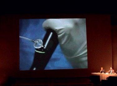
for communication (c), live presentation on the screen in the lecture hall (d).
TBNA by a dedicated ultrasonic bronchoscope, it is widely replacing mediastinoscopy for staging of lung cancer [29]. Early detection of lung cancer created further demand for new imaging modalities by high-power magnification videobronchoscopes and narrow band imaging (NBI) for analysis of subtle vascular structures [30]. Endoscopic optical coherence tomography (EOCT) providing information on the layer structure of the bronchial wall with higher resolution than EBUS is currently under investigation for bronchology [31].
Maneuvering smart diagnostic tools inside the airways beyond the visible range has been enhanced by smart electromagnetic navigation (EMN) [32,33], which also supported bronchoscopic treatment of peripheral lesions by insertion of brachytherapy catheters [34]. The first results of studies for treatment of asthma by thermic destruction of the bronchial muscles [35] and of emphysema by insertion of endobronchial valves have been published [36].
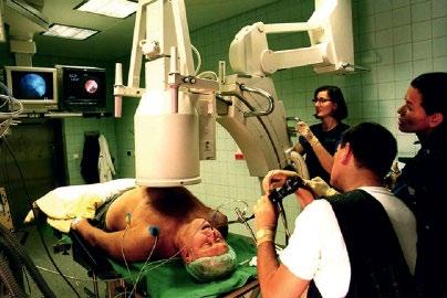
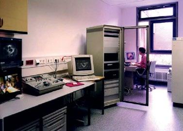
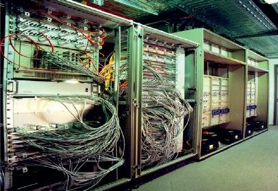
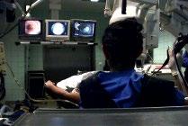
The rapid growth of internet communication opened new avenues for communication to enhance consulting, research, and teaching. The first long-distance live transmission on the occasion of the 12th World Congress for Bronchology and Bronchoesophagology was performed between Heidelberg, Germany, and Yokohama, Japan, in 2000 (Figure 1.9). Further details of all these innovations are beyond the scope of this article and interested readers will find suggestions for reading in the references.
1.5 Current Concepts and Strategies in Flexible Bronchoscopy
During my professional life, around 600 physicians from all over the world came to our institution to obtain experience in all kinds of procedures. Frequently, younger doctors
(a)
(b)
(c)
(d)
Figure 1.9 Live transmission of an EBUS procedure from Heidelberg, Germany, to Yokohama, Japan, at the WCB. Endoscopy setting (a), communication center in the endoscopy unit (b), hardware (“brain”) in the endoscopy
were surprised to observe that, apart from obtaining skills in a special technique, they learned that bronchoscopy is part of the wider concept of pulmonary medicine. This is why it might be useful to illustrate the evolution of flexible bronchoscopy in more detail by some current issues in interventional bronchoscopy.
1.6 Detection and Staging of Early Lung Cancer in the Central Airways
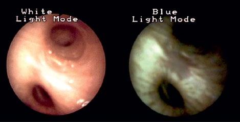
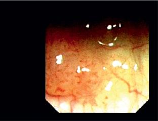
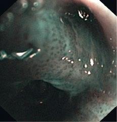
While applying PDT treatment of advanced lung cancer, it was observed that invisible small lesions in the bronchial mucosa became visible due to HPD fluorescence. This proved useful for detection of so-called early lung cancer, that is, in patients with positive sputum cytology with radiologically invisible lesions that could potentially be cured. In further studies, it was shown that by illumination with special light sources, the bronchial mucosa fluoresced without activation by HPD and several systems have been developed in fiberscopes and videoscopes for detection of early cancers by autofluorescence
As autofluorescence was unspecific for diagnosis of malignancy, methods for further analysis were added. Magnifying videobronchoscopes allow more detailed analysis of intra- and subepithelial structure, especially pathological vessels, that are characteristic of development of early bronchial cancer. By selecting smaller spectra in RGB imaging, narrow band imaging (NBI), the visualization of characteristic pathological vessels could be markedly improved. Further increase of resolution toward almost microscopy level could be achieved by EOCT in which an optical scanning beam is reflected from the different layers of the mucosa, providing optical histology images. The most recent technology, confocal microendoscopy, is finally bringing optical resolution to the cellular level [37].
It is expected that combination of these methods might finally replace histological examination on biopsy specimens. When early lesions were treated by very efficient local therapy, such as PDT, there were always a considerable number of patients who had only partial remission of their tumors. By definition, in situ carcinoma does not transgress the lamina propria of the mucosa and early lung cancer does not invade the cartilage and the connective tissue layer in between. The latter in particular could not be assessed by optical methods.
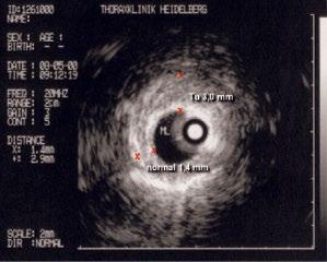
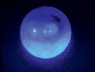
In the 1990s, together with Olympus, we developed a dedicated radial ultrasound (EBUS) probe of 20 MHz. The high resolution of its images clearly showed all the layers of the bronchial wall. When we analyzed “early cancers” detected by autofluorescence, frequently the
lesions extended further into the bronchial wall, even into the adjacent tissue and small lymph nodes that evaded diagnosis by computed tomography (CT) [38]. When by EBUS exploration only strictly localized tumors were treated by PDT, in all patients long-lasting complete remission was achieved [39]. Thus it was proven that it was not failure of the PDT, as had been assumed, but of local staging of these lesions. Which means that supposedly early lesions should be explored by the described methods, before a decision on the method of endoscopic treatment is made. Thus in a more extensive localized lesion, if surgery is not considered, endobronchial brachytherapy might be preferable because of its deeper penetration (Figure 1.10).
1.7 Diagnosing and Staging of Advanced Lung Cancer
Correct staging of lung cancer is the prerequisite for successful treatment [40]. The introduction of flexible bronchoscopy allowed visualization of segmental and subsegmental airways and in this way significantly helped to assess the endoluminal extension of cancers, which is especially important for delineation of the prospective resection lines for thoracic surgery (Figure 1.11). Also, preoperative bronchoscopy is essential to exclude additional endobronchial metastasis. As the vision of the bronchoscope is limited to the lumen and the internal surface of the bronchial wall, involvement of the deeper layers or penetration into the mediastinal structures could be only indirectly assessed. Because radiological exploration of the mediastinum depends on the contrast between water density, air, fat, or calcium, we experienced a lot of misdiagnosis because no such interface was present. Especially when in solid tumors adjacent to the mediastinum, infiltration of those structures was diagnosed by radiology, this could often not be confirmed during surgery. Also diagnosis of lymph node metastasis based on size was of only limited value.
This is why in 1989 I approached Olympus who were already active in gastrointestinal ultrasonography, to develop EBUS for exploration, especially with regard to staging of lymph nodes. During the following 10 years, many obstacles had to be overcome because of the special conditions in the airways. However, after many protototypes, two EBUS systems had been developed, the radial probe and the ultrasonic endoscope, that are now widely used in different indications. By using EBUS, tumor compression of the airways versus infiltration of the wall can be reliably differentiated in the preoperative bronchoscopic evaluation [41].
Most important for bronchoscopic staging is involvement of mediastinal lymph nodes. With the rigid bronchoscope
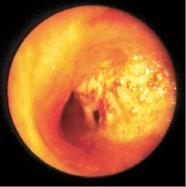
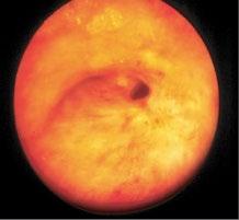
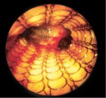
1.10 Early lung cancer. The slight discoloration on white light becomes very prominent by autofluorescence (a). By magnifying endoscopy, the pathological vascularization becomes visible (b), which is even more prominent under NBI (c). In the EBUS image, the superficial lesion ventrally is thickened (3 mm) compared to the normal wall on the left (1.4 mm), but well within the confines of the bronchiall wall and can be treated by bronchoscopic intervention (d).
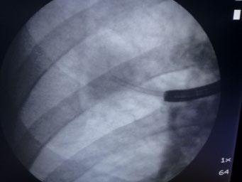
after 4 y After dilation and stenting
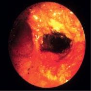
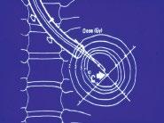
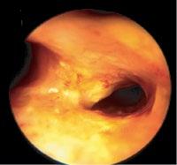
1.11 Extensive squamous cell cancer at the bifurcation extending into the trachea (a). Immediately after Nd:YAG laser resection (b). Isodose lines around the Ir 192 radioactive probe (c). Bifurcation two years after HDR therapy (d). Survival of patients with complete remission compared to partial remission after HDR (e). Recurrent high-grade radiogenic stenoses after complete remission (f). Recanalization after balloon dilation by insertion of three Nitinol stents (g).
Figure
Tumor at bifurcation
(a) (e)
at 2y
Figure
TBNA of mediastinal masses and enlarged lymph nodes had been performed but was never widely used. This changed with the introduction of the flexible bronchofiberscope. After localizing enlarged lymph nodes in relation to bronchial landmarks on CT, guided TBNA, such as the main carina and bronchial branchings, special flexible cytology or histology needles can be easily passed through the airway wall, even in a bent position, inaccessible for rigid needles. The method was very safe, complications mainly anecdotal and the results were so good that in many cases TBNA replaced mediastinoscopy, back then the gold standard for staging. But despite that, TBNA remained widely underused, mainly probably for fear of bleeding from larger mediastinal vessels.
With the introduction of the radial ultrasound probe, lymph node localization, especially in the paratracheal region where there are no clear landmarks, became more reliable. In the paratracheal region, results of EBUS-guided TBNA were significantly superior. However, only after the introduction of the dedicated ultrasound bronchoscope, by which realtime observation of the needle passing into the lesion is possible, did EBUS-guided TBNA become widely accepted and today it is the recommended standard for mediastinal staging and has widely replaced mediastinoscopy [42].
1.8 Tumors of the Central Airways:
From Palliation to Cure
With the increasing incidence of lung tumors, one main indication for bronchoscopy had become central airway obstruction. In contrast to rigid bronchoscopy, mechanical ablation by the flexible instrument itself, apart from necrotic fragile tissue, is mostly not successful. Resection by forceps, curette, or other mechanic instruments is tedious due to their small size. One exception in special cases is balloon dilation of malignant airway stenosis by compression [43]. The effect, however, is short-lived and not very efficient for improvement of dyspnea. For rapid desobliteration, several methods have been introduced. In 1982, Dumon described the application of the Nd:YAG laser for thermal destruction of cancer tissue for rapid relief of central airway obstruction. After attending Dumon’s lecture in 1980, Ikeda visited him in Marseille and immediately introduced the laser in Japan. The advantage was the noncontact destruction by vaporization of the tissue. By observation of risk factors, application of short impulses at maximum 40 W power setting and 50% oxygen supply, the method was very safe. In 1997, Homasson described high-frequency (HF) electrocautery that uses the thermal effect of electric current for the destruction of tissue and by its immediate effect is comparable to the
Nd:YAG laser. Sutedja called it “the poor man’s laser” as it has a similar effect at much lower costs [44 45]. A variation of electrodestruction is argon plasma coagulation (APC) [46]. In this noncontact method, the current is induced by heating argon gas to separate the electrons from the atoms and the resulting Ar + plasma is the medium for transfer of the electric current. In contrast to methods that destroy tissue by heat, cryotherapy uses the Joule–Thomson effect of creating cold by rapid expansion of liquefied gases in small catheters. This can be used for removal of the adhering frozen tissue by rapid extraction of the probe or for inducing delayed tumor necrosis due to intracellular ice crystal formation [47].
All the above methods provide immediate relief of symptoms and in malignancies have to be repeated after recurrence, unless definitive treatment for cure can be provided. Survival after laser resection of malignancies in my experience was 20% after three years. For patients who could not be cured by surgery or radiotherapy after local resection, endoscopic methods for long-term palliation were sought.
In the early 1990s, local long-term palliation of endobronchal cancer with high dose-rate intraluminal irradiation (HDR brachytherapy) by insertion of catheters armed with a radioactive Ir192 probe at the tip was investigated. The principle is application of a very high dose of radiation near the radiation source with a steep gradient to spare the surrounding tissues. We added HDR to laser treatment. When we were able to achieve endoscopic complete remission, the longterm effect lasted many years, in some patients even resulting in cure. Our strategy is application of 2–3 sessions of external radiation to reduce the tumor volume and then four sessions at 5 Gy by endoluminal radiation [48].
In patients with complete remission, long-term complications frequently included extensive scar formation resulting in recurrence of severe central airway stenoses. These can only be treated by internal support. In 1989, Dumon presented his dedicated silicone stent that could be placed by rigid bronchoscopy. Anecdotal reports described techniques for implantation with the fiberscope it but never gained wider use. Instead, endovascular stent systems were adapted for insertion into the airways. However, the first expandable and self-expanding metallic stents made from stainless steel and tantalum either collapsed due to missing internal support, as in blood vessels, or perforated due to excessive pressure on the airway wall. After negative experiences with tantalum stents, we developed the Boston Scientific Nitinol stent, later called the Ultraflex stent (Figure 1.12). After improving the insertion mechanism and development of an additional covered version, it became the standard model for flexible insertion. Some of my patients have been living with this stent for more than 20 years, one after insertion at the age of 2 years.

Parallel to laser therapy, PDT for bronchial cancer was introduced. By this treatment, cancer tissue is sensitized to light by injection of HPD which, during laser illumination, is degraded and obliterates capillary vessels and destroys cancer cells due to oxygen radical production. However, in contrast to Nd:YAG laser resection, the effect of treatment is delayed and not recommended for emergency situations. But it is very efficient as an adjunct to acute interventions and can also be applied in conjunction with brachytherapy or after placement of uncovered stents in order to treat tumor ingrowth.
A possibly promising recent approach for treatment of inoperable lung cancer is bronchoscopic intratumoral injection of anticancer agents such as ethanol, chemotherapeutics, or immunostimulatory genes. For better local control of the injection, EBUS-guided transbronchial needle injection (EBUS-TBNI) offers visual control by measuring tumor volume for dose calculation, avoiding intravascular injection and observing the hypoechoic swelling by fluid injection [49].
1.9 Diagnosis and Treatment of Peripheral Lung Cancer
Over recent decades, a gradual shift has occurred from centrally located lung cancer to peripheral lesions in the lung. As in early endobronchial cancer, the prognosis of
peripheral cancer improves the earlier it is detected. This is why screening programs for early detection by low-dose spiral CT have been investigated. After long-term followup, it could be shown that significantly more smaller lesions were found and that mortality could be reduced by 20% [50]. However, when SPN were routinely resected, it turned out that half were benign and in fact did not need surgery with its side-effects [51]. This is why there was a demand for preoperative confirmation of the histology. With flexible tools like washing, brushing, curettes, needles, and biopsy forceps, there exists a wide armamentarium for obtaining histopathological material.
The challenge is finding the path toward the lesion, because calculating the location of the lesion from X-ray or CT and trying to approach it merely by endoscopic view has a very low success rate. Real-time control of transbronchial biopsy under fluoroscopy was better [52]. But the visualization of lesions under 2 cm and ground glass opacities is very poor. Anecdotal reports of transbronchial biopies under real-time CT visualization were more successful, but the logistic challenges and radiation exposure prevented widespread application [53]. After introduction of radial EBUS, it became possible to confirm the exact position of the SPN and the path via which it could be approached. When the probe was introduced via a guide sheath that could be left in place as an “extended working channel” for introducing tools, the results became much
(a) (b)
Figure 1.12 Dumon silicone stent (a) and covered Ultraflex Nitinol stent (b).
bronchoscope
and the endoscopic view
www.aurishealth.com/monarch-platform
better [54]. However, especially in the upper lobes, the path toward the lesion is frequently hard to find and, moreover, the radial probe is not steerable. This is why we investigated a system for EMN, analogous to a GPS, to introduce a catheter into the lung periphery of the lung.
The patient is placed with the upper body within a lowintensity electromagnetic field (serving as “satellite”) and a sensor (board computer in GPS) can be followed on a monitor moving through the overlaid image of the patient’s CT scan (road map). The important feature is that the sensor is mounted on the tip of a steerable guide that can be turned 360° while moving it through an extended working channel to the periphery. After reaching the lesion, the sensor is removed and with EBUS the intralesional position can be confirmed. With an ultra-slim bronchoscope, observation
of the intrapulmonary airway is possible to assess whether forceps biopsy in endoluminal growth or needle biopsy and transbronchial biopsy in external compression is preferable. An alternative technique for navigation is introduction of a smaller bronchoscope along a path created by virtual bronchocopy and overlaid on the real endoscopy image [55].
Navigation has significantly improved diagnosis of SPN and has become the standard for approaching these lesions. Yet, as in all interventional bronchchoscopic procedures, success also depends on the individual skills of the bronchoscopist. Therefore, robotic bronchoscopy was developed to offer an alternative approach to address the limitations of current bronchoscopic techniques for biopsy of peripheral lung lesions. In a paper published recently by
Operator Console
Rack
Robotic Endoscope
(b)
(a)
(c)
lesion
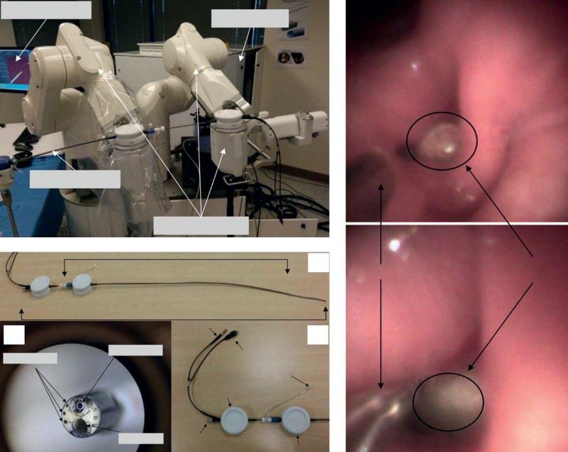
Figure 1.13 The Monarch robotic endoscope system (RES). The proximal controlling system and the robotic endoscope (a), the
(b)
of the peripheral tumor and the biopsy procedure (c). For an information video clip see
my former coworker Dr. Rojas-Solano, very promising results with the first commercially available robotic endoscopy (RES) (Auris Surgical Robotics, San Carlos, CA) were reported.
RES consists of a 3.2 mm videobronchoscope with a 1.2 mm working channel and an outer sheath, which both allow four-way steering by two robotic arms under continuous remote, direct, and visual front view control. The bronchoscope’s distal section can achieve 180° of deflection in any direction. The proximal section allows for control of irrigation and aspiration (Figure 1.13).
With reliable tools for the approach to SPN and EBUS control of stable positioning of the extended working channel, endoscopic treatment of inoperable peripheral cancer became feasible. Considering the most similar endoscopic treatment compared to curative surgery after our experience with localized central airway cancer, we decided to apply peripheral brachytherapy as we could easily match the dosage to the primary tumor and also to the peribronchial lymphatics and hilar lymph nodes when
moving the source centrally through the airways. Following our first experience in a small study, we could achieve longstanding complete remission in the majority of cases [56] (Figure 1.14). Other modalities in planning or currently under investigation are radiofrequency ablation (RFA), laser, cryotherapy, PDT, and vapor ablation.
1.10 Parenchymal Lung Disease
Although, with improved CT imaging, many causes of interstitial lung diseases display pathognomonic morphological patterns, frequently histomorphological or microbiological confirmation for diagnosis is necessary. Right from the beginning of fiberbronchoscopy, taking samples by washing and brushing was used for this purpose. Whereas this proved useful in diagnosing infectious lung diseases and lung cancer in cases of positive cytology, the small amounts of fluid did not suffice for diagnosis of parenchymal lung diseases.
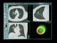
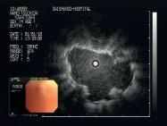
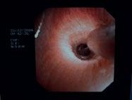

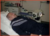

Figure 1.14 Diagnosis and treatment of peripheral lesions. Conventional navigation via three monitors for endoscopy, fluoroscopy, and EBUS (a). The electromagnetic navigation system (EMN, superDimension) with sensor, electromagnetic board and computer with control monitor (b). The sensor (green) is maneuvered into the target lesion (yellow) under three-plane CT control on the monitor (c). The intralesional position is confirmed by introducing the EBUS probe (d). To assess the best biopsy technique, an ultra-thin Olympus endoscope is introduced and forwarded toward the lesion (e–g). As the lesion protrudes into the lumen, forceps biopsy is the appropriate technique for obtaining tissue samples (h). In case of inoperability, by the same procedure a brachytherapy catheter can be inserted (i) and HDR therapy can be applied according to calculation by the isodose lines (j).
However, as sometimes characteristic cells were observed, in the early 1980s a method with instillation of larger amounts of fluid for sampling specimens from the bronchoalveolar space was introduced – bronchoalveolar lavage (BAL). Initially, it was expected that BAL provided a nonbloody biopsy method, for example by the relation of T-helper to T-suppressor cells for sarcoidosis. But in further studies this hope did not hold true, as the findings were frequently unspecific. Findings were characteristic only for limited, comparatively rarer diseases such as eosinophilic pneumonia or alveolar proteinosis. Only in severe infections, especially in immunocompromised patients, is it still widely used. In most cases, examination of lung tissue for diagnosis is needed.
After Anderson demonstrated the safety of taking transbronchial biopsies by forceps from lung tissue via the flexible bronchoscope, this method became state of the art in
diagnosis of lung diseases and SPN. As the specimens are pretty small, the sampling error can be reduced by taking several biopsies from different segments of one lung. In our experience, using a larger forceps that is not pulled out through the biopsy channel increases positive results. For this purpose, we introduce the flexible scope through an endotracheal tube, as Ikeda described in his first publication, and leave the forceps in front of the endoscope tip. This way, we are taking up to one specimen from each segment in one lung with a very low rate of complications, which also gives us information on disease activity in different parts of the lung [57].
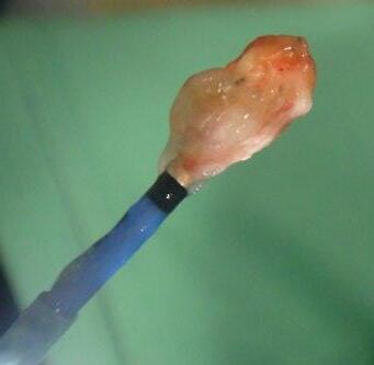
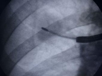
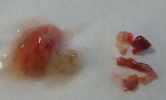
The safety of taking larger specimens encouraged the use of cryobiopsy probes, by which much larger tissue fragments can be obtained at an equally low complication rate. In addition, pathologists prefer these samples because they have much less mechanical damage than in forceps biopsies [58] (Figure 1.15).
(a) (b)
(c)
(d)
Figure 1.15 Transbronchial cryobiopsy. With EMN, a catheter is inserted into a peripheral lesion (a) and the cryoprobe is inserted (b). After defreezing the probe is removed together with the catheter and the bronchoscope (c). Comparison of biopsy sizes with cryoprobe and large biopsy forceps (d).