Basic guide to orthodontic dental nursing 2nd edition fiona grist all chapter instant download
Visit to download the full and correct content document: https://ebookmass.com/product/basic-guide-to-orthodontic-dental-nursing-2nd-edition -fiona-grist/
More products digital (pdf, epub, mobi) instant download maybe you interests ...
Basic guide to dental sedation nursing Second Edition
All rights reserved. No part of this publication may be reproduced, stored in a retrieval system, or transmitted, in any form or by any means, electronic, mechanical, photocopying, recording or otherwise, except as permitted by law. Advice on how to obtain permission to reuse material from this title is available at http://www.wiley.com/go/permissions.
The right of Fiona Grist to be identified as the author of this work has been asserted in accordance with law.
Registered Offices
John Wiley & Sons, Inc., 111 River Street, Hoboken, NJ 07030, USA
John Wiley & Sons Ltd, The Atrium, Southern Gate, Chichester, West Sussex, PO19 8SQ, UK
Editorial Office
9600 Garsington Road, Oxford, OX4 2DQ, UK
For details of our global editorial offices, customer services, and more information about Wiley products visit us at www.wiley.com.
Wiley also publishes its books in a variety of electronic formats and by print‐on‐demand. Some content that appears in standard print versions of this book may not be available in other formats.
Limit of Liability/Disclaimer of Warranty
The contents of this work are intended to further general scientific research, understanding, and discussion only and are not intended and should not be relied upon as recommending or promoting scientific method, diagnosis, or treatment by physicians for any particular patient. In view of ongoing research, equipment modifications, changes in governmental regulations, and the constant flow of information relating to the use of medicines, equipment, and devices, the reader is urged to review and evaluate the information provided in the package insert or instructions for each medicine, equipment, or device for, among other things, any changes in the instructions or indication of usage and for added warnings and precautions. While the publisher and authors have used their best efforts in preparing this work, they make no representations or warranties with respect to the accuracy or completeness of the contents of this work and specifically disclaim all warranties, including without limitation any implied warranties of merchantability or fitness for a particular purpose. No warranty may be created or extended by sales representatives, written sales materials or promotional statements for this work. The fact that an organization, website, or product is referred to in this work as a citation and/or potential source of further information does not mean that the publisher and authors endorse the information or services the organization, website, or product may provide or recommendations it may make. This work is sold with the understanding that the publisher is not engaged in rendering professional services. The advice and strategies contained herein may not be suitable for your situation. You should consult with a specialist where appropriate. Further, readers should be aware that websites listed in this work may have changed or disappeared between when this work was written and when it is read. Neither the publisher nor authors shall be liable for any loss of profit or any other commercial damages, including but not limited to special, incidental, consequential, or other damages.
Library of Congress Cataloging‐in‐Publication Data
Names: Grist, Fiona, author.
Title: Basic guide to orthodontic dental nursing / Fiona Grist. Other titles: Basic guide to dentistry series. Description: Second edition. | Hoboken, NJ : Wiley-Blackwell, 2020. | Series: Basic guide to dentistry series | Includes index. Identifiers: LCCN 2019035284 (print) | LCCN 2019035285 (ebook) | ISBN 9781119573692 (paperback) | ISBN 9781119573708 (adobe pdf) | ISBN 9781119573746 (epub)
LC record available at https://lccn.loc.gov/2019035284
LC ebook record available at https://lccn.loc.gov/2019035285
Cover Design: Wiley
Cover Image: Courtesy of David Morris
Set in 10/12.5pt Sabon by SPi Global, Pondicherry, India
Dedication
For Michael, with love
Foreword to Second Edition
The orthodontic team is in the privileged position of being able to significantly improve our patients’ lives almost on a daily basis. To do this efficiently and effectively requires not only an understanding of all the equipment and materials available, but precision teamwork practised day in, day out.
We, as orthodontists, are totally dependent upon our orthodontic assistants to understand exactly what we are doing and to be able to predict what we are going to require next, to ensure the correct instruments and materials are prepared accordingly. Working with a well‐prepared and conscientious assistant is a dream, and ensures high‐quality treatment is delivered to as many patients as effectively as possible.
This second edition of the Basic Guide to Orthodontic Dental Nursing by Fiona Grist allows the trainee orthodontic nurse to take the first tentative steps on a fascinating and rewarding lifelong journey. It will also provide extremely useful revision for experienced orthodontic assistants of many of the orthodontic concepts we are all required to know intimately.
With its recently updated photographs it now provides an invaluable reference book for all those wishing to learn and improve their orthodontic knowledge.
Professor P.J. Sandler BDS (Hons), MSc, PhD, FDSRCPS, MOrthRCS President, British Orthodontic Society
How to use this book
The aim of this book is to give the dental nurse in general practice an introduction to the world of orthodontics and orthodontic dental nursing. It may also be helpful to trainee nurses working in an orthodontic environment.
Orthodontics is specialist branch of dentistry and has its own vocabulary. The information in this book is a basic guide, so it does not set out to:
• examine clinical features (why the problem arose)
• cover treatment planning (what is the best choice of treatment)
• treatment mechanics (how the appliances achieve what they do).
Its objective is to describe what the dental nurse needs to know so they can work efficiently at the chairside when treating an orthodontic patient.
If you feel you want to develop your knowledge further there are several excellent orthodontic textbooks available. The career pathways for orthodontic dental nurses are now wide and the possibilities are extensive. Nurses have an important place in the dental team. This book aims to be a helpful first guide on what is hoped be a long and interesting journey.
Different procedures for various treatments are outlined in this book. While it is the nurse’s role to assist the clinician, there are areas that are their sole responsibility; these are highlighted in the text in italics.
A quick glance into the stock cupboards and cabinets in an orthodontic surgery reveals quite different contents from that of a general dental surgery. There will be nothing with which to fill teeth or fissure seal, or root canal trays. Anything that helps to irrigate a periodontal pocket, whiten a tooth, prepare abutments for a bridge or fit veneers will be missing. Cupboards in orthodontic units and practices may share the basics, such as mirrors, probes and College tweezers, and use the same alginates and disposable sundries, but beyond that they have very little in common. However, these cupboards are full and it is not possible to cover all materials or equipment that is in use, or every method or procedure.
Just as we had to learn what was needed for restorative, endodontic and prosthetic procedures, we need to learn what is needed for orthodontic treatment, which instruments are used for what procedure and why they are used.
Each chapter will cover a topic, with a short background and guide to what you will need to prepare so that the treatment can be undertaken as efficiently as possible. It is hoped that the photographic examples are helpful, the aim being to show the instruments as clearly as possible. The photographs are not all on the same scale.
This book does not go into detail regarding decontamination and sterilisation. The areas to focus on are those that concern the effect repeated sterilisation has on stiffening
box joints on pliers. It can also have a detrimental effect on pliers that have cutting edges. When sterilising pliers and instruments with beaks, always have the beaks open. The same procedures and protocols apply in orthodontics as in other specialties. These you already know. As dental care professionals it is up to the nurse to ensure that they are fully aware and comply with all the current legislation, standards and codes of practice.
As with every skill, be it orthodontic treatment or baking a cake, everyone will have their individual method of working and their favourite tools. There is no hard‐and‐fast rule that says each procedure must be carried out using only certain instruments in the same way or in an exact order. Every clinician has their preferred methods of working and each and every nurse organises the layout of their trays as they like them. This is as it should be – do what works best for you.
There is a saying,
You don’t know what you don’t know
This book contains a lot of information but at the same time there will certainly be omissions. Every day brings new materials, new techniques and new treatment philosophies. Orthodontics is inevitably becoming split into specialties within a specialty. The pace of development and change ensures that what is current today is not so tomorrow.
I hope that this book achieves what it sets out to do, which is to provide enough written and visual information for a reasonable grounding of basic knowledge. Its aim is to encourage dental care professionals, especially dental nurses, to understand more about orthodontic nursing.
As trained or trainee dental nurses there is so much that you are already expert at doing, so this book will not cover knowledge you already have or skills you already possess. It does not set out to be comprehensive, but aims to give you a basic insight into the world of orthodontic nursing – it is merely a guide.
Acknowledgements
This is the second time I have written acknowledgements for this book and there are now so many more people to thank! So many that is seems like a mini chapter in itself. Firstly, the tremendous support from my home team: my husband Michael and grand‐daughter Kate had unlimited patience when computers, cameras and all manner of technology was out to get me. They just quietly sorted it out. I could not have done it without them.
The format and structure of the original book, which benefited from the expertise and enthusiasm of Alan Hall, has remained, enlarged and hopefully improved. Jo Clark has generously taken over the task as my ‘go‐to’ clinical guru. She has been helpful with providing material for new photographs, advice and encouragement, not least in offering her proofreading skills. Her expert eye looked over my shoulder to ensure I had not got my clinical wires crossed. Also thanks to Maureen Dickinson who tried to make sure I did not leave out major facts whilst busily including the minor ones. They devoted many hours to this and I am truly grateful. Colin Anderson was my ‘lay’ proofreader, who also spent hours crossing the t’s and dotting the i’s. My thanks to you all for sharing your expertise so generously and for giving the book the benefit of your time, knowledge and experience with such graciousness.
Special thanks must also go to my young photographer, Kate Meheux, whose contribution to the aesthetic appeal and clarity of this book was huge. She was efficient, knowledgeable and enthusiastic and was a pleasure to work with over the many hours we spent chasing our vision.
I must thank David Morris who again gave permission for the images on the cover, Steven Jones who allowed me to re-use his photographs of TADs, Paul Ward who supplied photographs of fixed lingual appliances, Simon Littlewood who supplied the image of a Barrer spring retainer and Daljit Gill for his RME and cone beam CT images. Tracey Buckfield at NEBDN was helpful with permission to reproduce the Certificate in Orthodontic Nursing Syllabus as was Elena Scherbatykh at the GDC with the Certificate of Orthodontic Therapists Syllabus. The Occlusal Indices are reproduced by kind permission of Professor Steve Richmond and Ortho‐Care (UK) Ltd. There were also many images supplied by Alan Hall, and Jo Clark let me photograph a wide variety of her orthodontic goodies. I appreciate the kindness of Alison Williams in sharing her knowledge of aligners with me. My sincere thanks also to Alex Cash, and Wendy Bull in the office, for sending me full records of four of his cleft patients, Douglas, Emily, Georgia and Harvey. I want to thank them specially for kindly agreeing to be part of this book. I have tried to give an idea of their treatment journey and show you how great they look now.
Without a doubt one of the most noticeable aspects of this edition is the updating of many of the clinical photographs. This has been made possible by a generous offer from Jonathan Sandler to access his vast database. I am more than grateful for this and his
permission to use these photos in the book and the help given by Sue Mallender and Anne McTighe: merely looking at his seemingly endless files was a masterclass in clinical photography.
Orthodontics has some of the very best supply companies and I appreciate their encouragement and willingness to help. These include Richard Garford, Kelvin Scott and Lisa Howorth at Ortho‐Care, David Rees and his helpful staff at TOC, Justyn Gumienna at TB Orthodontics, and Mandy Mills at 3M Unitek. All have been really generous with their time.
I have had the pleasure of working with the Orthodontic National Group for Nurses and Therapists from the beginning. Their contribution to the role of dental nurses today was encouraged by their vision. It would be impossible to include everyone but special mention must go to Janet Robins, Maureen Dickinson, Alex Moss and Sally Dye.
I am grateful to Anjli Patel, Chair of Publications and Joe Noar, Head of Clinical Governance at BOS for their help with permission to use PILs and Guidelines. Anshu Sood and Rod Ferguson kindly allowed me to use the BOS Courses on Impression Taking and Clinical Photography. Ann Wright used all her co-ordinating skills with this too!
My respect for the British Orthodontic Society is unquantifiable. They have long been in the forefront of fostering the ‘team’ approach in orthodontics in the UK and have blazed a trail for other specialties to follow. The Society has always, and continues to be, hugely supportive of orthodontic nurses and therapists. Thanks to Professor Jonathan Sandler for generously agreeing to write a foreword for this book. Special thanks to Ann Wright, Ann Humphrys and Tony Kearney at BOS headquarters in Bridewell Place for their unflagging good humour and willingness to help and share their expertise. You may not realise it but your bar is high, you really do set the standard.
Caroline Holland first encouraged me to write an article on orthodontic nursing. Initially sure I couldn’t, she encouraged me to give it a try, for which I will always be in her debt, as without her I would never have written a word.
Nearly all the names on this page are members of the orthodontic family; they share a passion for their work and have themselves made a significant contribution to their specialty, not to mention their patients, colleagues and the sphere of research. They have been gracious in sharing their expertise. All omissions and errors are down to me.
Huge thanks to the team at Wiley‐Blackwell, especially Loan Nguyen, Susan Engelken, Jayadivya Saiprasod, Jolyon Phillips, Baskar Anandraj and Nick Morgan. Knowing you were there was good, but hearing your voices at the end of the phone was better. Thanks for all the hand holding.
Last, but by no means least, to you, who have made it to the bottom of the page. Teachers of English will say this piece is woeful as it repeats the words ‘generous’, ‘thanks’ and ‘appreciate’ too often. They are correct but these words are precisely what this page is all about. I hope that you feel inspired to keep turning the pages and that you begin, or are continuing, to enjoy your work in orthodontics, probably the best job in the world!
Chapter 1
Definition of orthodontics and factors influencing orthodontic treatment
Orthodontics is a specialised branch of dentistry. The name comes from two Greek words:
• orthos, meaning straight or proper
• odons, meaning teeth.
So the meaning is clear – ‘straight teeth’.
Orthodontics is the study of the variations that occur in the development and growth of the structures of the face, jaws and teeth and of how they affect the occlusion of the teeth. Ideally there should be the same number of permanent teeth in each arch.
Any deviation from the norm that affects teeth alignment and the bite relationship is called a malocclusion. Most malocclusions are genetic – they are inherited (e.g. missing teeth or a protruding mandible). Other malocclusions can be caused by the patient, for example digit sucking, or by external factors such as trauma.
Orthodontic treatment can correct a malocclusion by restoring the teeth to their normal position and occlusal relationship (with surgical help if needed) so that:
• the bite is fully functioning and the patient can bite and chew properly
• oral hygiene is made easier, so helping to prevent caries and gingivitis
• the malocclusion does not cause other damage, often to soft tissues
• the patient looks better and has better self‐esteem.
Orthodontic treatment in conjunction with orthognathic (maxillofacial) surgery can correct an underlying jaw discrepancy or facial asymmetry. Orthodontic planning is done in conjunction with the surgeons using clinical and radiographic assessment, with a cephalometric tracing (Figure 1.1) often analysed using a computer software program.
So orthodontists set out to:
• straighten teeth
• improve the bite
• improve the function
• improve oral hygiene (making teeth easier to clean)
Source: Reproduced by kind permission of Alan Hall.
CLASSIFICATION OF OCCLUSION
When assessing occlusion there are two aspects to classification:
• incisor relationship
• buccal segment occlusion, left and right.
Both are recorded on a patient’s orthodontic assessment form.
Incisor classification
• classes have Roman numerals, e.g. I, II, III.
• divisions do not, e.g. Class II/1 or Class II/2. The incisor classification (Figure 1.2):
• relates to the bite of the tip of the lower central incisors onto the back of the upper central incisors
• it is divided into three horizontal sections and where the lower incisor occludes will determine the classification.
Class I
• the incisal edge of the lower incisors bite on or below the cingulum plateau of the upper incisors.
Class I
Class II/1
Figure 1.2 Incisor classification. Source: Reproduced by kind permission of Alan Hall.
Class II/1
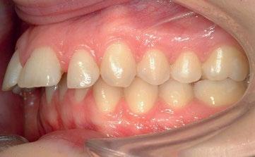
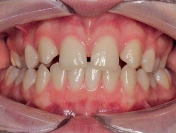
• the upper incisors are proclined or upright (Figures 1.3 and 1.4).
Class III
• the lower incisors bite behind the cingulum plateau of the upper incisors.
• the position of these front teeth means they can be damaged more easily because of their vulnerable position.
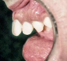
Class II/2
• the upper incisors are retroclined.
• the lower incisors bite behind the cingulum plateau.
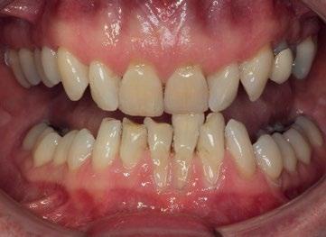
• the position of the teeth can, when closed, lead to trauma to the lower labial gingivae and the upper palatal gingivae (Figures 1.5–1.7).
Class III
• the bite is edge to edge or reversed.
• the incisal edge of the upper incisors can bite into the back (lingual) surface of the lower incisor (Figure 1.8).
• a horizontal overlap is called an overjet
• a vertical overlap is called an overbite.
Buccal segment occlusion
The buccal segment occlusion:
• was devised by Edward Angle in 1890
• is still widely used today
Class II/2
Figure 1.3 Large overjet. Source: Reproduced by kind permission of Jonathan Sandler.
Figure 1.4 Side view of severe overjet. Source: Reproduced by kind permission of Alan Hall.
Figure 1.5 Bite stripping lower gingivae. Source: Reproduced by kind permission of Jonathan Sandler.
Figure 1.6 Damage to labial gingivae caused by the bite. Source: Reproduced by kind permission of Jonathan Sandler.
• is based on the occlusion between the first permanent molar teeth, which erupt when the patient is about 6 years old.
There are three classes:
• class I: this is near to the correct relationship
• class II: this is at least half a cusp width behind the ideal relationship
• class III: this is at least half a cusp width in front of the ideal relationship (Figure 1.9).
Figure 1.7 Bite causing trauma to the palate.
Source: Reproduced by kind permission of Alan Hall.
Figure 1.8 Class III. Source: Reproduced by kind permission of Alan Hall.
Class I½ unit Class II
Class III
Class II
Figure 1.9 Diagram of buccal segment occlusion. Source: Reproduced by kind permission of Alan Hall.
THE MIXED
DENTITION
Sometimes parents see their child’s perfectly straight deciduous teeth fall out only to be replaced by a ‘jumble’ of crowded permanent teeth. This often prompts them to want early treatment because permanent teeth can look huge in little faces.
Hypodontia
Patients with hypodontia do not have the full complement of teeth. This can occur in the deciduous and permanent dentition. In some cases, if it is just a single tooth, it is possible to close the space orthodontically. If there are too many missing this may require a solution involving replacements such as bridges and implants, with orthodontic treatment being used to position the teeth in the correct spaces (Figure 1.10).
The average times for permanent tooth eruption are as follows.
• age 6
• 1/1 lower central incisors
• 6/6 lower first molars
• 6/6 upper first molars
• age 7
• 1/1 upper central incisors
• 2/2 lower lateral incisors
• age 8
• 2/2 upper lateral incisors
• age 11
• 3/3 lower canines (cuspids)
• 4/4 lower first premolars (bicuspids)
• 4/4 upper first premolars (bicuspids)
• age 12
• 3/3 upper canines (cuspids)
• 5/5 lower second premolars (bicuspids)
• 5/5 upper second premolars (bicuspids)
• 7/7 upper second molars
• 7/7 lower second molars
Figure 1.10 Hypodontia. Source: Reproduced by kind permission of Jonathan Sandler.
• age 18–25
• 8/8 upper third molars (wisdom teeth)
• 8/8 lower third molars (wisdom teeth).
Normally, patients begin orthodontic treatment between the ages of 10 and 13 years old. At 10–11 years they are still in the mixed dentition, with
• some deciduous teeth
• some permanent teeth
• some teeth yet to erupt.
INDICATIONS FOR TREATMENT
Clinical indications for orthodontic treatment may be because the teeth:
• are overcrowded
• may have erupted out of position
• are protruding (Class II/l)
• exhibit reverse bite
• exhibit self‐damaging bite (Figure 1.11)
• are spaced
• are absent (hypodontia)
• are damaged.
Mild malocclusions, for example:
• with only very small irregularities
• where the tooth position does not compromise oral hygiene
• which do not interfere with function, such as biting off food and eating, may not be merit orthodontic treatment, as it may not be seen to significantly improve dental health.
However, some presentations, for example:
• with overcrowded, protruding teeth
• with rotated teeth that make oral hygiene difficult and cause problems with caries
• which visually deviate from average, e.g. a reverse bite
Figure 1.11 Lower incisor trapped outside the bite.
Source: Reproduced by kind permission of Alan Hall.
• which look unattractive and affect the smile
• which seriously affect function, e.g. make chewing food difficult, are classed as malocclusions warranting treatment.
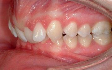
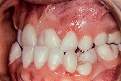
Scissors bite
This is a lingual crossbite, where the buccal cusps of the lower premolars and molars occlude palatal to their opposing upper tooth (Figure 1.12).
UNDERLYING CAUSES OF MALOCCLUSION OF THE TEETH
There may also be:
• underlying skeletal abnormalities
• facial asymmetries.
These can be:
• hereditary (e.g. tendency to being Class III)
• a result of injury
• a result of illness affecting facial or skeletal growth
• a result of a syndrome or cleft.
These may require orthodontic treatment as part of a multidisciplinary care treatment pathway.
Multidisciplinary approach
In cases requiring a multidisciplinary approach, patients receive their orthodontic treatment in co-ordination with other specialties.
These specialties include:
• restorative (e.g. hypodontia patients requiring implants/bridges or microdontia patients needing veneers or crowns)
• surgical (e.g. patients needing an osteotomy)
• cleft (e.g. patients requiring alveolar bone grafting).
Figure 1.12 Scissors bite. Source: Reproduced by kind permission of Jonathan Sandler.
PROBLEMS WHEN THE ARCH IS NOT INTACT
One of the aims of orthodontic treatment is to have each tooth in its correct place within the dental arch.
If a tooth is malaligned (out of its correct position), it is not necessarily an isolated problem – it has a ‘domino’ effect. The teeth either side of it may also be out of their correct position and the opposing tooth does not have the correct occlusion.
If there is no tooth to oppose it, a tooth may supra‐erupt. Contact points are lost, teeth rotate and, because they are no longer self‐cleansing, food traps are created where fibres can get lodged or packed. As a consequence of this, plaque is encouraged to accumulate, which in turn:
• inflames the gingivae
• encourages periodontal pockets.
In the young patient this is not too drastic, as it probably has not yet become a significant issue.
In adult patients, however, following orthodontic treatment it may be necessary to restore incisal edges or fill cervical abrasion cavities, which only become apparent when the teeth have been corrected.
BRUXISM
• young patients, towards the end of the deciduous dentition, can often present with teeth almost ground down to gingival level. It may continue into the mixed dentition and is often quite noisy and noticeable when it occurs in sleep.
• for some older patients with severe bruxism, an occlusal guard can be made to be worn at night during sleep. This attempts to limit the damage that is done to the incisal and occlusal surfaces of the teeth.
• anxious patients also grind and clench their teeth during the day when under stress. Patients often also clench their teeth when doing weight training at the gym.
DIGIT SUCKING
Some patients continue to suck their fingers or thumbs well beyond the age when their deciduous teeth have been replaced by their permanent successors. A prolonged habit is one which exists until the age of 7 years.
It may adversely affect the bite and position of the anterior teeth and can produce:
• a unilateral buccal crossbite
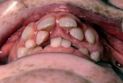
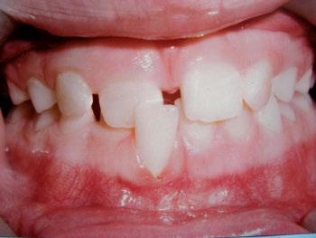
• an asymmetrical anterior open bite where the finger or thumb enters the mouth (Figure 1.13)
• an increased overjet.
Figure 1.13 Anterior open bite due to digit sucking. Source: Reproduced by kind permission of Alan Hall.
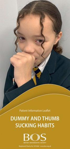
Figure 1.14 Leaflet on digit sucking. Source: Reproduced by kind permission of British Orthodontic Society.
How much damage is caused depends on how long, and how frequently, the thumb or finger is sucked and how strong the habit is (i.e. occurs not just when alone going to sleep but also during the day when tired, bored or stressed).
These patients try really hard to break this habit but sometimes they need a bit of help (Figure 1.14). It is possible to fit a removable upper anti‐habit appliance, which has prongs in the centre of the palate that act as a positive deterrent for the thumb or finger. This is worn full time or only when the individual is asleep.
Once the habit is broken, the problem is often solved. However, some patients experience strong emotional comfort from digit sucking and this compulsion may need to be assessed in detail. An image of an anti‐habit device is provided in Chapter 7.
DENTAL HEALTH
Some problems are caused by:
• diet (too much sugary or acidic food or drink, causing dental caries)
• tooth brushing (the wrong technique, too hard a brush)
• acid reflux (a symptom of bulimia in anorexic patients)
• medication (side effect of some medication inhalers).
Damage to teeth resulting in tooth surface loss comes under the following general headings:
• attrition: bruxism (patients who grind their teeth, often during sleep)
• erosion: of the enamel by acid, found in fresh fruit juice, diet drinks and stomach acid (found in reflux)
• abfraction: a tooth being ‘high on the bite’ and being overloaded.
Charting of teeth is an area you all know well, and follows the standard numbering commonly used in the UK.
Permanent dentition (as the clinician looks at the patient)
Upper right
upper left
87654321 12345678
Lower right lower left
87654321 12345678
Deciduous dentition (as the clinician looks at the patient)
There are other methods of tooth numbering, of which the World Dental Federation (FDI) and the universal numbering systems, are notable examples. Sometimes you may receive transfer cases which use an alternative method to the one you are used to, so it is good to know the alternatives.
The FDI code is one most commonly used and it uses the existing numbers but just adds an extra number:
• upper right is 1, so upper right canine would be 13
• upper left is 2, so upper left lateral would be 22
• lower left is 3, so lower left wisdom tooth would be 38
• lower right is 4, so lower right first premolar would be 44. And in the deciduous dentition
• upper right is 5, so upper right lateral is 52
• upper left is 6, so upper left canine is 63
• lower left is 7, so lower left central 71
• lower right is 8, so first lower right molar is 84.
CONDITION OF THE SURROUNDING SOFT TISSUES
Lips
• competent: when they are at rest and come together easily and form a good oral seal.
• incompetent: when at rest do not close, or if they are closed, the lips are strained, often as a result of posturing. This closure is only temporary.
Tongue
• the tongue works with the lower lip to form a seal when swallowing.
• a tongue which tends to thrust can push forward and ‘splay’ the front teeth out.
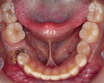
The position of the teeth and the form of the dental arches are determined by the balance of the soft tissues between tongue and lips/cheek. If the tongue’s free movement is restricted, when the lingual frenum is attached too far forward on the tongue, this interferes with, and restricts, function and is called a tongue tie. It can also interfere with speech, hence the expression ‘to be tongue tied’ (Figure 1.15).
Figure 1.15 Tongue tie. Source: Reproduced by kind permission of Jonathan Sandler.
The first appointment Chapter 2
Orthodontic patients are usually referred by their own dentist (their general dental practitioner) for specialist orthodontic treatment.
These referrals can be sent to:
• an orthodontic specialist practitioner
• a community orthodontist
• a consultant orthodontist
• a GDP with enhanced skills.
Some adult patients may choose to self‐refer.
The referring dentist may wish to send the patient to an orthodontist to:
• see and advise
• if there are teeth that are slow to erupt
• if there are teeth that have submerged
• if there are teeth in a self‐damaging position
• see and monitor
• if the patient is dentally too young for treatment
• if there are already signs of adverse dental development, i.e. growth, facial asymmetry or crowding
• see and treat
• if the second dentition has developed but is overcrowded
• if there is a complex problem
• if there is a multidisciplinary need.
The referral letter needs to contain as much relevant information for the orthodontic pra ctitioner as possible.
• clinical reason for referral (what the dentist feels is the problem)
• medical history (if it is helpful to know in advance, e.g. attention deficit hyperactivity disorder, autism, deafness, dental phobia)
• dental history (good oral hygiene, high caries level, etc.)
• any previous orthodontic history (e.g. previous assessment or treatment)
• social history (e.g. supportive family, regular check‐ups)
• what concerns the patient/parent (‘fangs’, teasing)
• whether the patient is bothered at all (quite happy to stay as they are)
• likely compliance (supportive family, the patient is keen).
When the referral letter is received, the patient (or their parent or guardian if they are under age) is sent an appointment.
On the first visit a full orthodontic assessment is carried out (Figures 2.1 and 2.2). This includes:
• checking the name and age of the patient
• what is of concern to the patient
• full medical history, including whether the patient
• has any known allergies (nickel, latex, etc.)
• is currently under the care of a doctor for any reason
• has had any operations
• is taking medication of any kind
• has asthma; if so, which type of inhalers
• has diabetes
• has or had chest or heart conditions.
Patients and/or parents are also asked to fill in and sign a health questionnaire. This should be updated and checked regularly. This also contains details of:
• school or college (day or boarding)
• work
• contact sports
• musical instruments played by mouth
• any digit (thumb or finger) sucking, bruxism (tooth clenching or grinding)
• lip, cheek or tongue jewellery, e.g. studs
• any known allergies, e.g. latex, nickel.
THE CLINICAL ASSESSMENT RECORDS
These begin with extra‐oral features and then move on to intra‐oral ones.
NAME D of B REF/GDP
SKEL PATT
FM ANGLE
ASYMMETRY HABITS/MUSIC/SPORTS
INCS REL
OVERJET normal increased (mm) reduced e/e reversed