Full download Radiography essentials for limited practice e book 5th edition, (ebook pdf) pdf docx
Visit to download the full and correct content document: https://ebookmass.com/product/radiography-essentials-for-limited-practice-e-book-5th -edition-ebook-pdf/
More products digital (pdf, epub, mobi) instant download maybe you interests ...
Lavinu2019s Radiography for Veterinary Technicians E Book 6th Edition, (Ebook PDF)
Community/Public Health Nursing Practice E Book: Health for Families and Populations (Maurer, Community/ Public Health Nursing Practice) 5th Edition, (Ebook PDF)
No part of this publication may be reproduced or transmitted in any form or by any means, electronic or mechanical, including photocopying, recording, or any information storage and retrieval system, without permission in writing from the publisher. Details on how to seek permission, further information about the Publisher’s permissions policies and our arrangements with organizations such as the Copyright Clearance Center and the Copyright Licensing Agency, can be found at our website: www.elsevier.com/permissions.
This book and the individual contributions contained in it are protected under copyright by the Publisher (other than as may be noted herein).
The ARRT does not review evaluate, or endorse publications. Permission to reproduce ARRT copyrighted materials within this publication should not be construed as an endorsement of the publication by the ARRT.
Notices
Knowledge and best practice in this field are constantly changing. As new research and experience broaden our understanding, changes in research methods, professional practices, or medical treatment may become necessary.
Practitioners and researchers must always rely on their own experience and knowledge in evaluating and using any information, methods, compounds, or experiments described herein. In using such information or methods they should be mindful of their own safety and the safety of others, including parties for whom they have a professional responsibility.
With respect to any drug or pharmaceutical products identified, readers are advised to check the most current information provided (i) on procedures featured or (ii) by the manufacturer of each product to be administered, to verify the recommended dose or formula, the method and duration of administration, and contraindications. It is the responsibility of practitioners, relying on their own experience and knowledge of their patients, to make diagnoses, to determine dosages and the best treatment for each individual patient, and to take all appropriate safety precautions.
To the fullest extent of the law, neither the Publisher nor the authors, contributors, or editors, assume any liability for any injury and/or damage to persons or property as a matter of products liability, negligence or otherwise, or from any use or operation of any methods, products, instructions, or ideas contained in the material herein.
Previous editions copyrighted 2013, 2010, 2006, and 2002.
Library of Congress Cataloging-in-Publication Data
Names: Long, Bruce W., author. | Frank, Eugene D., author. | Ehrlich, Ruth
Ann, 1938- author.
Title: Radiography essentials for limited practice / Bruce W. Long, Eugene D.
Frank, Ruth Ann Ehrlich.
Description: Fifth edition. | St. Louis, Missouri : Elsevier, [2017] | Includes bibliographical references and index.
Identifiers: LCCN 2016015047 | ISBN 9780323356237 (pbk. : alk. paper)
Classification: LCC RC78 | NLM WN 200 | DDC 616.07/572 dc23 LC record available at https://lccn.loc.gov/2016015047
Executive Content Strategist: Sonya Seigafuse
Content Development Manager: Billie Sharp
Associate Content Development Specialist: Laurel Shea
Publishing Services Manager: Jeff Patterson
Book Production Specialist: Carol O’Connell
Design Direction: Ashley Miner
Printed in the United States of America
Review board
Kellie S. Cranfill, MSRS, RT(R)(BD), Assistant Professor of Clinical Radiologic and Imaging Sciences, Indiana University
Radiologic and Imaging Sciences Programs, Indianapolis, Indiana
Kathleen M. Drotar, MEd, RT(R)(N)(T), University Department
Chair and Sarasota Campus Program Director, Keiser University Department of Radiologic Technology and Radiation Therapy, Sarasota, Florida
Mary Alice Statkiewicz Sherer, AS, RT(R), FASRT, Medical author, Mount Juliet, Tennessee
Contributing Author
Sharon R. Wartenbee RT(R)(BD), CBDT, FASRT, Senior Diagnostic and Bone Densitometry Technologist, Avera Medical Group McGreevy, Sioux Falls, South Dakota
Preface
Since the publication of the first edition of Radiography Essentials for Limited Practice, information has been continually gathered about the education and work of the limited x-ray machine operator (LXMO). The term “limited operator” and the acronym “LXMO” will be used throughout this book, since these have become the generally accepted terms. However, each state with a licensure law may use different terms. We have included a list of these most current terms, by state, in Appendix A near the end of this book.
Several significant events have impacted the limited operator’s status in the diagnostic imaging profession. The 2004 publication by the American Society of Radiologic Technologists (ASRT) of the Limited X-ray Machine Operator Curriculum was intended to establish national standardized educational guidelines for LXMOs, including clinical and didactic components. An updated ASRT Limited X-ray Machine Operator Curriculum was adopted in 2009. In 2013, the American Registry of Radiologic Technologists (ARRT) adopted a new set of Content Specifications for the Limited Scope of Practice in Radiography examination. A new version is scheduled for adoption in 2018. This examination is used by nearly all states with LXMO licensure laws as their validation of a LXMO’s competency to hold that license. The material in this fifth edition of Radiography Essentials for Limited Practice has been updated to encompass all the concepts included in the current ARRT Content Specifications, providing the most up-to-date learning source for LXMO examination candidates.
National accreditation of LXMO educational programs is now a reality. In January 2012, the Joint Review Committee on Education in Radiologic Technology (JRCERT) began to accredit these programs.
This national organization, itself accredited by the U.S. Department of Education, serves as the programmatic accreditation agency for radiography education programs. It now offers accreditation services to LXMO education programs to promote academic excellence, patient safety, and quality health care delivery. Since the JRCERT will require accredited LXMO programs to adhere to the ASRT LXMO Curriculum, Radiography Essentials for Limited Practice will meet the educational content needs of these programs.
The radiography profession is accepting the limited operators as an integral part of the radiology department by hiring them and allowing them to perform a limited range of radiography examinations. Although many LXMOs work alone or with other LXMOs in small clinics and hospitals, chiropractic clinics, and so forth, an increasing number of them are working alongside radiologic technologists in large clinics, imaging centers, and hospitals throughout the United States. As this new health care worker emerges, strong emphasis is being placed on the education and certification of these individuals. The need for a comprehensive textbook written specifically for limited practice has never been greater.
For this fifth edition we worked with an advisory board and instructors in limited operator schools throughout the United States to determine the content needed to educate limited operators and were challenged by our discovery of the extremely wide range of curriculums. We found schools whose didactic curriculum presentation ranged from as few as 3 days to as many as 9 months. Many had clinical components, and many had none. Obviously, the lack of a national standardized curriculum made our revision work all the more difficult. As we revised the text, we determined that it was best to produce a comprehensive text that would serve the schools that had the most comprehensive curriculums. We used the latest ASRT Limited X-ray Machine Operator Curriculum as a guide. Schools with abbreviated curriculums can use this textbook by selecting those chapters or sections of chapters that apply to their programs.
Our foremost goal for this edition was to ensure that the content of
this book matches that of the textbooks used in the JRCERT-accredited radiography or radiologic technology programs. Information taught to LXMOs should not differ from that taught to radiographers. The same technical exposure factors, radiation protection, and positioning of body parts should be used by both LXMOs and radiographers when performing radiographic procedures. Therefore we strove to make sure that the content of this text paralleled the mainstream radiography textbooks. This also ensures that those graduates of limited schools who must take a limited scope state licensure examination will be prepared to pass the Limited Scope of Practice in Radiography examination administered by the ARRT.
For instructors who teach limited operators, the comprehensive coverage of radiographic topics in this book should facilitate the teaching of any aspect of limited practice. However, because of the wide range of limited operator curriculums, it is up to the instructor, or more specifically the school or its curriculum committee, to determine the scope of what is taught. Some will present all the chapters in this book, whereas others will have time to cover only selected chapters or portions of chapters. To assist instructors, we have developed a comprehensive workbook and licensure exam prep that will help students learn radiologic concepts and prepare for a limited scope state licensure examination. A comprehensive test bank is also available for instructors to use in evaluating student outcomes.
Content and organization
Radiography Essentials for Limited Practice is organized in five parts, each with a different focus.
• Part I, Introduction to Limited Radiography, covers the role of the LXMO as a health care worker, introduces radiographic equipment, and includes material to refresh or improve math skills needed later in the book.
• Part II, X-ray Science, contains clear and concise discussion of all physical principles related to the production or control of the x-ray exposure needed to create medical radiographs. The expanding
world of digital radiography has prompted many updates to chapters in this section.
• Part III, Radiographic Anatomy, Positioning, and Pathology, presents the essential procedural steps required to produce quality radiographs of all body structures.
• Part IV, Professionalism and Patient Care, covers the attitudes, behaviors, knowledge, and skills required to deliver safe and effective patient care.
• Part V, Ancillary Clinical Skills, includes chapters presenting other diagnostic and patient care skills that may be needed in the practice environment where the limited operator frequently works. In addition, a collection of appendices has been included to provide information that an instructor or limited operator may find useful during the educational program or during performance of job duties.
Features
Simplified concepts
Simplified math and physics concepts are presented throughout the text. Many chapters were written to ensure that students focus on the most relevant information. Added art and radiographs augment the text for ease of learning. Over 900 illustrations are provided for reference purposes.
Special boxes
Throughout the text, special boxes are inserted that contain information to reinforce important points in the text. The addition of many new boxes ensures that important concepts are identified.
Step-by-step procedures
The position and procedures chapters contain step-by-step instructions on how to perform each projection. The projections have
been completely revised in this edition to provide quick, easy-tounderstand reference. All the projections were revised to ensure consistency with radiologic technologist textbooks and to ensure that LXMOs perform x-ray procedures exactly the same way.
Learning objectives, key terms, glossary
Learning objectives and key terms highlight important information in each chapter and can be used as review tools. The key terms are bolded throughout the text. Definitions for these terms can be found in the glossary at the end of the book.
New to this edition
The fifth edition incorporates many outstanding features. Up-to-date information from the new ASRT Limited X-ray Machine Operator Curriculum covers limited practice radiography and state-by-state guidelines for licensure and testing.
ARRT limited scope of practice in radiography examination
The text was revised so that complete coverage of all the subjects students need to know to pass the ARRT Limited Scope of Practice in Radiography examination is included. Although the text covers the Content Specifications of the Limited Scope of Practice in Radiography examination, most chapters contain additional relevant information for limited practice.
New content
The introductory and x-ray science sections of the book have been extensively updated to reflect the current practice environment of the Limited Operator, the latest ASRT Limited X-ray Machine Operators Curriculum, and the latest ARRT Content Specifications for the Limited Scope of Practice in Radiography examination. Photos of
modern digital radiography equipment have been added to augment the more extensive coverage of digital imaging principles. All chapters have been updated with the international system of units (SI), which is broadly used. All screen-film imaging and film processing material has been moved to an appendix, if needed. It should be noted that screen-film imaging and film processing will be removed from the ARRT Contents in 2018.
The radiographic positioning material has been updated to include the latest recommendations of image receptor and radiation field sizes. All positioning instructions reflect the latest material contained in the mainstream positioning and procedures textbooks used in accredited radiography programs. Recommendations have been added for modifications to procedures needed to properly image the obese patient. Positioning photographs in Chapters 13, 14, and 15 have been updated to reflect use of digital image receptors.
The patient care and ancillary skills sections have been entirely updated to reflect current national standards and recognized best practices.
A final note on the use of this text: The ARRT does not review, evaluate, or endorse publications. Permission to reproduce ARRT copyrighted material should not be construed as an endorsement of the publication by the ARRT.
Learning aids for the student
Workbook and licensure examination prep for radiography essentials for limited practice
This workbook matches the textbook chapter for chapter, challenging students with a variety of exercises. Included are multiple-choice and fill-in-the-blank questions, matching exercises, and numerous labeling exercises of illustrations and radiographic images. The workbook is designed specifically to focus on the most important concepts contained in each chapter of the textbook. A Challenge Exercise is
featured at the end of most chapters to assist the student in reviewing the key concepts presented in the workbook chapter. In addition, a Study Plan has been added to help students stay organized and on track as they prepare for their state licensure examination. The workbook also contains full-length, simulated limited licensure examinations to provide practice and identify deficiencies in knowledge for students preparing to take a state licensure examination. The answers for the workbook questions can be found on the free Evolve site.
Teaching aids for the instructor
Evolve
Evolve is an interactive learning environment designed to work in coordination with Radiography Essentials for Limited Practice. This website contains the Image Collection (all images in book), Microsoft PowerPoint slide presentations (for each book chapter), Test Bank, and the answers for the workbook questions. In addition, instructors may use Evolve to provide an Internet-based course component that reinforces and expands on the concepts delivered in class. Evolve may be used to publish the class syllabus, outlines, and lecture notes; set up “virtual office hours” and email communication; share important dates and information through the online class calendar; and encourage student participation through chat rooms and discussion boards. Evolve allows instructors to post examinations and manage their grade books online. For more information about how to register for access to these free Evolve resources, visit http://evolve.elsevier.com/Long/radiographylimited or contact an Elsevier sales representative.
PART I Introduction to Limited Radiography
OUTLINE
1. Role of the limited X-ray machine operator
2. Introduction to radiographic equipment
3. Basic mathematics for limited operators
CHAPTER 1
Role of the limited X-ray machine operator
Learning objectives
At the conclusion of this chapter, you will be able to:
• Compare the role of the limited x-ray machine operator with that of the registered radiologic technologist
• Identify the discoverer of x-rays and the date of the discovery
• Explain the primary purposes of the American Registry of Radiologic Technologists, American Society of Radiologic Technologists, and Joint Review Committee on Education in Radiologic Technology
• Determine the legal requirements for the practice of radiography in your state
• Describe the typical work environment of the limited x-ray machine operator
• Describe in a general way the duties of a limited x-ray machine operator
KEY TERMS
American Registry of Radiologic Technologists (ARRT)
American Society of Radiologic Technologists (ASRT)
back office
front office
Joint Review Committee on Education in Radiologic Technology (JRCERT)
limited operator
limited x-ray
limited x-ray machine operator (LXMO)
medical assistant (MA)
radiograph
radiographer
radiologist
reciprocity
Welcome to the fascinating field of radiography! You are beginning a study of the art and science needed to create images of the internal structures of the human body. The images you create will aid physicians in diagnosis and will help patients receive treatment needed to promote or regain health. This is a vital role in the health care delivery system, one that requires knowledge, skill, judgment, integrity, and dedication.
Radiography
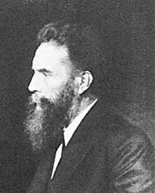
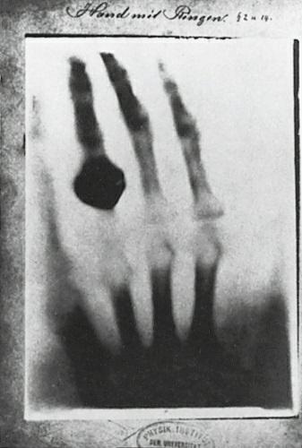
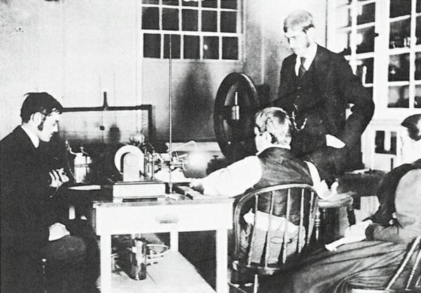
X-rays were discovered on November 8, 1895, by Wilhelm Conrad Roentgen (Fig. 1-1) at the University of Würzburg in Germany. Roentgen was a teacher and researcher with a special interest in the conduction of high-voltage electricity through low-vacuum tubes. His discovery of the x-ray during his regularly planned experiments was accidental. The first x-ray image by Roentgen was of his wife Bertha’s hand using a 15-minute exposure (Fig. 1-2). The first radiographers were scientists and physicians who experimented with primitive x-ray
apparatus to make x-ray images of the human body (Fig. 1-3). Soon these pioneers trained their assistants to make these “x-ray pictures,” now called radiographs, and the profession of radiography was born.
FIG. 1-1 Wilhelm Conrad Roentgen (1845-1923) He discovered xrays on November 8, 1895
1-2 The
FIG.
first radiograph, which demonstrates the bones of the hand of Roentgen’s wife Bertha with a ring on one finger.
American Society of Radiologic Technologists
Early radiographers soon began meeting to share their knowledge. The organization, now called the American Society of Radiologic Technologists (ASRT), was founded in Chicago in 1920. It is the world’s oldest and largest professional radiologic science organization. The ASRT provides many services to its members, including continuing education, a professional journal, a newsletter, guidelines and assistance for radiography educators, and an annual national meeting. The ASRT publishes a Code of Ethics that can be found at www.asrt.org.
American Registry of Radiologic Technologists
Through the efforts of this organization, the American Registry of
FIG. 1-3 The first clinical radiograph in the United States was made at Dartmouth College in 1896.
Radiologic Technologists (ARRT) was formed in 1922 to establish standards and examinations necessary to certify radiologic technologists. Radiologic technologists certified by ARRT use the initials RT(R) after their names. This abbreviation means registered technologist (radiography). Registered technologists who have passed the ARRT examination in radiography are referred to as radiographers. Do not get radiographer mixed up with radiologist. A radiologist is a physician who specializes in radiography. Radiographers and radiologists work together in radiology departments. The ARRT publishes a Code of Ethics, which are aspirational statements. This can be found in Chapter 20 or at www.arrt.org. It also publishes an important document called the Rules of Ethics, which are mandatory and enforceable statements. This document, found in Appendix B, discusses the minimally acceptable professional conduct that those in radiology should adhere to. An important document, the Task Inventory for Limited Scope of Practice in Radiography, is also published by the ARRT. This document, which identifies for the limited operator all the tasks that are tested on the certification examination, can be found in Appendix M.
The profession of radiography has expanded to include a variety of imaging and treatment modalities, including those listed in Box 1-1. Many of these modalities require specialized training beyond that needed for certification by the ARRT in radiography. The newest role for the radiographer is that of radiologist assistant (RA). These radiographers obtain additional schooling, usually at the graduate level, and perform limited duties that a radiologist typically carries out, such as fluoroscopy and initial readings of radiographs.
Box 1-1
Imaging and Treatment Modalities in Radiology
Angiography: imaging of blood vessels with the injection of special
compounds called contrast media
Bone densitometry (BD): art and science of measuring the bone mineral content and density of specific skeletal sites or the whole body
Computed tomography (CT): computerized x-ray system that provides axial images (transverse “slices”) of all parts of the body
Fluoroscopy: real-time viewing of x-ray images in motion
Magnetic resonance imaging (MRI): computerized imaging system that uses a powerful magnetic field and radiofrequency pulses to produce images of all parts of the body
Mammography: x-ray imaging of the breast using a special x-ray machine
Nuclear medicine (NM): injection or ingestion of radioactive materials and recording of their uptake in the body using a gamma camera
Positron emission tomography (PET): highly sophisticated computerized form of nuclear medicine imaging
Radiation therapy: treatment of malignant diseases using radiation
Sonography: imaging of soft tissue structures using sound echoes. This modality is also referred to as “ultrasound”
Joint review committee on education in radiologic technology
The Joint Review Committee on Education in Radiologic Technology (JRCERT) is the national organization that formally conducts the accreditation of schools of radiologic technology. The JRCERT was formed in 1969 and accredits, as of this printing, 637 radiography programs in the United States. The JRCERT publishes the Standards for an Accreditation Program in Radiography document, which tells colleges what standards are required to be accredited. Certification in radiography requires at least 2 years of education (six college semesters) in an accredited program that includes
comprehensive academic coursework in the sciences. These programs are affiliated with acute care general hospitals to provide extensive clinical experience in the care of patients who are severely ill or injured. A growing number of programs are changing to a 4-year bachelor’s degree curriculum.
Effective January 1, 2012, the JRCERT began to accredit limited scope x-ray machine operator educational programs. This accreditation is designed to promote academic excellence, patient safety, and quality health care. Many states currently have limited xray machine operator programs of anywhere from 10 weeks to 9 months. Many of these programs are expected to apply for this national accreditation and recognition. Documents from the JRCERT can be obtained at www.jrcert.org.
To understand how these three organizations work together to develop a professional radiographer, the following scenario is provided. Individuals interested in becoming a radiographer must qualify and enter a JRCERT-accredited radiography program. While in the program they complete a comprehensive curriculum developed by the ASRT. On graduation, these students take the certification examination in radiography given by the ARRT. ARRT-registered technologists can perform all diagnostic x-ray examinations and operate complex radiography equipment in all 50 states. The titles registered technologist and radiographer are used throughout this text to denote these professionals.
Limited X-ray machine operator
The ASRT recently published the Limited X-ray Machine Operator Curriculum to support the education of limited operators of x-ray equipment. The curriculum allows states and faculty flexibility in the development of limited curricula to meet the needs of individuals performing diagnostic x-ray procedures within a limited scope. The ASRT officially terms the limited operator, a limited x-ray machine operator (LXMO). The titles limited operator and LXMO are used throughout this text to denote limited operators of x-ray equipment. The ARRT and JRCERT also use these titles in their publications.
LXMOs are encouraged to join the ASRT to support the profession and to obtain continuing education.
Limited x-ray work is regulated within the offices of each state’s Department of Health. The majority of states that have regulations for those who operate x-ray equipment use the above titles for individuals who perform limited x-rays. However, a few states use other terms such as basic x-ray machine operator, practical x-ray machine operator, limited radiologic technologist, or limited radiographer.
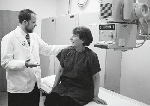
Limited x-ray is practiced primarily in clinics and physicians’ offices (Fig. 1-4). In some areas, however, limited operators are employed in hospitals, a practice that is expanding.
Limited x-ray developed as nurses, medical assistants, chiropractic assistants, laboratory technologists, and health care office personnel were trained to perform limited aspects of radiography in addition to their primary duties. It is called limited because the scope of practice is restricted compared with that of registered technologists. Limited xray practice does not involve the use of contrast media for the imaging