University of Hertfordshire Hatfield, Hertfordshire, UK
For further volumes: http://www.springer.com/series/7651
For over 35 years, biological scientists have come to rely on the research protocols and methodologies in the critically acclaimed Methods in Molecular Biology series. The series was the first to introduce the step-by-step protocols approach that has become the standard in all biomedical protocol publishing. Each protocol is provided in readily-reproducible step-bystep fashion, opening with an introductory overview, a list of the materials and reagents needed to complete the experiment, and followed by a detailed procedure that is supported with a helpful notes section offering tips and tricks of the trade as well as troubleshooting advice. These hallmark features were introduced by series editor Dr. John Walker and constitute the key ingredient in each and every volume of the Methods in Molecular Biology series. Tested and trusted, comprehensive and reliable, all protocols from the series are indexed in PubMed.
Chaperones
Methods and Protocols
Second Edition
Edited by Stuart K. Calderwood
Harvard Medical School, Beth Israel Deaconess Medical Center, Boston, MA, USA
Thomas L. Prince
Danville, PA, USA
Editors
Stuart K. Calderwood
Harvard Medical School
Beth Israel Deaconess Medical Center
Boston, MA, USA
Thomas L. Prince Danville, PA, USA
ISSN 1064-3745ISSN 1940-6029 (electronic)
Methods in Molecular Biology
ISBN 978-1-0716-3341-0ISBN 978-1-0716-3342-7 (eBook) https://doi.org/10.1007/978-1-0716-3342-7
This work is subject to copyright. All rights are solely and exclusively licensed by the Publisher, whether the whole or part of the material is concerned, specifically the rights of translation, reprinting, reuse of illustrations, recitation, broadcasting, reproduction on microfilms or in any other physical way, and transmission or information storage and retrieval, electronic adaptation, computer software, or by similar or dissimilar methodology now known or hereafter developed.
The use of general descriptive names, registered names, trademarks, service marks, etc. in this publication does not imply, even in the absence of a specific statement, that such names are exempt from the relevant protective laws and regulations and therefore free for general use.
The publisher, the authors, and the editors are safe to assume that the advice and information in this book are believed to be true and accurate at the date of publication. Neither the publisher nor the authors or the editors give a warranty, expressed or implied, with respect to the material contained herein or for any errors or omissions that may have been made. The publisher remains neutral with regard to jurisdictional claims in published maps and institutional affiliations.
Cover Caption: The predicted protein structure of HSF1, a key component of the heat shock response and guardian of the proteome, is shown on the cover. Model structure generated by AlphaFold (Jumper, J. et al. Nature, 2021; Varadi, M. et al. Nucleic Acid Research, 2021). HSF1 color scheme based on studies by Kijima, T. et al. Scientific Reports, 2018.
This Humana imprint is published by the registered company Springer Science+Business Media, LLC, part of Springer Nature.
The registered company address is: 1 New York Plaza, New York, NY 10004, U.S.A.
Dedication
(SKC) This volume is dedicated to the memory of my sister Carole who passed away in 2021. I would particularly like to thank my wife Laura for her patience and support in the long hours science demands.
Preface
Life is a dynamic and complex interplay of energy and mass within space and time. Biological life depends on proteins to facilitate the processes that allow communities of cells to cooperate and compete to propagate their genetic information. Proteins are linear polymers that fold into intricate three-dimensional structures that provide the essential functions of life. Here nature demonstrates the common notion that structure determines function. Most proteins are able to spontaneously fold into their proper structure given adequate space. However, in the crowded confines of the cell, many proteins require the aid of a special cadre of proteins known as molecular chaperones. These chaperone proteins help other client proteins avoid misfolding, aggregation, and loss of function. Furthermore, molecular chaperones maintain cellular proteostasis by facilitating protein transport and degradation. The cellular stress response is the biological process for enabling the explosive expression of molecular chaperones in response to proteostatic perturbations. And since heat is one of the oldest and most studied forms of proteotoxic stress, many molecular chaperones are referred to as heat shock proteins (HSPs). Expression of HSP genes is initiated by the activation of heat shock factors (HSFs) that bind to the DNA promoters of chaperones and other stress-related genes to initiate or extend transcription. HSPs once expressed may be post-translationally modified as they reestablish proteostasis while also contributing to the modulation of immunity, extracellular communication, and cell growth. Life is shaped by molecular chaperones and the stress response. Almost all aspects of health and disease are affected by the ability to maintain proteostasis. Unchecked chaperone and HSF activity enables malignant cell growth that gives way to cancer. In contrast, reduced chaperone activity allows for accumulated protein aggregation that leads to neurodegeneration. All while, the stress response influences inflammation and immunity.
Our pursuit to better understand molecular chaperones and the stress response along with the desire to pass this knowledge on to future generations of scientist is represented by this book. In this volume, we have complied 23 chapters of methods and topics focused on investigating the role of chaperones. Some chapters are succinct; some chapters are extensive; all address important topics within the field. We appreciate the expertise, time, and effort the authors have provided.
We commence the volume analyzing the initiation and regulation of the stress response. In Chap. 1, Ackerman et al. describe the use of a plasmid-based reporter for monitoring the heat shock response. In Chaps. 2 and 3, Heyoun Bunch analyzes the stress response at the level of transcription and RNA elongation. Holton et al. in Chap. 4 describes their workflow for studying RNA-seq data relative to the stress response. Ritwick Sawarkar in Chap. 5 explores the role of Hsp90 in gene expression through chromosome-immunoprecipitation. Yuka Okusha next details the use of microRNA to regulate HSP levels.
Chapters 7 through 14 revolve around major features of chaperone function and biology: post-translational modifications and protein-protein interactions. Jin et al. explain the effects of key HSF1 phosphorylation sites on transcriptional activity. In Chap. 8, Chakraborty et al. describe a fluorescent complementation assay to analyze Hsp90 and client protein interactions. Edkins and Blatch further elucidate and compare complementation assays for protein-protein interactions studies. Tonui et al. in Chap. 10 explain their protein aggregation assay for measuring chaperone holdase activity. Sager et al. explore the vii
detectionofHsp90pos thechallengesofaddre Chap. 13 describetheir dynamics.Thestudyof amidegelelectrophores t-translational modifications. In Chap. 12, Ono and Eguchi discuss ssing HSP depletion and isoform compensation. Baldan et al. in proximity ligation assay to analyze HSP protein-protein interaction the epichaperone complexes through the use of native polyacrylis is described by Roychowdhury et al in Chap. 14
Chapter 15 through 21 focus on the emerging role of extracellular HSPs. Borges et al. provide an update on molecular chaperone and extracellular receptor interactions. In Chap. 15, Weng et al. expound the use of Hsp70 in antigen presentation and possible vaccine generation. Votra et al. assess the effect of Hsp90 activity on extracellular proteases. Ono and Eguchi follow up with proteomic profiling of chaperone-rich extracellular vesicles with mass spectrometry. In Chap. 19, Chang et al. provide a protocol for analyzing extracellular Hsp90 levels. Bavisotto et al. in Chap. 20 characterize the role of extracellular Hsp60 in health and disease.
The final Chaps. 22, 23, and 24 explore the use chaperones as biomarkers. Kawai et al develop a method for profiling HSP isoforms in cancer specimen. This is followed by a chapter on chaperone focused cancer database analysis for prognostic biomarkers. Finally, Dezfouli et al. utilize flow cytometry and enzyme-linked immunoassays to profile Hsp70 localization in cancer.
Boston, MA, USA Stuart K. Calderwood Danville, PA, USA
Thomas L. Prince
Acknowledgments
This book is dedicated to the next generation of scientists including my (T.L.P.) boys Jessup and Lincoln along with my wife, Bourdana, and to the memory of my mother Suzanne Prince. I would also like to acknowledge the Thomas Beaver Free Library in Danville, PA, for providing me a quiet place to get work done.
1 Monitoring of the Heat Shock Response with a Real-Time Luciferase Reporter
Andrew Ackerman, Toshiki Kijima, Takanori Eguchi, and Thomas L. Prince
2 Studying RNA Polymerase II Promoter-Proximal Pausing by In Vitro Immobilized Template and Transcription Assays
Heeyoun Bunch
3 Role of Heat Shock Factors in Stress-Induced Transcription: An Update
Heyoun Bunch and Stuart K. Calderwood
4 A Workflow Guide to RNA-Seq Analysis of Chaperone
Kristina M. Holton, Richard M. Giadone, Benjamin J. Lang, and Stuart K. Calderwood
5 Chromatin Immunoprecipitation (ChIP) of Heat Shock Protein 90 (Hsp90)
Ritwick Sawarkar
6 Transfection and Thermotolerance Methods for Analysis of miR-570 Targeting the HSP Chaperone Network
Yuka Okusha and Stuart K. Calderwood
7 Targeted Replacement of HSF1 Phosphorylation Sites at S303/S307 with Alanine Residues in Mice Increases Cell Proliferation and Drug Resistance
Xiongjie Jin, Demetrius Moskophidis, and Nahid F. Mivechi
8 Bimolecular Fluorescence Complementation Assay to Evaluate HSP90-Client Protein Interactions in Cells
Abir Chakraborty, Gregory L. Blatch, and Adrienne L. Edkins
9 Complementation Assays for Co-chaperone Function
Adrienne L. Edkins and Gregory L. Blatch
10 Optimized Microscale Protein Aggregation Suppression Assay: A Method for Evaluating the Holdase Activity of Chaperones
Ronald Tonui, Ruth O. John, and Adrienne L. Edkins
11 Detecting Posttranslational Modifications of Hsp90 Isoforms .
Rebecca A. Sager, Sarah J. Backe, Len Neckers, Mark R. Woodford, and Mehdi Mollapour
12 Multiple Targeting of HSP Isoforms to Challenge Isoform Specificity and Compensatory Expression
Kisho Ono and Takanori Eguchi
13 Using a Modified Proximity Ligation Protocol to Study the Interaction Between Chaperones and Associated Proteins . . . . .
Simone Baldan, Anatoli B. Meriin, and Michael Y. Sherman
163
14 Use of Native-PAGE for the Identification of Epichaperomes in Cell Lines . . . . 175
Tanaya Roychowdhury, Anand R. Santhaseela, Sahil Sharma, Palak Panchal, Anna Rodina, and Gabriela Chiosis
15 Molecular Chaperone Receptors: An Update 193
Thiago J. Borges, Ayesha Murshid, Jimmy Theriault, and Stuart K. Calderwood
16 A Novel Heat Shock Protein 70-Based Vaccine Prepared from DC Tumor Fusion Cells: An Update .
Desheng Weng, Stuart K. Calderwood, and Jianlin Gong
17 Methods to Assess the Impact of Hsp90 Chaperone Function on Extracellular Client MMP2 Activity 221
SarahBeth D. Votra, Deema Alsalih, and Dimitra Bourboulia
18 Proteomic Profiling of the Extracellular Vesicle Chaperone in Cancer 233
Kisho Ono and Takanori Eguchi
19 A Modified Differential Centrifugation Protocol for Isolation and Quantitation of Extracellular Heat Shock Protein 90 (eHsp90) .
Cheng Chang, Xin Tang, and Wei Li
20 Immunohistochemistry of Human Hsp60 in Health and Disease: Recent Advances in Immunomorphology and Methods for Assessing the Chaperonin in Extracellular Vesicles 263
Celeste Caruso Bavisotto, Francesco Cappello, Everly Conway de Macario, Alberto J. L. Macario, and Francesca Rappa
21 Multiplex Immunostaining Method to Distinguish HSP Isoforms in Cancer Tissue Specimens 281
Hotaka Kawai, Kisho Ono, and Takanori Eguchi
22 Large-Scale Databases and Portals on Cancer Genome to Analyze Chaperone Genes Correlated to Patient Prognosis
Kisho Ono and Takanori Eguchi
23 Immunohistochemical, Flow Cytometric, and ELISA-Based Analyses of Intracellular, Membrane-Expressed, and Extracellular Hsp70 as Cancer Biomarkers
Ali Bashiri Dezfouli, Stefan Stangl, Gemma A. Foulds, Philipp Lennartz, Geoffrey J. Pilkington, A. Graham Pockley, and Gabriele Multhoff
293
307
Contributors
ANDREW ACKERMAN • Geisinger Clinic, Danville, PA, USA
DEEMA ALSALIH • Department of Urology, SUNY Upstate Medical University, Syracuse, NY, USA
SARAH J. BACKE • Department of Urology, SUNY Upstate Medical University, Syracuse, NY, USA
SIMONE BALDAN • MRC Toxicology Unit, University of Cambridge, Cambridge, United Kingdom
GREGORY L. BLATCH • Biomedical Biotechnology Research Unit (BioBRU), Department of Biochemistry and Microbiology, Rhodes University, Makhanda, South Africa; Biomedical Research and Drug Discovery Research Group, Faculty of Health Sciences, Higher Colleges of Technology, Sharjah, United Arab Emirates; The Vice Chancellery, The University of Notre Dame Australia, Fremantle, WA, Australia
GREGORY L. BLATCH • Biomedical Biotechnology Research Unit (BioBRU), Department of Biochemistry and Microbiology, Rhodes University, Makhanda, South Africa; Biomedical Research and Drug Discovery Research Group, Faculty of Health Sciences, Higher Colleges of Technology, Sharjah, United Arab Emirates; The University of Notre Dame Australia, Fremantle, WA, Australia
THIAGO J. BORGES • Center for Transplantation Sciences, Department of Surgery, Massachusetts General Hospital, Harvard Medical School, Boston, MA, USA
DIMITRA BOURBOULIA • Department of Urology, SUNY Upstate Medical University, Syracuse, NY, USA; Upstate Cancer Center, SUNY Upstate Medical University, Syracuse, NY, USA; Department of Biochemistry and Molecular Biology, SUNY Upstate Medical University, Syracuse, NY, USA
HEEYOUN BUNCH • Department of Applied Biosciences, Kyungpook National University, Daegu, Republic of Korea
STUART K. CALDERWOOD • Molecular and Cellular Radiation Oncology, Beth Israel Deaconess Medical Center, Harvard Medical School, Boston, MA, USA; Department of Radiation Oncology, Beth Israel Deaconess Medical Center, Boston, MA, USA; Beth Israel Deaconess Medical Center, Harvard Medical School, Boston, MA, USA
FRANCESCO CAPPELLO • Department of Biomedicine, Neurosciences and Advanced Diagnostic (BiND), Human Anatomy Section, University of Palermo, Palermo, Italy
CELESTE CARUSO BAVISOTTO • Department of Biomedicine, Neurosciences and Advanced Diagnostic (BiND), Human Anatomy Section, University of Palermo, Palermo, Italy
ABIR CHAKRABORTY • Biomedical Biotechnology Research Unit (BioBRU), Department of Biochemistry and Microbiology, Rhodes University, Makhanda, South Africa
CHENG CHANG • Department of Dermatology the Norris Comprehensive Cancer Centre, University of Southern California Keck Medical Center, Los Angeles, CA, USA
GABRIELA CHIOSIS • Program in Chemical Biology, Memorial Sloan Kettering Cancer Center, New York, NY, USA
EVERLY CONWAY DE MACARIO • Department of Microbiology and Immunology, School of Medicine, University of Maryland at Baltimore-Institute of Marine and Environmental Technology (IMET) – Rita Rossi Colwell Center, Baltimore, MD, USA
ALI BASHIRI DEZFOULI • Radiation Immuno-Oncology Group, Center for Translational Cancer Research Technische Universit € at Mu¨nchen (TranslaTUM), Department of Radiation Oncology, Klinikum rechts der Isar, Technische Universit € at Mu¨nchen (TUM), Munich, Germany
ADRIENNE L. EDKINS • Biomedical Biotechnology Research Unit (BioBRU), Department of Biochemistry and Microbiology, Rhodes University, Makhanda, South Africa
TAKANORI EGUCHI • Department of Dental Pharmacology, Graduate School of Medicine, Dentistry and Pharmaceutical Sciences, Okayama University, Okayama, Japan; Department of Dental Pharmacology, Faculty of Medicine, Dentistry and Pharmaceutical Sciences, Okayama University, Okayama, Japan
GEMMA A. FOULDS • John van Geest Cancer Research Centre, School of Science and Technology, Nottingham Trent University, Nottingham, UK
RICHARD M. GIADONE • Department of Stem Cell and Regenerative Biology, Harvard University, Cambridge, MA, USA; Harvard Stem Cell Institute, Cambridge, MA, USA; Stanley Center for Psychiatric Research, Broad Institute of MITand Harvard, Cambridge, MA, USA
JIANLIN GONG • Department of Medicine, Boston University School of Medicine, Boston, MA, USA
KRISTINA M. HOLTON • Department of Stem Cell and Regenerative Biology, Harvard University, Cambridge, MA, USA; Harvard Stem Cell Institute, Cambridge, MA, USA; Stanley Center for Psychiatric Research, Broad Institute of MITand Harvard, Cambridge, MA, USA
XIONGJIE JIN • Molecular Chaperone Biology, Medical College of Georgia, Augusta, GA, USA; Georgia Cancer Center, Augusta University, Augusta, GA, USA
RUTH O. JOHN • Biomedical Biotechnology Research Unit (BioBRU), Department of Biochemistry and Microbiology, Rhodes University, Makhanda, South Africa
HOTAKA KAWAI • Department of Oral Pathology and Medicine, Faculty of Medicine, Dentistry and Pharmaceutical Sciences, Okayama University, Okayama, Japan
TOSHIKI KIJIMA • Department of Urology, Dokkyo Medical University, Tochigi, Japan
BENJAMIN J. LANG • Department of Radiation Oncology, Beth Israel Deaconess Medical Center, Boston, MA, USA
PHILIPP LENNARTZ • Radiation Immuno-Oncology Group, Center for Translational Cancer Research Technische Universit € at Mu¨nchen (TranslaTUM), Department of Radiation Oncology, Klinikum rechts der Isar, Technische Universitat Mu¨nchen (TUM), Munich, Germany
WEI LI • Department of Dermatology the Norris Comprehensive Cancer Centre, University of Southern California Keck Medical Center, Los Angeles, CA, USA
ALBERTO J. L. MACARIO • Department of Microbiology and Immunology, School of Medicine, University of Maryland at Baltimore-Institute of Marine and Environmental Technology (IMET) – Rita Rossi Colwell Center, Baltimore, MD, USA
ANATOLI B. MERIIN • Department of Biochemistry, Boston University, Boston, MA, USA
NAHID F. MIVECHI • Molecular Chaperone Biology, Medical College of Georgia, Augusta, GA, USA; Georgia Cancer Center, Augusta University, Augusta, GA, USA; Department of Radiation Oncology, Augusta University, Augusta, GA, USA
MEHDI MOLLAPOUR • Department of Urology, SUNY Upstate Medical University, Syracuse, NY, USA; Department of Biochemistry and Molecular Biology, SUNY Upstate Medical University, Syracuse, NY, USA
DEMETRIUS MOSKOPHIDIS • Molecular Chaperone Biology, Medical College of Georgia, Augusta, GA, USA; Georgia Cancer Center, Augusta University, Augusta, GA, USA; Department of Medicine, Augusta University, Augusta, GA, USA
GABRIELE MULTHOFF • Radiation Immuno-Oncology Group, Center for Translational Cancer Research Technische Universit € at Mu¨nchen (TranslaTUM), Department of Radiation Oncology, Klinikum rechts der Isar, Technische Universit € at Mu¨nchen (TUM), Munich, Germany
AYESHA MURSHID • Molecular and Cellular Radiation Oncology, Beth Israel Deaconess Medical Center, Harvard Medical School, Boston, MA, USA
LEN NECKERS • Urologic Oncology Branch, Center for Cancer Research, National Cancer Institute, Bethesda, MD, USA
YUKA OKUSHA • Beth Israel Deaconess Medical Center, Harvard Medical School, Boston, MA, USA; JSPS Overseas Research Fellow, Tokyo, Japan
KISHO ONO • Department of Oral and Maxillofacial Surgery, Okayama University Hospital, Okayama, Japan
PALAK PANCHAL • Program in Chemical Biology, Memorial Sloan Kettering Cancer Center, New York, NY, USA
GEOFFREY J. PILKINGTON • Brain Tumour Research Centre, School of Pharmacy and Biomedical Sciences, Institute of Biomedical and Biomolecular Sciences, University of Portsmouth, Portsmouth, UK
A. GRAHAM POCKLEY • John van Geest Cancer Research Centre, School of Science and Technology, Nottingham Trent University, Nottingham, UK
THOMAS L. PRINCE • Ranok Therapeutics, Danville, PA, USA
FRANCESCA RAPPA • Department of Biomedicine, Neurosciences and Advanced Diagnostic (BiND), Human Anatomy Section, University of Palermo, Palermo, Italy
ANNA RODINA • Program in Chemical Biology, Memorial Sloan Kettering Cancer Center, New York, NY, USA
TANAYA ROYCHOWDHURY • Program in Chemical Biology, Memorial Sloan Kettering Cancer Center, New York, NY, USA
REBECCA A. SAGER • Department of Urology, SUNY Upstate Medical University, Syracuse, NY, USA
ANAND R. SANTHASEELA • Program in Chemical Biology, Memorial Sloan Kettering Cancer Center, New York, NY, USA
RITWICK SAWARKAR • Department of Genetics, Medical Research Council (MRC), University of Cambridge, Cambridge, UK
SAHIL SHARMA • Program in Chemical Biology, Memorial Sloan Kettering Cancer Center, New York, NY, USA
MICHAEL Y. SHERMAN • Department of Molecular Biology, Ariel University, Ariel, Israel
STEFAN STANGL • Radiation Immuno-Oncology Group, Center for Translational Cancer Research Technische Universit € at Mu¨nchen (TranslaTUM), Department of Radiation Oncology, Klinikum rechts der Isar, Technische Universitat Mu¨nchen (TUM), Munich, Germany
XIN TANG • Department of Dermatology the Norris Comprehensive Cancer Centre, University of Southern California Keck Medical Center, Los Angeles, CA, USA
JIMMY THERIAULT • Molecular and Cellular Radiation Oncology, Beth Israel Deaconess Medical Center, Harvard Medical School, Boston, MA, USA
RONALD TONUI • Biomedical Biotechnology Research Unit (BioBRU), Department of Biochemistry and Microbiology, Rhodes University, Makhanda, South Africa
xviContributors
SARAHBETH D. VOTRA • Department of Urology, SUNY Upstate Medical University, Syracuse, NY, USA
DESHENG WENG • Department of Medicine, Boston University School of Medicine, Boston, MA, USA
MARK R. WOODFORD • Department of Urology, SUNY Upstate Medical University, Syracuse, NY, USA; Department of Biochemistry and Molecular Biology, SUNY Upstate Medical University, Syracuse, NY, USA
Chapter 1
Monitoring of the Heat Shock Response with a Real-Time Luciferase Reporter
Andrew Ackerman, Toshiki Kijima, Takanori Eguchi, and Thomas L. Prince
Abstract
The heat shock response (HSR) is a cellular mechanism for counteracting acute proteotoxic stress. In eukaryotes, transcriptional activation of the HSR is regulated by heat shock factor 1 (HSF1). Activation of HSF1 induces the expression of heat shock proteins (HSPs) that function as molecular chaperones to fold and maintain the three-dimensional structure of misfolded proteins. The regulation of the degree and duration of the HSR is controlled by multiple biochemical mechanisms that include posttranslational modification of HSF1 and numerous protein–protein interactions. In this chapter, we describe a method to evaluate the activation and deactivation of the HSR at the transcriptional level using a short half-life luciferase reporter assay. This assay can be used to further characterize the HSR or as a screen for small molecule inducers, amplifiers, or repressors.
Key words Heat shock response, Heat shock factor 1 (HSF1), Heat shock protein 90 (HSP90), Luciferase assay, Drug screen
1 Introduction
The heat shock response (HSR) is an evolutionarily conserved cytoprotective mechanism induced by proteotoxic stress. The HSR is essential for maintaining proteostasis and plays a role in overall health and longevity [1]. Heat shock factor 1 (HSF1) is the master transcription factor responsible for initiating the HSR and inducing the expression of a variety of genes, most notably heat shock proteins (HSP) [2, 3]. Through a mechanism incompletely understood, proteotoxic stress initiates the release of HSF1 from bound HSP complexes to homotrimerize, translocates from the cytosol into the nucleus, and binds degenerate heat shock elements (HSE, ideally represented as GAAnnTTCnnGAA) across the genome and within promoters of transcriptional target genes [4, 5]. Preloaded and paused RNA polymerase II within HSP
genes are activated by HSF1-binding to resume transcription elongation resulting in the rapid production of mRNA transcripts [6, 7]. Once stress has abated and proteostasis restored, HSF1trimers are dissembled by newly synthesized HSPs such as HSP90 and HSP70 along with other cellular components [8–10]. Throughout this process, HSF1 is heavily posttranslationally modified and interacts with numerous cellular components that may also be modified [11–14]. These modifications and interactions af fect the degree and duration of the HSR that is often amplified in cancer and reduced in neurodegenerative diseases [15, 16]. Understanding how to effectively modulate the HSR through small molecule therapies and lifestyle has the potential to positively influence disease outcomes and improve overall health.
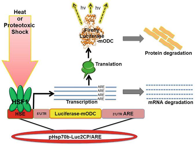
Being able to monitor real-time expression of HSP gene products is essential for determining the degree and duration of the HSR. Previous versions of luciferase reporter assays are useful for measuring the induction and degree of the HSR but not effective at determining the attenuation of transcription and the actual duration of the response. Here we describe the use of a destabilized luciferase reporter assay driven by the heat-inducible HSP70B gene promoter for real-time monitoring of the HSR in mammalian cell culture (Fig. 1). The HSP70B gene (HSPA7 locus) is one of the most strongly expressed human transcripts induced by proteotoxic stress, despite not encoding a known functional protein [17]. The reporter assay is derived from a plasmid developed by Younis et al.
Fig. 1 Cartoon of real-time detection of induction of the HSR using destabilized luciferase reporter
[18]. For this real-time HSR reporter assay, a plasmid encoding an HSP70B promoter-driven firefly luciferase linked to a mouse ornithine decarboxylase (mODC) domain open reading frame (ORF) followed by a 3′untranslated region (UTR) AU-rich element (ARE) was constructed. The MODC contains a PEST sequence that is promptly ubiquitinated resulting in degradation of the luciferase, while the 3′-UTR ARE induces decay of the mRNA transcript (Fig. 1). Together these features promote rapid turnover of the luciferase within cells and thereby provide a real-time readout of HSR transcription. With this assay, screens for the effects of small molecule drugs, natural products, or transiently expressed HSPs and HSF1 mutants on the HSR can readily be developed.
Our method essentially involves the transfection of plasmids into cells, treating with drugs (optional), heat shock, and then allowing the cells to recover for a specific period of time before harvesting and assaying for luciferase activity. Differences in luciferase activity at each time point will reflect the intensity and stage of the HSR. In human embryonic kidney (HEK), 293 cell transcription of the HSP70B promoter typically attenuates after 2 h when heat shocked for 30 min at 42 °C; however, in other cell lines, these times may vary. Treating the cells with drugs before, during, or after the heat shock may alter the HSR by shortening, extending, and/or amplifying it. Some drugs such as HSP90 and proteasome inhibitors may also induce the HSR, while other small molecules may inhibit it [19–22]. Furthermore, transiently over-expressing other protein components such as HSF1 mutants or HSPs can alter the HSR and provide a mechanism for evaluating the effects of genetic alterations or posttranslational modifications.
2 Materials
2.1 Cell Culture and Transfection
All solutions should be prepared with double-deionized water. Chemicals should be molecular biology grade or above. Follow your institution’s waste disposal guidelines when discarding used reagents.
1. HEK293 cells
2. Dulbecco’s modified Eagle medium (DMEM), serum-free for transfecting and supplemented with 10% fetal calf serum (FCS), and 100 U/mL penicillin–streptomycin (optional) for growing cells
3. X-tremeGENE 9 DNA Transfection Reagent (Roche)
2.2 Plastic Ware
1. 96-well clear cell culture plates
2. 96-well white assay plates
3. Parafilm
2.3 Plasmids
2.4 Buffers and Reagents
2.5 Equipment
1. Standard positive control luciferase, pGL3-CMV-Luc (Promega).
3. HSP70B promoter luciferase reporter plasmid, pHsp70b-Luc, was made for an earlier project [23].
4. Short half-life luciferase expression plasmid, pCMV-Luc2CP/ ARE, with two protein-destabilizing sequences (2CP) at the C-terminus of a luciferase protein along with a 3′UTR AU-rich element (ARE) for rapid mRNA turnover, was made and deposited into Addgene (#62857) by Dr. Gideon Dreyfuss [18].
5. The heat shock-inducible reporter plasmid, pHsp70bLuc2CP/ARE, was constructed by substituting the CMV promoter sequence in pCMV-Luc2CP/ARE with the HSP70B gene promoter sequence by Gibson Assembly [14, 24].
1. Reporter lysis buffer (RLB): 25 mM bicine (pH 7.6), 0.05% Tween 20, 0.05% Tween 80. Store at 4 °C.
2. Luciferase assay reagent (LAR): 25 mM glycine, 15 mM KPO4, 15 mM MgSO4, 4 mM EGTA, 2 mM ATP, 1 mM DTT, 1.7 mM K-luciferin. Aliquot and store at -20 °C.
3. β-Galactosidase assay reagent (BAR): 200 mM NaPO4 (pH 7.3), 2 mM MgCl2, 100 mM β-mercaptoethanol, 1.33 mg/mL o-nitrophenyl-β-D-galactopyranoside (ONPG). Aliquot and store at -20 °C.
4. β-Galactosidase stop (BGS): 1 M Na2CO3. Store at 4 °C.
5. 17-AAG (N-terminal HSP90 inhibitor): 1 μM in DMSO.
1. Set of single and multichannel pipettes
2. Cell culture incubator (37 °C, 5% CO2)
3. Laboratory water bath (42 °C)
4. 96-well plate reader with a luminometer and UV-Vis absorbance
5. -20 °C and/or -80 °C freezer
3 Methods
Care should be taken when working with any biological material. The plasmids and cell lines used in this method are not known to be infectious; however, proper personal protective equipment should be worn. All cell lysate, media, and chemicals should be treated and disposed of according to the institution’s guidelines. Prepare all reagents in a sterile environment such as a cell culture hood.
3.1 Cell Plating
3.2 Transfection
A separate 96-well plate is needed for each time point in the experiment. For studying the HSR with this assay, 5 time points suffice: control no HS, 1 h post HS, 2 h post HS, 4 h post HS, and 6 h post HS. This allows the experiment to be done in single a workday; however, as for most experiments, more time points are better. The density at which the cells are plated may influence transfection levels and overall HSR reporter signal. Moreover, each cell line divides at a different rate. Varying the cell plating density to optimize the HSR signal may be a worthwhile preliminary experiment. As a general rule, plating HEK293 cells at 30–40% confluency the night before transfecting works well.
1. Aliquot 12,000 cells in 100 μL of supplemented DMEM per well into each 96-well plate.
2. Incubated overnight this gives a confluency of 50–80% the next day at the time of transfection (see Note 1).
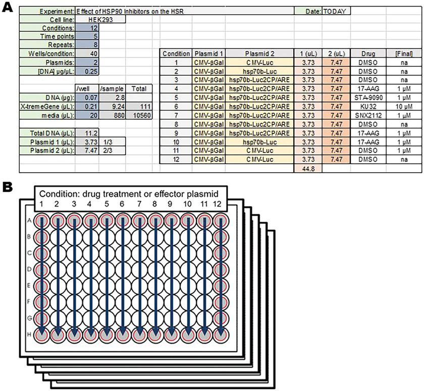
Transfect cells with a mixture of the following: 0.05 μg of DNA + 0.15 μL of X-tremeGENE +20 μL of serum-free DMEM for each well (see Note 2). The 0.05 μg of DNA includes all plasmids to be transfected. Adjusting all plasmids to the same concentration is rather helpful. The ratio of each plasmid can be varied to optimize the assay. Typically, less pCMV-βGal is required compared to other plasmids in the experiment. The ratio of phsp70b-Luc2CP/ARE and effector HSF1 or HSP plasmid can be varied to test for mutation or regulatory effects. It is advised to repeat each sample 4 or 8 times per plate when assaying 24 or 12 respective conditions (see Note 3). Setting up a spreadsheet to calculate plasmid and reagent amounts is advised (Fig. 2a). Plasmids to be used: pCMV-βGal (transfection normalization control), pCMV-LUC (positive luciferase control), phsp70B-Luc (stable HSR control), phsp70bLuc2CP/ARE (experimental, real-time reporter), and optional pcDNA3-HSF1 or –HSP (effector constructs).
Fig. 2 Preparation of 96-well plates for transfection and drug treatment. (a) Sample spreadsheet for aliquoting reagents and arranging conditions. (b) Schematic for 12 drug treatment conditions across 5 time points. Gray circles indicate perimeter wells that should not typically be used for experimental testing
1. Aliquot and mix X-tremeGENE, serum-free DMEM, and plasmids in a separate sterile 96-well plate with each well corresponding to a different sample condition.
2. Using a multichannel pipette, distribute the ~20 μL of transfection mix into each well on the 96-well plates containing the cultured cells.
3. Incubate the cells and transfection mix overnight for 16–24 h.
3.3 Drug Treatment (Optional)
To test the effects of drugs on the HSR, transfected cells can be treated with small molecule inhibitors for 1 h before heat shocking. Treatment time can be varied but at least 45 min is needed to allow the drugs to accumulate in the cells. The number of plates and wells
M per plate to be treated will determine the number of drugs to be pre-diluted in DMEM and aliquoted.
1. For 17-AAG at a final concentration of ~1 μM in 16 wells (2 columns of 8) across 5 plates, a total of 12 μLof1m stock 17-AAG dissolved in DMSO should be added to 1700 μL of DMEM.
2. Once mixed, 20 μL of pre-diluted 17-AAG is added to each well.
3. DMSO is used as the vehicle control (Fig. 2a, b).
3.4 Heat Shock There are a few different ways to heat shock cells depending on available equipment (see Note 4):
1. Wrap each plate tightly with Parafilm.
2. Float each plate in a water bath heated to 42 °C for 30 min.
3. If using a common lab water bath, before unwrapping and placing back into the 37 °C incubator, each plate must be thoroughly sprayed and wiped with 70% ethanol and then dried to prevent contaminating the cell culture incubator. Or
1. Preheat a separate incubator to 43 °C.
2. Place the plates in the incubator for 45–60 min to induce a robust heat shock.
3. Place the cells back into the 37 °C incubator to recover and be harvested.
3.5 Cell Lysis and Sample Storage To harvest the cells at each time point:
1. Remove the plate from the incubator and aspirate off all media, but be careful not to dislodge the cells from the plate.
2. Add 110 μL of RLB to each well and place in a freezer for at least 45 min. Samples can also be stored until the time course is complete. For longer-term storage, samples should be placed in an -80 °C freezer.
3. A single freeze-thaw cycle should efficiently lyse the cells.
3.6 Luciferase Assay To determine the level of induced gene expression:
1. Allow all plates to completely thaw on ice.
2. Once thawed, plates can be spun down on a large benchtop centrifuge at 4 °C to aggregate cell debris and ensure uniform lysis (optional).
3. Thaw both LAR and BAR making sure both reagents are warm and completely dissolved.
3.7 β-Galactosidase Assay
4. Turn on the plate reader luminometer and load the proper program for analysis.
5. When ready use a multichannel pipette to transfer 50 μLo lysate from each well to the corresponding well in a clean white 96-well plate.
6. Add 50 μL of LAR to each well. Let the plate set for 10 min at room temperature.
7. Read the plate on the luminometer and record the results.
8. Repeat the process of aliquoting the lysate, adding the LAR, allowing it to set for 10 min, and reading the luciferase activity for each plate in the time course.
9. Due to changes in luciferase activity over time, make sure the timing between adding the LAR and reading luciferase activity is consistent for each plate. This will ensure consistent and accurate readings of HSR induction at each time point (see Note 5).
To determine the transfection efficiency for each well:
1. For each plate pipette 50 μL of lysate from each well into a corresponding well in a clear 96-well plate. Do this for all the plates in the time course.
2. Add 35 μL of BAR to each well, and incubate all the plates at 37 °C for 20 min or until the lysate mix turns yellow.
3. Add 50 μL of BGS to all wells to terminate the enzyme reaction. This will ensure that the β-galactosidase reaction time is the same across all plates.
4. Read each plate for absorbance at 420 nM and record the results (see Note 6).
3.8 Normalization and Statistics
To determine the luciferase activity relative to the amount of plasmid transfected into the cells:
1. Divide the luciferase readout value by the β-galactosidase value using a spreadsheet program.
2. Identify the baseline reporter signal, in this case, the control no HS plate and no drugs sample set, and then calculate the average.
3. Divide all the other wells in all other plates by this control value. This makes all reporter readout signals relative to the average of the baseline signal.
4. For each sample condition set of repeats, determine the average and standard deviation.
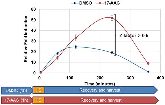
Fig. 3 Effect of 17-AAG pretreatment on HSR degree and duration. Cells were incubated with DMSO or 1 μM 17-AAG for 1 h, then heat shocked for 30 min at 42 °C, allowed to recover, and then harvested at designated time points
5. Sample values can finally be graphed +/- standard deviation at each time point and p-values calculated using Student’s t-test between each time point and/or condition (Fig. 3).
6. If utilizing the assay as a high-throughput screen, a Z-factor score can be calculated for a specific time point where a value greater than 0.5 indicates significance [25].
4 Results
Differences in HSR transcription rates were observed between DMSO and 17-AAG pretreatments in HEK293 cells. For DMSO control cells, the HSR increased over time with a maximum at 2 h then decreased to baseline at 6 h. For 17-AAG pretreated cells, the HSR increased over time with a maximum of around 4 h and remained above baseline at 6 h, indicating that HSP90 N-terminal inhibition increased the degree and duration of the HSR. At 4 h the difference between DMSO and 17-AAG treated cells gave a Z-factor of 0.65 suggesting that 17-AAG warrants further attention as an amplifier of the HSR (Fig. 3). Only the results for DMSO and 17-AAG treatment are shown.
5
Notes
1. Transfection and luciferase/β-galactosidase assays work well in 96-well plates with HEK293 cells. For cell lines with low transfection efficiency, scale up to 12-or 24-well plates to increase cell numbers.
2. We have used X-tremeGENE 9 and lipofectamine 3000 as transfection reagents with equal success.
3. Due to exposure and evaporation issues, use the wells on the perimeter of the plate as control repeats and not for experimental testing.
4. Each cell line is different in regards to inducing the HSR; some cell lines may require higher temperatures for longer periods of time. Preliminary testing may be required using the 2 h postHS time point.
5. It is essential that all plates are assayed and read in the same manner and timing after the addition of LAR. This is to ensure that the substrate concentration and luciferase activity are the same for each time point. The pre-read incubation time can be adjusted to 5 min and staggered depending on setup.
6. In HEK293 cells, 10–20 min incubation is enough for the β-galactosidase assay. Do not incubate too long because the reaction can be saturated. If you have multiple plates for timecourse assay, it is very important to measure the β-galactosidase activity of each plate with the same incubation time.
References
1. Murshid A, Eguchi T, Calderwood SK (2013) Stress proteins in aging and life span. Int J Hyperth 29(5):442–447
2. Prince TL, Lang BJ, Guerrero-Gimenez ME, Fernandez-Munoz JM, Ackerman A, Calderwood SK (2020) HSF1: primary factor in molecular chaperone expression and a major contributor to cancer morbidity. Cell 9(4): 1046
3. Lang BJ, Guerrero ME, Prince TL, Okusha Y, Bonorino C, Calderwood SK (2021) The functions and regulation of heat shock proteins; key orchestrators of proteostasis and the heat shock response. Arch Toxicol 95(6):1943–1970
4. Mendillo ML, Santagata S, Koeva M, Bell GW, Hu R, Tamimi RM, Fraenkel E, Ince TA, Whitesell L, Lindquist S (2012) HSF1 drives a transcriptional program distinct from heat shock to support highly malignant human cancers. Cell 150(3):549–562
5. Schmauder L, Sima S, Hadj AB, Cesar R, Richter K (2022) Binding of the HSF-1 DNA-binding domain to multimeric C. elegans consensus HSEs is guided by cooperative interactions. Sci Rep 12(1):1–19
6. Bunch H, Zheng X, Burkholder A, Dillon ST, Motola S, Birrane G, Ebmeier CC, Levine S, Fargo D, Hu G, Taatjes DJ (2014) TRIM28 regulates RNA polymerase II promoterproximal pausing and pause release. Nat Struct Mol Biol 21(10):876–883
7. Vihervaara A, Mahat DB, Guertin MJ, Chu T, Danko CG, Lis JT, Sistonen L (2017) Transcriptional response to stress is pre-wired by promoter and enhancer architecture. Nat Commun 8(1):1–6
8. Kijima T, Prince TL, Tigue ML, Yim KH, Schwartz H, Beebe K, Lee S, Budzynski MA, Williams H, Trepel JB, Sistonen L (2018) HSP90 inhibitors disrupt a transient HSP90-
HSF1 interaction and identify a noncanonical model of HSP90-mediated HSF1 regulation. Sci Rep 8(1):1–3
9. Kmiecik SW, Le Breton L, Mayer MP (2020) Feedback regulation of heat shock factor 1 (Hsf1) activity by Hsp70-mediated trimer unzipping and dissociation from DNA. EMBO J 39(14):e104096
10. Pernet L, Faure V, Gilquin B, DufourGue ´ rin S, Khochbin S, Vourc’h C (2014) HDAC6–ubiquitin interaction controls the duration of HSF1 activation after heat shock. Mol Biol Cell 25(25):4187–4194
11. Guettouche T, Boellmann F, Lane WS, Voellmy R (2005) Analysis of phosphorylation of human heat shock factor 1 in cells experiencing a stress. BMC Biochem 6(1):1–4
12. Neef DW, Jaeger AM, Gomez-Pastor R, Willmund F, Frydman J, Thiele DJ (2014) A direct regulatory interaction between chaperonin TRiC and stress-responsive transcription factor HSF1. Cell Rep 9(3):955–966
14. Kmiecik SW, Drzewicka K, Melchior F, Mayer MP (2021) Heat shock transcription factor 1 is SUMOylated in the activated trimeric state. J Biol Chem 296:100324
15. Gomez-Pastor R, Burchfiel ET, Thiele DJ (2018) Regulation of heat shock transcription factors and their roles in physiology and disease. Nat Rev Mol Cell Biol 19(1):4–19
16. Cyran AM, Zhitkovich A (2022) Heat shock proteins and HSF1 in cancer. Front Oncol 12: 860320
17. Parsian AJ, Sheren JE, Tao TY, Goswami PC, Malyapa R, Van Rheeden R, Watson MS, Hunt CR (2000) The human Hsp70B gene at the
HSPA7 locus of chromosome 1 is transcribed but non-functional. Biochim Biophys Acta 1494(1–2):201–205
18. Younis I, Berg M, Kaida D, Dittmar K, Wang C, Dreyfuss G (2010) Rapid-response splicing reporter screens identify differential regulators of constitutive and alternative splicing. Mol Cell Biol 30(7):1718–1728
19. West JD, Wang Y, Morano KA (2012) Small molecule activators of the heat shock response: chemical properties, molecular targets, and therapeutic promise. Chem Res Toxicol 25(10):2036–2053
20. Kurop MK, Huyen CM, Kelly JH, Blagg BS (2021) The heat shock response and small molecule regulators. Eur J Med Chem 226: 113846
21. Kim D, Kim SH, Li GC (1999) Proteasome inhibitors MG132 and lactacystin hyperphosphorylate HSF1 and induce hsp70 and hsp27 expression. Biochem Biophys Res Commun 254(1):264–268
22. Kijima T, Prince T, Neckers L, Koga F, Fujii Y (2019) Heat shock factor 1 (HSF1)-targeted anticancer therapeutics: overview of current preclinical progress. Expert Opin Ther Targets 23(5):369–377
23. Murshid A, Chou SD, Prince T, Zhang Y, Bharti A, Calderwood SK (2010) Protein kinase A binds and activates heat shock factor 1. PLoS One 5(11):e13830
24. Gibson DG, Young L, Chuang RY, Venter JC, Hutchison CA, Smith HO (2009) Enzymatic assembly of DNA molecules up to several hundred kilobases. Nat Methods 6(5):343–345
25. Zhang JH, Chung TD, Oldenburg KR (1999) A simple statistical parameter for use in evaluation and validation of high throughput screening assays. J Biomol Screen 4(2):67–73
Studying RNA Polymerase II Promoter-Proximal Pausing by In Vitro Immobilized Template and Transcription Assays
Heeyoun Bunch
Abstract
The immobilized template assay is a versatile biochemical method for studying protein–nucleic acid interactions. Using this method, immobilized nucleic acid-associated or specific proteins can be identified and quantified by techniques such as mass spectrometry and immunoblotting. Here, a modified immobilized template assay combined with in vitro transcription assay to study the function of transcription factors and transcriptional activities at the human heat shock protein 70 (HSP70) gene is described. Notably, this method can be applied to study other important genes and transcription factors in vitro.
Key words Transcriptional regulation, In vitro transcription, Immobilized template assay, Transcription factors, Gene regulation
1 Introduction
Immobilized template assays followed by in vitro transcription assays have been used for over a couple of decades [1–3]. The development of one such method for isolating HeLa nuclei and producing the nuclear extracts (HeLa NE) enabled us to study transcription in vitro. This is mainly attributed to the ability of the immobilized template, including the promoter of a gene to assemble the pre-initiation complex (PIC) from HeLa NE. In the case of bacterial in vitro transcription, recombinant 5-subunit-RNA polymerase (2α, β, β′, and ω subunits typically associated with σ factor) [4], a DNA template, and dNTPs under proper buffer conditions are sufficient for de novo synthesis of RNA molecules [5]. However, eukaryotic transcription is much more complex and involves an array of general and gene-specific transcription factors and at least 12 subunit-RNA polymerase II (Pol II) [6, 7]. This complexity imposes practical difficulties in studying eukaryotic transcription in vitro. The immobilized template assay to assemble the PIC on a DNA template of interest from HeLa NE bypasses the
need to purify all the protein components. To be immobilized, the DNA template requires to be conjugated on the collectable beads. With elaborative modifications, this powerful method can be used to understand the function and interaction of transcription factors in gene regulation in vitro.
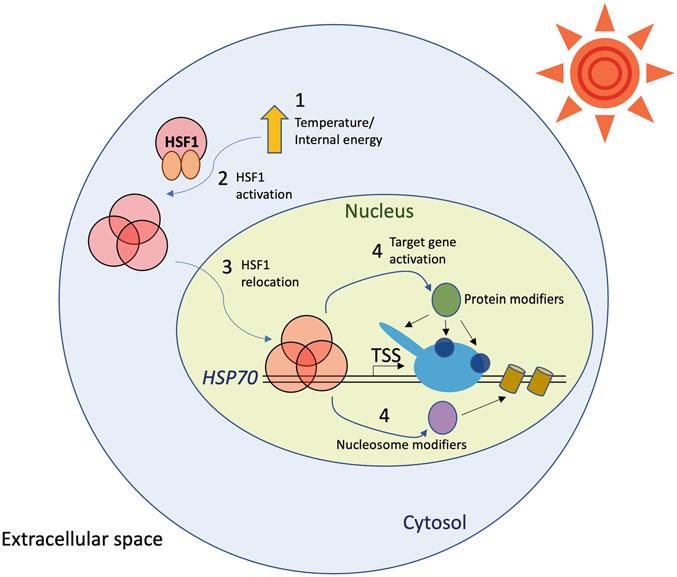
HSP70 is one of the most representative and well-studied stimulus-inducible genes [8–11]. Gene regulation of HSP70 is instantaneous and dramatical, provoking a burst of mRNA synthesis. This rapid response to the increased internal energy is ensured due to a single transcription factor, HSF1, serving as a primary signal transduction regulator that transfers the cytosolic signal to the target gene activation in the nucleus [11–13]. However, upon HSF1 binding to the HSP70 promoter, the activated gene undergoes several changes in terms of its interacting transcription factors, Pol II modifications, DNA structure, and nucleosome architecture [2, 9, 10, 14–25] (Fig. 1). Notably, many features of transcriptional regulation at HSP70 are conserved and common in diverse classes of stimulus-inducible genes, thereby establishing HSP70 as a model gene to study gene regulation mechanisms. Pol II promoterproximal pausing is one of the critical regulatory features of
Fig. 1 HSF1-mediated transcriptional activation at HSP70. The numbers indicate the order of HSP70 transcriptional activation. Proteins are shown in circular shapes. Nucleosomes are shown in cylinders. The sun is shown in a circle surrounded by triangles, a source of heat
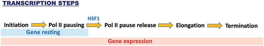
Fig. 2 Transcriptional regulation at HSP70. Pol II pausing is an additional critical regulatory step in transcriptional regulation in a large number of genes in metazoan cells
HSP70 and other stimulus-inducible genes, including serum-, neurotransmitter-, and hypoxia-inducible genes [8, 14, 26–28]. In this phenomenon, Pol II is paused at +25–+100 from the transcription start site (TSS) before receiving gene activating signal, which is recognized as a rate-limiting step in transcriptional activation [29, 30] (Fig. 2). In Drosophila, it was estimated that approximately 30% of protein-coding genes are estimated to harbor Pol II pausing [29]. It reportedly occurs in both protein-coding and nonproteincoding genes at relatively higher percentages in higher organisms like mice and humans [28, 31, 32].
In our previous studies, we demonstrated Pol II pausing at the human HSP70 gene by combining an immobilized template and in vitro transcription assay [10, 33]. Furthermore, we identified a novel Pol II pausing regulator, TRIM28, and characterized its function [33]. These results led us to reveal the noteworthy and critical cross talk between DNA break and damage response signaling and transcriptional activation [14, 34, 35]. Other research groups have employed similar methods to investigate transcriptional regulation at human HSP70 [2, 36, 37]. Here, I describe the methods used to perform the immobilized template assay and in vitro transcription assay to study Pol II pausing and transcriptional regulation at HSP70.
2 Materials
2.1 Preparation of the HSP70 Template DNA
1. PCR with a set of primers, including a 5′-biotinylated forward primer (see Note 1) that can amplify the HSP70 segment of interest: For the PCR template in the reaction, the promoter and TSS of HSP70 (-467 – +216) were amplified from the HeLa cells and inserted into a pCR4-TOPO vector to generate pTOPO-HSP70, as described in our previous study [33].
2. Agarose gel electrophoresis: The PCR reaction is run on a 0.8% agarose gel to run in 1× Tris–HCl–acetate–EDTA (TAE) buffer.
3. DNA extraction from the gel using a Qiagen gel extraction kit. Elute the DNA using ultrapure deionized water.
2.2 Preparation of the HSP70 Immobilized Template
2.3 Performing the Immobilized Template Assay
1. Dynabeads M-280 Streptavidin (Invitrogen) are washed and equilibrated with 2× B & W buffer (10 mM Tris-HCl, pH 7.5, 1 mM EDTA, 2 M NaCl, filtered using 0.2 μm filter) (see Note 2) and incubated with the biotin-conjugated HSP70 template.
2. The template–bead complex is washed with 1× B & W buffer (5 mM Tris-HCl, pH 7.5, 0.5 mM EDTA, 1 M NaCl, filtered using 0.2 μm filter) and resuspended in 0.1 M Buffer D (20 mM HEPES, 20% glycerol, pH 7.6, pH 7.9, 0.1 mM EDTA, 100 mM KCl, filtered using 0.2 μm filter) to a final DNA concentration of 10 ng/μL.
1. HeLa NE (>80 mg/mL) can be prepared using the traditional method described in [33, 38, 39]. Per a reaction, 100–120 ng of the HSP70 template DNA is used and mixed with transcription factor (TF) buffer (12.5 ng/μL dI-dC, 0.075% NP40, 5 mM MgCl2, 250 ng/μL BSA, 12.5% glycerol, 100 mM KCl, 12.5 mM HEPES, pH 7.6, 62.5 μM EDTA, 10 μM ZnCl2; the mixture is filtered through a 0.2 μm filter before supplementing with BSA and dI-dC) to incubate the DNA–bead complex with a DNA binding transcription factor(s) (e.g., HSF1). Recombinant HSF1 can be purified as previously described [33].
2. The protein-bound DNA–bead complex is pulled down using a magnetic stand and resuspended in nuclear extract (NE) buffer (17.5 ng/μL dI-dC, 0.1% NP40, 7.5 mM MgCl2, 1.25 μg/μL BSA, 8.7% glycerol, 8.7 mM HEPES, pH 7.6, 44 μM EDTA, 130 mM KCl, 10 μM ZnCl2; the mixture is filtered through a 0.2 μm filter before supplementing with BSA and dI-dC) (see Note 3).
f
3. After incubating the complex with approximately 100 μgo HeLa NE, the bead–DNA–protein complex is washed with transcription wash 1 (TW1) buffer (12.5 mM HEPES, pH 7.6, 12.5% glycerol, 500 mM KCl, 833 nM MgCl2, 62.5 μM EDTA, 0.0125% NP40; the buffer is filtered through a 0.2 μm filter). NE and TW1 buffers are freshly supplemented with protease inhibitors (1 mM benzamidine, 0.25 mM PMSF, aprotinin [1:1000, Sigma A6279], and 1 mM sodium metabisulfite). The specific transcription factor of investigation can be depleted from HeLa NE by a series of immunoprecipitation assays as described in our previous study [33].
4. The DNA-bound proteins are eluted by 2% sarkosyl dissolved in TW1 buffer and are shown by silver staining and quantified by immunoblotting.